
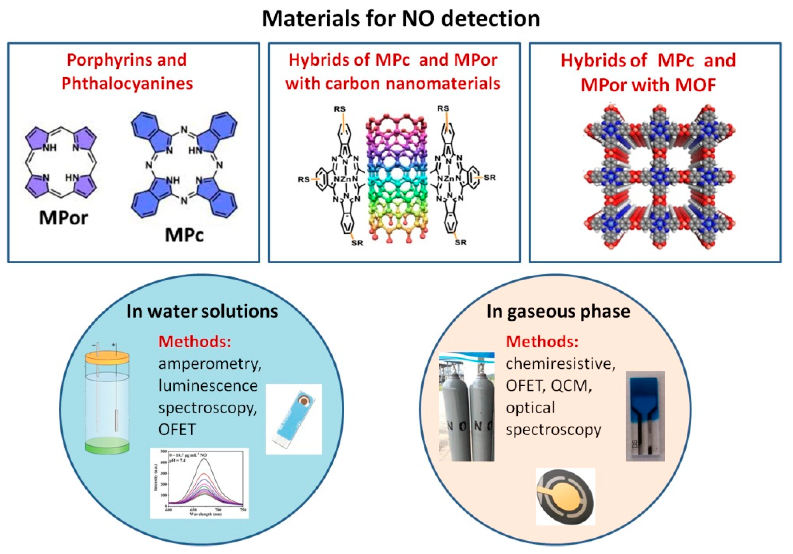
Recent Advances in Phthalocyanine and Porphyrin-Based Materials as Active Layers for Nitric Oxide Chemical Sensors


Abstract
:1. Introduction
2. Sensing Layers for Detecting NO in Aqueous Media
2.1. Porphyrins and Phthalocyanines
2.2. Hybrids of Phthalocyanines and Porphyrins with Carbon Nanomaterials
2.3. Hybrid Materials of Phthalocyanines and Porphyrins with MOF
3. Sensing Layers for the Detection of Gaseous NO
4. Advances, Current Issues and Future Scope
Author Contributions
Funding
Institutional Review Board Statement
Informed Consent Statement
Data Availability Statement
Acknowledgments
Conflicts of Interest
References
- Kim, D.H.; Ringe, S.; Kim, H.; Kim, S.; Kim, B.; Bae, G.; Oh, H.S.; Jaouen, F.; Kim, W.; Kim, H.; et al. Selective electrochemical reduction of nitric oxide to hydroxylamine by atomically dispersed iron catalyst. Nat. Commun. 2021, 12, 1856. [Google Scholar] [CrossRef] [PubMed]
- Bobrovskaya, A.N.; Simonov, P.A.; Kvon, R.I.; Bukhtiyarov, A.V.; Romanenko, A.V. Synthesis of Hydroxylamine Sulfate via NO Hydrogenation over Pt/Graphite Catalysts, Part 2: Effect of the Reaction Conditions and the Physicochemical State of a Catalyst on the Yield of Products. Catal. Ind. 2020, 12, 16–28. [Google Scholar] [CrossRef]
- Kumar, G.; Dey, S.K.; Kundu, S. Functional implications of vascular endothelium in regulation of endothelial nitric oxide synthesis to control blood pressure and cardiac functions. Life Sci. 2020, 259, 118377. [Google Scholar] [CrossRef]
- Liu, W.; Wang, Y.; Leng, Z.; Wang, Q.; Duan, X.; Luo, Y.; Jiang, Y.; Qin, L. Nitric oxide plays a crucial role in midgut immunity under microsporidian infection in Antheraea pernyi. Mol. Immunol. 2020, 126, 65–72. [Google Scholar] [CrossRef] [PubMed]
- Morales-Medina, J.C.; Aguilar-Alonso, P.; Di Cerbo, A.; Iannitti, T.; Flores, G. New insights on nitric oxide: Focus on animal models of schizophrenia. Behav. Brain Res. 2021, 409, 113304. [Google Scholar] [CrossRef]
- Smith, O. Nobel Prize for NO research. Nat. Med. 1998, 4, 1215. [Google Scholar] [CrossRef] [PubMed]
- Pisi, R.; Aiello, M.; Tzani, P.; Marangio, E.; Olivieri, D.; Chetta, A. Measurement of fractional exhaled nitric oxide by a new portable device: Comparison with the standard technique. J. Asthma 2010, 47, 805–809. [Google Scholar] [CrossRef]
- Schneider, A.; Tilemann, L.; Schermer, T.; Gindner, L.; Laux, G.; Szecsenyi, J.; Meyer, F.J. Diagnosing asthma in general practice with portable exhaled nitric oxide measurement—Results of a prospective diagnostic study. Respir. Res. 2009, 10, 15. [Google Scholar] [CrossRef] [Green Version]
- Jackson, D.J.; Virnig, C.M.; Gangnon, R.E.; Evans, M.D.; Roberg, K.A.; Anderson, E.L.; Burton, R.M.; Salazar, L.P.; DaSilva, D.F.; Shanovich, K.M.; et al. Fractional exhaled nitric oxide measurements are most closely associated with allergic sensitization in school-age children. J. Allergy Clin. Immunol. 2009, 124, 949–953. [Google Scholar] [CrossRef] [Green Version]
- Lim, K.G. Nitric oxide measurement in chronic cough. Lung 2010, 188, 20–23. [Google Scholar] [CrossRef]
- Das, S.; Pal, M. Review—Non-Invasive Monitoring of Human Health by Exhaled Breath Analysis: A Comprehensive Review. J. Electrochem. Soc. 2020, 167, 037562. [Google Scholar] [CrossRef]
- Baptist, A.P.; Khan, F.I.; Wang, Y.; Ager, J. Exhaled nitric oxide measurements in hospitalized children with asthma. J. Asthma 2008, 45, 670–674. [Google Scholar] [CrossRef] [PubMed]
- Tadaki, H.; Mochizuki, H.; Muramastu, R.; Hagiwara, S.; Mizuno, T.; Arakawa, H.; Morikawa, A. A flow- and pressure-controlled offline method of exhaled nitric oxide measurement in children. Ann. Allergy Asthma Immunol. 2008, 100, 308–313. [Google Scholar] [CrossRef]
- Gupta, R.; Gupta, N.; Turner, S.W. A methodology for measurements of nasal nitric oxide in children under 5 yr. Pediatr. Allergy Immunol. 2008, 19, 233–238. [Google Scholar] [CrossRef]
- Hewitt, R.S.; Smith, A.D.; Cowan, J.O.; Schofield, J.C.; Herbison, G.P.; Taylor, D.R. Serial exhaled nitric oxide measurements in the assessment of laboratory animal allergy. J. Asthma 2008, 45, 101–107. [Google Scholar] [CrossRef]
- Oh, M.J.; Lee, J.Y.; Lee, B.J.; Choi, D.C. Exhaled nitric oxide measurement is useful for the exclusion of nonasthmatic eosinophilic bronchitis in patients with chronic cough. Chest 2008, 134, 990–995. [Google Scholar] [CrossRef]
- Cameli, P.; Bargagli, E.; Bergantini, L.; D’alessandro, M.; Giugno, B.; Gentili, F.; Sestini, P. Alveolar nitric oxide as a biomarker of COVID-19 lung sequelae: A pivotal study. Antioxidants 2021, 10, 1350. [Google Scholar] [CrossRef]
- Núñez-Fernández, M.; Ramos-Hernández, C.; García-Río, F.; Torres-Durán, M.; Nodar-Germiñas, A.; Tilve-Gómez, A.; Rodríguez-Fernández, P.; Valverde-Pérez, D.; Ruano-Raviña, A.; Fernández-Villar, A. Alterations in respiratory function test three months after hospitalisation for covid-19 pneumonia: Value of determining nitric oxide diffusion. J. Clin. Med. 2021, 10, 2119. [Google Scholar] [CrossRef]
- Pizzimenti, S.; Bugiani, M.; Piccioni, P.; Heffler, E.; Carosso, A.; Guida, G.; Rolla, G. Exhaled nitric oxide measurements: Correction equation to compare hand-held device to stationary analyzer. Respir. Med. 2008, 102, 1272–1275. [Google Scholar] [CrossRef] [Green Version]
- Korn, S.; Telke, I.; Kornmann, O.; Buhl, R. Measurement of exhaled nitric oxide: Comparison of different analysers. Respirology 2010, 15, 1203–1208. [Google Scholar] [CrossRef]
- Bryan, N.S.; Grisham, M.B. Methods to detect nitric oxide and its metabolites in biological samples. Free Radic. Biol. Med. 2007, 43, 645–657. [Google Scholar] [CrossRef] [PubMed] [Green Version]
- Nagasaka, Y.; Fernandez, B.O.; Steinbicker, A.U.; Spagnolli, E.; Malhotra, R.; Bloch, D.B.; Bloch, K.D.; Zapol, W.M.; Feelisch, M. Pharmacological preconditioning with inhaled nitric oxide (NO): Organ-specific differences in the lifetime of blood and tissue NO metabolites. Nitric Oxide 2018, 80, 52–60. [Google Scholar] [CrossRef] [PubMed]
- Balamurugan, M.; Madasamy, T.; Pandiaraj, M.; Bhargava, K.; Sethy, N.K.; Karunakaran, C. Electrochemical assay for the determination of nitric oxide metabolites using copper(II) chlorophyllin modified screen printed electrodes. Anal. Biochem. 2015, 478, 121–127. [Google Scholar] [CrossRef] [PubMed]
- Vargas-Maya, N.I.; Padilla-Vaca, F.; Romero-González, O.E.; Rosales-Castillo, E.A.S.; Rangel-Serrano, Á.; Arias-Negrete, S.; Franco, B. Refinement of the Griess method for measuring nitrite in biological samples. J. Microbiol. Methods 2021, 187, 106260. [Google Scholar] [CrossRef]
- Green, L.C.; Wagner, D.A.; Glogowski, J.; Skipper, P.L.; Wishnok, J.S.; Tannenbaum, S.R. Analysis of nitrate, nitrite, and [15N]nitrate in biological fluids. Anal. Biochem. 1982, 126, 131–138. [Google Scholar] [CrossRef]
- Wang, J.; Lu, M.; Yang, F.; Zhang, X.; Baeyens, W.R.G.; García Campaña, A.M. Microdialysis with on-line chemiluminescence detection for the study of nitric oxide release in rat brain following traumatic injury. Anal. Chim. Acta 2001, 428, 173–181. [Google Scholar] [CrossRef]
- Fink, B.; Dikalov, S.; Fink, N. ESR techniques for the detection of nitric oxide in vivo as an index of endothelial function. Pharmacol. Rep. 2006, 58, 8–15. [Google Scholar]
- Xu, Y.C.; Cao, Y.L.; Guo, P.; Tao, Y.; Zhao, B.L. Detection of nitric oxide in plants by electron spin resonance. Phytopathology 2004, 94, 402–407. [Google Scholar] [CrossRef] [Green Version]
- Öter, Ö.; Aydin, A.C.; Zeyrek Ongun, M.; Celik, E. Development of a nanoscale-based optical chemical sensor for the detection of no radical. Turk. J. Chem. 2018, 42, 1056–1071. [Google Scholar] [CrossRef]
- Ding, L.; Fan, C.; Zhong, Y.; Li, T.; Huang, J. A sensitive optic fiber sensor based on CdSe QDs fluorophore for nitric oxide detection. Sens. Actuators B Chem. 2013, 185, 70–76. [Google Scholar] [CrossRef]
- Privett, B.J.; Shin, J.H.; Schoenfisch, M.H. Electrochemical nitric oxide sensors for physiological measurements. Chem. Soc. Rev. 2010, 39, 1925–1935. [Google Scholar] [CrossRef] [PubMed] [Green Version]
- Bedioui, F.; Griveau, S. Electrochemical Detection of Nitric Oxide: Assessement of Twenty Years of Strategies. Electroanalysis 2013, 25, 587–600. [Google Scholar] [CrossRef]
- Paleczek, A.; Grochala, D.; Staszek, K.; Wincza, K.; Gruszczynski, S.; Rydosz, A. Microwave-based nitrogen dioxide gas sensor for automotive applications. In Proceedings of the 2021 International Conference on Electrical, Computer, Communications and Mechatronics Engineering (ICECCME), Mauritius, 7–8 October 2021; pp. 1–4. [Google Scholar] [CrossRef]
- Singh, S.K.; Azad, P.; Akhtar, M.J.; Kar, K.K. High-sensitive nitrogen dioxide and ethanol gas sensor using a reduced graphene oxide-loaded double split ring resonator. Mater. Res. Express 2017, 4, 086301. [Google Scholar] [CrossRef]
- Zhou, X.; Xue, Z.; Chen, X.; Huang, C.; Bai, W.; Lu, Z.; Wang, T. Nanomaterial-based gas sensors used for breath diagnosis. J. Mater. Chem. B 2020, 8, 3231–3248. [Google Scholar] [CrossRef] [PubMed]
- Dey, A. Semiconductor metal oxide gas sensors: A review. Mater. Sci. Eng. B 2018, 229, 206–217. [Google Scholar] [CrossRef]
- Qin, R.; Shan, G.; Hu, M.; Huang, W. Two-dimensional transition metal carbides and/or nitrides (MXenes) and their applications in sensors. Mater. Today Phys. 2021, 21, 100527. [Google Scholar] [CrossRef]
- Lv, W.; Shi, K.; Li, L.; Shao, S. Nitrogen-doped multiwalled carbon nanotubes and their electrocatalysis towards oxidation of NO. Microchim. Acta 2010, 170, 91–98. [Google Scholar] [CrossRef]
- Wenninger, N.; Bračič, U.; Kollau, A.; Pungjunun, K.; Leitinger, G.; Kalcher, K.; Ortner, A. Development of an electrochemical sensor for nitric oxide based on carbon paste electrode modified with Nafion, gold nanoparticles and graphene nanoribbons. Sens. Actuators B Chem. 2021, 346, 130532. [Google Scholar] [CrossRef]
- Klyamer, D.; Bonegardt, D.; Basova, T. Fluoro-Substituted Metal Phthalocyanines for Active Layers of Chemical Sensors. Chemosensors 2021, 9, 133. [Google Scholar] [CrossRef]
- Bouvet, M.; Gaudillat, P.; Suisse, J.M. Phthalocyanine-based hybrid materials for chemosensing. J. Porphyr. Phthalocyanines 2013, 17, 913–919. [Google Scholar] [CrossRef]
- Giancane, G.; Valli, L. State of art in porphyrin Langmuir–Blodgett films as chemical sensors. Adv. Colloid Interface Sci. 2012, 171–172, 17–35. [Google Scholar] [CrossRef] [PubMed]
- Klyamer, D.; Sukhikh, A.; Gromilov, S.; Krasnov, P.; Basova, T. Fluorinated metal phthalocyanines: Interplay between fluorination degree, films orientation, and ammonia sensing properties. Sensors 2018, 18, 2141. [Google Scholar] [CrossRef] [PubMed] [Green Version]
- Gounden, D.; Nombona, N.; van Zyl, W.E. Recent advances in phthalocyanines for chemical sensor, non-linear optics (NLO) and energy storage applications. Coord. Chem. Rev. 2020, 420, 213359. [Google Scholar] [CrossRef]
- Fan, F.; Zhang, J.; Li, J.; Zhang, N.; Hong, R.R.; Deng, X.; Tang, P.; Li, D. Hydrogen sensing properties of Pt-Au bimetallic nanoparticles loaded on ZnO nanorods. Sens. Actuators B Chem. 2017, 241, 895–903. [Google Scholar] [CrossRef]
- Yang, S.; Yu, Y.; Gao, X.; Zhang, Z.; Wang, F. Recent advances in electrocatalysis with phthalocyanines. Chem. Soc. Rev. 2021, 50, 12985–13011. [Google Scholar] [CrossRef]
- Brown, M.D.; Schoenfisch, M.H. Catalytic selectivity of metallophthalocyanines for electrochemical nitric oxide sensing. Electrochim. Acta 2018, 273, 98–104. [Google Scholar] [CrossRef]
- Dang, X.; Hu, H.; Wang, S.; Hu, S. Nanomaterials-based electrochemical sensors for nitric oxide. Microchim. Acta 2015, 182, 455–467. [Google Scholar] [CrossRef]
- Zagal, J.H.; Griveau, S.; Silva, J.F.; Nyokong, T.; Bedioui, F. Metallophthalocyanine-based molecular materials as catalysts for electrochemical reactions. Coord. Chem. Rev. 2010, 254, 2755–2791. [Google Scholar] [CrossRef]
- Goshi, E.; Zhou, G.; He, Q. Nitric oxide detection methods in vitro and in vivo. Med. Gas Res. 2019, 9, 192–207. [Google Scholar] [CrossRef]
- Fukuto, J.M.; Cho, J.Y.; Switzer, C.H. Chapter 2—The chemical properties of nitric oxide and related nitrogen oxides. In Nitric Oxide; Academic Press: San Diego, CA, USA, 2000; pp. 23–40. ISBN 978-0-12-370420-7. [Google Scholar]
- Li, C.M.; Zang, J.; Zhan, D.; Chen, W.; Sun, C.Q.; Teo, A.L.; Chua, Y.; Lee, V.; Moochhala, S. Electrochemical Detection of Nitric Oxide on a SWCNT/RTIL Composite Gel Microelectrode. Electroanalysis 2006, 18, 713–718. [Google Scholar] [CrossRef]
- Hu, F.X.; Xie, X.; Wang, D.; Bin Yang, H.; Gu, Y.; Chen, B.; Zhang, C.; Rao, Q.; Li, Q.; Guo, C. Three-dimensional cell-adhesive matrix of silk cocoon derived carbon fiber assembled with iron-porphyrin for monitoring cell released signal molecules. Sens. Actuators B Chem. 2021, 334, 129594. [Google Scholar] [CrossRef]
- Wang, M.; Zhu, L.; Zhang, S.; Lou, Y.; Zhao, S.; Tan, Q.; He, L.; Du, M. A copper(II) phthalocyanine-based metallo-covalent organic framework decorated with silver nanoparticle for sensitively detecting nitric oxide released from cancer cells. Sens. Actuators B Chem. 2021, 338, 129826. [Google Scholar] [CrossRef]
- Yan, Y.; Yao, P.; Mu, Q.; Wang, L.; Mu, J.; Li, X.; Kang, S.Z. Electrochemical behavior of amino-modified multi-walled carbon nanotubes coordinated with cobalt porphyrin for the oxidation of nitric oxide. Appl. Surf. Sci. 2011, 258, 58–63. [Google Scholar] [CrossRef]
- Sivanesan, A.; John, S.A. Highly sensitive electrochemical sensor for nitric oxide using the self-assembled monolayer of 1,8,15,22-tetraaminophthalocyanatocobalt(II) on glassy carbon electrode. Electroanalysis 2010, 22, 639–644. [Google Scholar] [CrossRef]
- Yu, A.; Liang, Z.; Cho, J.; Caruso, F. Nanostructured Electrochemical Sensor Based on Dense Gold Nanoparticle Films. Nano Lett. 2003, 3, 1203–1207. [Google Scholar] [CrossRef]
- Matsuoka, R.; Kobayashi, C.; Nakagawa, A.; Aoyagi, S.; Aikawa, T.; Kondo, T.; Kasai, S.; Yuasa, M. A reactive oxygen/nitrogen species sensor fabricated from an electrode modified with a polymerized iron porphyrin and a polymer electrolyte membrane. Anal. Sci. 2017, 33, 911–915. [Google Scholar] [CrossRef] [Green Version]
- Chandra, S.; Mende, C.; Bahadur, D.; Hildebrandt, A.; Lang, H. Fabrication of a porphyrin-based electrochemical biosensor for detection of nitric oxide released by cancer cells. J. Solid State Electrochem. 2015, 19, 169–177. [Google Scholar] [CrossRef]
- Liu, M.Y.; Punckt, C.; Pope, M.A.; Gelperin, A.; Aksay, I.A. Electrochemical Sensing of Nitric Oxide with Functionalized Graphene Electrodes. ACS Appl. Mater. Interfaces 2013, 5, 12624–12630. [Google Scholar] [CrossRef]
- Biesaga, M.; Pyrzyńska, K.; Trojanowicz, M. Porphyrins in analytical chemistry. A review. Talanta 2000, 51, 209–224. [Google Scholar] [CrossRef]
- Okada, Y.; Hoshi, T.; Kobayashi, N. Recent Progress in Optically-Active Phthalocyanines and Their Related Azamacrocycles. Front. Chem. 2020, 8, 595998. [Google Scholar] [CrossRef]
- Orzeł, Ł.; Polaczek, J.; Procner, M. Review: Recent advances in the investigations of no activation on cobalt and manganese porphyrins: A brief review. J. Coord. Chem. 2015, 68, 2971–2989. [Google Scholar] [CrossRef]
- Xu, H.; Liao, C.; Liu, Y.; Ye, B.C.; Liu, B. Iron Phthalocyanine Decorated Nitrogen-Doped Graphene Biosensing Platform for Real-Time Detection of Nitric Oxide Released from Living Cells. Anal. Chem. 2018, 90, 4438–4444. [Google Scholar] [CrossRef] [PubMed]
- Caro, C.A.; Zagal, J.H.; Bedioui, F. Electrocatalytic Activity of Substituted Metallophthalocyanines Adsorbed on Vitreous Carbon Electrode for Nitric Oxide Oxidation. J. Electrochem. Soc. 2003, 150, E95–E103. [Google Scholar] [CrossRef]
- Li, J.; Xie, J.; Gao, L.; Li, C.M. Au Nanoparticles—3D Graphene Hydrogel Nanocomposite to Boost Synergistically in Situ Detection Sensitivity toward Cell-Released Nitric Oxide. ACS Appl. Mater. Interfaces 2015, 7, 2726–2734. [Google Scholar] [CrossRef] [PubMed]
- Mumtarin, Z.; Rahman, M.M.; Marwani, H.M.; Hasnat, M.A. Electro-kinetics of conversion of NO3− into NO2− and sensing of nitrate ions via reduction reactions at copper immobilized platinum surface in the neutral medium. Electrochim. Acta 2020, 346, 135994. [Google Scholar] [CrossRef]
- Nguyen, T.Q.; Padama, A.A.B.; Escano, M.C.S.; Kasai, H. Theoretical Study on The Adsorption of NO on Metal Macrocycles, Metal=Mn,Fe,Co,Ni,Cu,Zn. ECS Trans. 2013, 45, 91–100. [Google Scholar] [CrossRef]
- Nguyen, T.Q.; Escaño, M.C.S.; Kasai, H. Nitric oxide adsorption effects on metal phthalocyanines. J. Phys. Chem. B 2010, 114, 10017–10021. [Google Scholar] [CrossRef]
- Liao, M.S.; Scheiner, S. Electronic structure and bonding in metal phthalocyanines, metal=Fe, Co, Ni, Cu, Zn, Mg. J. Chem. Phys. 2001, 114, 9780–9791. [Google Scholar] [CrossRef] [Green Version]
- Nyokong, T.; Vilakazi, S. Phthalocyanines and related complexes as electrocatalysts for the detection of nitric oxide. Talanta 2003, 61, 27–35. [Google Scholar] [CrossRef]
- Silva, V.H.C.; Martins, M.P.; De Oliveira, H.C.B.; Camargo, A.J. Theoretical investigation of nitric oxide interaction with aluminum phthalocyanine. J. Mol. Graph. Model. 2011, 29, 777–783. [Google Scholar] [CrossRef]
- Oliveira, T.I.S.; dos Santos, V.N.; Lomonaco, D.; Correia, A.N.; Mazetto, S.E.; de Lima-Neto, P. Gold Electrode Modified with Cu-Porphyrin Derived from Cardanol as Electrochemical Sensor for Nitric Oxide. J. Electrochem. Soc. 2013, 160, B113–B118. [Google Scholar] [CrossRef]
- Yap, C.M.; Xu, G.Q.; Ang, S.G. Amperometric nitric oxide sensor based on nanoporous platinum phthalocyanine modified electrodes. Anal. Chem. 2013, 85, 107–113. [Google Scholar] [CrossRef] [PubMed]
- Ramirez-Garcia, G.; Martinez-Alfaro, M.; Gutierrez-Granados, S.; Alatorre-Ordaz, A.; Griveau, S.; Bedioui, F. Electrochemical assessment of possible melatonin effect on nitric oxide production from kidneys of sub-acute lead treated rats. Electrochim. Acta 2015, 166, 88–92. [Google Scholar] [CrossRef]
- Li, Y.; Liu, Q.; Liang, X.; Xiao, Q.; Fang, Y.; Wu, Y. A new fluorescence biosensor for nitric oxide detection based on cytochrome P450 55B1. Sens. Actuators B Chem. 2016, 230, 405–410. [Google Scholar] [CrossRef]
- Suhag, D.; Sharma, A.K.; Patni, P.; Garg, S.K.; Rajput, S.K.; Chakrabarti, S.; Mukherjee, M. Hydrothermally functionalized biocompatible nitrogen doped graphene nanosheet based biomimetic platforms for nitric oxide detection. J. Mater. Chem. B 2016, 4, 4780–4789. [Google Scholar] [CrossRef]
- Xie, H.; Li, Y.T.; Lei, Y.M.; Liu, Y.L.; Xiao, M.M.; Gao, C.; Pang, D.W.; Huang, W.H.; Zhang, Z.Y.; Zhang, G.J. Real-Time Monitoring of Nitric Oxide at Single-Cell Level with Porphyrin-Functionalized Graphene Field-Effect Transistor Biosensor. Anal. Chem. 2016, 88, 11115–11122. [Google Scholar] [CrossRef]
- Ye, Y.; Liu, H.; Li, Y.; Zhuang, Q.; Liu, P.; Gu, J. One-pot doping platinum porphyrin recognition centers in Zr-based MOFs for ratiometric luminescent monitoring of nitric oxide in living cells. Talanta 2019, 200, 472–479. [Google Scholar] [CrossRef]
- Basova, T.V.; Polyakov, M.S. Hybrid materials based on carbon nanotubes and polyaromatic molecules: Methods of functionalization and sensor properties. Macroheterocycles 2020, 13, 91–112. [Google Scholar] [CrossRef]
- Liu, Y.L.; Wang, X.Y.; Xu, J.Q.; Xiao, C.; Liu, Y.H.; Zhang, X.W.; Liu, J.T.; Huang, W.H. Functionalized graphene-based biomimetic microsensor interfacing with living cells to sensitively monitor nitric oxide release. Chem. Sci. 2015, 6, 1853–1858. [Google Scholar] [CrossRef] [Green Version]
- Park, S.; Gildersleeve, J.C.; Blixt, O.; Shin, I. Carbohydrate microarrays. Chem. Soc. Rev. 2013, 42, 4310–4326. [Google Scholar] [CrossRef]
- Cheng, W.; Tang, X.; Zhang, Y.; Wu, D.; Yang, W. Applications of metal-organic framework (MOF)-based sensors for food safety: Enhancing mechanisms and recent advances. Trends Food Sci. Technol. 2021, 112, 268–282. [Google Scholar] [CrossRef]
- Abrahams, B.F.; Hoskins, B.F.; Michail, D.M.; Robson, R. Assembly of porphyrin building blocks into network structures with large channels. Nature 1994, 369, 727–729. [Google Scholar] [CrossRef]
- Abrahams, B.F.; Hoskins, B.F.; Robson, R. A new type of infinite 3D polymeric network containing 4-connected, peripherally-linked metalloporphyrin building blocks. J. Am. Chem. Soc. 2002, 113, 3606–3607. [Google Scholar] [CrossRef]
- Li, H.Y.; Zhao, S.N.; Zang, S.Q.; Li, J. Functional metal-organic frameworks as effective sensors of gases and volatile compounds. Chem. Soc. Rev. 2020, 49, 6364–6401. [Google Scholar] [CrossRef]
- Zhang, L.T.; Zhou, Y.; Han, S.T. The Role of Metal–Organic Frameworks in Electronic Sensors. Angew. Chem. Int. Ed. 2021, 60, 15192–15212. [Google Scholar] [CrossRef] [PubMed]
- Koo, W.-T.; Jang, J.-S.; Kim, I.-D. Metal-Organic Frameworks for Chemiresistive Sensors. Chem 2019, 5, 1938–1963. [Google Scholar] [CrossRef]
- American Thoracic Society; European Respiratory Society. ATS/ERS recommendations for standardized procedures for the online and offline measurement of exhaled lower respiratory nitric oxide and nasal nitric oxide, 2005. Am. J. Respir. Crit. Care Med. 2005, 171, 912–930. [Google Scholar] [CrossRef] [PubMed]
- Miki, H.; Matsubara, F.; Nakashima, S.; Ochi, S.; Nakagawa, K.; Matsuguchi, M.; Sadaoka, Y. A fractional exhaled nitric oxide sensor based on optical absorption of cobalt tetraphenylporphyrin derivatives. Sens. Actuators B Chem. 2016, 231, 458–468. [Google Scholar] [CrossRef]
- Su, P.G.; Li, M.C. Recognition of binary mixture of NO2 and NO gases using a chemiresistive sensors array combined with principal component analysis. Sens. Actuators A Phys. 2021, 331, 112980. [Google Scholar] [CrossRef]
- Palaniappan, A.; Moochhala, S.; Tay, F.E.H.; Su, X.; Phua, N.C.L. Phthalocyanine/silica hybrid films on QCM for enhanced nitric oxide sensing. Sens. Actuators B Chem. 2008, 129, 184–187. [Google Scholar] [CrossRef]
- Ho, K.-C.; Tsou, Y.-H. Chemiresistor-type NO gas sensor based on nickel phthalocyanine thin films. Sens. Actuators B Chem. 2001, 77, 253–259. [Google Scholar] [CrossRef]
- Magori, E.; Hiltawsky, K.; Fleischer, M.; Simon, E.; Pohle, R.; Von Sicard, O.; Tawil, A. Fractional exhaled nitric oxide measurement with a handheld device. J. Breath Res. 2011, 5, 027104. [Google Scholar] [CrossRef] [PubMed]
- Andringa, A.-M.; Spijkman, M.-J.; Smits, E.C.; Mathijssen, S.G.; van Hal, P.A.; Setayesh, S.; Willard, N.P.; Borshchev, O.; Ponomarenko, S.; Blom, P.W.; et al. Gas sensing with self-assembled monolayer field-effect transistors. Org. Electron. 2010, 11, 895–898. [Google Scholar] [CrossRef] [Green Version]
- Meng, Z.; Aykanat, A.; Mirica, K.A. Welding Metallophthalocyanines into Bimetallic Molecular Meshes for Ultrasensitive, Low-Power Chemiresistive Detection of Gases. J. Am. Chem. Soc. 2019, 141, 2046–2053. [Google Scholar] [CrossRef] [PubMed]
- Tao, W.; Lin, P.; Ai, Y.; Wang, H.; Ke, S.; Zeng, X. Multichannel quartz crystal microbalance array: Fabrication, evaluation, application in biomarker detection. Anal. Biochem. 2016, 494, 85–92. [Google Scholar] [CrossRef] [PubMed]
- Shiba, S.; Yamada, K.; Matsuguchi, M. Humidity-resistive optical NO gas sensor devices based on cobalt tetraphenylporphyrin dispersed in hydrophobic polymer matrix. Sensors 2020, 20, 1295. [Google Scholar] [CrossRef] [Green Version]
- Knoben, W.; Crego-Calama, M.; Brongersma, S.H. Comparison of nitric oxide binding to different pure and mixed protoporphyrin IX monolayers. Sens. Actuators B Chem. 2012, 166–167, 349–356. [Google Scholar] [CrossRef]
- Ho, K.C.; Chen, C.M.; Liao, J.Y. Enhancing chemiresistor-type NO gas-sensing properties using ethanol-treated lead phthalocyanine thin films. Sens. Actuators B Chem. 2005, 108, 418–426. [Google Scholar] [CrossRef]
- Amao, Y.; Nakamura, N. Optical CO2 sensor with the combination of colorimetric change of α-naphtholphthalein and internal reference fluorescent porphyrin dye. Sens. Actuators B Chem. 2004, 100, 347–351. [Google Scholar] [CrossRef]


| Sensing Layer | Method/Electrode | Sensing Layer Preparation/Deposition | Analyte | Linear Range, μM | LOD, μM | Ref. |
|---|---|---|---|---|---|---|
| 1,8,15,22-tetraamino- phthalocyanatocobalt(II) (4α-CoIITAPc) | Amperometry, DPV/GCE | Immersion of the electrode in 4α-CoIITAPc solution in DMF | NO in PBS (0.2 M, pH 2) | 0.003–0.03 | 1.4 × 10⁻⁴ | [56] |
| Fe tetrakis(3-thienyl)porphy- rin (FeT3ThP) | Amperometry, DPV/GCE | Electropolymerization/coating with Nafion | NO in PBS (0.15 M) | 0.5–10 | 0.003 | [58] |
| Cu(II)Por complexes (Figure 5) | CV/Au electrode | Electropolymerization | NO, aqueous solution with electrolytes (Na2SO4, TBAP) | 0.282–2.85 | 0.0618 | [73] |
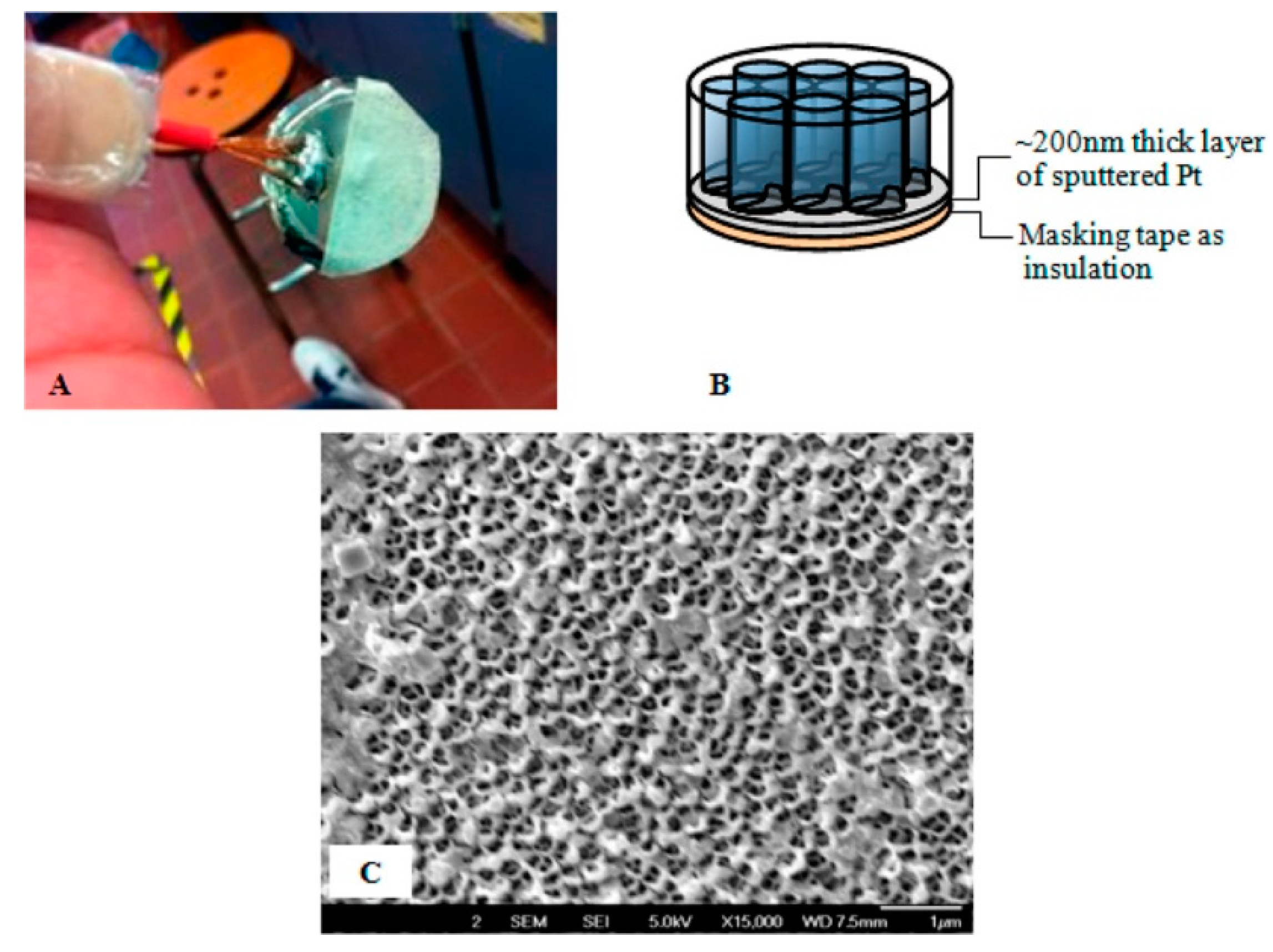
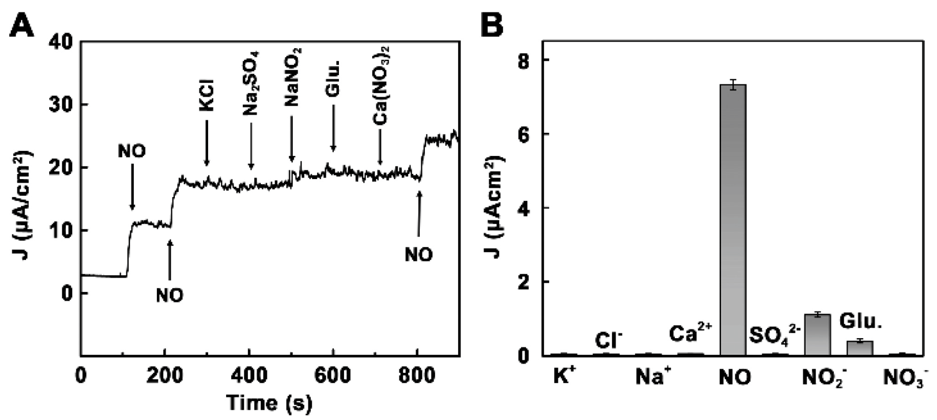
| Pt 4′, 4″, 4‴, 4′′′′-tetraamine phthalocyanine | Pt-coated Anodisc nanoporous membrane | Electropolymerization/coating with Nafion | NO in PBS (pH 7.4) | 0.01–0.1 | 0.01 | [74] |
| 5,10,15,20-tetrakis(4-me- thoxyphenyl)porphyrin (H2TMPP) | Amperometry/GCE | Drop casting | NO released from HeLa cells | - | 1 × 10⁻⁴ | [59] |
| Nickel tetrasulfonated phthalocyanine (NiTSPc) | CV/Ultramicroelectrode (25 μm Pt wire) | Electropolymerization/coating with polyphenol | NO released from HeLa cells | <50 | 0.0254 | [75] |
| Iron porphyrin of cytochrome P450 55B1 | Fluorescence spectroscopy | - | NO in rat liver homogenate | <22.4 | 0.15 | [76] |
| N-G/FePc | Amperometry/ITO electrode | Non-covalent functionalization of N-G/drop casting | NO in PBS (0.01 M, pH 7.4) | 0.18−400 | 0.18 | [77] |
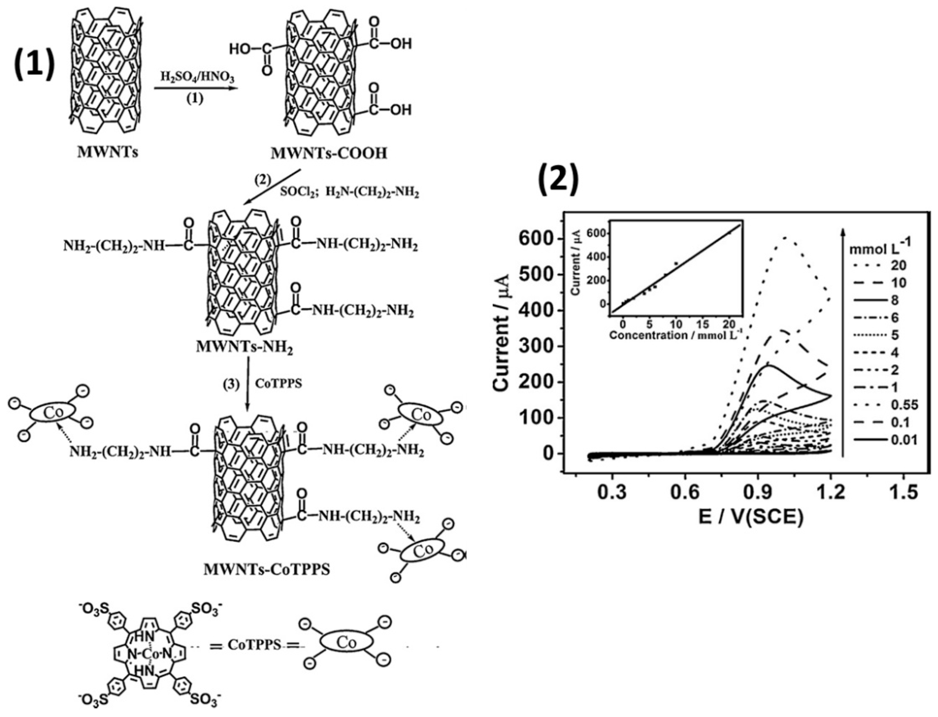
| MWCNT/Co(II) tetrakis(4-sulfonatophenyl)por-phyrin (CoTPPS) | Amperometry/GCE | CoTPPS was attached on MWCNT bearing -CO-NH-(CH2)2-NH2 via their axial coordination to Co/drop casting | NaNO2 in PBS (pH 2.0) was used as a source of NO | 10–20,000 | 6.6 | [55] |
| N-doped graphene nanosheets (PFNGS)/5,10,15,20-tetra- kis(1-methyl-4-pyridino) porphyrin tetra (p-toluenesulfonate) | Amperometry/Pt electrode | Non-covalent functionalization of PFNGS/drop casting | NO in PBS | 0.001–10 | 0.001 | [77] |
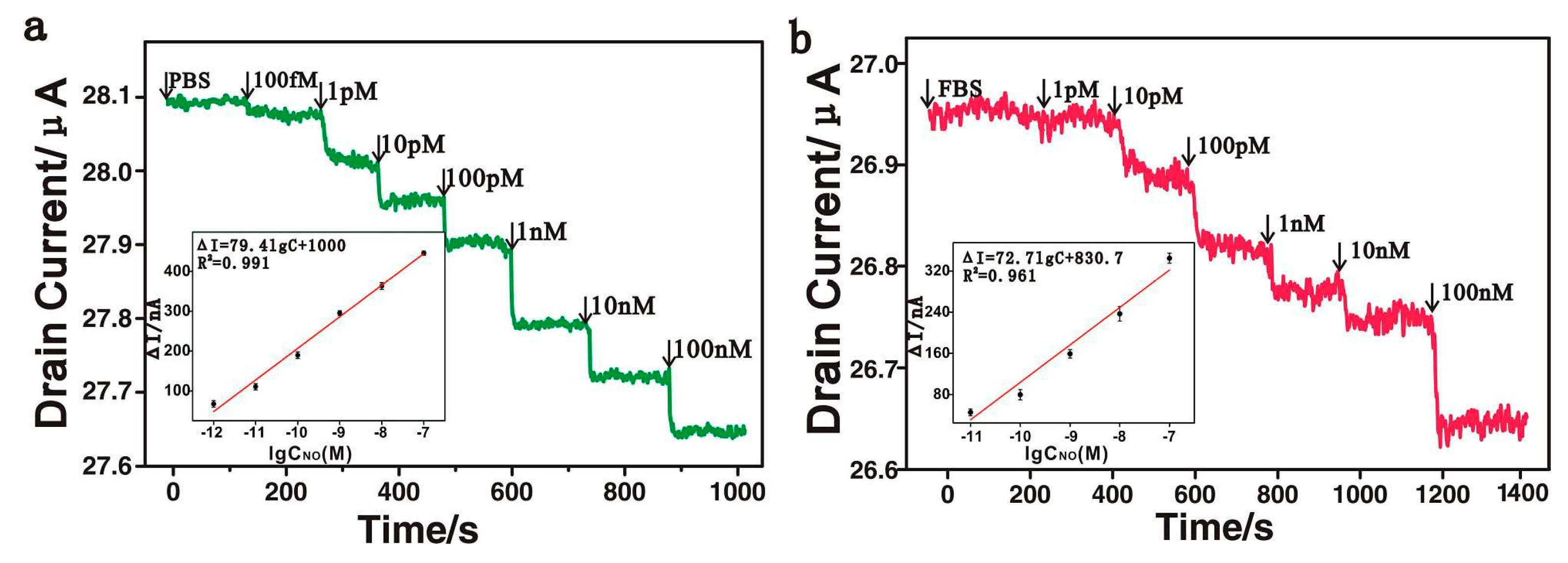
| rGO/Fe(III) meso-tetra(4-carboxyphenyl)porphyrin | Field-effect transistor | Non-covalent functionalization of rGO/drop casting | NO in PBS NO in Human Umbilical Vein Endothelial cells | - | 1 × 10⁻⁶ 1 × 10⁻⁵ | [78] |
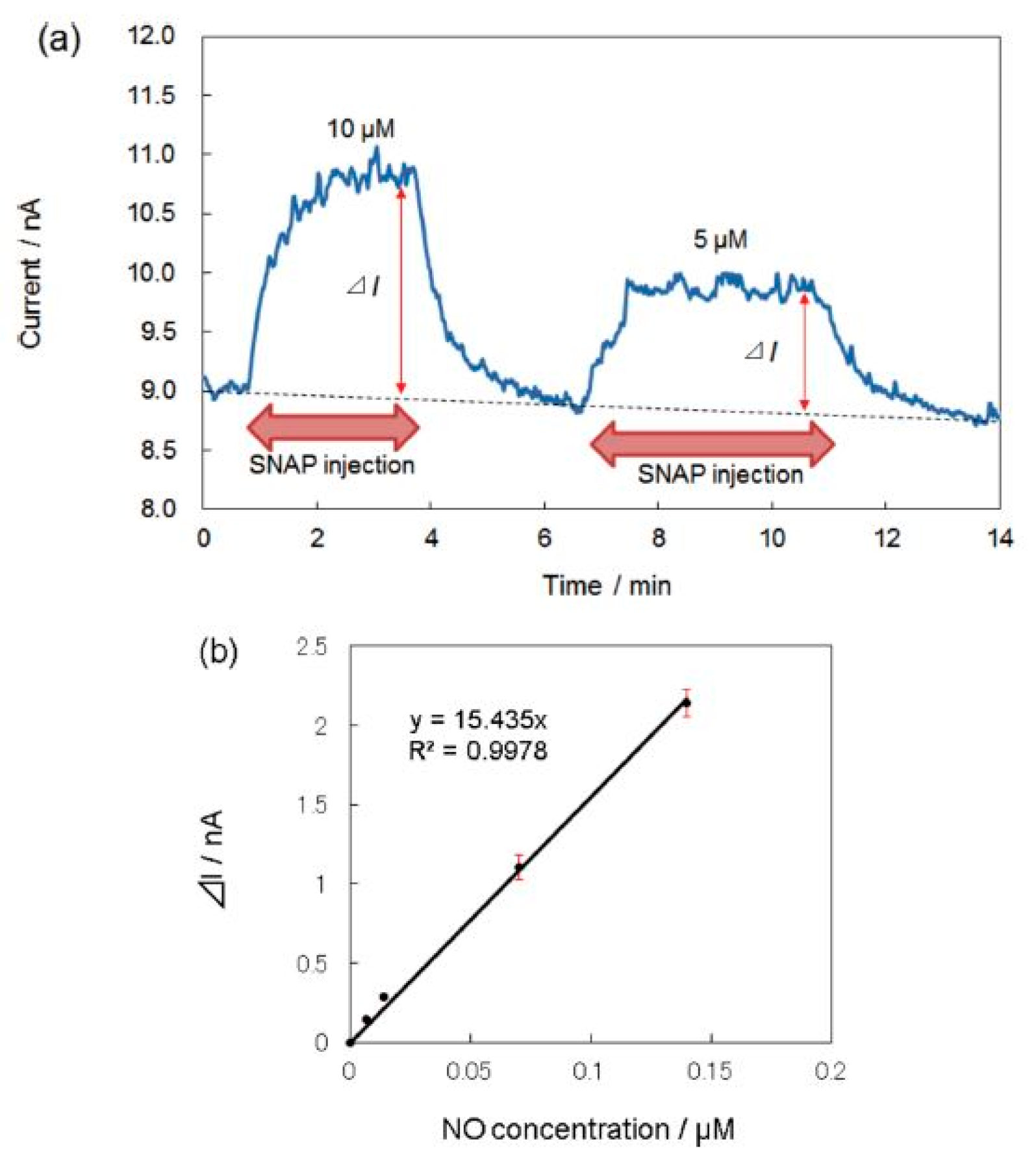
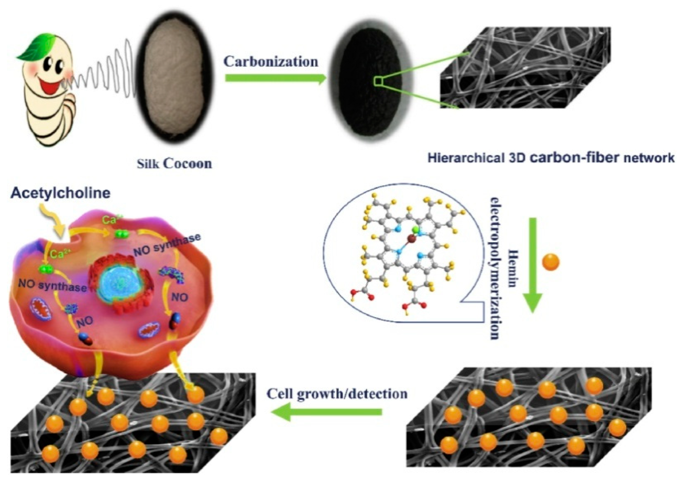
| Iron porphyrin of hemin/CFN | Amperometry/carbon fiber networks | Electrodeposition | NO in PBS (pH 7.4) | 0.024–70.9 | 0.008 | [53] |
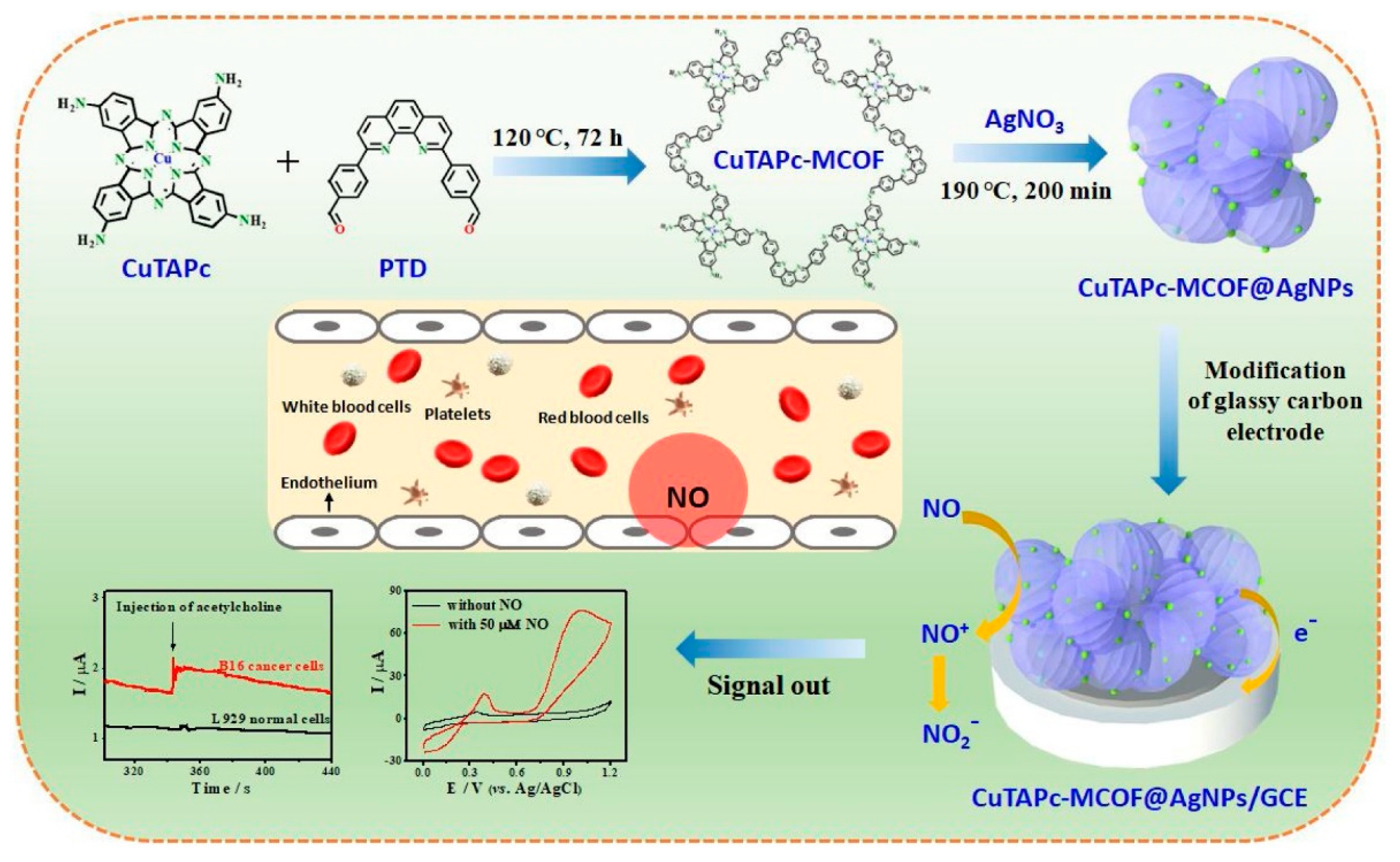
| Cu(II) 4′,4″,4‴,4⁗-tetra-amino-phthalocyanine/MCOF@AgNPs | Amperometry/GCE | CuPc-based covalent-organic framework doped with Ag nanoparticles/drop casting | NO in PBS (pH 7.4) | 0.18–17.1 | 0.0126 | [54] |
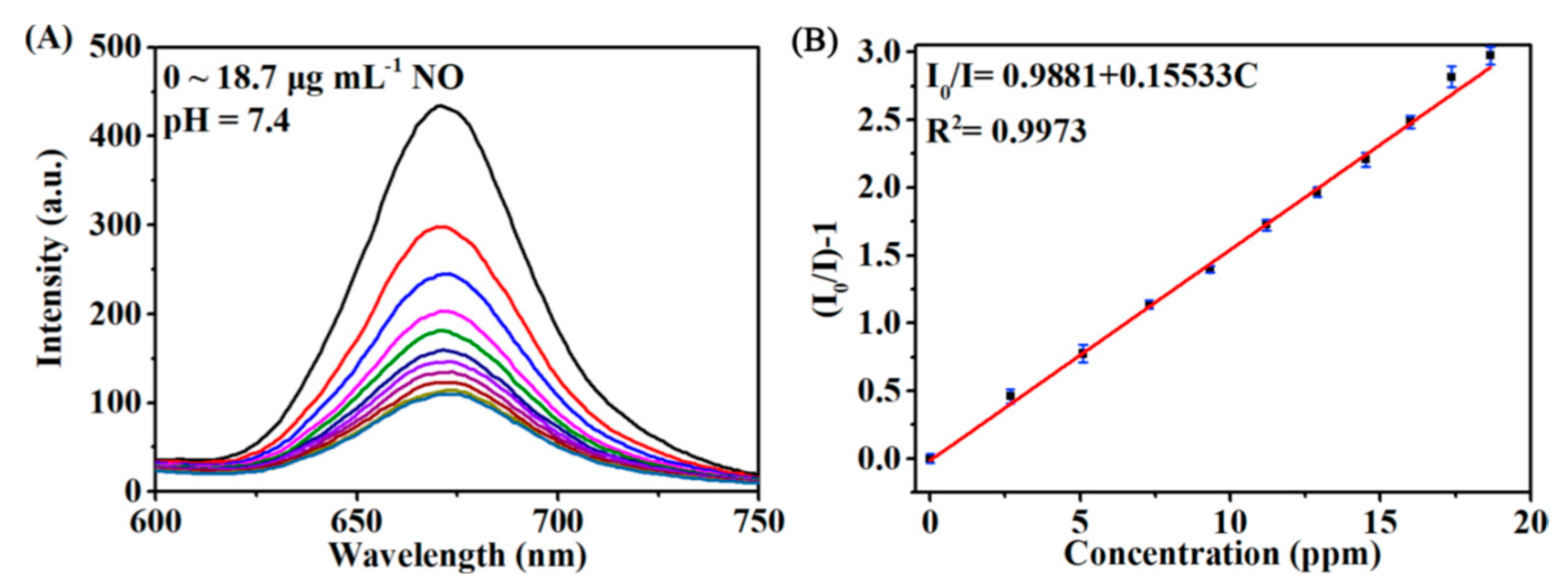
| Pt meso-tetra(4-carboxyphenyl)porphyrin/UiO-66 | Luminescence spectroscopy | Pt-TCPP and H4TCPE integrated in MOFs UiO-66 | NO in HEPES buffer (pH 7.4) | 16–623.3 | 4.73 | [79] |
| Sensing Layer | Method | Linear Range, ppb | LOD, ppb | Ref. |
|---|---|---|---|---|
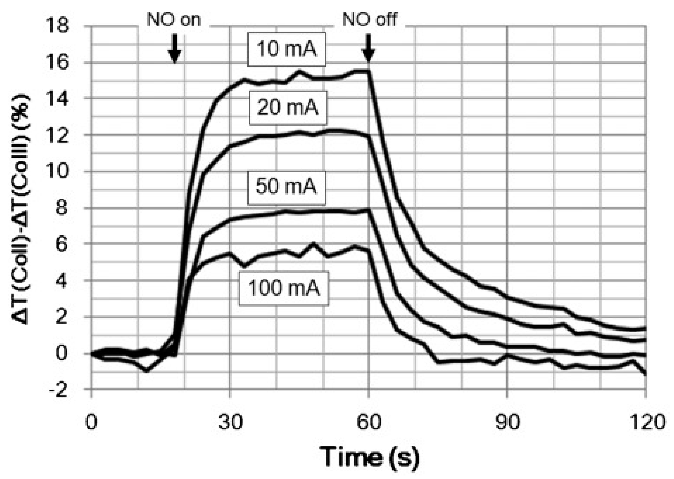
| Co(II) tetraphenylporphyrin (CoTPP)/porous nonwoven materials | Optical absorption spectroscopy | - | 1 | [90] |
| α-phase NiPc films (100 nm) | Chemiresistive | 5000–50,000 | - | [93] |
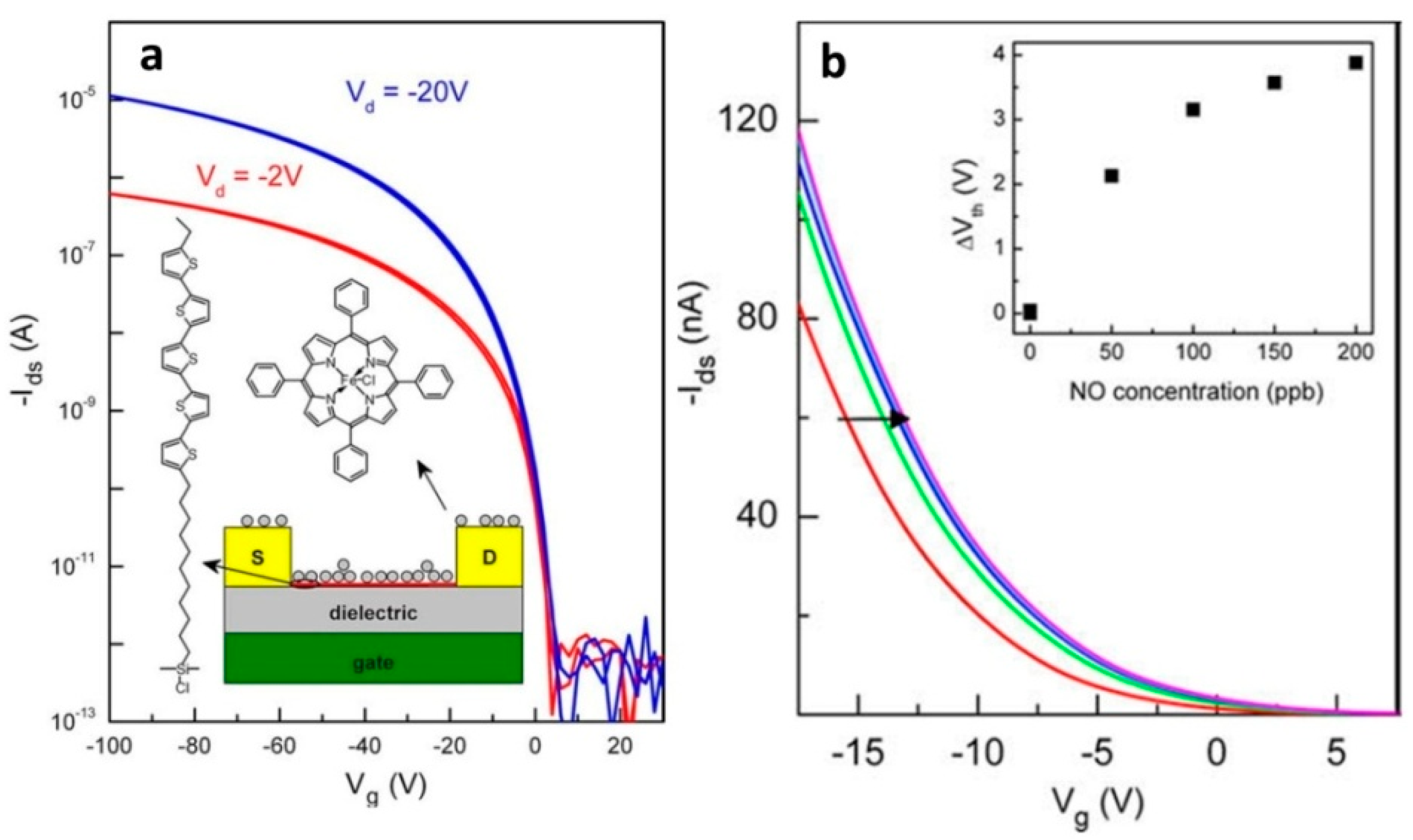
| Fe(III) tetraphenylporphyrin chloride (Fe(TPP)Cl) | Self-assembled monolayer field-effect transistor (SAMFET) | - | 100 | [95] |
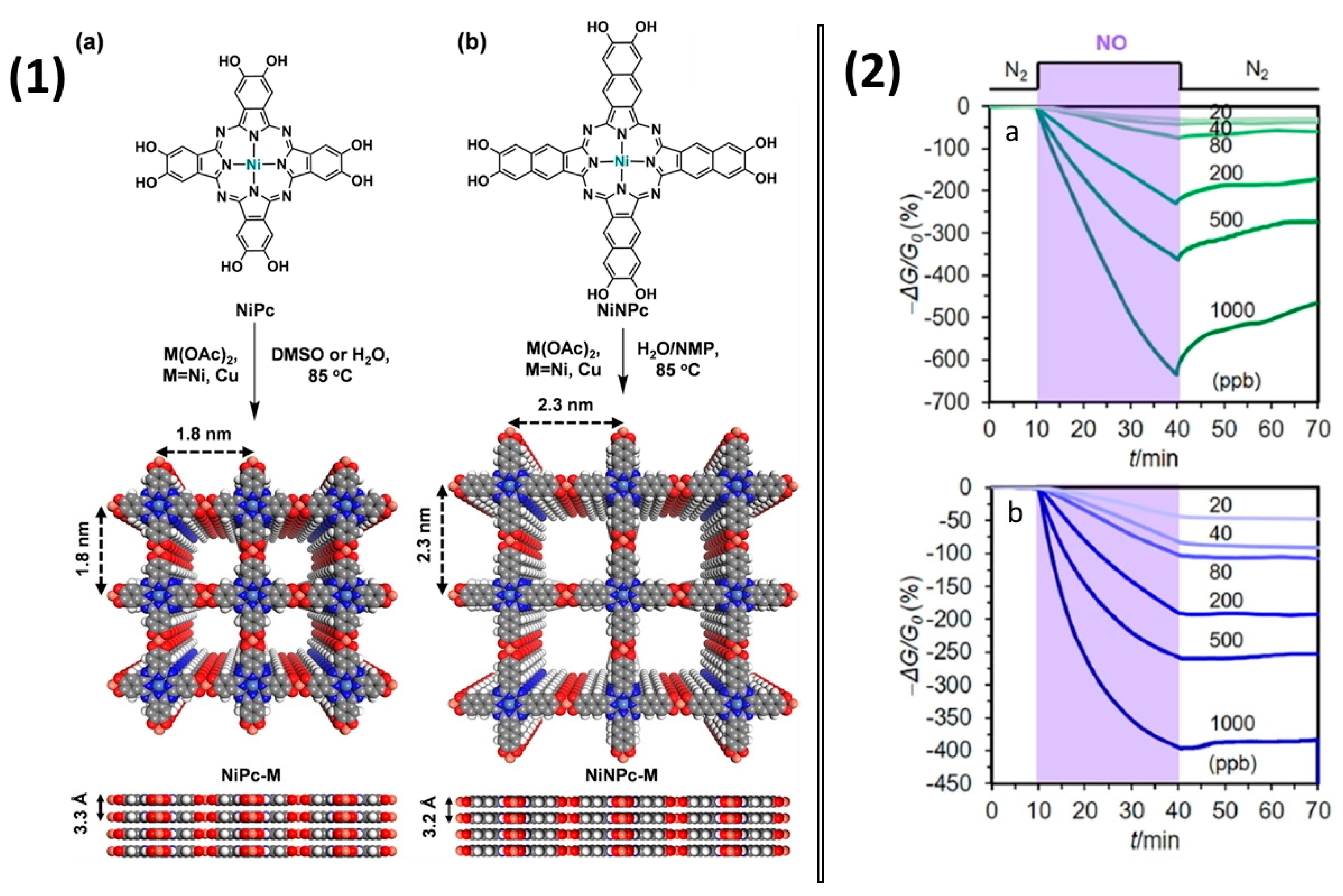
| Phthalocyanine and naphthalocyanine bimetallic 2D MOFs NiPc-M and NiNPc-M (M = Ni, Cu) (Figure 18) | Chemiresistive | 20–1000 | 1.0–1.1 (for 1.5 min exposure) | [96] |
| CoPc-silica hybrid material | QCM | 5.75–103.45 | 5.75 | [97] |
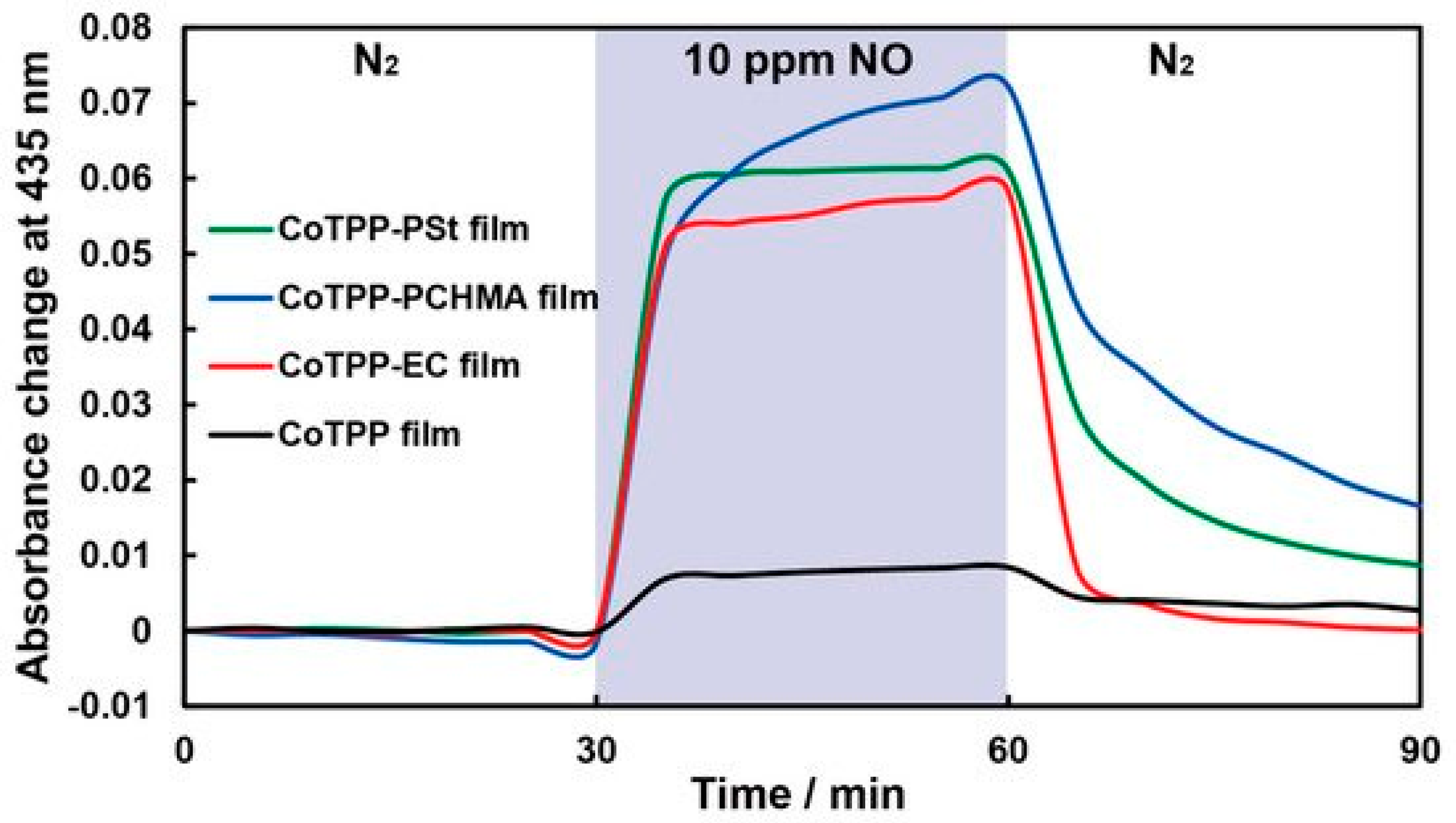
| Co(II) tetraphenylporphyrin (CoTPP)/polymer (polystyrene (PSt), ethylcellulose (EC), polycyclohexyl methacrylate (PCHMA)) | Optical absorption spectroscopy | 100–1000 | 33 (for CoTPP dispersed in EC) | [98] |
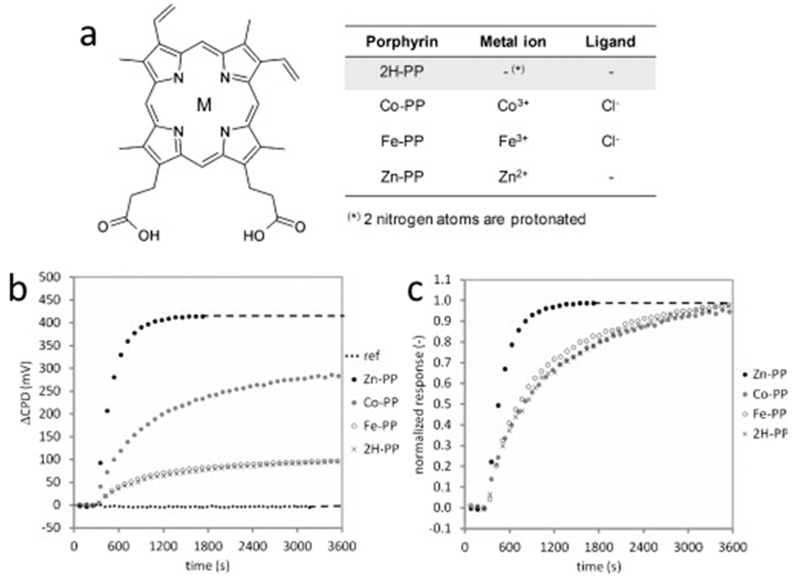
| 2H-PP, Co-PP, Fe-PP and Zn-PP on Al2O3 | Kelvin probe technique | 100–2000 | 5 | [99] |
Publisher’s Note: MDPI stays neutral with regard to jurisdictional claims in published maps and institutional affiliations. |
© 2022 by the authors. Licensee MDPI, Basel, Switzerland. This article is an open access article distributed under the terms and conditions of the Creative Commons Attribution (CC BY) license (https://creativecommons.org/licenses/by/4.0/).
Share and Cite
Klyamer, D.; Shutilov, R.; Basova, T. Recent Advances in Phthalocyanine and Porphyrin-Based Materials as Active Layers for Nitric Oxide Chemical Sensors. Sensors 2022, 22, 895. https://doi.org/10.3390/s22030895
Klyamer D, Shutilov R, Basova T. Recent Advances in Phthalocyanine and Porphyrin-Based Materials as Active Layers for Nitric Oxide Chemical Sensors. Sensors. 2022; 22(3):895. https://doi.org/10.3390/s22030895
Chicago/Turabian StyleKlyamer, Darya, Roman Shutilov, and Tamara Basova. 2022. "Recent Advances in Phthalocyanine and Porphyrin-Based Materials as Active Layers for Nitric Oxide Chemical Sensors" Sensors 22, no. 3: 895. https://doi.org/10.3390/s22030895
APA StyleKlyamer, D., Shutilov, R., & Basova, T. (2022). Recent Advances in Phthalocyanine and Porphyrin-Based Materials as Active Layers for Nitric Oxide Chemical Sensors. Sensors, 22(3), 895. https://doi.org/10.3390/s22030895