Abstract
The emerging literature suggests that implantable functional electrical stimulation may improve gait performance in stroke survivors. However, there is no review providing the possible therapeutic effects of implanted functional electrical stimulation on gait performance in stroke survivors. We performed a web-based, systematic paper search using PubMed, the Cochrane Library, and EMBASE. We limited the search results to human subjects and papers published in peer-reviewed journals in English. We did not restrict demographic or clinical characteristics. We included 10 papers in the current systematic review. Across all included studies, we found preliminary evidence of the potential therapeutic effects of functional electrical stimulation on walking endurance, walking speed, ankle mobility, and push-off force in stroke survivors. However, due to the heterogeneity between the included studies, small sample size, and lack of randomized controlled trials, more studies are critically needed to confirm whether implanted functional electrical stimulation can improve gait performance in stroke survivors.
1. Introduction
In the United States (US), nearly 800,000 people experience a stroke each year [1], and approximately 4% of the US population will have experienced a stroke by 2030 [2]. Globally, stroke is ranked as the second leading cause of mortality, with more than 5 million deaths attributed to stroke in 2016 [3]. Stroke is also ranked as the second most common cause of disability worldwide, accounting for more than 116 million disability-adjusted life-years in 2016 [3].
Approximately 80% of strokes are ischemic, caused by blood clots blocking blood flow to the brain, and 20% are hemorrhagic, caused when a blood vessel leaks or ruptures [4]. The treatment of ischemic stroke is time-sensitive and requires thrombolytic therapy (delivered intravenously). However, for hemorrhagic stroke, thrombolytic therapy is contraindicated. Patients with hemorrhagic stroke commonly have inferior neurological outcomes and a lower survival rate within the first 30 days post-stroke than those with ischemic stroke [5]. Treatment of either type of stroke is time-sensitive, and if recognized early, is treatable.
It has been estimated that more than 70% of people who survive a stroke sustain some feature of gait impairment post-stroke [6]. This gait impairment results in increased risk of falls in stroke survivors [7]. Indeed, studies have reported that almost 50% of stroke patients fall during inpatient rehabilitation [8], and that up to 73% of stroke survivors fall during the first six months after rehabilitation [9]. The high risk of falls in stroke survivors, in turn, leads to serious problems such as fracture injuries [10], an onset of a fear of falling [11], and further restrictions on activity and mobility [12,13].
One expression of gait impairment in stroke is the significant reduction in distal lower limb strength [14]. While walking, this reduction results in the inability to properly dorsiflex the ankle joint immediately after the push-off and safely clear the ground, traditionally known as ”foot drop” (i.e., difficulty in lifting up the front part of the foot) [15].
Functional electrical stimulation using surface electrodes is often used as a rehabilitation technique to help manage foot drop and improve gait performance. Functional electrical stimulation provides electrical currents through transcutaneous electrodes to stimulate peripheral nerves that activate the ankle dorsiflexor muscles [16]. A tilt sensor or in-shoe sensor is commonly used to wirelessly control the stimulation. After the initial introduction of this approach [17], functional electrical stimulation has been continuously studied, developed, and utilized to restore lower limb functions in stroke survivors [18]. However, recent human-centric examinations reported some limitations related to the surface-based functional electrical stimulation such as changes in skin resistance, skin irritation, and a physical and cosmetic discomfort regarding the external device [19,20,21].
In an attempt to address the aforementioned limitations, implanted functional electrical approaches were developed. Since the official approval in Germany in 2007 for stroke survivors [22], several trials investigated the effect of implanted functional electrical stimulation on treating gait impairment in stroke survivors. However, to date, there is no published review of the therapeutic effect of the implementable approach on gait impairment. Accordingly, we aimed to systematically review the effect of implanted functional electrical stimulation on gait performance in stroke survivors. The research question posed in this review was: Does implanted functional electrical stimulation improve gait performance in stroke survivors?
2. Materials and Methods
2.1. Search Strategy
In this systematic review, we followed the Preferred Reporting Items for Systematic Reviews and Meta-Analyses (PRISMA) guidelines [23]. We performed a web-based, systematic paper search using PubMed, the Cochrane Library, and EMBASE for papers published before 5 October 2021. The keywords that were used for the systematic paper search are as follows: “electrical stimulation”, “foot drop stimulator”, “gait”, “gait parameters”, “gait speed”, “gait stability”, “gait initiation”, “gait kinematics”, “gait kinetics”, “stroke survivors”, and “stroke rehabilitation” (see Table 1 for the full search strategy). We limited search results to peer-reviewed journal papers published in English, and studies with human subjects.

Table 1.
Full search strategy.
2.2. Study Selection
We included papers that reported the therapeutic efficacy of implanted functional electrical stimulation on gait performance in stroke survivors with foot drop as randomized controlled trials or observational studies. Outcomes of this systematic review were quantitative measures of gait including spatiotemporal parameters (e.g., gait speed), gait stability (e.g., stride-to-stride asymmetry, movement smoothness, obstacle negotiation), and gait kinetics (e.g., plantar load). We excluded papers that provided a qualitative description of gait performance using subjective methods. Additionally, we excluded conference abstracts, editorial papers, case studies, and review papers from the final study selection.
Two independent reviewers (Rebecca Frederick and Brandon Nunley) conducted an initial screening of the searched papers based on title and abstract. Another reviewer (Gu Eon Kang) independently provided the deciding vote on disagreements (n = 43).
3. Results
3.1. Search Results
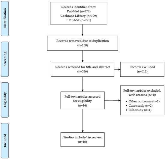
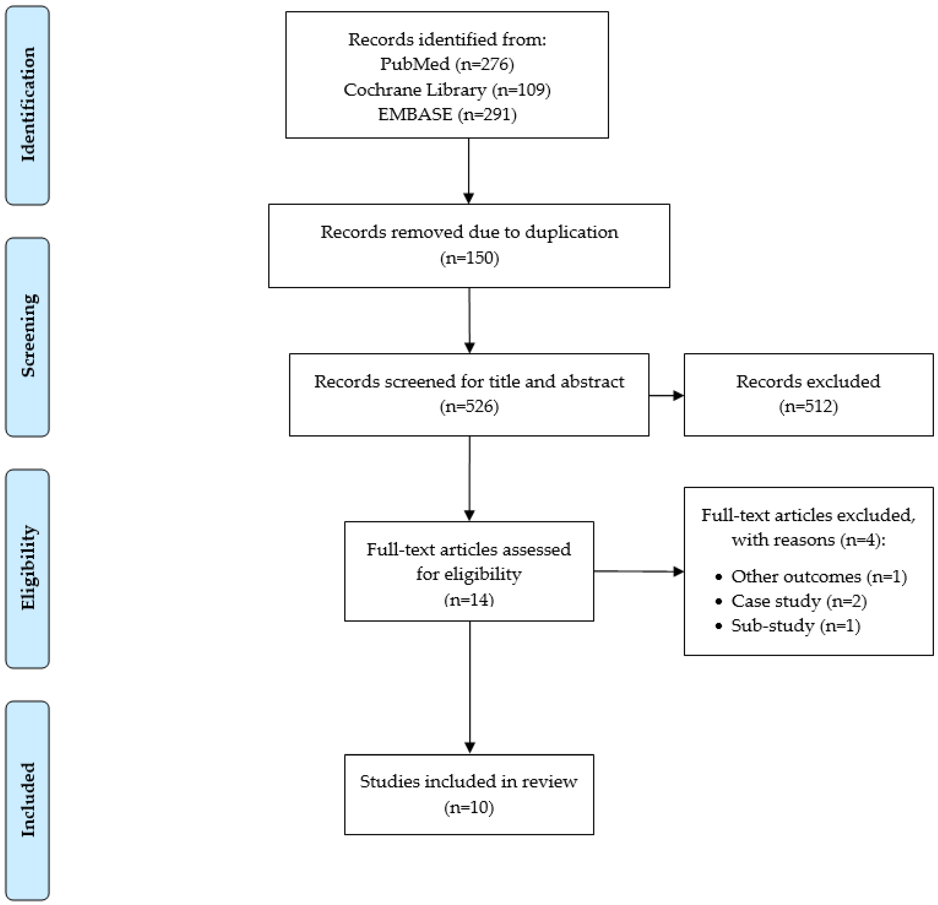
The flow diagram for study selection is shown in Figure 1. We identified 676 papers through PubMed, the Cochrane Library, and EMBASE. After obtaining the initial record of papers, we excluded 150 papers due to duplication using a reference management software (EndNote, Philadelphia, PA, USA). We screened the title and abstract for the remaining 526 papers and excluded 512 papers that did not report the efficacy of implantable functional electrical stimulation on gait performance in stroke survivors. We excluded another 4 papers after assessing eligibility criteria based on full-text review. Consequently, we included 10 papers in the current systematic review [24,25,26,27,28,29,30,31,32,33].

Figure 1.
PRISMA consort diagram for paper selection.
3.2. Study Design and Countries
Our findings are summarized in Table 2. All included studies were published in the last 15 years and were conducted in European countries (n = 5 in Germany; n = 4 in the Netherlands; n = 1 in Denmark). Two studies (20.0%) were randomized controlled trials and eight studies (80.0%) were single arm trials. The vast majority of the included studies (90.0%) followed the study participants longitudinally: Follow-up periods ranged from 1–12 months. One study reported an immediate effect of the implanted functional electrical stimulation (i.e., ON vs. OFF), and did not follow their study participants over the longitudinal course.

Table 2.
Characteristics of studies included for full review.
3.3. Characteristics of Stroke Survivors
The total number of recruited participants across studies was 161 at the baseline. Dropout rates at the conclusion of these studies ranged between 0% and 26.7%. The mean age of the recruited stroke survivors was early to mid-50s for the vast majority of the studies (80.0%) and late 40s for the remaining studies (20.0%). Across all studies, slightly more than half of the recruited participants (n = 81; 50.3%) had right hemiplegia. Types of strokes included in the studies were ischemic (n = 85), hemorrhage (n = 50), and unknown (n = 1). One participant in Bucklitsch et al. (2019) and four participants in Buentjen et al. (2019) had foot drop due to multiple sclerosis. Kottink et al. (2007 and 2012) and Ernst et al. (2013) did not report types of strokes. The time since the onset of stroke ranged, approximately, between 5 and 15 years.
3.4. Intervention
Eight studies (80.0%) utilized the ActiGait foot drop stimulator (Otto Bock, Duderstadt, Germany), which consists of an implantable 4-channel peroneal nerve stimulator, 12-contact electrode cuff, an external control unit, and a heel switch to activate the stimulator (see Burridge et al. (2007) for more details). For the ActiGait foot drop stimulator, the location of the implant was proximal to the common peroneal nerve’s bifurcation into the deep and superficial branches of the nerve that innervate the ankle dorsiflexors.
Two studies (20.0%; Kottink et al. (2007 and 2012)) utilized a foot drop stimulator (it was not reported whether the stimulator was commercially available or was developed by the authors), which consisted of an implantable 2-channel peroneal nerve stimulator, bipolar intraneural electrodes, an external transmitter, a heel switch to activate the transmitter (see Kottink et al. (2007) for more details). The location of the implant was within the epineurium of the superficial peroneal nerve.
For most of studies (n = 7), surgical procedures for implantation were performed under general anesthesia. In four studies (40.0%), adverse events including minor wound infection, delayed wound healing, hematoma, bleeding, and neurodermatitis were reported. One study reported that a participant died but it was not related to the implant. Six studies (60.0%) did not report adverse events.
3.5. Gait Outcomes
Four studies (40.0%) reported walking endurance based on the 6-min walk test (with the exception of one study employing the 4-min walk test). Eight studies (80.0%) reported walking speed at a comfortable pace and/or fast pace. Among the eight studies that reported walking speed, two studies reported other spatiotemporal gait parameters such as stride length, cadence, double support phase, and single support phase. Two studies (20.0%) reported outcomes related to gait stability: step width, step length asymmetry, and effective foot length. Four studies (40.0%) reported joint kinematics, such as range of motion, in the hip, knee, or ankle during walking. Four studies (40.0%) reported kinetic outcomes during gait: plantar pressure, ground reaction forces, and ankle power. Two studies reported other outcomes including objective measures of physical activity level and the duration of timed up and go.
3.6. Summary of Key Results
Across the four studies that reported endurance measures, 4-min or 6-min walk distances were improved at follow-up. Significant improvement was reported by Martin et al. (2016) at 6-weeks, and by Burridge et al. (2007) at 15-months. Some studies did not report p-values for their walking endurance data.
In terms of walking speed and other spatiotemporal gait parameters, overall, most studies reported improvements (either significant or non-significant) in these outcomes. Significant improvements were reported by Burridge et al. (2007) at 90-day and 15-month assessments, by Martin et al. (2016) after 6-weeks, and by Buenijen et al. (2019) at 12-months. Gait stability was also improved with implanted functional electrical stimulation, but the improvements were non-significant.
As for joint kinematics, Daniilidis et al. (2017) and Berenpas et al. (2018) reported significant improvements in joint kinematics such as sagittal knee and ankle angles. Daniilidis et al. (2017) also reported significant improvements in gait kinetics (peak vertical ground reaction force).
4. Discussion
In this paper, we aimed to provide a systematic review of the effect of implanted functional electrical stimulation on gait performance in stroke survivors. We found a total of 10 papers that were within inclusion and exclusion criteria. Preliminary findings from this examination suggest stroke survivors may express some level of improvement in walking endurance, walking speed, ankle mobility, and push-off after receiving functional electrical stimulation from an implanted stimulator.
In the context of assessing the efficacy of implanted functional electrical stimulation for ankle control, no randomized controlled trials were identified. Most of the included studies used a single arm trial design. Since the primary purpose of using implanted functional electrical stimulation would be an alternative to ankle–foot orthoses and surface-based functional electrical stimulation, there is a critical need to conduct randomized controlled trials comparing the therapeutic effects of implanted functional electrical stimulation and conventional first-line treatments.
Our review also highlights a few issues regarding study participants: (1) Small sample size, (2) varied demographic (e.g., age) and clinical characteristics (time since stroke), and (3) varied follow-up time points. Furthermore, across all studies, we found a lack of measuring possible common covariates that could mitigate the effect of the experimental intervention such as frailty, fear of falling, and cognitive status [34,35].
Another important limitation of the included papers is the heterogeneity in the type of stroke. Several previous papers reported mixed results about the effect of other rehabilitation techniques on functional outcomes between ischemic and hemorrhagic patients [36,37]. However, based on our systematic review, we found that the reported effects of implanted functional electrical stimulation may have been confounded by different types of strokes included in the patient population studied. It will be important to investigate the influence of the type and severity of stroke on functional outcomes after being treated with implanted functional electrical stimulation.
We also found another important limitation regarding the stimulation specifications (amplitude, frequency, duration), which was found only in half of the included studies. As these specifications may have affected the results of gait performance tested in the included studies, it will be critical to clearly report in future studies.
We also found issues regarding gait outcome measures. In terms of walking endurance tests (i.e., either 4-min walk distance vs. 6-min walk distance), although it may be too early to determine based on the limited number of included studies, given the validity and popularity [38], the 6-min walk test may better reflect walking endurance in stroke survivors. Furthermore, gait speed during level short-distance walking that was reported in 80% of the included studies may not provide a comprehensive view of gait performance in stroke survivors because this walking condition may not best represent gait performance in natural circumstances. To address this issue, we recommend investigating other gait outcomes such as gait stability and gait initiation under various walking conditions (e.g., dual-task walking, changing directions during walking).
Additionally, although a few studies reported ankle mobility during walking, we noticed heterogeneity in the reported outcome variables for ankle mobility. A direct quantitative measure of foot drop, namely, changes in sagittal ankle angle around push-off and the associated compensatory movements like hip hiking is lacking. Further, no studies addressed other types of gait dysfunction besides foot drop, i.e., dysfunction in muscle groups other than the dorsiflexors.
5. Conclusions
Based on our systematic review, we found preliminary evidence of the therapeutic effects of implanted functional electrical stimulation on gait performance in stroke survivors. However, it seems premature to positively assert the therapeutic benefit of implantable functional electrical stimulation due to the limited number of examinations and the corresponding design limitations that were identified in this literature. Additional studies are needed to further investigate the therapeutic effects of implanted functional stimulation in stroke survivors. Furthermore, future work will need to evaluate the effects of implanted functional electrical stimulation not only for correcting foot drop (evoking the contraction of dorsiflexor muscles), but also for addressing dysfunction in other muscles or muscle groups in the lower limb that can contribute to overall gait dysfunction following stroke.
Author Contributions
Conceptualization, G.E.K. and S.C.; methodology, G.E.K., R.F. and B.N. (Brandon Nunley); writing—original draft preparation, G.E.K.; writing—review and editing, G.E.K., R.F., B.N. (Brandon Nunley), L.L., Y.D., B.N. (Bijan Najafi) and S.C. All authors have read and agreed to the published version of the manuscript.
Funding
This research received no external funding.
Institutional Review Board Statement
Not applicable.
Informed Consent Statement
Not applicable.
Data Availability Statement
Not applicable.
Conflicts of Interest
The authors declare no conflict of interest.
References
- Virani, S.S.; Alonso, A.; Benjamin, E.J.; Bittencourt, M.S.; Callaway, C.W.; Carson, A.P.; Chamberlain, A.M.; Chang, A.R.; Cheng, S.; Delling, F.N. Heart Disease and Stroke Statistics—2020 Update: A Report from the American Heart Association. Circulation 2020, 141, e139–e596. Available online: https://pubmed.ncbi.nlm.nih.gov/31992061/ (accessed on 9 December 2021). [CrossRef]
- Ovbiagele, B.; Goldstein, L.B.; Higashida, R.T.; Howard, V.J.; Johnston, S.C.; Khavjou, O.A.; Lackland, D.T.; Lichtman, J.H.; Mohl, S.; Sacco, R.L. Forecasting the future of stroke in the United States: A policy statement from the American Heart Association and American Stroke Association. Stroke 2013, 44, 2361–2375. [Google Scholar] [CrossRef] [Green Version]
- Johnson, C.O.; Nguyen, M.; Roth, G.A.; Nichols, E.; Alam, T.; Abate, D.; Abd-Allah, F.; Abdelalim, A.; Abraha, H.N.; Abu-Rmeileh, N.M. Global, regional, and national burden of stroke, 1990–2016: A systematic analysis for the Global Burden of Disease Study 2016. Lancet Neurol. 2019, 18, 439–458. [Google Scholar] [CrossRef] [Green Version]
- Virani, S.S.; Alonso, A.; Aparicio, H.J.; Benjamin, E.J.; Bittencourt, M.S.; Callaway, C.W.; Carson, A.P.; Chamberlain, A.M.; Cheng, S.; Delling, F.N. Heart Disease and Stroke Statistics—2021 Update: A Report from the American Heart Association. Circulation 2021, 143, e254–e743. Available online: https://pubmed.ncbi.nlm.nih.gov/33501848/ (accessed on 9 December 2021). [CrossRef] [PubMed]
- Henriksson, K.M.; Farahmand, B.; Åsberg, S.; Edvardsson, N.; Terént, A. Comparison of cardiovascular risk factors and survival in patients with ischemic or hemorrhagic stroke. Int. J. Stroke 2012, 7, 276–281. [Google Scholar] [CrossRef] [PubMed]
- Lawrence, E.S.; Coshall, C.; Dundas, R.; Stewart, J.; Rudd, A.G.; Howard, R.; Wolfe, C.D. Estimates of the prevalence of acute stroke impairments and disability in a multiethnic population. Stroke 2001, 32, 1279–1284. [Google Scholar] [CrossRef] [Green Version]
- Cho, K.H.; Bok, S.K.; Kim, Y.-J.; Hwang, S.L. Effect of lower limb strength on falls and balance of the elderly. Ann. Rehabil. Med. 2012, 36, 386. [Google Scholar] [CrossRef] [PubMed]
- Suzuki, T.; Sonoda, S.; Misawa, K.; Saitoh, E.; Shimizu, Y.; Kotake, T. Incidence and consequence of falls in inpatient rehabilitation of stroke patients. Exp. Aging Res. 2005, 31, 457–469. [Google Scholar] [CrossRef]
- Mackintosh, S.F.; Hill, K.D.; Dodd, K.J.; Goldie, P.A.; Culham, E.G. Balance score and a history of falls in hospital predict recurrent falls in the 6 months following stroke rehabilitation. Arch. Phys. Med. Rehabil. 2006, 87, 1583–1589. [Google Scholar] [CrossRef]
- Teasell, R.; McRae, M.; Foley, N.; Bhardwaj, A. The incidence and consequences of falls in stroke patients during inpatient rehabilitation: Factors associated with high risk. Arch. Phys. Med. Rehabil. 2002, 83, 329–333. [Google Scholar] [CrossRef]
- Schmid, A.A.; Rittman, M. Fear of falling: An emerging issue after stroke. Top. Stroke Rehabil. 2007, 14, 46–55. [Google Scholar] [CrossRef] [PubMed]
- Kang, G.E.; Murphy, T.K.; Kunik, M.E.; Badr, H.J.; Workeneh, B.T.; Yellapragada, S.V.; Sada, Y.H.; Najafi, B. The detrimental association between fear of falling and motor performance in older cancer patients with chemotherapy-induced peripheral neuropathy. Gait Posture 2021, 88, 161–166. [Google Scholar] [CrossRef]
- Kang, G.E.; Najafi, B. Sensor-based daily physical activity: Towards prediction of the level of concern about falling in peripheral neuropathy. Sensors 2020, 20, 505. [Google Scholar] [CrossRef] [PubMed] [Green Version]
- Aguiar, L.T.; Camargo, L.B.A.; Estarlino, L.D.; Teixeira-Salmela, L.F.; de Morais Faria, C.D.C. Strength of the lower limb and trunk muscles is associated with gait speed in individuals with sub-acute stroke: A cross-sectional study. Braz. J. Phys. Ther. 2018, 22, 459–466. [Google Scholar] [CrossRef] [PubMed]
- Kluding, P.M.; Dunning, K.; O’Dell, M.W.; Wu, S.S.; Ginosian, J.; Feld, J.; McBride, K. Foot drop stimulation versus ankle foot orthosis after stroke: 30-week outcomes. Stroke 2013, 44, 1660–1669. [Google Scholar] [CrossRef] [Green Version]
- Ho, C.; Adcock, L. Foot Drop Stimulators for Foot Drop: A Review of Clinical, Cost-Effectiveness, and Guidelines; Canadian Agency for Drugs and Technologies in Health: Ottawa, ON, Canada, 2018. Available online: https://www.ncbi.nlm.nih.gov/books/NBK537874/ (accessed on 9 December 2021).
- Liberson, W. Functional electrotherapy: Stimulation of the peroneal nerve synchronized with the swing phase of the gait of hemiplegic patients. Arch. Phys. Med. 1961, 42, 101–105. [Google Scholar]
- Melo, P.; Silva, M.; Martins, J.; Newman, D. Technical developments of functional electrical stimulation to correct drop foot: Sensing, actuation and control strategies. Clin. Biomech. 2015, 30, 101–113. [Google Scholar] [CrossRef] [PubMed] [Green Version]
- Bulley, C.; Mercer, T.H.; Hooper, J.E.; Cowan, P.; Scott, S.; van der Linden, M.L. Experiences of functional electrical stimulation (FES) and ankle foot orthoses (AFOs) for foot-drop in people with multiple sclerosis. Disabil. Rehabil. Assist. Technol. 2015, 10, 458–467. [Google Scholar] [CrossRef]
- Yao, D.; Jakubowitz, E.; Tecante, K.; Lahner, M.; Ettinger, S.; Claassen, L.; Plaass, C.; Stukenborg-Colsman, C.; Daniilidis, K. Restoring mobility after stroke: First kinematic results from a pilot study with a hybrid drop foot stimulator. Musculoskelet. Surg. 2016, 100, 223–229. [Google Scholar] [CrossRef]
- van Swigchem, R.; van Duijnhoven, H.J.; den Boer, J.; Geurts, A.C.; Weerdesteyn, V. Effect of peroneal electrical stimulation versus an ankle-foot orthosis on obstacle avoidance ability in people with stroke-related foot drop. Phys. Ther. 2012, 92, 398–406. [Google Scholar] [CrossRef]
- Kenney, L.; Bultstra, G.; Buschman, R.; Taylor, P.; Mann, G.; Hermens, H.; Holsheimer, J.; Nene, A.; Tenniglo, M.; Van der Aa, H. An implantable two channel drop foot stimulator: Initial clinical results. Artif. Organs 2002, 26, 267–270. [Google Scholar] [CrossRef] [PubMed]
- Page, M.J.; McKenzie, J.E.; Bossuyt, P.M.; Boutron, I.; Hoffmann, T.C.; Mulrow, C.D.; Shamseer, L.; Tetzlaff, J.M.; Akl, E.A.; Brennan, S.E. The PRISMA 2020 statement: An updated guideline for reporting systematic reviews. BMJ 2021, 372, 71. [Google Scholar] [CrossRef] [PubMed]
- Burridge, J.; Haugland, M.; Larsen, B.; Pickering, R.M.; Svaneborg, N.; Iversen, H.K.; Christensen, P.B.; Haase, J.; Brennum, J.; Sinkjaer, T. Phase II trial to evaluate the ActiGait implanted drop-foot stimulator in established hemiplegia. J. Rehabil. Med. 2007, 39, 212–218. [Google Scholar] [CrossRef] [PubMed] [Green Version]
- Kottink, A.I.; Hermens, H.J.; Nene, A.V.; Tenniglo, M.J.; van der Aa, H.E.; Buschman, H.P.; IJzerman, M.J. A randomized controlled trial of an implantable 2-channel peroneal nerve stimulator on walking speed and activity in poststroke hemiplegia. Arch. Phys. Med. Rehabil. 2007, 88, 971–978. [Google Scholar] [CrossRef] [PubMed]
- Kottink, A.I.; Tenniglo, M.J.; de Vries, W.H.; Hermens, H.J.; Buurke, J.H. Effects of an implantable two-channel peroneal nerve stimulator versus conventional walking device on spatiotemporal parameters and kinematics of hemiparetic gait. J. Rehabil. Med. 2012, 44, 51–57. [Google Scholar] [CrossRef] [Green Version]
- Ernst, J.; Grundey, J.; Hewitt, M.; von Lewinski, F.; Kaus, J.; Schmalz, T.; Rohde, V.; Liebetanz, D. Towards physiological ankle movements with the ActiGait implantable drop foot stimulator in chronic stroke. Restor. Neurol. Neurosci. 2013, 31, 557–569. [Google Scholar]
- Schiemanck, S.; Berenpas, F.; van Swigchem, R.; van den Munckhof, P.; de Vries, J.; Beelen, A.; Nollet, F.; Geurts, A.C. Effects of implantable peroneal nerve stimulation on gait quality, energy expenditure, participation and user satisfaction in patients with post-stroke drop foot using an ankle-foot orthosis. Restor. Neurol. Neurosci. 2015, 33, 795–807. [Google Scholar] [CrossRef] [PubMed]
- Martin, K.D.; Polanski, W.H.; Schulz, A.-K.; Jöbges, M.; Hoff, H.; Schackert, G.; Pinzer, T.; Sobottka, S.B. Restoration of ankle movements with the ActiGait implantable drop foot stimulator: A safe and reliable treatment option for permanent central leg palsy. J. Neurosurg. 2016, 124, 70–76. [Google Scholar] [CrossRef] [PubMed] [Green Version]
- Daniilidis, K.; Jakubowitz, E.; Thomann, A.; Ettinger, S.; Stukenborg-Colsman, C.; Yao, D. Does a foot-drop implant improve kinetic and kinematic parameters in the foot and ankle? Arch. Orthop. Trauma Surg. 2017, 137, 499–506. [Google Scholar] [CrossRef]
- Berenpas, F.; Schiemanck, S.; Beelen, A.; Nollet, F.; Weerdesteyn, V.; Geurts, A. Kinematic and kinetic benefits of implantable peroneal nerve stimulation in people with post-stroke drop foot using an ankle-foot orthosis. Restor. Neurol. Neurosci. 2018, 36, 547–558. [Google Scholar] [CrossRef]
- Bucklitsch, J.; Müller, A.; Weitner, A.; Filmann, N.; Patriciu, A.; Behmanesh, B.; Seifert, V.; Marquardt, G.; Quick-Weller, J. Significant impact of implantable functional electrical stimulation on gait parameters: A kinetic analysis in foot drop patients. World Neurosurg. 2019, 127, e236–e241. [Google Scholar] [CrossRef]
- Buentjen, L.; Kupsch, A.; Galazky, I.; Frantsev, R.; Heinze, H.-J.; Voges, J.; Hausmann, J.; Sweeney-Reed, C.M. Long-term outcomes of semi-implantable functional electrical stimulation for central drop foot. J. Neuroeng. Rehabil. 2019, 16, 72. [Google Scholar] [CrossRef] [PubMed]
- Kang, G.E.; Yang, J.; Najafi, B. Does the presence of cognitive impairment exacerbate the risk of falls in people with peripheral neuropathy? An application of body-worn inertial sensors to measure gait variability. Sensors 2020, 20, 1328. [Google Scholar] [CrossRef] [PubMed] [Green Version]
- Kang, G.E.; Naik, A.D.; Ghanta, R.K.; Rosengart, T.K.; Najafi, B. A Wrist-Worn Sensor-Derived Frailty Index Based on an Upper-Extremity Functional Test in Predicting Functional Mobility in Older Adults. Gerontology 2021, 67, 753–761. [Google Scholar] [CrossRef] [PubMed]
- Perna, R.; Temple, J. Rehabilitation outcomes: Ischemic versus hemorrhagic strokes. Behav. Neurol. 2015, 2015, 891651. [Google Scholar] [CrossRef] [PubMed] [Green Version]
- Katrak, P.H.; Black, D.; Peeva, V. Do stroke patients with intracerebral hemorrhage have a better functional outcome than patients with cerebral infarction? PMR 2009, 1, 427–433. [Google Scholar] [CrossRef]
- Cheng, D.K.; Nelson, M.; Brooks, D.; Salbach, N.M. Validation of stroke-specific protocols for the 10-meter walk test and 6-minute walk test conducted using 15-meter and 30-meter walkways. Top. Stroke Rehabil. 2020, 27, 251–261. [Google Scholar] [CrossRef] [PubMed]
Publisher’s Note: MDPI stays neutral with regard to jurisdictional claims in published maps and institutional affiliations. |
© 2021 by the authors. Licensee MDPI, Basel, Switzerland. This article is an open access article distributed under the terms and conditions of the Creative Commons Attribution (CC BY) license (https://creativecommons.org/licenses/by/4.0/).

