Review

DNA/RNA Electrochemical Biosensing Devices a Future Replacement of PCR Methods for a Fast Epidemic Containment

Departments of Life Sciences, Chemistry and Ilse Katz Institute for Nanoscale Science and Technology, PO Box 653, Ben-Gurion University of the Negev, Beer-Sheva 8410501, Israel
*
Author to whom correspondence should be addressed.
Sensors 2020, 20(16), 4648; https://doi.org/10.3390/s20164648
Submission received: 11 July 2020 / Revised: 16 August 2020 / Accepted: 17 August 2020 / Published: 18 August 2020
(This article belongs to the Special Issue Biosensors – Recent Advances and Future Challenges)

Abstract

:
Pandemics require a fast and immediate response to contain potential infectious carriers. In the recent 2020 Covid-19 worldwide pandemic, authorities all around the world have failed to identify potential carriers and contain it on time. Hence, a rapid and very sensitive testing method is required. Current diagnostic tools, reverse transcription PCR (RT-PCR) and real-time PCR (qPCR), have its pitfalls for quick pandemic containment such as the requirement for specialized professionals and instrumentation. Versatile electrochemical DNA/RNA sensors are a promising technological alternative for PCR based diagnosis. In an electrochemical DNA sensor, a nucleic acid hybridization event is converted into a quantifiable electrochemical signal. A critical challenge of electrochemical DNA sensors is sensitive detection of a low copy number of DNA/RNA in samples such as is the case for early onset of a disease. Signal amplification approaches are an important tool to overcome this sensitivity issue. In this review, the authors discuss the most recent signal amplification strategies employed in the electrochemical DNA/RNA diagnosis of pathogens.

1. Introduction

Rapid, specific and sensitive detection is a goal in emerging biosensor technology. Detection of pathogens using their genomes becomes a central strategy due to advancement of nucleic acid sequencing technologies [1]. Nucleic acid-based detection of pathogens provides more flexibility compared to other biomolecules, as they are present in all living organisms, while every organism or a virus encode their genes with a distinct genome and sequences. However, access to the DNA/RNA sequences in a viral pathogen is not straight forward; they are buried inside bilayers of lipids and proteins of the virion particle. Thus, DNA/RNA should be efficiently extracted from a potential sample. Additionally, the copy number of a given virion may vary, depending on the stages of the infection, virulence and the host cells’ replication efficiency. In the case of RNA targets, RNA is reverse transcribed to complementary DNA using reverse transcriptase and then it is quantified.
Currently, quantitative real time-polymerase chain reaction (qPCR) is a standard method where a fluorescent signal is coupled with DNA polymerase chain reaction for quantification of DNA [2,3,4,5,6]. Though this method affords the detection of the presence of 1–10 copies/mL of DNA sample, PCR is still restricted to professional laboratories due to the need for specialized instrumentation [7,8]. To simplify DNA detection for point-of-care testing, other alternative approaches are being developed, namely, colorimetric [9], microfluidic platform based optical detection [10] and electrochemical methods [11]. Among these methods, electrochemical methods are ultrasensitive and well established [12]. In electrochemical DNA sensors, nucleic acid hybridization is coupled with the electrochemical reaction for selective detection of target DNA [13,14]. However, electrochemical methods may not be employed directly for the detection of a single copy of DNA sensing in biological samples. Thus, signal amplification approaches are employed to increase the sensitivity and selectivity towards the sensing of low concentration targets [15]. Additionally, in terms of the possibility to miniaturize as well as to make a quantitative measurement, electrochemical sensors is currently the most promising route for laboratory as well as for the point-of-care approaches [16,17].
Comprehensive reviews on bacterial, protozoan, viral and clinical diagnostics through a variety of electrochemical systems have been published before [18,19,20]. Additionally, different aspects of DNA electrochemical sensors, such as novel materials and electroanalytical methods, were reviewed in detail [21,22,23,24,25,26]. In this review, developments in signal amplification approaches for enhanced detection of DNA–DNA or DNA–RNA hybridization events using electrochemical approaches are discussed and directed to non-specialized readers. Specifically, signal amplification approaches with synthetic and/or real samples of pathogens are discussed.

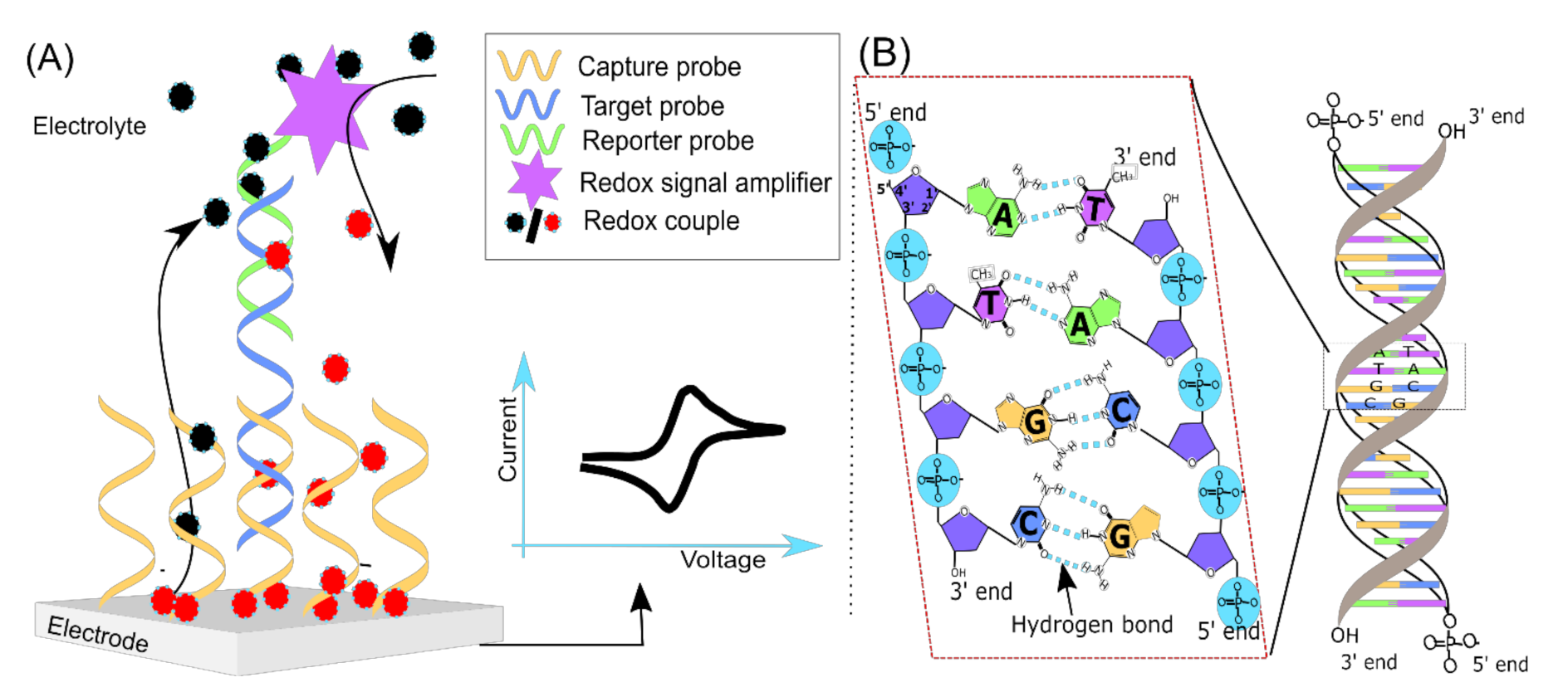
2. Signal Transduction

DNA electrochemical biosensors consist of (i) a DNA recognition element where the target DNA is recognized by a probe/capture DNA(s) strand and (ii) a signal transduction part where the molecular recognition event is translated into an electrical response by an electrochemical reaction [27] (Figure 1A). Two complementary strands of DNA specifically hybridize with each other; thus detection of this hybridization event has become a central theme in biosensor studies [14,26]. The DNA hybridization is based on Watson and Crick base pairing rules (Figure 1B), i.e., specific hydrogen bond formation between two (target DNA and probe DNA) complementary single strands of DNA (ssDNA) [28]. In a sensor detection scheme, ssDNA(s) specifically hybridizes with a target DNA sequence that is being employed as a probe(s): a capture probe used to attach the target DNA to the surface of materials and/or a reporter probe labeled with signaling molecules, e.g., redox-active molecules. The hybridization reaction occurs in solution (homogeneous) or at an electrode/transducer surface (heterogeneous) [12]. The advantage of hybridization in the solution phase is that it is well controlled using known properties such as melting temperature (Tm) and ionic strength of the buffer [14,29]. In the case of hybridization on an electrode surface, the probe DNA is attached to the electrode surface in such a way that the sequence is available to target DNA in the hybridization solution. Additionally, non-specific interactions of DNA with the electrode should be avoided along with other optimization processes such as surface coverage and incubation time [30,31], these will not be discussed herein.
The DNA hybridization product could be selectively and electrochemically quantified on the electrode surface. DNA adsorbed on electrode surfaces are stable below guanine oxidation potential (Table 1), this stability is advantageous for immobilization of DNA directly on the electrode surface. To make a quantitative measurement, the DNA hybridization event is coupled with electrochemical reactions, in a way that a probe-target complex increases/decreases a coupled redox reaction at the electrode surface. It can also be achieved by measuring the changes in the electrode/electrolyte interface properties due to a DNA hybridization event. In general, DNA sensors are categorized into several types based on what kind of probe DNA is being used (label-free/labeled) and how is signal transduction achieved (reagent-free or reagent-dependent). The detailed information on DNA sensor history, principles and fabrication approaches is thoroughly reviewed elsewhere [12,16,26,32,33]. In this review, approaches specific to signal amplification that involves pathogenic DNA/RNA detection is reviewed.

3. Signal Amplification Approaches for the DNA/RNA Electrochemical Sensor

DNA hybridization is a selective process where even a single mismatch between the target and probe can be differentiated in most cases [14,29]. However, often, sensitive detection of the capture-target hybridization event is challenging. Since clinical samples may have very low copies of a pathogen in the early stages of infection, as low as 1–10 colony forming units (CFU/mL) [8]. Electrochemical methods are inherently very sensitive to detect even fM target DNA concentrations [41,42,43,44,45,46,47]. To overcome the limitation of instrumentation requirement for sensor deployment, signal amplification strategies have been investigated and developed to enhance the electrochemical signal (Table 2). Amplified signals are quantified using electrochemical analytical techniques such as chronoamperometry (CA), differential pulse voltammetry (DPV), square wave voltammetry (SWV), cyclic voltammetry (CV) and electrochemical impedance spectroscopy (EIS; Table 2). Signal amplification methods in combination with electrochemical analytical techniques were demonstrated for the detection of femto- and attomolar concentrations of DNA. Herein, we illustrate and discuss several signal amplification methods that were reported for pathogen detection. The methods are categorized into (i) enzyme mediated signal amplification, (ii) nanomaterials-based approaches and (iii) nucleic acid-based approaches.

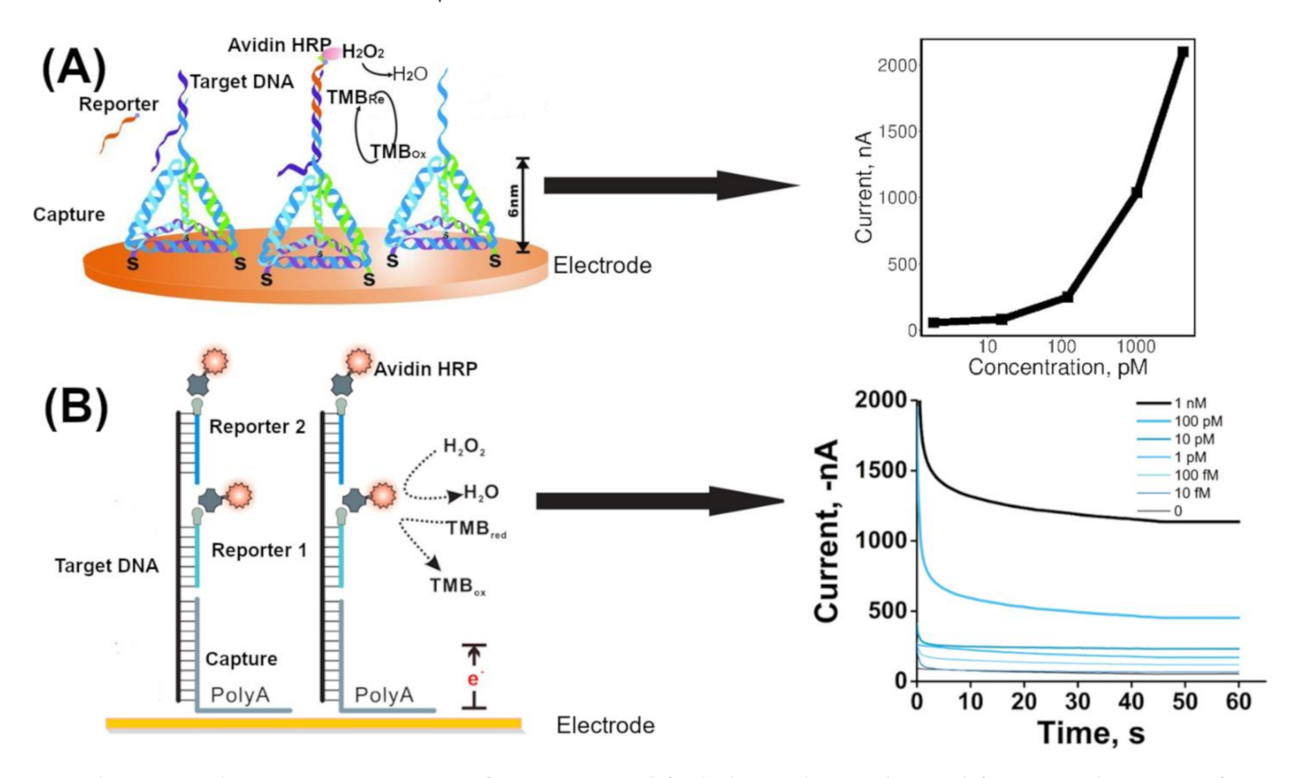
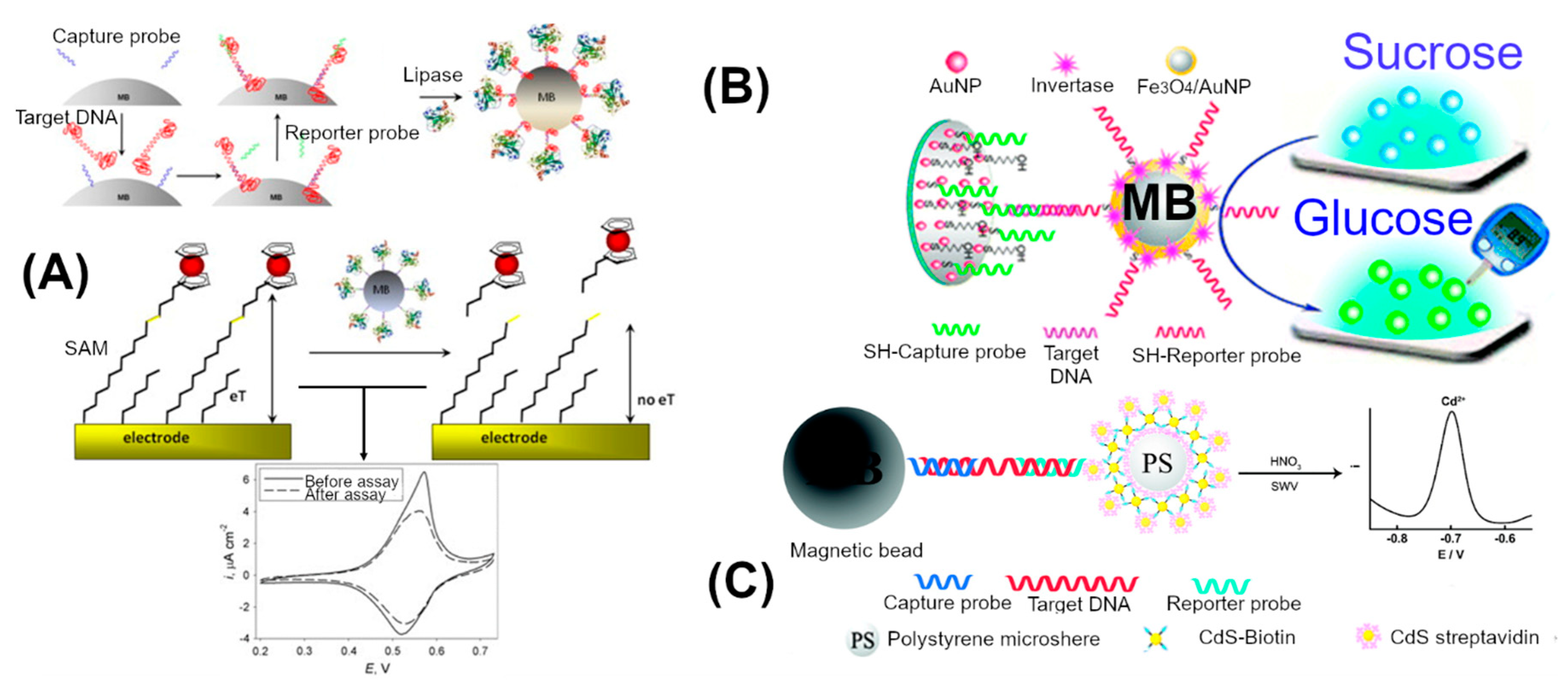
3.1. Enzyme Mediated Signal Amplification

The use of an enzyme for signal amplification can aid in increasing sensitivity of a sensor in that a single recognition event that can be sensed only stoichiometrically could be transduced and recycled several times by the biocatalytic reaction mediated by an enzyme that is coupled to this recognition event. Several enzymes have been strategically conjugated with DNA hybridization complexes to amplify electrochemical signals. When an enzyme is tagged with a probe DNA, each hybridization event is coupled to an enzyme molecule. Each enzyme can produce multiple (10–1000) fold higher redox-active products. This can result in a multifold enhanced redox current at the electrode surface. Horseradish peroxidase (HRP) [70], alkaline phosphatase [64], lipase [54], invertase [11] and glucose oxidase [68] were successfully employed for signal amplification of pathogen detection studies. Different methods have been employed in signal amplification approaches to detect a low copy number of target DNA on the electrode surface. Application of magnetic beads and advancement in functionalization of nano/micro bead structures provides the ability to specifically enrich the target DNA from the background matrix components. Bioconjugates (e.g., biotin-avidin) are utilized as molecular binders with high affinity for building a network of molecular conjugations. Alzate et al. demonstrated a magnetic bead-based approach to quantify the Zika virus [51]. First, a biotinylated capture probe was immobilized on streptavidin-coated magnetic beads. Second, the target was prehybridized with the Digoxigenin (Dig)-labeled reporter probe and then added to the capture probe-coated magnetic beads to hybridize. Third, the reporter probe was recognized by an anti-Dig monoclonal antibody labeled with horseradish peroxidase (HRP). The final bead complex was magnetically attracted to the surface of a screen-printed electrode. H2O2 and 3,3′,5,5′-tetramethylbenzidine (TMB) were added as the HRP substrate. This strategy achieved the detection of 10 pM synthetic target ssDNA. Dong et al. reported the use of a DNA tetrahedral nanostructure-based electrochemical biosensor to detect avian influenza A (H7N9) virus [49]. The tetrahedral nanostructure was used as a biomolecule-confined surface to increase molecular recognition at the biosensing interface (Figure 2A) [71]. First, the DNA tetrahedral structure was immobilized onto a gold electrode surface via an Au-thiol bond. A single strand part of the tetrahedral DNA acted as the capture DNA to hybridize with a target ssDNA. The capture-target sequence was hybridized with a biotinylated reporter DNA sequence. Then streptavidin-horseradish peroxidase was introduced to bind the biotinylated reporter–target DNA hybrids. The reduction current for HRP oxidized TMB substrate was measured using an amperometric method. When this sensor was used for the detection of PCR products (ssDNA) amplified from cDNA isolated from positive patients, the 1.2–1200 pM range was detected. It was also shown that 1–5 cycles of asymmetric PCR generated enough target DNA for the experiment. Wang et al. reported a multiple-reporter probe approach for detection of the 16S rRNA gene of different bacteria [50] (Figure 2B). In this approach, A high-adsorption affinity of the polyA tail towards the Au surface was used to immobilize the molecular recognition complex on an Au electrode [72]. First, the target DNA was hybridized with a multiple biotinylated reporter probe. Second, the prehybridized target-reporter probe was hybridized with the capture probe immobilized at the Au electrode. The capture DNA sequence was designed to have a polyA tail. Then, an Avidin-HRP conjugate was bound to the biotinylated groups of reporter probes in the hybrid complex. The usage of multiple-reporter probes enhanced the number of HRP molecules per hybridization event [73]. The detection range was reported to be 10 fM to 1 nM of synthetic targets. The sensor was also successfully tested for specific detection of denatured genomic DNA from bacterial samples.
Walter et al. presented a simple approach in which signal amplification was achieved by redox cycling of p-aminophenol phosphate (p-AP) using nicotinamide adenine dinucleotide [53]. A molecular recognition complex based on sandwich-type hybridization and reporter probe was tagged with alkaline phosphatase. The electrochemically inert p-AP was converted to an electrochemically active form of p-AP by tagged alkaline phosphatase. Enzymatically generated p-AP was electro-oxidized at an Au electrode to p-quinone imine (p-QI) and in the presence of NADH, p-QI was reduced back to p-AP, which was reoxidized on the electrode. This approach overcame the drawbacks associated with the stability of p-AP. It has allowed reaching a detection limit of 1 pM of target DNA. When it was applied for the monitoring of the 16S rRNA of E. coli pathogenic bacteria it had a detection limit of 250 CFU μL−1.
The signal turn-off mode system was employed for enhanced detection with enzyme-mediated signal amplification. In a signal-off mode, the current signal decreases as a function of DNA concentration. Shipovskov et al. demonstrated lipase chemistry to detect the low amount of target DNA in a signal turn-off mode (Figure 3A) [54]. They established an ester bond containing self-assembled monolayer (SAM) of 9-mercaptononyl, 4-ferrocene aminobutanoate (Fc-alkanethiol ester) on a gold surface, which exhibited high surface redox current using CVs [74]. Lipase was used to cleave the ester bond to remove ferrocene (Fc), a redox-active molecule, from a SAM layer, which resulted in a decrease in current in this system. First, a streptavidin-coated magnetic bead was decorated with biotinylated capture DNA. Then, step by step, it was allowed to react with a target DNA, biotinylated reporter probe and a streptavidin-lipase conjugate. At the end of the hybridization step, the final complex was immobilized on a magnetic bead, which was then applied on an Fc-alkanethiol ester SAM on a gold electrode. The lipase coupled to the DNA recognition complex removed the Fc from the SAM. This resulted in a decrease in the CV peak current. The lowest detected signal peak was 4 fM of the synthetic target DNA. Further, the assay could be used for the detection of down to 16 aM of denatured RNA and their cDNA copies prepared from Lactobacillus brevis. In a similar turn off mode, Luo et al. has employed exonuclease III for the detection of low levels of E. coli in milk samples [64]. First, a capture DNA was immobilized through its 5′-end. Second, the target DNA was hybridized with the capture probe to form a double-stranded structure, which resulted in a blunt end at the 3′-end of the capture probe. Then Exo III, an exonuclease, was introduced to catalytically remove the mononucleotides from the 3′-hydroxyl termini of DNA duplexes. The Exo III activity degraded the capture DNA strand and released the target DNA. The released target was recycled for more capture DNA degradation. After a fixed duration of treatment with the Exo III treatment, the capture DNAs that were not degraded on the sensor surface hybridized with the biotinylated reporter probe. The reporter probe was linked to streptavidin-alkaline phosphatase to produce an enzymatic electrochemical guanine signal for quantitative detection of Enterobacteriaceae bacteria. Using this approach about 40 CFU/mL of E. coli was electrochemically detected where a single strand PCR product was used as a target.
Screen-printed electrodes, which require a small sample volume, are widely employed in sensor development studies. However, the electrochemical response analysis for the screen-printed electrode is still limited to high-end laboratory-based instrumentation. Instead of conventional laboratory-based electrochemical techniques, the commercial glucose meter was also successfully demonstrated for the detection of pathogen’s DNA. Xu et al. demonstrated multiple invertase-mediated signal amplification and the use of a glucometer as an electrochemical device for the detection of HIV DNA [11] (Figure 3B). First, a mixed layer of thiolated capture probes and 6-mercaptoethanol were self-assembled on the AuNPs via thiol–Au attachment. The capture probe coated AuNPs were applied on the glassy carbon electrode. Then target DNA was hybridized with capture probes. The reporter probe was tagged with multiple-invertase coated-Fe3O4/AuNPs using thiol chemistry. Hybridization of invertase coated reporter probes has led to massive quantities of invertase on the electrode surface. Glassy carbon electrode was used to characterize the loading of the probe and target DNA using [Fe(CN)63−/4−] redox couple. To quantify the target DNA, sucrose was introduced onto the glassy carbon electrode surface containing a molecular recognition complex. Upon introducing sucrose, invertase converted the sucrose into glucose molecules, which were measured by a glucometer. Due to several numbers of tagged invertase per hybridization event and its high turnover number, glucose in millimolar concentrations was produced. Using this approach, about 0.5 pM to 1 nM concentration of synthetic HIV DNA was detected using a standard glucose meter sensitivity range.

3.2. Nanomaterial Enhanced Signal

The nanomaterials are used as reporter molecules and high surface area materials for high loading of probe DNA. In case of a nanomaterial as a reporter, metal-based nanoparticles were tagged with DNA for hybridization. Xiang et al. reported CdS quantum dot decorated polystyrene (PS-(CdS)4) as a signal amplifier for the detection of urinary tract pathogen (Figure 3C) [61]. PS-(CdS)4 was built using biotin and streptavidin functionalized PS and CdS nanoparticles. First, the biotin-capture probe was immobilized on streptavidin-magnetic beads and then incubated with the target DNA. The magnetic bead–target complex was then hybridized with a reporter probe, which was immobilized on polystyrene-CdS spheres (PS-(CdS)4). The resulting complex was selectively separated by magnetic separation and was treated with nitric acid to dissolve the CdS nanoparticles. Cd ions in the solution were measured using square wave voltammetry. Using this approach, 0.5 fM to 10 pM of synthetic DNA was detected. In a similar metal nanoparticle-based approach, Zhang et al. demonstrated detection of multiple pathogens using nanoparticle-based biobarcoded electrochemical sensors [75]. Each pathogen-specific probe sequence was tagged to specific nanoparticles. The detection limit of bio-barcoded DNA sensor was 0.5 ng/mL for the insertion element (Iel) gene of Salmonella enteritidis using CdS, and 50 pg/mL for the pagA gene of Bacillus anthracis using PbS. As an alternative to toxic metal-based nanoparticles, Wang et al. reported a strategy for the detection of low levels of E. coli using liposome ‘nanocarriers’ loaded with Ca2+ ions [55]. Upon the successful formation of a recognition event, calcium-loaded liposomes were bound to the reporter DNA after which they were lysed by a surfactant. In this approach, sub-fmol DNA detection limit was achieved by employing Ca2+ ion-sensitive electrodes.
The high surface area of nanoparticles was exploited for loading of a high amount of capture DNA. Chowdhury et al. had detected Dengue virus DNA using nanocomposites of gold nanoparticles (AuNP) with nitrogen and sulfur co-doped graphene quantum dots (N,S-GQDs@AuNP) [56]. First, N,S-GQDs@AuNP were coated with a capture DNA (polydA) using Au-thiol bond formation. This led to the accumulation of a large number of single-stranded (ssDNA) capture DNA. Second, polydA was used to hybridize with a polydT tail of the target viral DNA. Finally, the complex was subjected to electrochemical quantification using methylene blue as a reporter molecule. In the presence of a target, hybridization resulted in a double-stranded DNA (dsDNA), which did not bind methylene blue effectively and resulted in low peak current. In the absence of the target, the dye binds to the capture ssDNA and gave high current in differential pulse voltammetry analysis. This signal-off mode analysis detected a synthetic target in the range of 10 fM to μM. Xu et al. employed AuNPs and exonuclease I for detection of uropathogen’s DNA using [Ru(NH3)6]3+ redox molecules [62]. The AuNPs were coated with multiple ssDNA that was used as the signal probe, this resulted in a greater number of DNA molecules per molecular recognition event. [Ru(NH3)6]3+ bound to the excess DNA molecules electrostatically, which ultimately amplified the redox signal for every target DNA. Furthermore, exonuclease I (Exo I) treatment removed the unhybridized single-stranded capture DNA probes, which minimized the background current. The combination of signal amplification and background current reduction resulted in 1 fM detection limit. Chen et al. reported the use of redox active carbon nanotubes (CNTs) doped with polyaniline (PANI) and endonuclease mediated target recycling approach for detection of Mycobacterium tuberculosis [63]. In the presence of target DNA and an assistant probe that hybridized to the capture probe, a hairpin structure has opened to form a Y-shaped junction. Endonuclease recognized the sequence in the Y-shaped junction and released the assistant probe and target DNA. Released target DNA triggered the next cycle of cleavage. After hybridization between CNTs-PANI tagged reporter probe and the cleaved capture probe on the electrode surface, the electrochemical signal of CNTs-PANI was used as a readout. This strategy detected the target in a range between 1 fM to 10 nM.

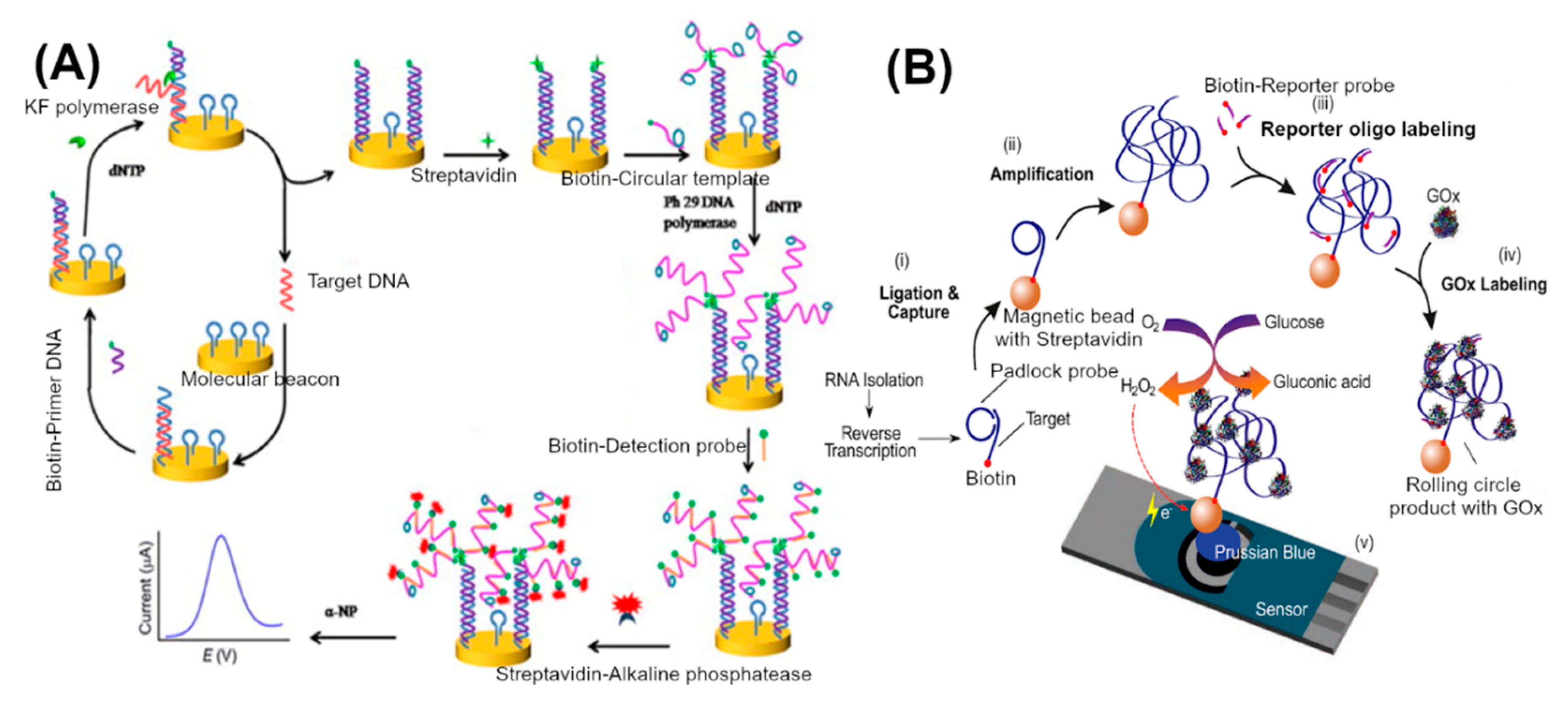
3.3. Nucleic Acid Amplification and Processing Based Approaches

Though enzyme-based and nanomaterial-based signal amplification approaches reach sensitivity in the femtomolar regime, they still depend on PCR to produce sufficient target DNA. In nucleic acid-based approaches, the enzyme-mediated isothermal amplification of nucleic acids plays an important role in sample amplification and detection. Unlike PCR, which requires specialized thermal cycler instruments to mediate denaturation, annealing and subsequent extension steps, isothermal amplification could be carried out at a constant temperature to produce about a million copies of the target DNA. For isothermal amplification, in addition to DNA polymerase, ligase, nicking enzymes and helicases are employed for specific amplification of target DNA molecules. A detailed review of the method can be found in the following references [76,77]. Simple temperature control makes isothermal amplification an attractive alternative to PCR for point-of-care applications. There are several choices of methods that are available for isothermal amplification, depending on the length, secondary structures and nature of the target (RNA or DNA) [76]. The challenging aspects are electrochemical detection of specific targets amplified by the isothermal method. Cheng et al. reported a method combining circular strand displacement polymerization reaction (CSD), rolling circle amplification (RCA) and enzymatic amplification to enhance the electrochemical sensing of a target DNA (Figure 4A) [78]. First, the capture probe (SH-ssDNA with a hairpin loop structure-molecular beacon) was immobilized on a gold electrode. Second, the strand displacement (CSD) reaction was carried out by adding the target DNA, and biotinylated-primer DNA to the electrode. In the presence of the target DNA, the hairpin structure of the molecular beacon opens and parts the sequence that was hybridized with the target DNA. Another part of the capture probe sequence binds specifically with a biotinylated-primer DNA. The primer sequence was extended towards the target DNA binding region by a DNA polymerase (KF exo-), which led to the release of the target DNA. At this stage, freed target DNA binds to another capture probe to trigger another strand displacement reaction, which results in multiple biotin-tagged DNA duplexes on the electrode surface. This biotin-tagged DNA duplex anchored with another streptavidin-primer specific for rolling circle amplification. Upon addition of specific circular ssDNA templates, deoxynucleotide triphosphates and phi29 DNA polymerase, the RCA reaction produces long ssDNA molecules with tandem repeated sequences. Then alkaline phosphatase tagged reporter probe DNA was added and hybridized with repeated sequences of RCA products. Alkaline phosphatase was used as a final redox signal amplifier at the electrode surface. Using this approach, 1 fM to 100 pM of synthetic target DNA was detected. Huang et al. used a similar strand displacement and rolling circle amplification approach—without biotin and streptavidin tags—for the detection of synthetic DNA sequences specific to the hepatitis B virus [65]. The rolling circle amplification resulted in long ssDNA. They detected the final rolling circle amplification product using methylene blue and reported detection in the range of 10 aM to 0.7 fM. Yanyan et al. reported a DNA detection approach for the avian influenza virus based on isothermal exponential amplification coupled with hybridization chain reaction [67]. Catalytic G-quadruplex–hemin, HRP-mimicking DNAzymes, was tagged to the final molecular recognition complex. Electrochemical signals obtained by measuring the increase in the reduction current of oxidized 3,3′,5,5′-tetramethylbenzidine sulfate, which was generated by DNAzyme in the presence of H2O2. This method exhibited detection limits of 9.4 fM. In a similar approach, exonuclease III mediated target DNA recycling and G-quadruplex–hemin reported for the detection of HIV gene sequence with a detection limit of 3.6 pM [79].
Ciftci et al. reported a method for tagging multiple glucose oxidase (GoX) enzymes using rolling circle amplification for the detection of Ebolavirus (Figure 4B) [68]. In the first step (i) biotinylated primers were used to reverse transcribe the RNA target to cDNA. Then the biotinylated cDNA target hybridized with linear pad-lock probes (PLPs). PLPs have a special sequence feature that renders the probe circular upon hybridization. These circular PLPs are ligated using the enzyme ligase; then biotinylated cDNA target-PLP complex captured on streptavidin-functionalized magnetic beads. Magnetic beads were used for the separation of target DNA from the sample. In the second step, RCA reaction was carried out to produce bulky tandem repeats of DNA coils. In the third step, RCA products were hybridized with the biotinylated-reporter probe, which was then bound to streptavidin-GoX. The final DNA recognition complex was quantified by GoX activity using chronoamperometry. The product, H2O2, of glucose oxidation by GoX in the presence of oxygen was electrochemically measured. In this method, 1–100 pM of synthetic target DNA was measured. An Ebolavirus positive clinical sample was also successfully differentiated from negative samples using this method. A similar Pad-lock-probe approach was demonstrated for the detection of Ebolavirus using HRP as a signal amplifier [80].
Helicase dependent amplification (HDA) is another widely employed isothermal system for amplification of target DNA [81]. Helicase is being used to unwind the double-stranded DNA instead of temperature-dependent denaturation during polymerase mediated amplification of the target gene [82]. Barreda-García et al. reported an asymmetric HDA for that resulted in a single-stranded target DNA from Mycobacterium tuberculosis [69]. The amplified ssDNA was selectively quantified using enzyme-mediated signal amplification assay. The system was sensitive to 0.5 aM of target DNA. In another study, HDA was also demonstrated for hybridizing the double-stranded target generated from Salmonella genome to a single-stranded capture DNA bound to indium-tin-oxide electrodes [83].
Yan et al. reported the detection of pathogenic DNA directly by transcription of RNA from the target DNA [66]. In this approach, hairpin structured primers were designed to open and bind to the target DNA specifically. The primer was extended using DNA polymerase (KF exo-) at 37 °C. The primer has efficiently triggered the circular primer extension reaction, i.e., the resultant dsDNA was further amplified by another primer binding and extension cycles. Additionally, the primer was designed to have a T7 RNA polymerase promoter, which served as a template for in vitro transcription of target DNA. The RNA products from the transcription reaction were directly hybridized with immobilized capture probes. The enzyme tagged signal probe was used to detect the hybridized products. With this approach, Salmonella’s invA gene from genomic DNA extract was successfully detected. Limit of detection was about 1 fM.

4. Conclusions and Future Perspectives

In nanomaterials and enzyme-mediated amplification approaches, DNA/RNA isolation and amplification of targets by PCR are commonly required for real sample electrochemical detection to increase the number of specific target DNA molecules. However, PCR can only be carried out using a specialized thermocycler. Thus, to reduce the dependence on PCR, isothermal amplification methods are explored in clinical sample target amplification in combination with an electrochemical sensor. While isothermal amplification provides the advantage of in situ amplification of target DNA at a constant temperature, more exploration of novel approaches for amplification and hybridization on-electrode surfaces is still needed to achieve practical electrochemical DNA sensors.
The requirement for single-stranded target DNA for hybridization is another constraint for electrochemical DNA sensors. To counter this issue, asymmetric PCR, thermal denaturation of target DNA followed by abrupt cooling, helicase mediated asymmetric DNA amplification, DNA to RNA transcription using RNA polymerase have been employed to yield single-stranded targets. However, novel approaches should be explored for the detection of double-stranded genomic DNA or structured single-stranded cDNA/RNA at room temperature.
Electrochemical analytical methods reached a “glass ceiling” with a limit of detection in the femtomolar concentration range. Enzymes, nanomaterials and molecular tools are successfully engineered for fM detection of DNA on an electrode surface using signal amplification approaches. With this, (i) future progress in the simplification of sample processing steps, (ii) developing approaches to detect double-stranded target DNA and (iii) making advancement in the sensitivity of instrumentation or handheld electrochemical devices will be crucial for achieving practical point-of-care electrochemical devices for pathogen detection in low titers.
In addition, for fast epidemic containment, in order to make sure that highly trained personnel will not be needed for point of care detection, it is highly needed to be able to interface such sensors with already existing simple devices such as glucometers, so it will be easy to sample (such as in non-invasive devices) and simple to read-out the signal for less trained users (e.g., airports personnel).

Author Contributions

Conceptualization, M.S. and L.A.; writing—original draft preparation, M.S. and I.A.; writing—review and editing L.A.; supervision, L.A.; project administration, L.A.; funding acquisition, All authors have read and agreed to the published version of the manuscript.

Funding

This research was funded by Israel Ministry of Science and Technology, grant number 88702. M.S. was funded by an Israeli planning and budgeting committee (PBC) post-doctoral fellowship for excellent Indian and Chinese candidates. I.A. gratefully acknowledges a Kreitman School fellowship for Biotechnology research at BGU.

Conflicts of Interest

The authors declare no conflict of interest.

References

  1. Wittwer, C.T.; Makrigiorgos, G.M. Nucleic acid techniques. In Principles and Applications of Molecular Diagnostics; Elsevier: Amsterdam, The Netherlands, 2018; pp. 47–86. ISBN 978-0-12-816061-9. [Google Scholar]
  2. Cheng, V.C.C.; Wong, S.C.; Chen, J.H.K.; Yip, C.C.Y.; Chuang, V.W.M.; Tsang, O.T.Y.; Sridhar, S.; Chan, J.F.W.; Ho, P.L.; Yuen, K.Y. Escalating infection control response to the rapidly evolving epidemiology of the coronavirus disease 2019 (COVID-19) due to SARS-CoV-2 in Hong Kong. Infect. Control Hosp. Epidemiol. 2020, 41, 493–498. [Google Scholar] [CrossRef] [Green Version]
  3. Fritsch, A.; Schweiger, B.; Biere, B. Influenza c virus in pre-school children with respiratory infections: Retrospective analysis of data from the national influenza surveillance system in germany, 2012 to 2014. Eurosurveillance 2019, 24. [Google Scholar] [CrossRef] [Green Version]
  4. Sonawane, A.A.; Shastri, J.; Bavdekar, S.B. Respiratory pathogens in infants diagnosed with acute lower respiratory tract infection in a tertiary care hospital of western India using multiplex Real Time PCR. Indian J. Pediatr. 2019, 86, 433–438. [Google Scholar] [CrossRef] [PubMed]
  5. Deghmane, A.E.; Hong, E.; Taha, M.K. Diagnosis of meningococcal infection using internally controlled multiplex real-time PCR. In Neisseria Meningitidis: Methods and Protocols; Humana Press Inc.: New York, NY, USA, 2019; Volume 1969, pp. 17–31. ISBN 978-1-4939-9202-7. [Google Scholar]
  6. Wlassow, M.; Poiteau, L.; Roudot-Thoraval, F.; Rosa, I.; Soulier, A.; Hézode, C.; Ortonne, V.; Pawlotsky, J.M.; Chevaliez, S. The new Xpert HCV viral load real-time PCR assay accurately quantifies hepatitis C virus RNA in serum and whole-blood specimens. J. Clin. Virol. 2019, 117, 80–84. [Google Scholar] [CrossRef] [PubMed]
  7. Zhang, C.; Zheng, X.; Zhao, C.; Li, Y.; Chen, S.; Liu, G.; Wang, C.; Lv, Q.; Liu, P.; Zheng, Y.; et al. Detection of pathogenic microorganisms from bloodstream infection specimens using TaqMan array card technology. Sci. Rep. 2018, 8. [Google Scholar] [CrossRef] [PubMed] [Green Version]
  8. Loonen, A.J.M.; Bos, M.P.; van Meerbergen, B.; Neerken, S.; Catsburg, A.; Dobbelaer, I.; Penterman, R.; Maertens, G.; van de Wiel, P.; Savelkoul, P.; et al. Comparison of pathogen DNA isolation methods from large volumes of whole blood to improve molecular diagnosis of bloodstream infections. PLoS ONE 2013, 8, e72349. [Google Scholar] [CrossRef] [PubMed] [Green Version]
  9. Teengam, P.; Siangproh, W.; Tuantranont, A.; Vilaivan, T.; Chailapakul, O.; Henry, C.S. Multiplex paper-based colorimetric DNA sensor using pyrrolidinyl peptide nucleic acid-induced AgNPs aggregation for detecting MERS-CoV, MTB, and HPV oligonucleotides. Anal. Chem. 2017, 89, 5428–5435. [Google Scholar] [CrossRef] [PubMed] [Green Version]
  10. Myers, F.B.; Lee, L.P. Innovations in optical microfluidic technologies for point-of-care diagnostics. Lab. Chip 2008, 8, 2015–2031. [Google Scholar] [CrossRef] [PubMed]
  11. Xu, J.; Jiang, B.; Xie, J.; Xiang, Y.; Yuan, R.; Chai, Y. Sensitive point-of-care monitoring of HIV related DNA sequences with a personal glucometer. Chem. Commun. 2012, 48, 10733–10735. [Google Scholar] [CrossRef]
  12. Trotter, M.; Borst, N.; Thewes, R.; von Stetten, F. Review: Electrochemical DNA sensing–Principles, commercial systems, and applications. Biosens. Bioelectron. 2020, 154, 112069. [Google Scholar] [CrossRef]
  13. Liu, G.; Wan, Y.; Gau, V.; Zhang, J.; Wang, L.; Song, S.; Fan, C. An enzyme-based E-DNA sensor for sequence-specific detection of femtomolar DNA targets. J. Am. Chem. Soc. 2008, 130, 6820–6825. [Google Scholar] [CrossRef] [PubMed]
  14. Zhang, D.Y.; Chen, S.X.; Yin, P. Optimizing the specificity of nucleic acid hybridization. Nat. Chem. 2012, 4, 208–214. [Google Scholar] [CrossRef] [PubMed]
  15. Wang, J. Nanomaterial-Based amplified transduction of biomolecular interactions. Small 2005, 1, 1036–1043. [Google Scholar] [CrossRef] [PubMed]
  16. Blair, E.O.; Corrigan, D.K. A review of microfabricated electrochemical biosensors for DNA detection. Biosens. Bioelectron. 2019, 134, 57–67. [Google Scholar] [CrossRef] [Green Version]
  17. Hoilett, O.S.; Walker, J.F.; Balash, B.M.; Jaras, N.J.; Boppana, S.; Linnes, J.C. KickStat: A coin-sized potentiostat for high-resolution electrochemical analysis. Sensors 2020, 20, 2407. [Google Scholar] [CrossRef] [Green Version]
  18. Chapman, J.; Power, A.; Kiran, K.; Chandra, S. Review—New twists in the plot: Recent advances in electrochemical genosensors for disease screening. J. Electrochem. Soc. 2017, 164, B665. [Google Scholar] [CrossRef]
  19. Ozer, T.; Geiss, B.J.; Henry, C.S. Review—Chemical and biological sensors for viral detection. J. Electrochem. Soc. 2019, 167, 037523. [Google Scholar] [CrossRef] [Green Version]
  20. Yáñez-Sedeño, P.; Campuzano, S.; Pingarrón, J.M. Pushing the limits of electrochemistry toward challenging applications in clinical diagnosis, prognosis, and therapeutic action. Chem. Commun. 2019, 55, 2563–2592. [Google Scholar] [CrossRef]
  21. Smith, S.J.; Nemr, C.R.; Kelley, S.O. Chemistry-Driven approaches for ultrasensitive nucleic acid detection. J. Am. Chem. Soc. 2017, 139, 1020–1028. [Google Scholar] [CrossRef]
  22. Bonanni, A.; del Valle, M. Use of nanomaterials for impedimetric DNA sensors: A review. Anal. Chim. Acta 2010, 678, 7–17. [Google Scholar] [CrossRef] [Green Version]
  23. Kupis-Rozmysłowicz, J.; Antonucci, A.; Boghossian, A.A. Review—Engineering the selectivity of the DNA-SWCNT sensor. ECS J. Solid State Sci. Technol. 2016, 5, M3067. [Google Scholar] [CrossRef]
  24. Rasheed, P.A.; Sandhyarani, N. Electrochemical DNA sensors based on the use of gold nanoparticles: A review on recent developments. Microchim. Acta 2017, 184, 981–1000. [Google Scholar] [CrossRef]
  25. Vikrant, K.; Bhardwaj, N.; Bhardwaj, S.K.; Kim, K.-H.; Deep, A. Nanomaterials as efficient platforms for sensing DNA. Biomaterials 2019, 214, 119215. [Google Scholar] [CrossRef] [PubMed]
  26. Pellitero, M.A.; Shaver, A.; Arroyo-Currás, N. Critical review—Approaches for the electrochemical interrogation of DNA-based sensors: A critical review. J. Electrochem. Soc. 2020, 167, 037529. [Google Scholar] [CrossRef]
  27. Drummond, T.G.; Hill, M.G.; Barton, J.K. Electrochemical DNA sensors. Nat. Biotechnol. 2003, 21, 1192–1199. [Google Scholar] [CrossRef] [Green Version]
  28. Leslie, A. Pray Discovery of DNA structure and function: Watson and Crick. Nat. Educ. 2008, 1, 100. [Google Scholar]
  29. Inouye, M.; Ikeda, R.; Takase, M.; Tsuri, T.; Chiba, J. Single-nucleotide polymorphism detection with “wire-like” DNA probes that display quasi “on-off” digital action. Proc. Natl. Acad. Sci. USA 2005, 102, 11606–11610. [Google Scholar] [CrossRef] [Green Version]
  30. Gong, P.; Levicky, R. DNA surface hybridization regimes. Proc. Natl. Acad. Sci. USA 2008, 105, 5301–5306. [Google Scholar] [CrossRef] [Green Version]
  31. Levicky, R.; Horgan, A. Physicochemical perspectives on DNA microarray and biosensor technologies. Trends Biotechnol. 2005, 23, 143–149. [Google Scholar] [CrossRef]
  32. Ferapontova, E.E. DNA Electrochemistry and Electrochemical Sensors for Nucleic Acids. Annu. Rev. Anal. Chem. 2018. [Google Scholar] [CrossRef]
  33. Paleček, E.; Bartošík, M. Electrochemistry of nucleic acids. Chem. Rev. 2012, 112, 3427–3481. [Google Scholar] [CrossRef] [PubMed]
  34. Ferapontova, E.E.; Shipovskov, S.V. Electrochemically induced oxidative damage to double stranded calf thymus DNA adsorbed on gold electrodes. Biochem. Mosc. 2003, 68, 99–104. [Google Scholar] [CrossRef] [PubMed]
  35. Yin, H.; Zhou, Y.; Ma, Q.; Ai, S.; Ju, P.; Zhu, L.; Lu, L. Electrochemical oxidation behavior of guanine and adenine on graphene-Nafion composite film modified glassy carbon electrode and the simultaneous determination. Process Biochem. 2010, 45, 1707–1712. [Google Scholar] [CrossRef]
  36. Zhou, M.; Zhai, Y.; Dong, S. Electrochemical sensing and biosensing platform based on chemically reduced graphene oxide. Anal. Chem. 2009, 81, 5603–5613. [Google Scholar] [CrossRef] [PubMed]
  37. Oliveira, S.C.B.; Oliveira-Brett, A.M. In situ DNA oxidative damage by electrochemically generated hydroxyl free radicals on a boron-doped diamond electrode. Langmuir 2012, 28, 4896–4901. [Google Scholar] [CrossRef] [PubMed]
  38. Özcan, A.; Şahin, Y.; Özsöz, M.; Turan, S. Electrochemical oxidation of ds-DNA on polypyrrole nanofiber modified pencil graphite electrode. Electroanalysis 2007, 19, 2208–2216. [Google Scholar] [CrossRef]
  39. Pogacean, F.; Biris, A.R.; Coros, M.; Watanabe, F.; Biris, A.S.; Clichici, S.; Filip, A.; Pruneanu, S. Electrochemical oxidation of adenine using platinum electrodes modified with carbon nanotubes. Phys. E Low Dimens. Syst. Nanostruct. 2014, 59, 181–185. [Google Scholar] [CrossRef]
  40. Wu, L.; Zhou, J.; Luo, J.; Lin, Z. Oxidation and adsorption of deoxyribonucleic acid at highly ordered pyrolytic graphite electrode. Electrochim. Acta 2000, 45, 2923–2927. [Google Scholar] [CrossRef]
  41. Wang, L.; Veselinovic, M.; Yang, L.; Geiss, B.J.; Dandy, D.S.; Chen, T. A sensitive DNA capacitive biosensor using interdigitated electrodes. Biosens. Bioelectron. 2017, 87, 646–653. [Google Scholar] [CrossRef] [Green Version]
  42. Manzano, M.; Viezzi, S.; Mazerat, S.; Marks, R.S.; Vidic, J. Rapid and label-free electrochemical DNA biosensor for detecting hepatitis A virus. Biosens. Bioelectron. 2018, 100, 89–95. [Google Scholar] [CrossRef]
  43. Subak, H.; Ozkan-Ariksoysal, D. Label-free electrochemical biosensor for the detection of Influenza genes and the solution of guanine-based displaying problem of DNA hybridization. Sens. Actuators B Chem. 2018, 263, 196–207. [Google Scholar] [CrossRef]
  44. Deshmukh, R.; Prusty, A.K.; Roy, U.; Bhand, S. A capacitive DNA sensor for sensitive detection of: Escherichia coli O157:H7 in potable water based on the z3276 genetic marker: Fabrication and analytical performance. Analyst 2020, 145, 2267–2278. [Google Scholar] [CrossRef] [PubMed]
  45. Grieshaber, D.; MacKenzie, R.; Vörös, J.; Reimhult, E. Electrochemical biosensors—Sensor principles and architectures. Sensors 2008, 8, 1400–1458. [Google Scholar] [CrossRef] [PubMed]
  46. Grabowska, I.; Malecka, K.; Stachyra, A.; Góra-Sochacka, A.; Sirko, A.; Zagórski-Ostoja, W.; Radecka, H.; Radecki, J. Single electrode genosensor for simultaneous determination of sequences encoding hemagglutinin and neuraminidase of avian influenza virus type H5N1. Anal. Chem. 2013, 85, 10167–10173. [Google Scholar] [CrossRef]
  47. Malecka, K.; Stachyra, A.; Góra-Sochacka, A.; Sirko, A.; Zagórski-Ostoja, W.; Radecka, H.; Radecki, J. Electrochemical genosensor based on disc and screen printed gold electrodes for detection of specific DNA and RNA sequences derived from Avian Influenza Virus H5N1. Sens. Actuators B Chem. 2016, 224, 290–297. [Google Scholar] [CrossRef] [Green Version]
  48. Ilkhani, H.; Farhad, S. A novel electrochemical DNA biosensor for Ebola virus detection. Anal. Biochem. 2018, 557, 151–155. [Google Scholar] [CrossRef]
  49. Dong, S.; Zhao, R.; Zhu, J.; Lu, X.; Li, Y.; Qiu, S.; Jia, L.; Jiao, X.; Song, S.; Fan, C.; et al. Electrochemical DNA biosensor based on a tetrahedral nanostructure probe for the detection of avian influenza A (H7N9) virus. ACS Appl. Mater. Interfaces 2015, 7, 8834–8842. [Google Scholar] [CrossRef]
  50. Wang, Q.; Wen, Y.; Li, Y.; Liang, W.; Li, W.; Li, Y.; Wu, J.; Zhu, H.; Zhao, K.; Zhang, J.; et al. Ultrasensitive electrochemical biosensor of bacterial 16S rRNA gene based on polyA DNA probes. Anal. Chem. 2019, 91, 9277–9283. [Google Scholar] [CrossRef]
  51. Alzate, D.; Cajigas, S.; Robledo, S.; Muskus, C.; Orozco, J. Genosensors for differential detection of Zika virus. Talanta 2020, 210, 120648. [Google Scholar] [CrossRef]
  52. Azek, F.; Grossiord, C.; Joannes, M.; Limoges, B.; Brossier, P. Hybridization assay at a disposable electrochemical biosensor for the attomole detection of amplified human cytomegalovirus DNA. Anal. Biochem. 2000, 284, 107–113. [Google Scholar] [CrossRef]
  53. Walter, A.; Wu, J.; Flechsig, G.-U.; Haake, D.A.; Wang, J. Redox cycling amplified electrochemical detection of DNA hybridization: Application to pathogen E. coli bacterial RNA. Anal. Chim. Acta 2011, 689, 29–33. [Google Scholar] [CrossRef] [PubMed] [Green Version]
  54. Shipovskov, S.; Saunders, A.M.; Nielsen, J.S.; Hansen, M.H.; Gothelf, K.V.; Ferapontova, E.E. Electrochemical sandwich assay for attomole analysis of DNA and RNA from beer spoilage bacteria Lactobacillus brevis. Biosens. Bioelectron. 2012, 37, 99–106. [Google Scholar] [CrossRef] [PubMed]
  55. Chumbimuni-Torres, K.Y.; Wu, J.; Clawson, C.; Galik, M.; Walter, A.; Flechsig, G.-U.; Bakker, E.; Zhang, L.; Wang, J. Amplified potentiometric transduction of DNA hybridization using ion-loaded liposomes. Analyst 2010, 135, 1618–1623. [Google Scholar] [CrossRef]
  56. Chowdhury, A.D.; Ganganboina, A.B.; Nasrin, F.; Takemura, K.; Doong, R.A.; Utomo, D.I.S.; Lee, J.; Khoris, I.M.; Park, E.Y. Femtomolar detection of Dengue virus DNA with serotype identification ability. Anal. Chem. 2018, 90, 12464–12474. [Google Scholar] [CrossRef]
  57. Khater, M.; de la Escosura-Muñiz, A.; Quesada-González, D.; Merkoçi, A. Electrochemical detection of plant virus using gold nanoparticle-modified electrodes. Anal. Chim. Acta 2019, 1046, 123–131. [Google Scholar] [CrossRef] [PubMed] [Green Version]
  58. Singhal, C.; Dubey, A.; Mathur, A.; Pundir, C.S.; Narang, J. Paper based DNA biosensor for detection of chikungunya virus using gold shells coated magnetic nanocubes. Process Biochem. 2018, 74, 35–42. [Google Scholar] [CrossRef]
  59. Shariati, M.; Ghorbani, M.; Sasanpour, P.; Karimizefreh, A. An ultrasensitive label free human papilloma virus DNA biosensor using gold nanotubes based on nanoporous polycarbonate in electrical alignment. Anal. Chim. Acta 2019, 1048, 31–41. [Google Scholar] [CrossRef]
  60. Lee, J.; Morita, M.; Takemura, K.; Park, E.Y. A multi-functional gold/iron-oxide nanoparticle-CNT hybrid nanomaterial as virus DNA sensing platform. Biosens. Bioelectron. 2018, 102, 425–431. [Google Scholar] [CrossRef]
  61. Xiang, Y.; Zhang, H.; Jiang, B.; Chai, Y.; Yuan, R. Quantum dot layer-by-layer assemblies as signal amplification labels for ultrasensitive electronic detection of uropathogens. Anal. Chem. 2011, 83, 4302–4306. [Google Scholar] [CrossRef]
  62. Xu, J.; Jiang, B.; Su, J.; Xiang, Y.; Yuan, R.; Chai, Y. Background current reduction and biobarcode amplification for label-free, highly sensitive electrochemical detection of pathogenic DNA. Chem. Commun. 2012, 48, 3309–3311. [Google Scholar] [CrossRef]
  63. Chen, Y.; Guo, S.; Zhao, M.; Zhang, P.; Xin, Z.; Tao, J.; Bai, L. Amperometric DNA biosensor for Mycobacterium tuberculosis detection using flower-like carbon nanotubes-polyaniline nanohybrid and enzyme-assisted signal amplification strategy. Biosens. Bioelectron. 2018, 119, 215–220. [Google Scholar] [CrossRef] [PubMed]
  64. Luo, C.; Tang, H.; Cheng, W.; Yan, L.; Zhang, D.; Ju, H.; Ding, S. A sensitive electrochemical DNA biosensor for specific detection of Enterobacteriaceae bacteria by Exonuclease III-assisted signal amplification. Biosens. Bioelectron. 2013, 48, 132–137. [Google Scholar] [CrossRef] [PubMed]
  65. Huang, S.; Feng, M.; Li, J.; Liu, Y.; Xiao, Q. Voltammetric determination of attomolar levels of a sequence derived from the genom of hepatitis B virus by using molecular beacon mediated circular strand displacement and rolling circle amplification. Microchim. Acta 2018, 185, 206. [Google Scholar] [CrossRef]
  66. Yan, Y.; Ding, S.; Zhao, D.; Yuan, R.; Zhang, Y.; Cheng, W. Direct ultrasensitive electrochemical biosensing of pathogenic DNA using homogeneous target-initiated transcription amplification. Sci. Rep. 2016, 6, 18810. [Google Scholar] [CrossRef] [Green Version]
  67. Yu, Y.; Chen, Z.; Jian, W.; Sun, D.; Zhang, B.; Li, X.; Yao, M. Ultrasensitive electrochemical detection of avian influenza A (H7N9) virus DNA based on isothermal exponential amplification coupled with hybridization chain reaction of DNAzyme nanowires. Biosens. Bioelectron. 2015, 64, 566–571. [Google Scholar] [CrossRef] [PubMed]
  68. Ciftci, S.; Cánovas, R.; Neumann, F.; Paulraj, T.; Nilsson, M.; Crespo, G.A.; Madaboosi, N. The sweet detection of rolling circle amplification: Glucose-based electrochemical genosensor for the detection of viral nucleic acid. Biosens. Bioelectron. 2020, 151, 112002. [Google Scholar] [CrossRef]
  69. Barreda-García, S.; González-Álvarez, M.J.; de-los-Santos-Álvarez, N.; Palacios-Gutiérrez, J.J.; Miranda-Ordieres, A.J.; Lobo-Castañón, M.J. Attomolar quantitation of Mycobacterium tuberculosis by asymmetric helicase-dependent isothermal DNA-amplification and electrochemical detection. Biosens. Bioelectron. 2015, 68, 122–128. [Google Scholar] [CrossRef]
  70. Li, L.; Wang, L.; Xu, Q.; Xu, L.; Liang, W.; Li, Y.; Ding, M.; Aldalbahi, A.; Ge, Z.; Wang, L.; et al. Bacterial analysis using an electrochemical DNA biosensor with poly-adenine-mediated DNA self-assembly. ACS Appl. Mater. Interfaces 2018, 10, 6895–6903. [Google Scholar] [CrossRef]
  71. Pei, H.; Lu, N.; Wen, Y.; Song, S.; Liu, Y.; Yan, H.; Fan, C. A DNA nanostructure-based biomolecular probe carrier platform for electrochemical biosensing. Adv. Mater. 2010, 22, 4754–4758. [Google Scholar] [CrossRef] [Green Version]
  72. Pei, H.; Li, F.; Wan, Y.; Wei, M.; Liu, H.; Su, Y.; Chen, N.; Huang, Q.; Fan, C. Designed diblock oligonucleotide for the synthesis of spatially isolated and highly hybridizable functionalization of DNA–gold nanoparticle nanoconjugates. J. Am. Chem. Soc. 2012, 134, 11876–11879. [Google Scholar] [CrossRef]
  73. Xu, L.; Liang, W.; Wen, Y.; Wang, L.; Yang, X.; Ren, S.; Jia, N.; Zuo, X.; Liu, G. An ultrasensitive electrochemical biosensor for the detection of mecA gene in methicillin-resistant Staphylococcus aureus. Biosens. Bioelectron. 2018, 99, 424–430. [Google Scholar] [CrossRef] [PubMed]
  74. Ferapontova, E.E.; Hansen, M.N.; Saunders, A.M.; Shipovskov, S.; Sutherland, D.S.; Gothelf, K.V. Electrochemical DNA sandwich assay with a lipase label for attomole detection of DNA. Chem. Commun. 2010, 46, 1836–1838. [Google Scholar] [CrossRef] [PubMed]
  75. Zhang, D.; Huarng, M.C.; Alocilja, E.C. A multiplex nanoparticle-based bio-barcoded DNA sensor for the simultaneous detection of multiple pathogens. Biosens. Bioelectron. 2010, 26, 1736–1742. [Google Scholar] [CrossRef]
  76. Asiello, P.J.; Baeumner, A.J. Miniaturized isothermal nucleic acid amplification, a review. Lab. Chip 2011, 11, 1420–1430. [Google Scholar] [CrossRef]
  77. Zhao, Y.; Chen, F.; Li, Q.; Wang, L.; Fan, C. Isothermal Amplification of Nucleic Acids. Chem. Rev. 2015, 115, 12491–12545. [Google Scholar] [CrossRef] [PubMed]
  78. Cheng, W.; Zhang, W.; Yan, Y.; Shen, B.; Zhu, D.; Lei, P.; Ding, S. A novel electrochemical biosensor for ultrasensitive and specific detection of DNA based on molecular beacon mediated circular strand displacement and rolling circle amplification. Biosens. Bioelectron. 2014, 62, 274–279. [Google Scholar] [CrossRef]
  79. Huang, Y.L.; Gao, Z.F.; Luo, H.Q.; Li, N.B. Sensitive detection of HIV gene by coupling exonuclease III-assisted target recycling and guanine nanowire amplification. Sens. Actuators B Chem. 2017, 238, 1017–1023. [Google Scholar] [CrossRef]
  80. Carinelli, S.; Kühnemund, M.; Nilsson, M.; Pividori, M.I. Yoctomole electrochemical genosensing of Ebola virus cDNA by rolling circle and circle to circle amplification. Biosens. Bioelectron. 2017, 93, 65–71. [Google Scholar] [CrossRef] [PubMed]
  81. Barreda-García, S.; Miranda-Castro, R.; de-los-Santos-Álvarez, N.; Miranda-Ordieres, A.J.; Lobo-Castañón, M.J. Helicase-dependent isothermal amplification: A novel tool in the development of molecular-based analytical systems for rapid pathogen detection. Anal. Bioanal. Chem. 2018, 410, 679–693. [Google Scholar] [CrossRef]
  82. Barreda-García, S.; Miranda-Castro, R.; de-los-Santos-Álvarez, N.; Lobo-Castañón, M.J. Sequence-specific electrochemical detection of enzymatic amplification products of Salmonella genome on ITO electrodes improves pathogen detection to the single copy level. Sens. Actuators B Chem. 2018, 268, 438–445. [Google Scholar] [CrossRef]
  83. Barreda-García, S.; Miranda-Castro, R.; de-los-Santos-Álvarez, N.; Miranda-Ordieres, A.J.; Lobo-Castañón, M.J. Solid-phase helicase dependent amplification and electrochemical detection of Salmonella on highly stable oligonucleotide-modified ITO electrodes. Chem. Commun. 2017, 53, 9721–9724. [Google Scholar] [CrossRef] [PubMed]
Figure 1. Nucleic acids electrochemical biosensor general principles. (A) A sandwich type genosensor model: A capture probe is employed to capture the target (DNA/RNA) from the solution phase to the electrode surface. The electrode bound target DNA is quantified indirectly by binding the reporter probes conjugated with a redox signal amplifier. The redox signal amplifier could be an enzyme or a nanomaterial, which produces the redox-active molecules. The redox-active molecules undergo an oxidation/reduction reaction, which is then quantified as an electrical response (current–voltage response) using electrochemical analytical methods. The whole strategy depends solely on hybridization efficiency between the nucleic acid probes and the target molecules (RNA/DNA/PNA). In this approach, target DNA does not need any modification. (B) The double-helical structure of DNA and Watson and Crick base pairing in DNA. DNA consists of two strands. The two strands are held together by complementary base pairing between the bases, i.e., hydrogen bonds (A with T and G with C). Two hydrogen bonds attach A to T; three hydrogen bonds attach G to C. High temperature can denature the double-stranded DNA into single-strands. These complementary single-stranded DNAs can specifically rehybridized to form a double-stranded helix by reducing the reaction temperature.
Sensors 20 04648 g001
Figure 2. Schematic presentation of an HRP amplified electrochemical signal for DNA detection yth of enzyme molecules for the electrochemical signal. (A) DNA tetrahedral nanostructure for enhanced signal detection on gold surfaces [71]. (B) PolyA–gold surface interaction for immobilization of capture DNA, which was combined with multiple reporter probes and was attached to multiple HRP enzyme copies for signal amplification [50]. Adapted with permission from cited sources.
Sensors 20 04648 g002
Figure 3. Schematic presentation of an electrochemical signal amplification for DNA detection. (A) DNA sandwich with a lipase labeled reporter probe for detection of Lactobacillus brevis DNA. Lipase was designed to bind with capture and target molecular recognition elements. During electrochemical analysis, lipase cleaves off the ferrocene from 9-mercaptononyl, 4-ferrocene aminobutanoate monolayer over the electrode surface. This results in the reduction of the observed current using cyclic voltammetry [74]. (B) Multiple invertase copies coated magnetic bead was conjugated with each capture and target molecular recognition element. The invertase was used to convert sucrose to glucose. Glucose was detected by a glucose meter. This system was reported for detection of HIV DNA [11]. (C) Similar to invertase, CdS coated polystyrene bead was used as a signal amplifier for the detection of urinary tract pathogens [61]. The Cds nanoparticle bound to the molecular recognition element was dissolved in the acid solution and resulting cadmium ions were quantified electrochemically. Adapted with permission from cited sources.
Sensors 20 04648 g003
Figure 4. Enhancement of nucleic acid detection by employing polymerase and other isothermal amplification approaches on the electrode surface. (A) Strand displacement reaction and rolling circle amplification coupled system [78]. (B) Ligation and rolling circle amplification coupled system [68]. Adapted with permission from cited sources.
Sensors 20 04648 g004
Table 1. Direct oxidation of DNA oxidation of guanine on different electrode supports.
ElectrodeReference ElectrodeElectrolyteGuanine Oxidation Peak (Ep) (V)Reference
GoldAg/AgClPBS, pH 7.4+0.7/+0.8[34]
Nafion/GrapheneSCE0.1 M PBS (pH 4.4)+0.8[35]
Glassy carbon electrodeAg/AgCl0.1 M PBS (pH 7.0)+0.6[36]
Boron doped diamondAg/AgCl0.1 M acetate buffer (pH 4.5)+0.9[37]
Pencil graphiteAg/AgCl0.5 M acetate buffer and 20 mM LiCIO4+0.76[38]
DWNTs, and MWNTsAg/AgClPBS (pH 6)+1[39]
HOPGEAg/AgCl0.1 M sodium acetate buffer (pH 7.6)0.9[40]
SCE—Saturated calomel electrode, PBS—Phosphate buffered saline, DWNTs—double-walled carbon nanotubes, MWNTs—multi-walled carbon nanotubes, HOPGE—Highly ordered pyrolytic graphite electrode.
Table 2. Various signal amplification strategies employed for the detection of pathogenic DNA using electrochemical analytical methods.
PathogenTargetCapture ProbeReporter ProbeElectrode ModificationAmplification StrategyRedox SignalLimit of Detection (LOD) *Analytical TechniqueReferences
EbolavirusBiotin-ssDNAHS-ssDNA NAAuStrep-alkaline phosphatase4-aminophenol4.7 nMDPV[48]
Avian influenza A (H7N9) virusssDNASH-tetrahedral DNABiotin-ssDNAAuStrep-HRPTMP0.75 pMAmperometric[49]
Bacteria 16s RNA genessDNA and genomic DNAssDNA (polydA SAM)Biotin-ssDNAAuStrep-HRPTMB10 fMAmperometric[50]
Zika virusssDNABiotin-ssDNA (Strept-magentic beads)Digoxigenin -ssDNAAuAnti- Digoxigenin coupled HRPTMB0.7 pMChronoamperometry[51]
HIV DNAssDNASH-ssDNASH-ssDNAGlucose meterInvertase-Fe3O4-AuGlucose0.5 pMAmperometry[11]
Human cytomegalovirusssDNA(PCR product)NABiotin-ssDNACarbonStrep-HRPOphenyldimine/2,2′-diaminoazobenene3.6 × 105 copies/mLDPV[52]
E. coligDNASH-ssDNABiotin-ssDNAAuStrep-HRP and redox cyclingp-aminophenyl phosphate0.5416667Chronoamperometric[53]
Lactobacillus brevisgDNA, RNABiotin-ssDNABiotin-ssDNAAuStrep-LipaseFerrocene16 amoleCV[54]
E. colissDNAHS-ssDNA Biotin-ssDNAAuLiposome loaded with Ca2+Ca2+ ion-selective electrode (No redox reaction)0.2 nMPotentiometric method using[55]
Dengue virusPCR amplified target with poly (dT)HS-ssPoly(dA)Fluro-ssDNAAu-Polyaniline/N,S-GQDs@AuNP-dANanomaterial as carrierMethylene blue-intercalation9.4 fMDPV[56]
Citrus tristeza virusssDNAHS-ssDNANAAu/AuNPsNanoparticle as carrier[Fe(CN)63−/4−]100 nMImpedance[57]
Chikungunya virusssDNAssDNANACarbon/Fe3O4@Au (+ and − charge interaction to accumulate the DNA)Nanoparticle as carrierMethylene blue0.1 nMDPV and CV[58]
Human papilloma virusssDNAHS-ssDNANANanoporous polycarbonate-AuNTs Nanoparticles as carrier[Fe(CN)63−/4−]1 fMImpedance[59]
Influenza and NorovirusssDNASH-ssDNANAPt-Au/Iron Oxide-CNTNanoparticles as carrierNA8.8 pMConductivity (the resistance change)[60]
E. coli uropathogensssDNABiotin-ssDNABiotin-ssDNAGlassy carbonCdS quantum dots as reporterCd2+0.22 fMSWV[61]
E. colissDNASH-ssDNAssDNAAuNP-deposited on glassy carbon electrodeNanoparticle as high amount reporter probe carrier[Ru(NH3)6]3+1 fMDPV[62]
Mycobacterium tuberculosisPCR productSH-ssDNASH-ssDNA loaded AuNPs@ CNT-PANIAuEndonucleasePolyaniline0.33 fMDPV[63]
EnterobacteriaceaessDNA and HAV cDNAHS-ssDNAbiotin-ssDNAAuExonuclease III and Strep-alkaline phosphataseα-naphthyl phosphate8.7 fMDPV[64]
Hepatitis B virus (HBV)ssDNAHS-ssDNAssDNA as primerAuCSD and RCAMethylene blue2.6 aMDPV[65]
SalmonellagDNASH-ssDNABiotin-ssDNAAuDNA polymerase, T4 RNA polymerase and Strep-alkaline phosphataseα-naphthyl phosphate0.97 fMDPV[66]
Avian influenza A (H7N9) virusssDNASH-ssDNAMolecular beaconsAuEXPAR-HCR and G-quadruplex–hemin-(HRP like catalysis)TMB9.4 fMDPV[67]
EbolavirusRNAcDNA synthesized from target RNABiotin-ssDNACarbonRCA and Strep-glucose oxidaseH2O21 pMChronoamperometry[68]
Mycobacterium tuberculosisgDNABiotin-PCR product from targetFluorescein-ssDNACarbonHDA and Antifluorescein-POD Fab TMP0.5 aMChronoamperometry[69]
DPV—Differential pulse voltammetry, CV—Cyclic Voltammetry, EIS—Electrochemical Impedance spectroscopy, CSD—Circular strand displacement, RCA—Rolling circle amplification, EXPAR—Isothermal exponential amplification, HCR—Hybridization chain reaction, HDA—Helicase dependent amplification, TMB—3,3′,5,5′-tetramethylbenzidine, N,S-GQDs@AuNP—Nitrogen, sulfur codoped graphene quantum, CNT-PANI—Carbon nanotube-polyanilline, NA—Not applicable, * If limit of detection is not reported, lowest detected value is provided. ssDNA—Single stranded DNA.

