Gamma Radiation Imaging System via Variable and Time-Multiplexed Pinhole Arrays


Abstract
1. Introduction
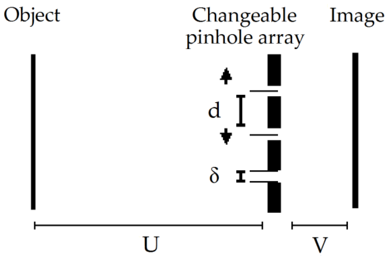
2. The Imaging Concept
3. Results
3.1. Simulation Results
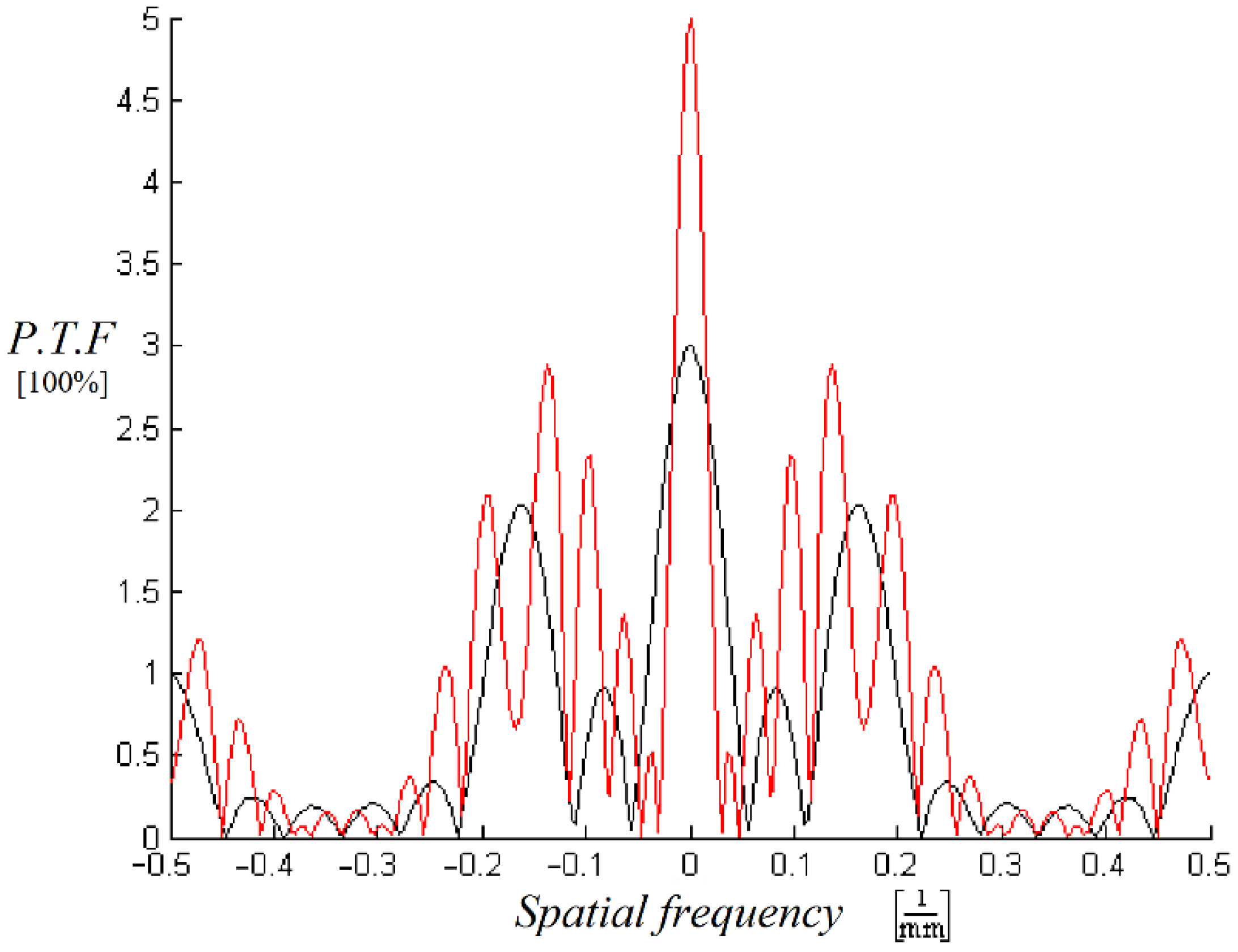
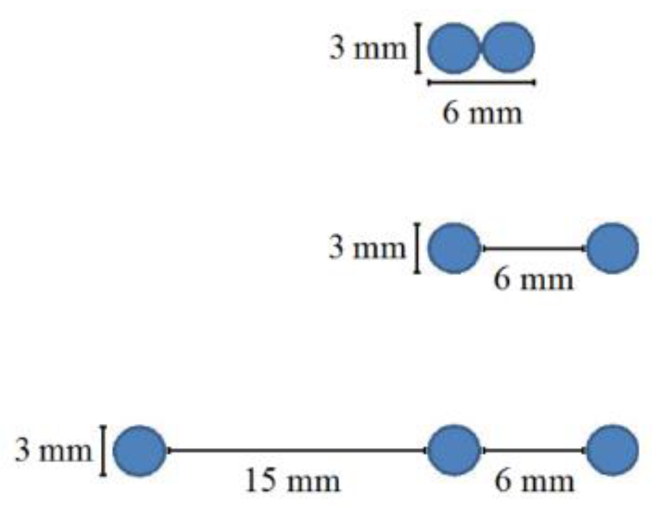
3.1.1. The Multipinhole Array Designs MATLAB Simulation
3.1.2. Geant4 Simulation
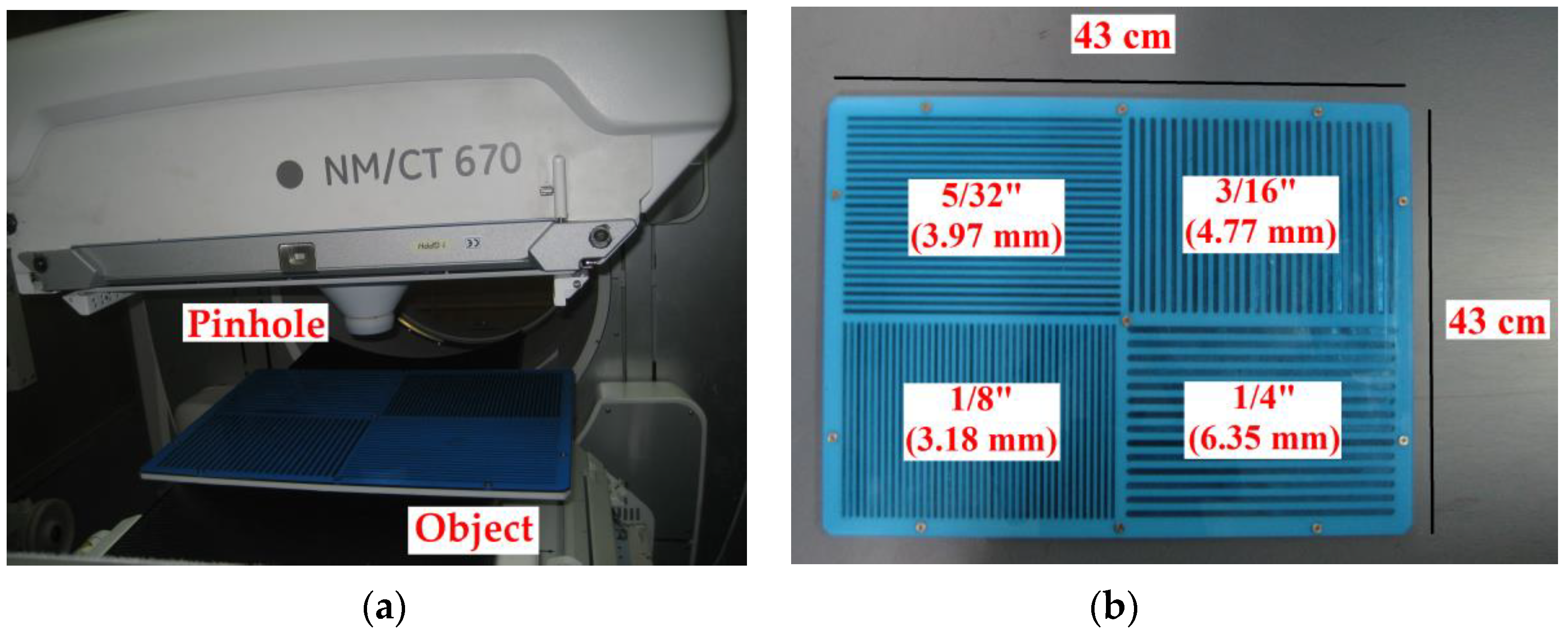
3.2. Experimental Results
4. Conclusions
Author Contributions
Funding
Acknowledgments
Conflicts of Interest
References
- Jaszczak, R.J.; Li, J.; Wang, H.; Zalutsky, M.R.; Coleman, R.E. Pinhole collimation for ultra-high-resolution, small-field-of-view SPECT. Phys. Med. Biol. 1994, 39, 425–437. [Google Scholar] [CrossRef]
- Hudson, H.; Larkin, R. Accelerated image reconstruction using ordered subsets of projection data. IEEE Trans. Med Imaging 1994, 13, 601–609. [Google Scholar] [CrossRef]
- Vogel, R.A.; Kirch, D.; Lefree, M.; Steele, P. A New Method of Multiplanar Emission Tomography Using a Seven Pinhole Collimator and an Auger Scintillation Camera. J. Nucl. Med. 1978, 19, 648–654. [Google Scholar]
- LeFree, M.T.; Vogel, R.A.; Kirch, D.L.; Steele, P.P. Seven-pinhole tomography—A technical description. J. Nucl. Med. 1981, 22, 48–54. [Google Scholar]
- Ivanovic, M.; Weber, D.; Loncaric, S. Multi-pinhole collimator optimization for high resolution SPECT imaging. IEEE Nuclear Sci. Symp. Conf. Record. 2002, 2, 1097–1101. [Google Scholar]
- Rowe, R.K.; Aarsvold, J.N.; Barrett, H.H.; Chen, J.C.; Klein, W.P.; Moore, B.A.; Pang, I.W.; Patton, D.D.; White, T.A. A stationary hemispherical SPECT imager for three-dimensional brain imaging. J. Nucl. Med. 1993, 34, 474–480. [Google Scholar] [PubMed]
- Klein, W.; Barrett, H.; Pang, I.; Patton, D.; Rogulski, M.; Sain, J.; Smith, W. FASTSPECT: Electrical and mechanical design of a high-resolution dynamic SPECT imager. IEEE Nucl. Sci. Symp. Med. Imag. Conf. Record. 2002, 2, 931–933. [Google Scholar]
- Furenlid, L.R.; Wilson, D.W.; Chen, Y.-C.; Kim, H.; Pietraski, P.J.; Crawford, M.J.; Barrett, H.H. FastSPECT II: A Second-Generation High-Resolution Dynamic SPECT Imager. IEEE Trans. Nucl. Sci. 2004, 51, 631–635. [Google Scholar] [CrossRef]
- Beekman, F.J.; Vastenhouw, B. Design and simulation of U-SPECT, an ultra-high resolution molecular imaging system. IEEE Nucl. Sci. Symp. Med. Imag. Conf. Record. 2004, 2, 792–796. [Google Scholar]
- Meikle, S.R.; Kench, P.L.; Weisenberger, A.; Wojcik, R.; Smith, M.; Majewski, S.; Eberl, S.; Fulton, R.R.; Rosenfeld, A.; Fulham, M. A prototype coded aperture detector for small animal SPECT. IEEE Trans. Nucl. Sci. 2002, 49, 2167–2171. [Google Scholar] [CrossRef][Green Version]
- Meikle, S.R.; Wojcik, R.; Weisenberger, A.G.; Smith, M.F.; Majewski, S.; Kench, P.; Eberl, S.; Fulton, R.R.; Lerch, M.; Rosenfeld, A.B. CoALA-SPECT: A coded aperture laboratory animal SPECT system for pre clinical imaging. IEEE Nucl. Sci. Symp. Med. Imag. Conf. Record. 2004, 2, 1061–1065. [Google Scholar]
- Wilson, D.W.; Barrett, H.H.; Furenlid, L.R. A new design for a SPECT small-animal imager. IEEE Nucl. Sci. Symp. Conf. Rec. 2002, 3, 1826–1829. [Google Scholar]
- Wilson, D.; Barrett, H.; Clarkson, E. Reconstruction of two- and three-dimensional images from synthetic-collimator data. IEEE Trans. Med. Imag. 2000, 19, 412–422. [Google Scholar] [CrossRef] [PubMed]
- Beekman, F.; Van Der Have, F.; Vastenhouw, B.; Van Der Linden, A.J.A.; Van Rijk, P.P.; Burbach, J.; Smidt, M. U-SPECT-I: A novel system for submillimeter-resolution tomography with radiolabeled molecules in mice. J. Nucl. Med. 2005, 46, 1194–1200. [Google Scholar] [PubMed]
- Van Der Have, F.; Vastenhouw, B.; Ramakers, R.M.; Branderhorst, W.; Krah, J.O.; Ji, C.; Staelens, S.; Beekman, F.J. U-SPECT-II: An Ultra-High-Resolution Device for Molecular Small-Animal Imaging. J. Nucl. Med. 2009, 50, 599–605. [Google Scholar] [CrossRef]
- McElroy, D.; Macdonald, L.; Beekman, F.; Wang, Y.; Patt, B.; Iwanczyk, J.; Tsui, B.; Hoffman, E. Performance evaluation of A-SPECT: A high resolution desktop pinhole SPECT system for imaging small animals. IEEE Trans. Nucl. Sci. 2002, 49, 2139–2147. [Google Scholar] [CrossRef]
- Lackas, C.; Schramm, N.; Hoppin, J.; Engeland, U.; Wirrwar, A.; Halling, H. T-SPECT: A novel imaging technique for small animal research. IEEE Trans. Nucl. Sci. 2005, 52, 181–187. [Google Scholar] [CrossRef]
- Ishizu, K.; Mukai, T.; Yonekura, Y.; Pagani, M.; Fujita, T.; Magata, Y.; Nishizawa, S.; Tamaki, N.; Shibasaki, H.; Konishi, J. Ultra-high resolution SPECT system using four pinhole collimators for small animal studies. J. Nucl. Med. 1995, 36, 2282–2287. [Google Scholar]
- Wu, M.C.; Hasegawa, B.H.; Dae, M.W. Performance evaluation of a pinhole SPECT system for myocardial perfusion imaging of mice. Med. Phys. 2002, 29, 2830–2839. [Google Scholar] [CrossRef]
- Schramm, N.; Ebel, G.; Engeland, U.; Schurrat, T.; Behe, M.; Behr, T. High-resolution SPECT using multipinhole collimation. IEEE Trans. Nucl. Sci. 2003, 50, 315–320. [Google Scholar] [CrossRef]
- Goertzen, A.L.; Jones, D.W.; Seidel, J.; Li, K.; Green, M.V. First results from the high-resolution mouseSPECT annular scintillation camera. IEEE Trans. Med. Imag. 2005, 24, 863–867. [Google Scholar] [CrossRef] [PubMed]
- Funk, T.; Després, P.; Barber, W.C.; Shah, K.S.; Hasegawa, B.H. A multipinhole small animal SPECT system with submillimeter spatial resolution. Med. Phys. 2006, 33, 1259–1268. [Google Scholar] [CrossRef] [PubMed]
- DiFilippo, F.P. Design and performance of a multi-pinhole collimation device for small animal imaging with clinical SPECT and SPECT–CT scanners. Phys. Med. Biol. 2008, 53, 4185–4201. [Google Scholar] [CrossRef] [PubMed]
- Miller, B.; Furenlid, L.R.; Moore, S.K.; Barber, H.B.; Nagarkar, V.V.; Barrett, H.H. System integration of FastSPECT III, a dedicated SPECT rodent-brain imager based on BazookaSPECT detector technology. In Proceedings of the 2009 IEEE Nuclear Science Symposium Conference Record (NSS/MIC), Orlando, FL, USA, 24 October–1 November 2009; pp. 4004–4008. [Google Scholar] [CrossRef]
- Peterson, T.; Shokouhi, S.; Furenlid, L.R.; Wilson, D.W. Multi-pinhole SPECT Imaging with Silicon Strip Detectors. IEEE Trans. Nucl. Sci. 2009, 56, 646–652. [Google Scholar] [CrossRef][Green Version]
- Shokouhi, S.; Metzler, S.D.; Wilson, D.W.; Peterson, T. Multi-pinhole collimator design for small-object imaging with SiliSPECT: A high-resolution SPECT. Phys. Med. Biol. 2008, 54, 207–225. [Google Scholar] [CrossRef]
- Shokouhi, S.; Wilson, D.W.; Metzler, S.D.; Peterson, T. Evaluation of image reconstruction for mouse brain imaging with synthetic collimation from highly multiplexed SiliSPECT projections. Phys. Med. Biol. 2010, 55, 5151–5168. [Google Scholar] [CrossRef][Green Version]
- Vanhove, C.; Defrise, M.; Lahoutte, T.; Bossuyt, A. Three-pinhole collimator to improve axial spatial resolution and sensitivity in pinhole SPECT. Eur. J. Nucl. Med. Mol. Imag. 2007, 35, 407–415. [Google Scholar] [CrossRef]
- Branderhorst, W.J. Focused Multi-pinhole SPECT. Ph.D. Thesis, Utrecht University, Utrecht, The Netherlands, 2011. [Google Scholar]
- Mertz, L.; Young, N.O. Fresnel transformations of images. In Proc. Int. Conf. Opt. Instrum. Techniques; Habell, K.J., Ed.; Chapman & Hall: London, UK, 1961; pp. 305–310. [Google Scholar]
- Barrett, H.; Wilson, D.; Demeester, G. The use of half-tone screens in Fresnel zone plate imaging of incoherent sources. Opt. Commun. 1972, 5, 398–401. [Google Scholar] [CrossRef]
- Barrett, H.H.; Horrigan, F.A. Fresnel zone plate imaging of gamma rays; theory. Appl. Opt. 1973, 12, 2686–2702. [Google Scholar] [CrossRef]
- Dicke, R.H. Scatter-Hole Cameras for X-Rays and Gamma Rays. Astrophys. J. 1968, 153, 101. [Google Scholar] [CrossRef]
- Skinner, G. Imaging with coded-aperture masks. Nucl. Instruments Methods Phys. Res. 1984, 221, 33–40. [Google Scholar] [CrossRef]
- Stroke, G.W.; Hayat, G.S.; Hoover, R.B.; Underwood, J.H. X-ray imaging with multiple-pinhole cameras using a posteriori holographic image synthesis. Opt. Commun. 1969, 1, 138–140. [Google Scholar] [CrossRef]
- Chou, C.; Barrett, H.H. Gamma-ray imaging in Fourier space. Opt. Lett. 1978, 3, 187. [Google Scholar] [CrossRef] [PubMed]
- Rosenfeld, D.; Macovski, A. Time Modulated Apertures for Tomography in Nuclear Medicine. IEEE Trans. Nucl. Sci. 1977, 24, 570–576. [Google Scholar] [CrossRef]
- Ohyama, N.; Honda, T.; Tsujiuchi, J. Tomogram reconstruction using advanced coded aperture imaging. Opt. Commun. 1981, 36, 434–438. [Google Scholar] [CrossRef]
- Ohyama, N.; Honda, T.; Tsujiuchi, J.; Matumoto, T.; Linuma, T.A.; Ishimatsu, K. Advanced coded-aperture imaging system for nuclear medicine. Appl. Opt. 1983, 22, 3555. [Google Scholar] [CrossRef] [PubMed]
- Chi, W.; George, N. Phase-coded aperture for optical imaging. Opt. Commun. 2009, 282, 2110–2117. [Google Scholar] [CrossRef]
- Chang, L.T.; Macdonald, B.; Perez-Mendez, V.; Shiraishi, L. Coded Aperture Imaging of Gamma-Rays Using Multiple Pinhole Arrays and Multiwire Proportional Chamber Detector. IEEE Trans. Nucl. Sci. 1975, 22, 374–378. [Google Scholar] [CrossRef][Green Version]
- Cherry, S.R.; Sorenson, J.A.; Phelps, M.E. Physics in Nuclear Medicine, 4th ed.; Elsevier: Philadelphia, PA, USA, 2012. [Google Scholar]
- Fenimore, E.E. Coded aperture imaging: Predicted performance of uniformly redundant arrays. Appl. Opt. 1978, 17, 3562–3570. [Google Scholar] [CrossRef]
- Accorsi, R.; Lanza, R.C. Near-field artifact reduction in planar coded aperture imaging. Appl. Opt. 2001, 40, 4697–4705. [Google Scholar] [CrossRef]
- Accorsi, R.; Gasparini, F.; Lanza, R.C. Optimal coded aperture patterns for improved SNR in nuclear medicine imaging. Nucl. Instruments Methods Phys. Res. Sect. A Accel. Spectrom. Detect. Assoc. Equip. 2001, 474, 273–284. [Google Scholar] [CrossRef]
- Accorsi, R.; Gasparini, F.; Lanza, R. A coded aperture for high-resolution nuclear medicine planar imaging with a conventional Anger camera: Experimental results. IEEE Trans. Nucl. Sci. 2001, 48, 2411–2417. [Google Scholar] [CrossRef]
- Schwarz, A.; Shemer, A.; Zalevsky, Z. Light intensity and SNR improvement for high-resolution optical imaging via time multiplexed pinhole arrays. Appl. Opt. 2014, 53, 4483–4492. [Google Scholar] [CrossRef]
- Schwarz, A.; Wang, J.; Shemer, A.; Zalevsky, Z.; Javidi, B. Lensless three-dimensional integral imaging using variable and time multiplexed pinhole array. Opt. Lett. 2015, 40, 1814–1817. [Google Scholar] [CrossRef] [PubMed]
- Tarantola, A. Inverse Problem Theory, 1st ed.; Elsevier: Philadelphia, PA, USA, 1987. [Google Scholar]
- Thiebaut, E. Introduction toimage reconstruction and inverse problems. In Optics in Astrophysics; Foy, R., Foy, F.C., Eds.; Springer: Dordrecht, The Netherlands, 2006; Volume 198, pp. 397–422. [Google Scholar]
- Daube-Witherspoon, M.E.; Muehllehner, G. An iterative image space reconstruction algorithm suitable for volume etc. IEEE Trans. Med. Imag. 1986, 5, 61–66. [Google Scholar] [CrossRef] [PubMed]
- Lanteri, H.; Soummer, R.; Aimé, C. Comparison between ISRA and RLA algorithms. Use of a Wiener Filter based stopping criterion. Astron. Astrophys. Suppl. Ser. 1999, 140, 235–246. [Google Scholar] [CrossRef][Green Version]
- Agostinelli, S.; Allison, J.; Amako, K.; Apostolakis, J.; Araujo, H.; Arce, P.; Asai, M.; Axen, D.; Banerjee, S.; Barrand, G.; et al. Geant4—A simulation toolkit. Nucl. Instrum. Methods Phys. Res. Sect. A Accel. Spectrom. Detect. Assoc. Equip. 2003, 506, 250–303. [Google Scholar] [CrossRef]
- Allison, J.; Amako, K.; Apostolakis, J.; Araujo, H.; Dubois, P.A.; Asai, M.; Barrand, G.; Capra, R.; Chauvie, S.; Chytracek, R.; et al. Geant4 developments and applications. IEEE Trans. Nucl. Sci. 2006, 53, 270–278. [Google Scholar] [CrossRef]
- Allison, J.; Amako, K.; Apostolakis, J.; Arce, P.; Asai, M.; Aso, T.; Bagli, E.; Bagulya, A.; Banerjee, S.; Barrand, G.; et al. Recent developments in Geant4. Nucl. Instrum. Methods Phys. Res. Sect. A Accel. Spectrom. Detect. Assoc. Equip. 2016, 835, 186–225. [Google Scholar] [CrossRef]


| One Pinhole System [Avg. Counts] Accumulation Time: 180 s | Multipinhole System [Avg. Counts] Accumulation Time: 180 s | |||||
|---|---|---|---|---|---|---|
| Object to Background Ratio 1:1.2 | Signal | Noise (Std) | SNR | Signal | Noise (Std) | SNR |
| Object | 13.56837806 | 3.88248836 | 3.49476336 | 31.57960458 | 5.62920882 | 5.60995436 |
| Background | 11.16794390 | 3.34176248 | 3.34193228 | 26.12460000 | 5.16068550 | 5.06223447 |
| One Pinhole System [Avg. Counts] Accumulation Time: 42 s | Multipinhole System [Avg. Counts] Accumulation Time: 18 s | |||
|---|---|---|---|---|
| Signal | Noise (Std) | Signal | Noise (Std) | |
| Object | 4.061305 | 0.313631 | 4.242393 | 0.222412 |
| Background | 4.475837 | 0.2993 | 4.564631 | 0.26209 |
| One Pinhole System | Multipinhole Array System | ||
|---|---|---|---|
| Accumulation time (s) | 18 | 42 | 18 |
| Relative CNR | 1 | 1.43 | 1.38 |
© 2020 by the authors. Licensee MDPI, Basel, Switzerland. This article is an open access article distributed under the terms and conditions of the Creative Commons Attribution (CC BY) license (http://creativecommons.org/licenses/by/4.0/).
Share and Cite
Schwarz, A.; Shemer, A.; Danan, Y.; Bar-Shalom, R.; Avraham, H.; Zlotnik, A.; Zalevsky, Z. Gamma Radiation Imaging System via Variable and Time-Multiplexed Pinhole Arrays. Sensors 2020, 20, 3013. https://doi.org/10.3390/s20113013
Schwarz A, Shemer A, Danan Y, Bar-Shalom R, Avraham H, Zlotnik A, Zalevsky Z. Gamma Radiation Imaging System via Variable and Time-Multiplexed Pinhole Arrays. Sensors. 2020; 20(11):3013. https://doi.org/10.3390/s20113013
Chicago/Turabian StyleSchwarz, Ariel, Amir Shemer, Yossef Danan, Rachel Bar-Shalom, Hemy Avraham, Alex Zlotnik, and Zeev Zalevsky. 2020. "Gamma Radiation Imaging System via Variable and Time-Multiplexed Pinhole Arrays" Sensors 20, no. 11: 3013. https://doi.org/10.3390/s20113013
APA StyleSchwarz, A., Shemer, A., Danan, Y., Bar-Shalom, R., Avraham, H., Zlotnik, A., & Zalevsky, Z. (2020). Gamma Radiation Imaging System via Variable and Time-Multiplexed Pinhole Arrays. Sensors, 20(11), 3013. https://doi.org/10.3390/s20113013