Detection of Cu2+ Ions with GGH Peptide Realized with Si-Nanoribbon ISFET

 , , , , and
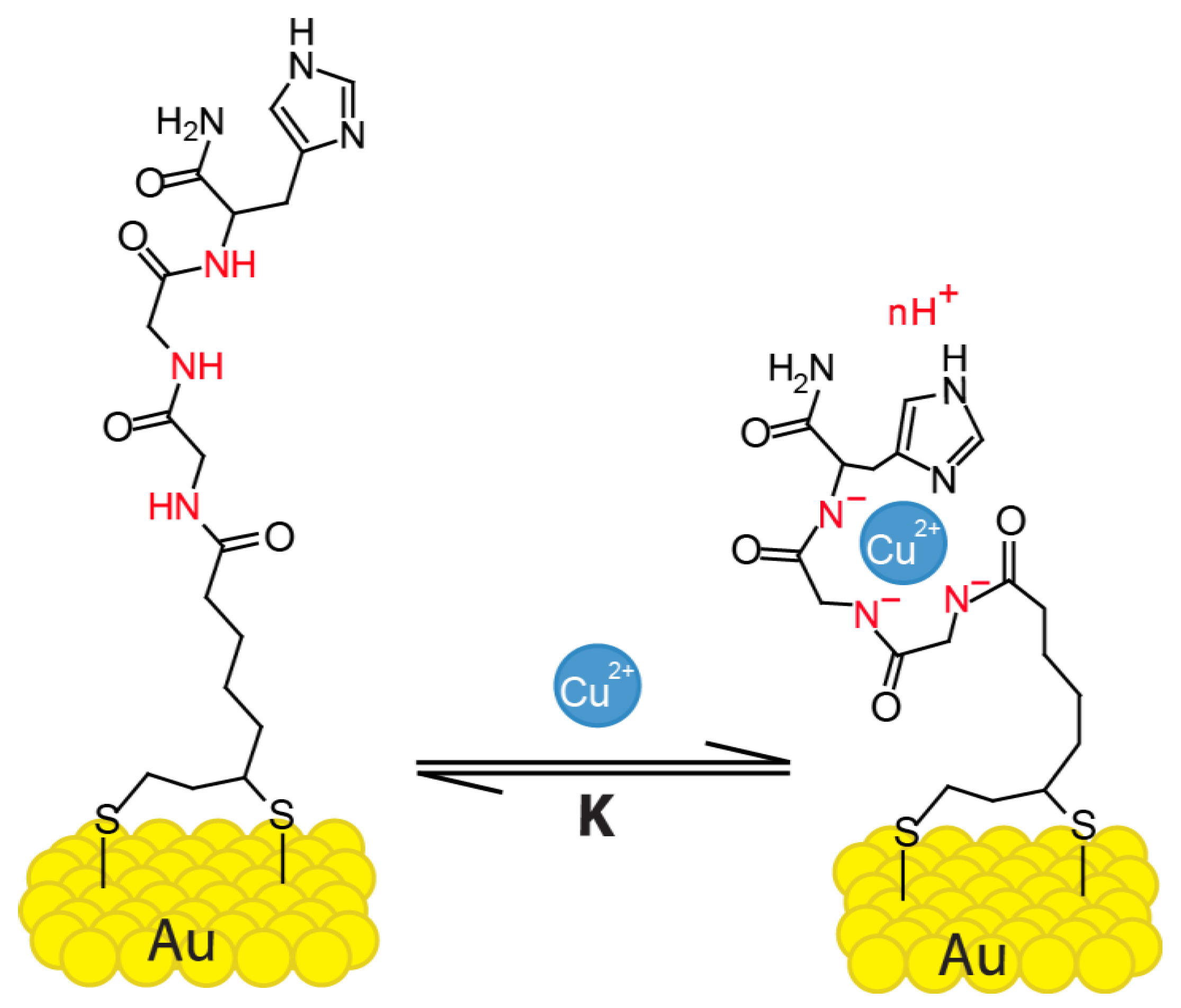
Abstract
1. Introduction
2. Materials and Methods
2.1. Device Fabrication
2.2. Microfluidic Channels
2.3. Surface Functionalization
2.4. Peptide, Electrolyte, and Cu2+ Ion Solutions
2.5. Measurement Procedure
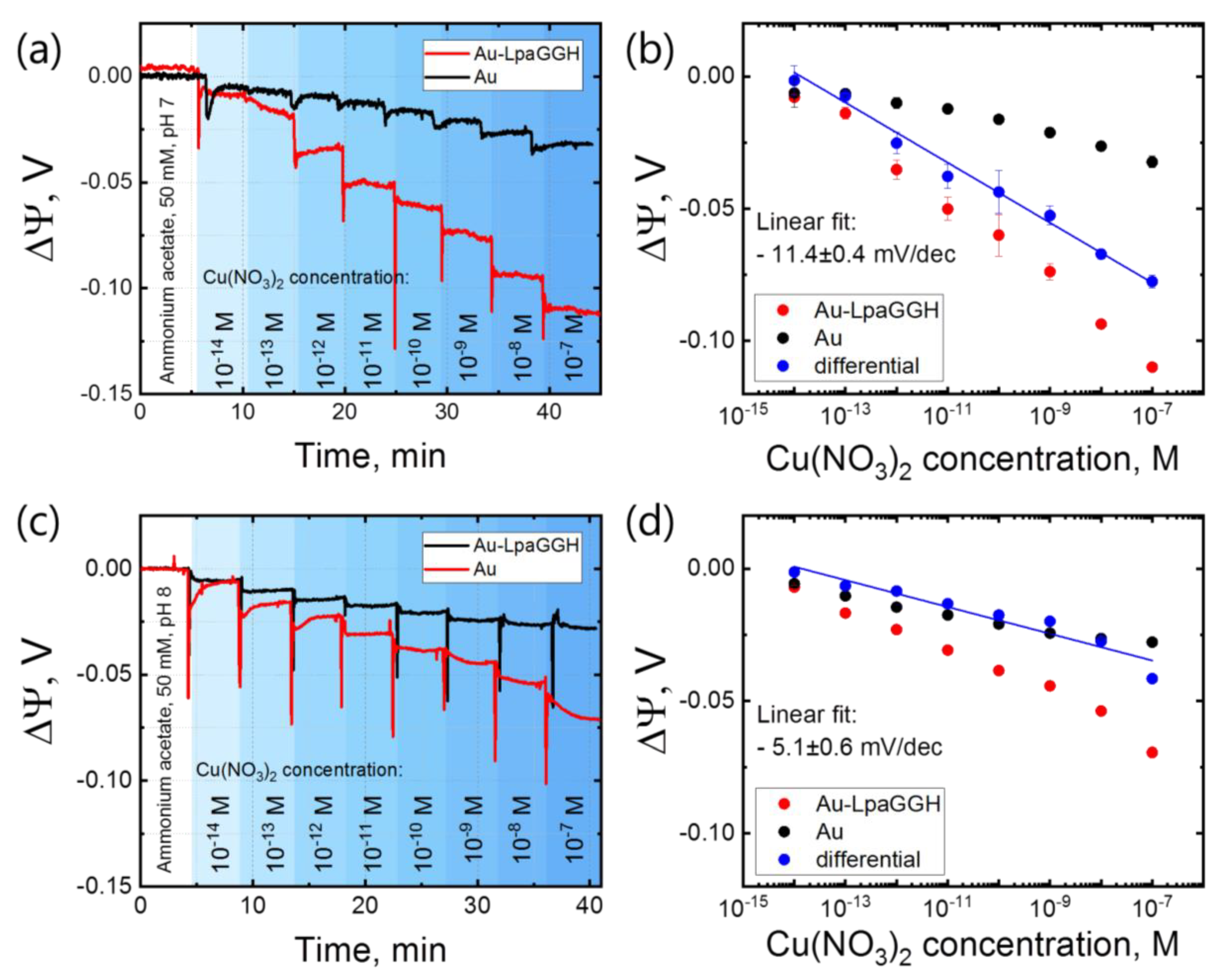
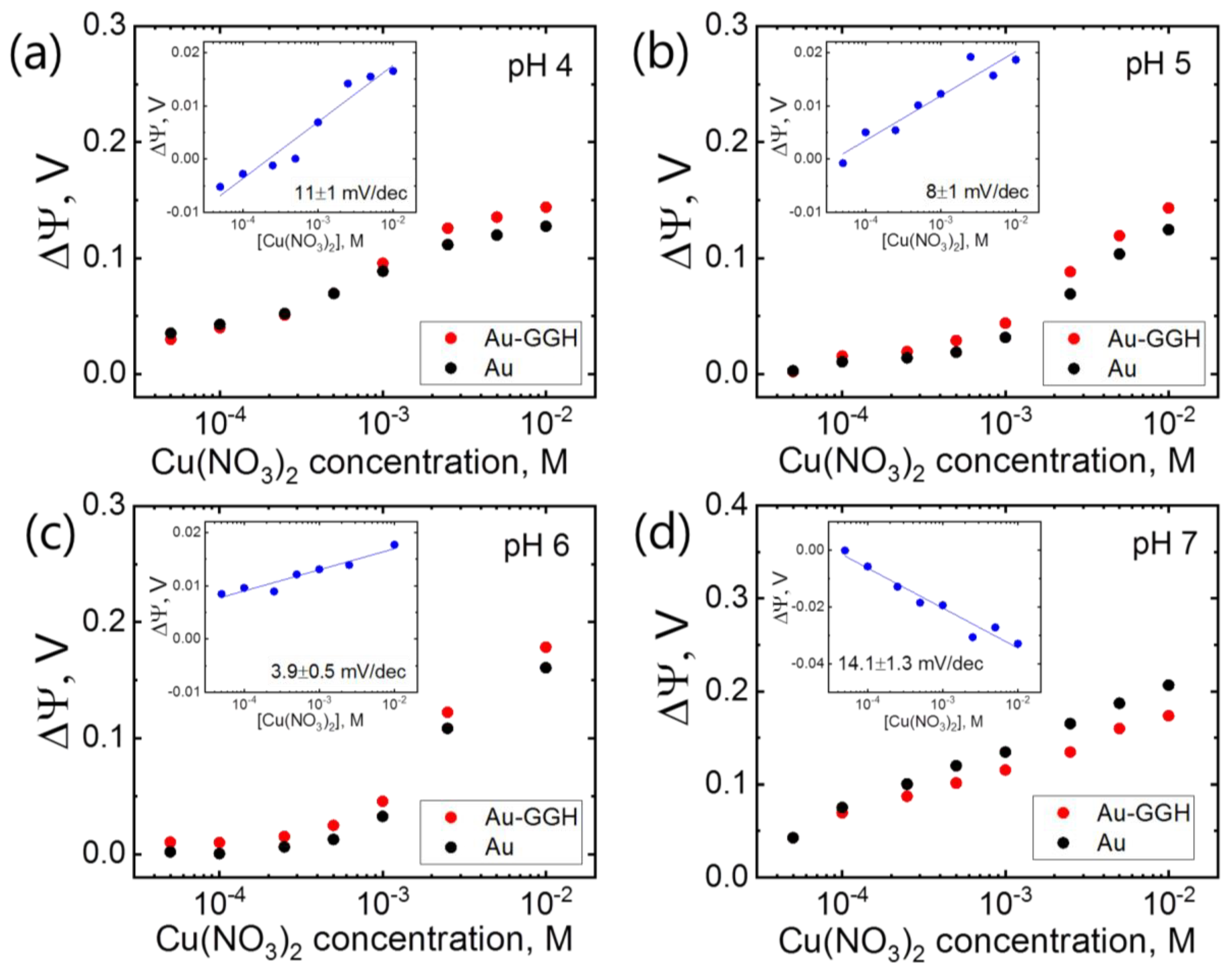
3. Results
4. Conclusions
Supplementary Materials
Author Contributions
Funding
Acknowledgments
Conflicts of Interest
References
- WHO. Trace Elements in Human Nutrition and Health. Available online: http://www.who.int/nutrition/publications/micronutrients/9241561734/en/ (accessed on 24 June 2019).
- Tchounwou, P.B.; Yedjou, C.G.; Patlolla, A.K.; Sutton, D.J. Heavy Metals Toxicity and the Environment. EXS 2012, 101, 133–164. [Google Scholar] [CrossRef] [PubMed]
- Emsley, J.; Emsley, S.W. Nature’s Building Blocks: An A–Z Guide to the Elements; Oxford University Press: Oxford, UK, 2001; ISBN 978-0-19-850341-5. [Google Scholar]
- Aragay, G.; Pons, J.; Merkoçi, A. Recent Trends in Macro-, Micro-, and Nanomaterial-Based Tools and Strategies for Heavy-Metal Detection. Chem. Rev. 2011, 111, 3433–3458. [Google Scholar] [CrossRef] [PubMed]
- World Health Organization. Copper in Drinking Water; World Health Organization: Geneva, Switzerland, 2004. [Google Scholar]
- US EPA. Lead and Copper Rule. Available online: https://www.epa.gov/dwreginfo/lead-and-copper-rule (accessed on 12 April 2019).
- Uauy, R.; Olivares, M.; Gonzalez, M. Essentiality of copper in humans. Am. J. Clin. Nutr. 1998, 67, 952S–959S. [Google Scholar] [CrossRef] [PubMed]
- Hedera, P.; Fink, J.K.; Bockenstedt, P.L.; Brewer, G.J. Myelopolyneuropathy and Pancytopenia Due to Copper Deficiency and High Zinc Levels of Unknown Origin: Further Support for Existence of a New Zinc Overload Syndrome. Arch. Neurol. 2003, 60, 1303–1306. [Google Scholar] [CrossRef] [PubMed]
- Brewer, G.J. Alzheimer’s disease causation by copper toxicity and treatment with zinc. Front Aging Neurosci. 2014, 6. [Google Scholar] [CrossRef] [PubMed]
- Bi, X.; Yang, K.-L. Complexation of Copper Ions with Histidine-Containing Tripeptides Immobilized on Solid Surfaces. Langmuir 2007, 23, 11067–11073. [Google Scholar] [CrossRef] [PubMed]
- Harford, C.; Sarkar, B. Amino Terminal Cu(II)- and Ni(II)-Binding (ATCUN) Motif of Proteins and Peptides: Metal Binding, DNA Cleavage, and Other Properties. Acc. Chem. Res. 1997, 30, 123–130. [Google Scholar] [CrossRef]
- Sanna, D.; Micera, G.; Kállay, C.; Rigó, V.; Sóvágó, I. Copper(II) complexes of N-terminal protected tri- and tetra-peptides containing histidine residues. Dalton Trans. 2004, 2702–2707. [Google Scholar] [CrossRef]
- Takehara, K.; Ide, Y. Electrochemistry of copper(II)-peptide complexes containing histidine residues. Inorg. Chim. Acta 1991, 183, 195–202. [Google Scholar] [CrossRef]
- Bal, W.; Dyba, M.; Kozłowski, H. The impact of the amino-acid sequence on the specificity of copper(II) interactions with peptides having nonco-ordinating side-chains. Acta Biochim. Pol. 1997, 44, 467–476. [Google Scholar]
- Lau, S.-J.; Kruck, T.P.A.; Sarkar, B. A Peptide Molecule Mimicking the Copper(II) Transport Site of Human Serum Albumin A Comparative Study between the Synthetic Site and Albumin. J. Biol. Chem. 1974, 249, 5878–5884. [Google Scholar] [PubMed]
- Angelova, M.; Asenova, S.; Nedkova, V.; Koleva-Kolarova, R. Copper in the Human Organism. Trakia J. Sci. 2011, 9, 11. [Google Scholar]
- Daniele, P.G.; Zerbinati, O.; Zelano, V.; Ostacoli, G. Thermodynamic and Spectroscopic Study of Copper(i1)-GIycyl-L-histidylglycine Complexes in Aqueous Solution. J. Chem. Soc. Dalton Trans. 1991, 5, 2711–2715. [Google Scholar] [CrossRef]
- Gonzalez, P.; Vileno, B.; Bossak, K.; El Khoury, Y.; Hellwig, P.; Bal, W.; Hureau, C.; Faller, P. Cu(II) Binding to the Peptide Ala-His-His, a Chimera of the Canonical Cu(II)-Binding Motifs Xxx-His and Xxx-Zzz-His. Inorg. Chem. 2017, 56, 14870–14879. [Google Scholar] [CrossRef] [PubMed]
- Mervinetsky, E.; Alshanski, I.; Hamo, Y.; Sandonas, L.M.; Dianat, A.; Buchwald, J.; Gutierrez, R.; Cuniberti, G.; Hurevich, M.; Yitzchaik, S. Copper Induced Conformational Changes of Tripeptide Monolayer Based Impedimetric Biosensor. Sci. Rep. 2017, 7, 9498. [Google Scholar] [CrossRef] [PubMed]
- Wawrzyniak, U.E.; Ciosek, P.; Zaborowski, M.; Liu, G.; Gooding, J.J. Gly-Gly-His Immobilized on Monolayer Modified Back-Side Contact Miniaturized Sensors for Complexation of Copper Ions. Electroanalysis 2013, 25, 1461–1471. [Google Scholar] [CrossRef]
- Gooding, J.J.; Hibbert, D.B.; Yang, W. Electrochemical Metal Ion Sensors. Exploiting Amino Acids and Peptides as Recognition Elements. Sensors 2001, 1, 75–90. [Google Scholar] [CrossRef]
- Yang, W.; Jaramillo, D.; Gooding, J.J.; Hibbert, D.B.; Zhang, R.; Willett, G.D.; Fisher, K.J. Sub-ppt detection limits for copper ions with Gly-Gly-His modified electrodes. Chem. Commun. 2001, 1982–1983. [Google Scholar] [CrossRef]
- Xu, Y.-M.; Pan, H.-Q.; Wu, S.-H.; Zhang, B.-L. Interaction Between Tripeptide Gly-Gly-His and Cu2+ Probed by Microcantilevers. Chin. J. Anal. Chem. 2009, 37, 783–787. [Google Scholar] [CrossRef]
- Wang, R.; Wang, W.; Ren, H.; Chae, J. Detection of copper ions in drinking water using the competitive adsorption of proteins. Biosens. Bioelectron. 2014, 57, 179–185. [Google Scholar] [CrossRef]
- Lee, C.-S.; Kim, S.; Kim, M. Ion-Sensitive Field-Effect Transistor for Biological Sensing. Sensors 2009, 9, 7111–7131. [Google Scholar] [CrossRef] [PubMed]
- Nguyen, T.T.K.; Tran, H.V.; Vu, T.T.; Reisberg, S.; Noël, V.; Mattana, G.; Pham, M.C.; Piro, B. Peptide-modified electrolyte-gated organic field effect transistor. Application to Cu2+ detection. Biosens. Bioelectron. 2019, 127, 118–125. [Google Scholar] [CrossRef] [PubMed]
- Wipf, M.; Stoop, R.L.; Navarra, G.; Rabbani, S.; Ernst, B.; Bedner, K.; Schönenberger, C.; Calame, M. Label-Free FimH Protein Interaction Analysis Using Silicon Nanoribbon BioFETs. ACS Sens. 2016, 1, 781–788. [Google Scholar] [CrossRef]
- Wipf, M.; Stoop, R.L.; Tarasov, A.; Bedner, K.; Fu, W.; Wright, I.A.; Martin, C.J.; Constable, E.C.; Calame, M.; Schönenberger, C. Selective Sodium Sensing with Gold-Coated Silicon Nanowire Field-Effect Transistors in a Differential Setup. ACS Nano 2013, 7, 5978–5983. [Google Scholar] [CrossRef] [PubMed]
- Stoop, R.L.; Wipf, M.; Müller, S.; Bedner, K.; Wright, I.A.; Martin, C.J.; Constable, E.C.; Fu, W.; Tarasov, A.; Calame, M.; et al. Competing surface reactions limiting the performance of ion-sensitive field-effect transistors. Sens. Actuators B Chem. 2015, 220, 500–507. [Google Scholar] [CrossRef]
- Stoop, R.L.; Wipf, M.; Müller, S.; Bedner, K.; Wright, I.A.; Martin, C.J.; Constable, E.C.; Fanget, A.; Schönenberger, C.; Calame, M. Implementing Silicon Nanoribbon Field-Effect Transistors as Arrays for Multiple Ion Detection. Biosensors 2016, 6, 21. [Google Scholar] [CrossRef] [PubMed]
- Hu, X.; Zhang, Q.; Wang, W.; Yuan, Z.; Zhu, X.; Chen, B.; Chen, X. Tripeptide GGH as the Inhibitor of Copper-Amyloid-β-Mediated Redox Reaction and Toxicity. ACS Chem. Neurosci. 2016, 7, 1255–1263. [Google Scholar] [CrossRef] [PubMed]
- Kulprachakarn, K.; Chen, Y.-L.; Kong, X.; Arno, M.C.; Hider, R.C.; Srichairatanakool, S.; Bansal, S.S. Copper(II) binding properties of hepcidin. J. Biol. Inorg. Chem. 2016, 21, 329–338. [Google Scholar] [CrossRef] [PubMed]
- Lin, M.; Cho, M.S.; Choe, W.S.; Lee, Y. Electrochemical analysis of copper ion using a Gly–Gly–His tripeptide modified poly(3-thiopheneacetic acid) biosensor. Biosens. Bioelectron. 2009, 25, 28–33. [Google Scholar] [CrossRef] [PubMed]
- Yang, W.; Chow, E.; Willett, G.; Brynn Hibbert, D.; Justin Gooding, J. Exploring the use of the tripeptide Gly–Gly–His as a selective recognition element for the fabrication of electrochemical copper sensors. Analyst 2003, 128, 712–718. [Google Scholar] [CrossRef] [PubMed]
- Papp, S.; Jágerszki, G.; Gyurcsányi, R.E. Ion-Selective Electrodes Based on Hydrophilic Ionophore-Modified Nanopores. Angew. Chem. 2018, 130, 4842–4845. [Google Scholar] [CrossRef]
- Pavan, S.; Berti, F. Short peptides as biosensor transducers. Anal. Bioanal. Chem. 2012, 402, 3055–3070. [Google Scholar] [CrossRef]
- Raics, M.; Sanna, D.; Sóvágó, I.; Kállay, C. Copper(II), nickel(II) and zinc(II) complexes of hexapeptides containing separate aspartyl and histidyl residues. Inorg. Chim. Acta 2015, 426, 99–106. [Google Scholar] [CrossRef]
- Knopfmacher, O.; Tarasov, A.; Fu, W.; Wipf, M.; Niesen, B.; Calame, M.; Schönenberger, C. Nernst Limit in Dual-Gated Si-Nanowire FET Sensors. Nano Lett. 2010, 10, 2268–2274. [Google Scholar] [CrossRef] [PubMed]
- Vacic, A.; Criscione, J.M.; Rajan, N.K.; Stern, E.; Fahmy, T.M.; Reed, M.A. Determination of Molecular Configuration by Debye Length Modulation. J. Am. Chem. Soc. 2011, 133, 13886–13889. [Google Scholar] [CrossRef]
- Hay, R.W.; Hassan, M.M.; You-Quan, C. Kinetic and thermodynamic studies of the copper(II) and nickel(II) complexes of glycylglycyl-L-histidine. J. Inorg. Biochem. 1993, 52, 17–25. [Google Scholar] [CrossRef]
- Vuceta, J.; Morgan, J.J. Hydrolysis of Cu(II)1. Limnol. Oceanogr. 1977, 22, 742–746. [Google Scholar] [CrossRef]
- Mervinetsky, E.; Alshanski, I.; Lenfant, S.; Guerin, D.; Medrano Sandonas, L.; Dianat, A.; Gutierrez, R.; Cuniberti, G.; Hurevich, M.; Yitzchaik, S.; et al. Electron Transport through Self-Assembled Monolayers of Tripeptides. J. Phys. Chem. C 2019, 123, 9600–9608. [Google Scholar] [CrossRef]
- Badruddoza, A.Z.M.; Tay, A.S.H.; Tan, P.Y.; Hidajat, K.; Uddin, M.S. Carboxymethyl-β-cyclodextrin conjugated magnetic nanoparticles as nano-adsorbents for removal of copper ions: Synthesis and adsorption studies. J. Hazard. Mater. 2011, 185, 1177–1186. [Google Scholar] [CrossRef]
- Licheri, G.; Musinu, A.; Paschina, G.; Piccaluga, G.; Pinna, G.; Sedda, A.F. Coordination of Cu(II) in Cu(NO3)2 aqueous solutions. J. Chem. Phys. 1984, 80, 5308–5311. [Google Scholar] [CrossRef]


© 2019 by the authors. Licensee MDPI, Basel, Switzerland. This article is an open access article distributed under the terms and conditions of the Creative Commons Attribution (CC BY) license (http://creativecommons.org/licenses/by/4.0/).
Share and Cite
Synhaivska, O.; Mermoud, Y.; Baghernejad, M.; Alshanski, I.; Hurevich, M.; Yitzchaik, S.; Wipf, M.; Calame, M. Detection of Cu2+ Ions with GGH Peptide Realized with Si-Nanoribbon ISFET. Sensors 2019, 19, 4022. https://doi.org/10.3390/s19184022
Synhaivska O, Mermoud Y, Baghernejad M, Alshanski I, Hurevich M, Yitzchaik S, Wipf M, Calame M. Detection of Cu2+ Ions with GGH Peptide Realized with Si-Nanoribbon ISFET. Sensors. 2019; 19(18):4022. https://doi.org/10.3390/s19184022
Chicago/Turabian StyleSynhaivska, Olena, Yves Mermoud, Masoud Baghernejad, Israel Alshanski, Mattan Hurevich, Shlomo Yitzchaik, Mathias Wipf, and Michel Calame. 2019. "Detection of Cu2+ Ions with GGH Peptide Realized with Si-Nanoribbon ISFET" Sensors 19, no. 18: 4022. https://doi.org/10.3390/s19184022
APA StyleSynhaivska, O., Mermoud, Y., Baghernejad, M., Alshanski, I., Hurevich, M., Yitzchaik, S., Wipf, M., & Calame, M. (2019). Detection of Cu2+ Ions with GGH Peptide Realized with Si-Nanoribbon ISFET. Sensors, 19(18), 4022. https://doi.org/10.3390/s19184022