Article

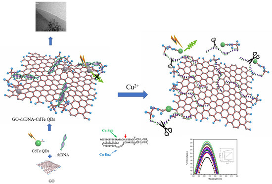
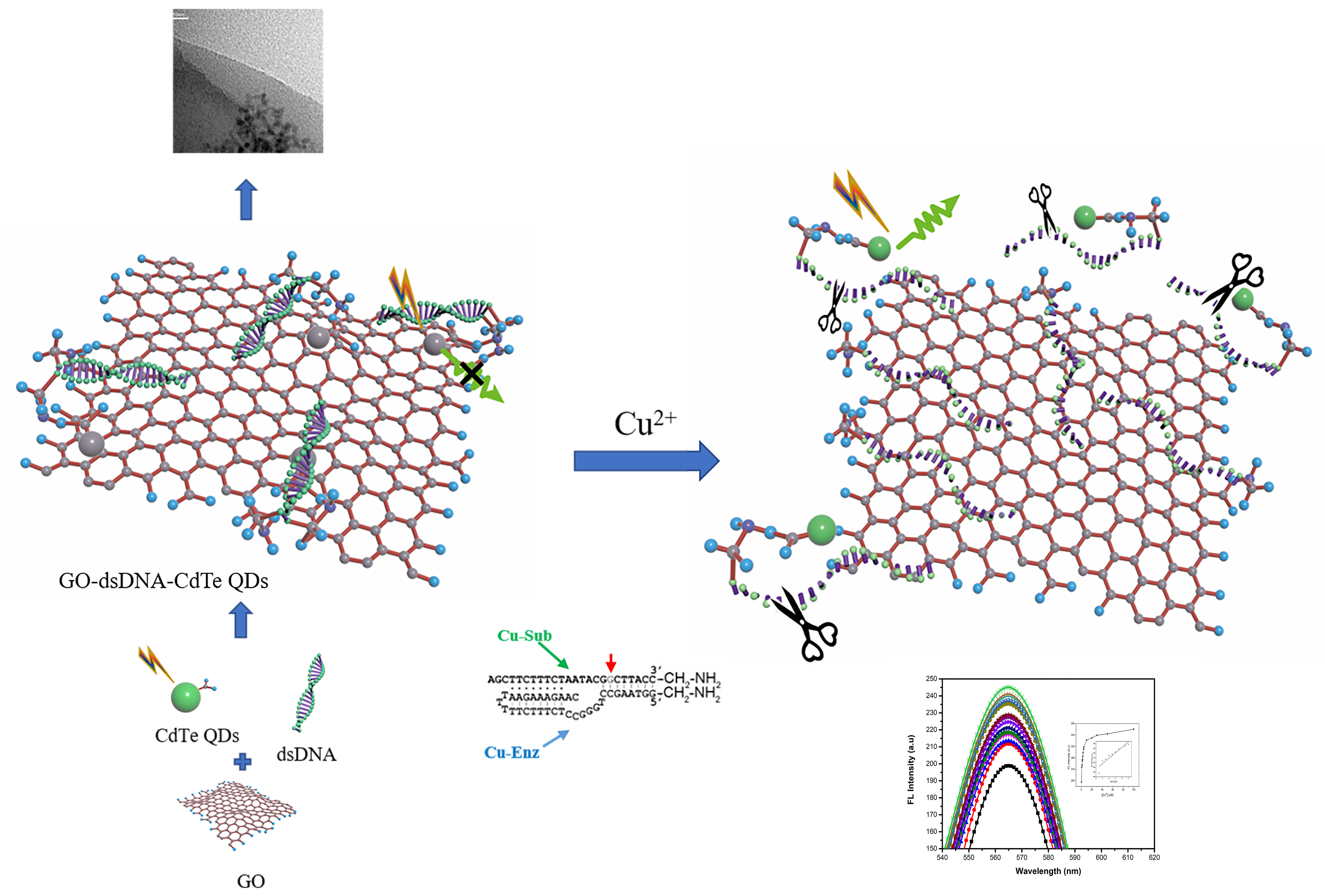
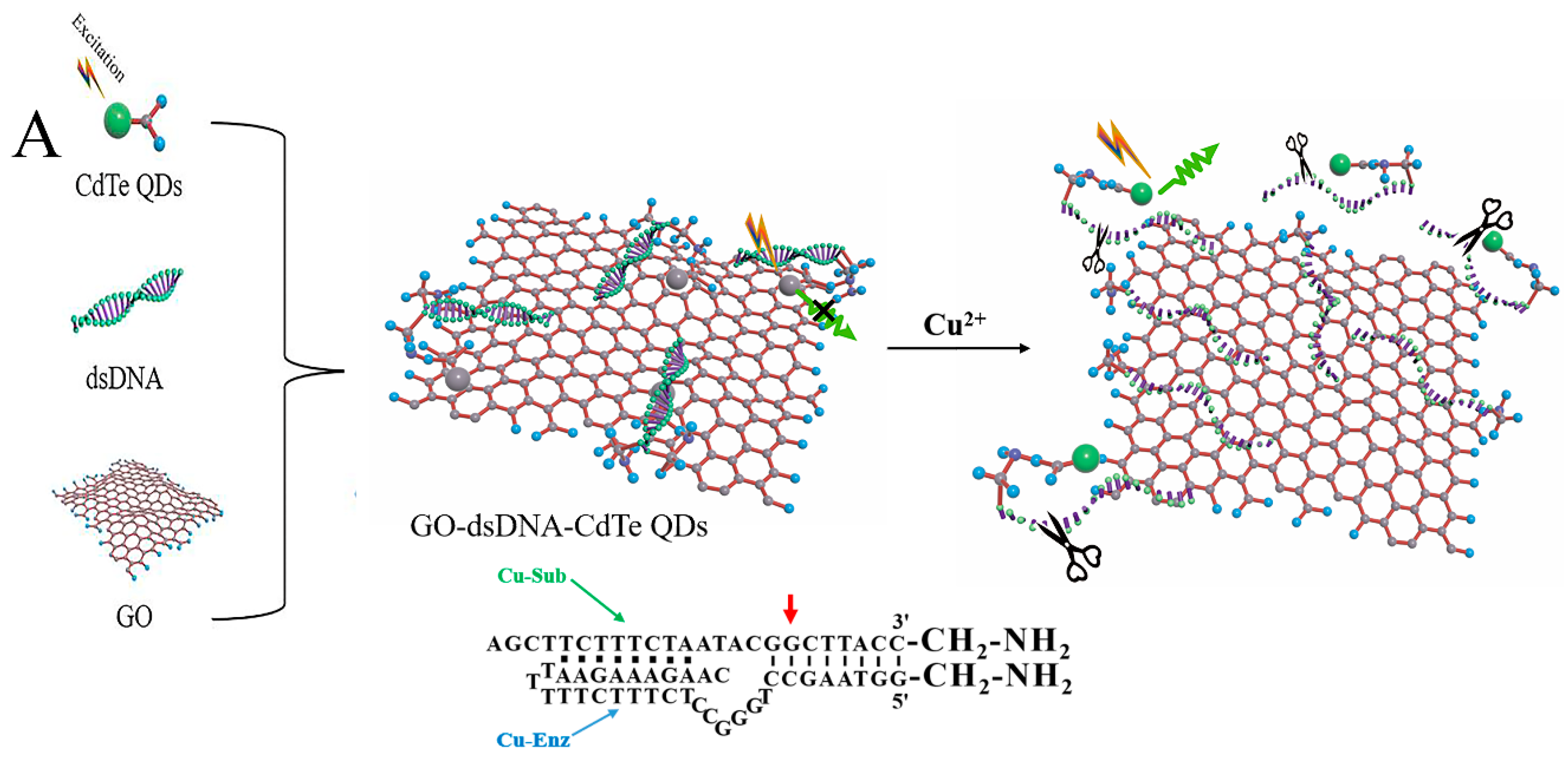
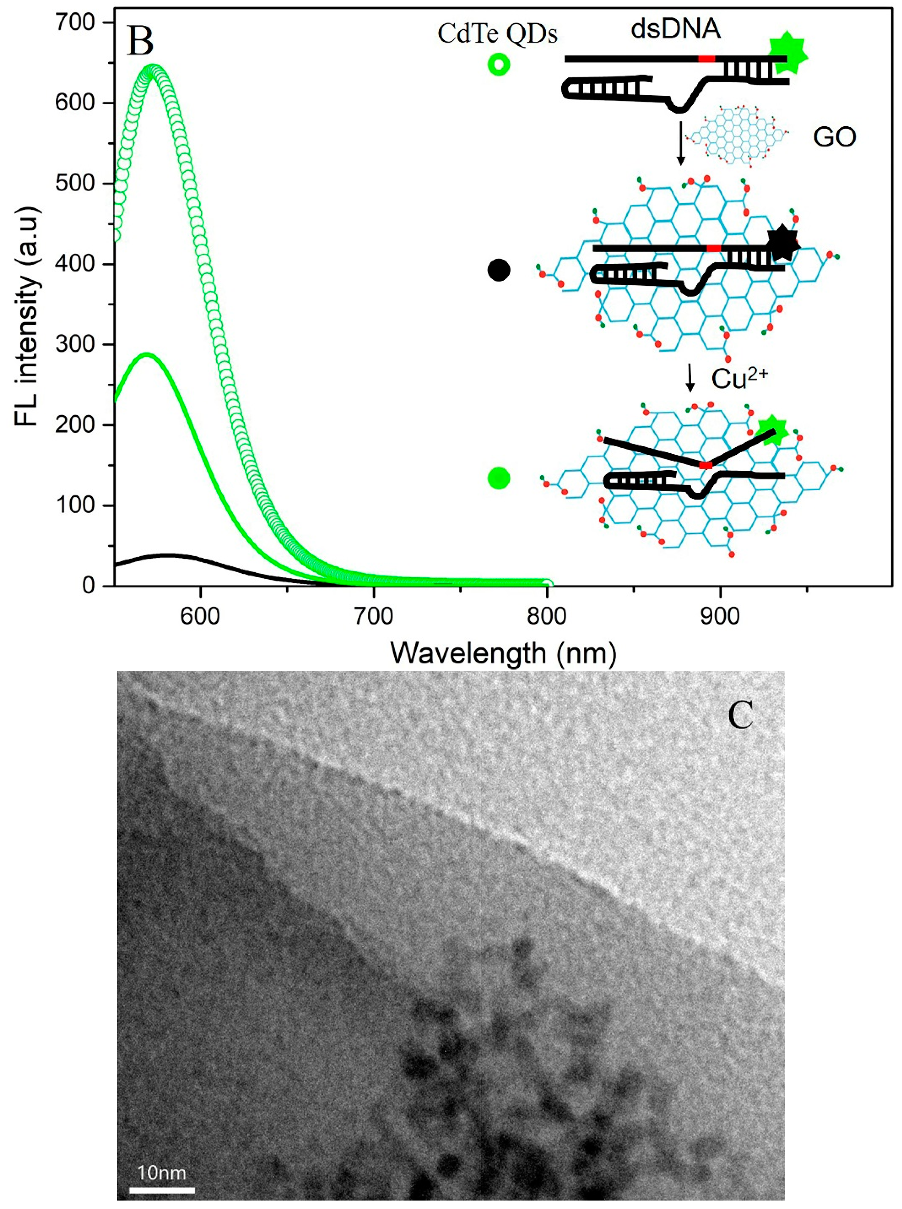
A “Turn-On” Fluorescence Copper Biosensor Based on DNA Cleavage-Dependent Graphene Oxide-dsDNA-CdTe Quantum Dots Complex

1
National Engineering Laboratory for Fiber Optic Sensing Technology, Wuhan University of Technology, Wuhan 430070, China
2
School of Information Engineering, Hubei University of Chinese Medicine, Wuhan 430065, China
*
Author to whom correspondence should be addressed.
Sensors 2018, 18(8), 2605; https://doi.org/10.3390/s18082605
Submission received: 9 July 2018 / Revised: 27 July 2018 / Accepted: 3 August 2018 / Published: 9 August 2018
(This article belongs to the Special Issue Graphene Based Sensors and Electronics)

Abstract

:
A novel “turn-on” fluorescent copper biosensor is developed successfully based on the graphene oxide (GO)-dsDNA-CdTe quantum dots (QDs) complex via chemical crosslink method. The optical and structure properties of GO-dsDNA-CdTe QDs complex are studied by fluorescence (FL) spectra and transmission electron microscopy (TEM) in detail. It is demonstrated that the fluorescence quenching of CdTe QDs is a process of fluorescence resonance energy transfer (FRET) due to the essential surface and quenching properties of two-dimensional GO. Copper ions induce the catalytic reaction of DNA chain and irreversibly break at the cleavage site, which will cause the G-quadruplex formation, moreover further result in the CdTe QDs separated from GO and restored its fluorescence. Therefore, a significant recovery effect on the fluorescence of the GO-dsDNA-CdTe QDs complex is observed in the presence of copper ions. The fluorescence responses are concentration-dependent and can be well described by a linear equation. Compared with other metal ions, the sensor performs good selectivity for copper ions.

Graphical Abstract

1. Introduction

Copper ions (Cu2+) are the most abundant essential nutrient in nature except zinc and iron ions, which plays a very important role for biological functions as a constituent of some enzymes [1]. However, excess Cu2+ is highly toxic to organism due to disordering gastrointestinal balance or damaging the kidneys and liver [2], as well as listed as priority pollutant in nature. Although several methods are currently employed to detect copper ions based on atomic absorption spectroscopy (AAS) [3], inductively coupled plasma mass spectrometry (ICP-MS) [4], UV-Vis spectroscopy [5], chemiluminescence detection [6] and electrochemical techniques [7,8,9,10], those methods generally need complex preparation process and precision instruments so that their practical applications are tremendously limited. While the use of fluorescent sensors offers distinct advantages, including high sensitivity, good selectivity, time efficiency and the possibility of performing real-time analysis. It is reported that the fluorescent sensors for copper ions detection based on semiconductor quantum dots [11,12], metal nanoclusters [13], upconverting luminescent nanoparticles and organic dyes [14,15,16,17], Ag nanoparticles [18], metal-organic compounds [19] and electrochemical means [20,21]. However, there are still the problems for these fluorescence technologies such as selectivity, water solubility, stability and potential toxicity.
DNAzymes have been regarded as novel and ideal platforms in sensing due to their catalytic ability or binding activity toward specific cofactors, including high binding affinity, easy accessibility and outstanding stability under harsh conditions [22]. So far, numerous sensitive and selective methods based on DNAzyme have been found for Cu2+ [23], Pb2+ [24,25,26,27,28], Hg2+ [29] and K+ [30] detection, respectively. The DNAzyme can be complementary to a specific substrate sequence, the substrate chain is cleaved into two fragments when metal ions are present as cofactors [31]. However, traditional DNAzymes-based fluorescent sensors require two or more quenchers labeled at one DNA strand in order to reduce the background, which will inevitably cause complicated or expensive operation and weaken the performance of DNAzymes [32]. It is necessary and urgent to focus on the designing label-free fluorescent sensors based on binding or dynamic cleavage-dependent fluorescence (FL) activation [33].
Recently, two-dimensional graphene-based materials have emerged as promising and novel platforms in sensors due to their unique surface and electronic properties [34]. In particular, graphene can act as an efficient quencher of an organic fluorophore or inorganic luminescent nanomaterial based on either an energy transfer or electron transfer process [35]. As a consequence, graphene has shown great potential in constructing fluorescent DNA biosensors by means of the external competition-dependent graphene/DNA interaction. In this work, by combining catalytic turnover inherent to DNAzymes and subsequent cleavage-induced G-quadruplex formation, a labeled Cu2+ fluorescent sensor based on GO-dsDNA-CdTe QDs complex constructed by Cu2+ specific DNAzyme was prepared, where CdTe quantum dots as fluorophore were labeled on DNAzyme and graphene oxide as the quencher. The structural transformation of GO-dsDNA-CdTe QDs complex was demonstrated by the change of the fluorescence intensity of CdTe quantum dots. When the CdTe quantum dots and graphene oxide were cross-linked with the end of functional DNAzyme, the CdTe quantum dots show a lower fluorescent in aqueous media. However, the addition of Cu2+ makes the fluorescence of the CdTe quantum dots change from “turn off” to “turn-on.” Meanwhile, the selective of the GO-dsDNA-CdTe QDs complex for Cu2+ were studied. Compared with other methods, this method has the characteristics of simple operation, high sensitivity and good selectivity.

2. Experimental

2.1. Materials and Methods

NaCl, KCl, CdCl2, Pb(NO3)2, MgCl2, Zn(NO3)2, CaCl2, FeCl3, CoCl2, Na2HPO4 and NaH2PO4 were of analytical grade and bought from Sinopharm Chemical Reagent Co, Ltd. (Shanghai, China). Cu2+ substrate sequence (Cu-Sub) (5′-AGCTTCTTTCT-AATACGGCTTACC-3′) (ssDNA) and enzyme sequence (Cu-Enz) (5′-GGTAAGCCTGGGCCTCTTTCTTTTTAAGAAAG-AAC-3′) (cDNA) were bought from Wuhan TianyiHuiyuan Life Science & technology Inc., Ltd. (Wuhan, China). Different pH values of phosphate buffer solutions (PBS, 0.05 M) were prepared using the stock solution of Na2HPO4 and NaH2PO4 with various volume ratios. GO was prepared in accordance with the well-documented Hummers methods [36] (1958, hummers). The water-soluble CdTe QDs are prepared by One-pot synthesis [37].

2.2. Apparatus

The F-4500 fluorescence spectrophotometer (Hitachi, CO., Tokyo, Japan) was used to measure the fluorescence of GO-dsDNA-CdTe QDs complex at excitation wavelength of 473 nm. UV-vis spectra were collected by the UV-2450 spectrophotometer (Shimad-zu, CO., Kyoto-fu, Japan). The morphology was studied in detail by a JEM-2100F STEM/EDS TEM (JEOL, Tokyo, Japan).

2.3. GO-dsDNA-CdTe QDs Complex Synthesis

25 mM EDC and 100 mM NHS was added to the GO solution in the PBS solution (10 mM Na2HPO4/NaH2PO4, 0.3 m NaCl and pH = 7.4) to activate the carboxyl group on GO sheet and dissolved and kept for 2 h. Then, 4 uM ssDNA was added to the activated carboxyl groups of GO solution (1 mL of 1 g/L) and cultivated overnight. Subsequently 4 uM cDNA was added to the GO-ssDNA solution and kept for 1 h at 95 °C in a water bath and then natural cooling to room temperature. 10 mM PBS solution was used to centrifuge and wash cDNA, ssDNA and GO solutions, respectively and then obtained GO-dsDNA product was stored at 4 °C for the future use. 25 mM EDC and 100 mM NHS was added to the CdTe QDs solution and cultivated for 2 h. GO-dsDNA was added the above CdTe QDs solution and incubated for 24 h, finally GO-dsDNA-CdTe complex was obtained.

3. Results and Discussion

3.1. Principle of the Cu2+ Sensing System

The design of the Cu2+ biosensor based on GO-dsDNA-CdTe complex is shown in Figure 1A. The system is a combination of two components that formed a self-assembled complex. The first component was the Cu2+ dependent DNAzyme containing a 3′-FAM-modified Cu-Sub and a Cu-Enz, which served as an effective recognition site. The second component was the FL transduction unit of graphene, which was employed as an independent quencher to suppress background signal. As shown in Figure 1B, initially, the CdTe QDs is fixed to one end of the ssDNA chain and CdTe QDs exhibits the strong fluorescence emission at 540 nm without GO. And as the GO mixed with the CdTe QDs–dsDNA, the fluorescence emission of the CdTe QDs will be quenching. It is because that sp2 bonded carbon atoms of GO and aromatic nucleobases of dsDNA will reveal π-π stacking interactions, which makes the distance between GO and CdTe QDs smaller than 10 nm [37,38,39,40] and such a short distance helps transfer the energy from excited state of CdTe QDs to GO. Therefore, the fluorescence emission of the CdTe QDs will be quenched. In the presence of Cu2+, the DNA substrate was irreversibly cleaved as a result of the specific DNAzyme-Cu2+ interaction, disturbing the self-assembled graphene–DNAzyme conformation. As a result, the quantum dots separated from the GO and restored the fluorescence. Hence, the GO-dsDNA-CdTe QDs complex could act as the sensor platform for Cu2+ detection. The FL spectra of GO-dsDNA-CdTe QDs complex in the absence or presence of Cu2+ is shown in Figure 1B. It can be demonstrated that the FL intensity of GO-dsDNA-CdTe QDs complex get effectively enhance when the Cu2+ was mixed with GO-dsDNA-CdTe QDs complex. It is verified that the CdTe nanoparticles are well dispersed and immobilized on GO surface as shown in the Figure 1C.

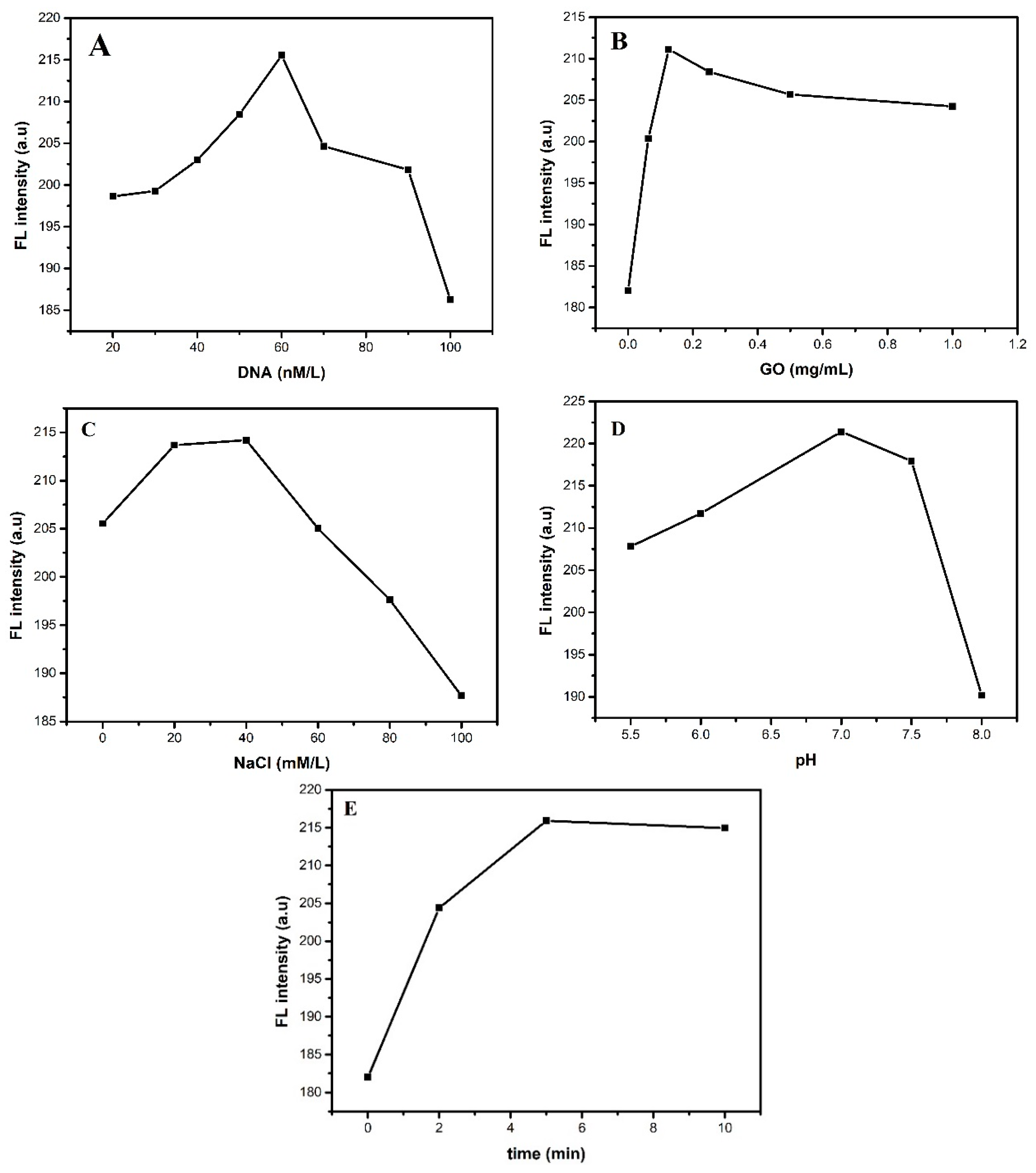
3.2. Optimization of Experimental Conditions

In order to improve the performance of the sensor for the Cu2+ detecting, the usage conditions of GO-dsDNA-CdTe QDs complex must be optimized. The concentration of DNA is an important factor affecting the performance of fluorescent sensor. Figure 2A shows the relationship between the DNA concentration and the FL intensity. It can be seen from Figure 2A that the FL intensity of the system increases continuously with the DNA concentration increasing from 0 to 60 nmol/L with the concentration of Cu2+ is 5 umol/L, while FL intensity decreased when DNA concentration increases from 60 to 100 nmol/L. When the concentration of DNA is low, increasing the amount of DNA is equivalent to increase of CdTe QDs connected with DNA, so the FL intensity of CdTe QDs enhanced obviously. However, the CdTe QDs released from the cleaved DNA and meet the GO sheet again if the concentration of DNA is excessive; resulting in the fluorescence was quenched. Therefore, the concentration of DNA in this experiment is chosen as 60 nmol/L.
The concentrations of GO as the quencher of the fluorescence probe affects the probe’s performance. Figure 2B shows that the fluorescence signal increased with increasing concentration of GO and reached the maximum intensity at 0.125 mg/mL with the concentration of Cu2+ is 5 umol/L. The reason is that the free CdTe QDs from the cleaved DNAzyme could meet GO again when the concentration of GO was too much. Thus, the optimum concentration of GO in this experiment is 0.125 mg/mL.
The ionic strength is a measure of the concentration of each ion in the solution. The catalytic activity of the DNA enzyme and the stability of the hybridization will be affected by the excessively ionic strength. In this study, ionic strength is regulated by the concentration of NaCl. In order to ensure the stability of the Cu2+ ion-specific DNA enzyme during the experiment, the high concentration of ion intensity in the buffer solution is very necessary. It can be seen from Figure 2C that the FL intensity increases continuously with the DNA concentration increasing from 0 to 40 mmol/L, while FL intensity decreased as DNA concentration increases from 40 to 100 mmol/L with the concentration of Cu2+ is 5 umol/L. Thus, the optimum concentration of NaCl is chosen as 40 mmol/L.
The pH value is one of the most important parameters influencing the capability of the sensor. On the one hand, strong acid condition will reduce the solubility of DNA, due to damaging the structure of DNA and bringing about the inactivation of DNA, which will eventually affect the number of DNA hybridized with CdTe QDs and further affect the fluorescence performance of CdTe QDs. On the other hand, Cu(OH)2 precipitation of Cu2+ ions will be formed under strong alkaline environment. Therefore, the range of pH is set from 5.5 to 8.0 in this experiment. As shown in Figure 2D, the FL intensity of the fluorescence signal increased gradually with pH and reached the maximum at pH = 7.0 and then decreased when pH further increased to 8.0. Thus, the optimum value of pH for Cu2+ detection is chosen as 7.
The reaction time of DNA catalyzed by Cu2+ ion depends on the concentration of Cu2+ ion. The Cu2+ ion concentration (100 μmol/L) is chosen as reference to analyze the reaction. The effect of test time on the performance of GO-dsDNA-CdTe QDs complex fluorescent probe is shown in Figure 2E. With the increase of reaction time from 0 to 10 min, the FL intensity increases rapidly and reaches the maximum at 5 min, then remained the same after 5 min. Therefore, 5 min is selected as the best time to detect the concentration of Cu2+ ions.

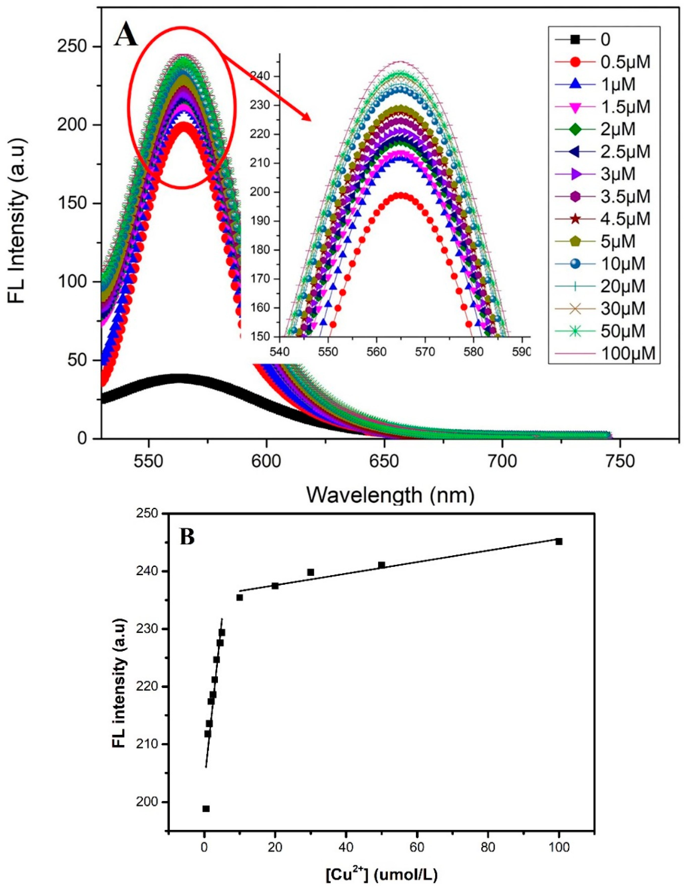
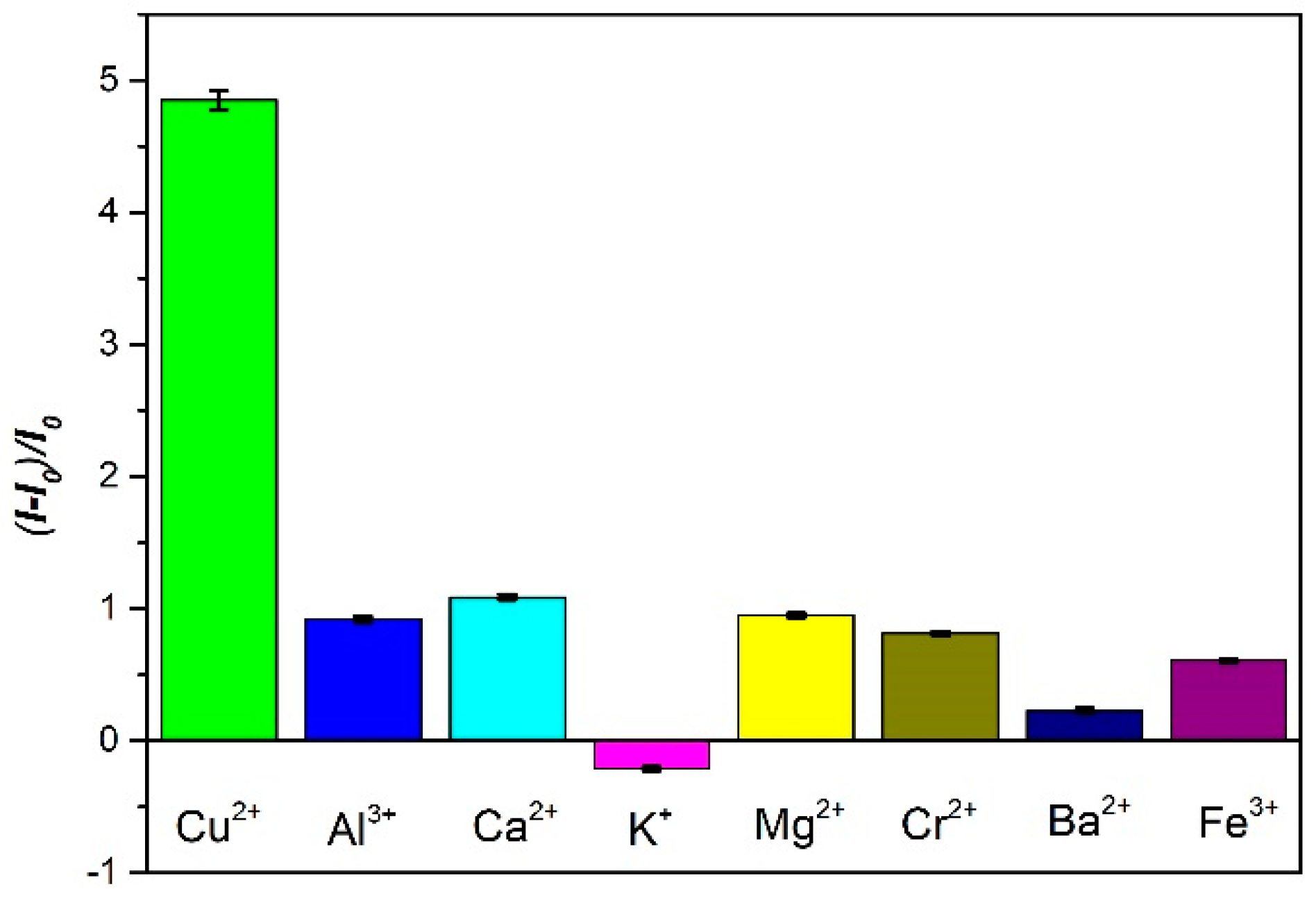
3.3. The Analytical Performance of the Sensor

Analytical performance is a key factor to measure a practical sensor. There are the fluorescence spectra of the GO-dsDNA-CdTe QDs complex upon addition of various concentration of Cu2+ show in the Figure 3A. With the addition of Cu2+, the FL intensity significantly increased, suggesting that the DNAzyme was activated and catalyze the cleavage of the hairpin substrate strand into inter-molecularly hybridized double-strand DNA with much less stability, resulting in the CdTe QDs far away from GO, thus producing an increased fluorescence signal. The FL intensity increased with the increase of Cu2+ concentration. It reached a plateau when the Cu2+ concentration increased to 5 μmol/L. It is mainly because the number of GO-dsDNA-CdTe QDs complex is limited, adding a small amount of Cu2+ ion can make it full contact with the GO-dsDNA-CdTe QDs complex and release CdTe QDs quickly, so that the fluorescence intensity fast recovered. Excessive Cu2+ may decrease the contact with the GO-dsDNA-CdTe QDs complex and decrease the speed of CdTe QDs released, so the FL intensity rises slowly. Figure 3B presented the calibration curve of the sensor in the range of 0.5 to 100 μmol/L. In the linear response range (0.5–5 μmol/L, 5–100 μmol/L) and the calibration curve can be expressed by the equation I = 5.724 Cu 2 + + 203.15755 (R2 = 0.865) and I = 0.10034 Cu 2 + + 235.574 (R2 = 0.919). The sensor shows a great linear relationship between the FL intensity and the concentration of Cu2+ because of the super quenching capability of GO and the significant difference in the affinity of dsDNA toward Cu2+ and GO.
In order to investigate the effective selectivity of GO-dsDNA-CdTe QDs complex to Cu2+ ions, the environmentally relevant metal ions solution of the same concentration (100 μmol/L), including Ag+, Ca2+, Cd2+, Pb2+, Fe3+ and K+, was respectively prepared under the best detection conditions. Here, we define a signal-to-background ratio (SBR) as ( I I 0 ) / I 0 to eliminate the influence of the original background intensity, which was used to estimate the FL response, where I and I0 were the FL signals of the CdTe QDs in the presence and absence of Cu2+, respectively. Selectivity is defined as SBR of the probe for Cu2+ detection against other metal ions. As shown in Figure 4, although any heavy metal ions except for Cu2+ are added in the solution, the FL intensity of GO-dsDNA-CdTe QDs complex still did not change. This result showed that compared with other metal ions, the GO-dsDNA-CdTe QDs complex has great selectivity to Cu2+ because of the G-quadruplex structure between dsDNA and Cu2+.

4. Conclusions

In this paper, a fluorescent Cu2+ sensor based on GO-dsDNA-CdTe QDs complex were prepared by chemical crosslinking method. We demonstrated the fluorescence emission GO-dsDNA-CdTe QDs complex can be “turned-on” by the presence of Cu2+ based on the FRET mechanism between CdTe QDs and GO. The optimum experiments conditions were determined, including the concentration of DNA (60 nmol/L), GO (0.125 mg/mL), NaCl (40 mmol/L) and pH (7) of solution and the best detection time is 5min. A linear correlation between Cu2+ and FL intensity was obtained with I = 5.72 Cu 2 + + 203.16 (R2 = 0.865) in the range from 0.5 to 5 μmol/L. The detection limit of the sensor presented is 0.5 uM (S/N = 3). The results show that the GO-dsDNA-CdTe QDs complex has good recognition ability of Cu2+. The sensing mechanism is suitable for the detection of other heavy metal ions, small molecules and biomolecules.

Author Contributions

L.D., J.H. and B.X. conceived and designed the experiments; B.X. and T.L. performed the experiments; B.X. and T.L. analyzed the data; B.X., T.L. and W.B. wrote the paper.

Funding

This work is supported by Financial Support from the National Natural Science Foundation of China (No. 61575150, No. 61377092) and the Fundamental Research Funds for the Central Universities (WUT: 2017II46GX).

Acknowledgments

This work is supported by Financial Support from the National Natural Science Foundation of China (No. 61575150, No. 61377092) and the Fundamental Research Funds for the Central Universities (WUT: 2017II46GX).

Conflicts of Interest

The authors declare no conflict of interest.

References

  1. Wu, S.P.; Huang, R.Y.; Du, K.J. Colorimetric sensing of Cu(ii) by 2-methyl-3-[(pyridin-2-ylmethyl)-amino]-1,4-naphthoquinone: Cu(ii) induced deprotonation of NH responsible for color changes. Dalton Trans. 2009, 1164, 4735–4740. [Google Scholar] [CrossRef] [PubMed]
  2. Sheline, C.T.; Choi, D.W. Cu2+ toxicity inhibition of mitochondrial dehydrogenases in vitro and in vivo. Ann. Neurol. 2004, 55, 645–653. [Google Scholar] [CrossRef] [PubMed]
  3. Faghihian, H.; Hajishabani, A.; Dadfarnia, S.; Zamani, H. Use of clinoptilolite loaded with 1-(2-pyridylazo)-2-naphthol as a sorbent for preconcentration of Pb(II), Ni(II), Cd(II) and Cu(II) prior to their determination by flame atomic absorption spectroscopy. Int. J. Environ. Anal. Chem. 2009, 89, 223–231. [Google Scholar] [CrossRef]
  4. Reddy, D.K.; Anil, G.; Reddy, M.R.P.; Rao, T.G. A DRC ICP-MS Based Screening Method for Heavy Metals Contamination in Proton Pump Inhibitor Compounds and Their Intermediates. At. Spectrosc. 2008, 29, 201–209. [Google Scholar]
  5. Zhang, J.F.; Zhou, Y.; Yoon, J.; Kim, Y.; Kim, S.J.; Kim, J.S. Naphthalimide Modified Rhodamine Derivative: Ratiometric and Selective Fluorescent Sensor for Cu2+ Based on Two Different Approaches. Org. Lett. 2010, 12, 3852–3855. [Google Scholar] [CrossRef] [PubMed]
  6. Tang, B.; Niu, J.; Yu, C.; Zhao, L.; Ge, J. Highly luminescent water-soluble CdTe nanowires as fluorescent probe to detect copper(II). Chem. Commun. 2005, 33, 4184–4186. [Google Scholar] [CrossRef] [PubMed]
  7. Berchmans, S.; Vergheese, T.M.; Kavitha, A.L.; Veerakumar, M.; Yegnaraman, V. Electrochemical preparation of copper–dendrimer nanocomposites: Picomolar detection of Cu2+, ions. Anal. Bioanal. Chem. 2008, 390, 939–946. [Google Scholar] [CrossRef] [PubMed]
  8. Mülazımoğlu, I.E.; Solak, A.O. A novel apigenin modified glassy carbon sensor electrode for the determination of copper ions in soil samples. Anal. Methods 2011, 3, 2534–2539. [Google Scholar] [CrossRef]
  9. Shao, X.; Gu, H.; Wang, Z.; Chai, X.; Tian, Y.; Shi, G. Highly selective electrochemical strategy for monitoring of cerebral Cu2+ based on a carbon Dot-TPEA hybridized surface. Anal. Chem. 2013, 85, 418–425. [Google Scholar] [CrossRef] [PubMed]
  10. Fu, X.C.; Wu, J.; Li, J.; Xie, C.G.; Liu, Y.S.; Zhong, Y.; Liu, J.H. Electrochemical determination of trace copper(II) with enhanced sensitivity and selectivity by gold nanoparticle/single-wall carbon nanotube hybrids containing three-dimensional l -cysteine molecular adapters. Sens. Actuators B Chem. 2013, 182, 382–389. [Google Scholar] [CrossRef]
  11. Guo, C.; Wang, J.; Cheng, J.; Dai, Z. Determination of trace copper ions with ultrahigh sensitivity and selectivity utilizing CdTe quantum dots coupled with enzyme inhibition. Biosens. Bioelectron. 2012, 36, 69–74. [Google Scholar] [CrossRef] [PubMed]
  12. Wu, C.S.; Khaing Oo, M.K.; Fan, X. Highly sensitive multiplexed heavy metal detection using quantum-dot-labeled DNAzymes. ACS Nano 2010, 4, 5897–5904. [Google Scholar] [CrossRef] [PubMed]
  13. Zhang, M.; Ye, B.C. Colorimetric chiral recognition of enantiomers using the nucleotide-capped silver nanoparticles. Anal. Chem. 2011, 83, 1504–1509. [Google Scholar] [CrossRef] [PubMed]
  14. Zhang, J.; Li, B.; Zhang, L.; Jiang, H. An optical sensor for Cu(II) detection with upconverting luminescent nanoparticles as an excitation source. Chem. Commun. 2012, 48, 4860–4862. [Google Scholar] [CrossRef] [PubMed]
  15. Cao, H.; Shi, W.; Xie, J.; Huang, Y. Highly sensitive and selective fluorescent assay for quantitative detection of divalent copper ion in environmental water samples. Anal. Methods 2011, 3, 2102–2107. [Google Scholar] [CrossRef]
  16. Kumar, M.; Kumar, N.; Bhalla, V.; Sharma, P.R.; Kaur, T. Highly selective fluorescence turn-on chemodosimeter based on rhodamine for nanomolar detection of copper ions. Org. Lett. 2012, 14, 406–409. [Google Scholar] [CrossRef] [PubMed]
  17. Jiao, L.; Li, J.; Zhang, S.; Wei, C.; Hao, E.; Vicente, M.G.H. A selective fluorescent sensor for imaging Cu2+ in living cells. New J. Chem. 2009, 33, 1888–1893. [Google Scholar] [CrossRef]
  18. Zhou, Y.; Zhao, H.; He, Y.; Cao, Q. Colorimetric detection of Cu2+ using 4-mercaptobenzoic acid modified silver nanoparticles. Colloids Surf. A 2011, 391, 179–183. [Google Scholar] [CrossRef]
  19. Wang, Y.; Wang, L.; Shi, L.L.; Shang, Z.B.; Zhang, Z.; Jin, W.J. Colorimetric and fluorescence sensing of Cu2+ in water using 1,8-dihydroxyanthraquinone-β-cyclodextrin complex with the assistance of ammonia. Talanta 2012, 94, 172–177. [Google Scholar] [CrossRef] [PubMed]
  20. Huang, J.F.; Lin, B.T. Application of a nanoporous gold electrode for the sensitive detection of copper via mercury-free anodic stripping voltammetry. Analyst 2009, 134, 2306–2313. [Google Scholar] [CrossRef] [PubMed]
  21. Li, L.; Luo, L.; Mu, X.; Sun, T.; Guo, L. A reagentless signal-on architecture for electronic, real-time copper sensors based on self-cleavage of DNAzymes. Anal. Methods 2010, 2, 627–630. [Google Scholar] [CrossRef]
  22. Liu, M.; Zhao, H.; Chen, S.; Yu, H.; Zhang, Y.; Quan, X. A “turn-on” fluorescent copper biosensor based on DNA cleavage-dependent graphene-quenched DNAzyme. Biosens. Bioelectron. 2011, 26, 4111–4116. [Google Scholar] [CrossRef] [PubMed]
  23. Liu, J.; Lu, Y. Colorimetric Cu2+ detection with a ligation DNAzyme and nanoparticles. Chem. Commun. 2007, 68, 4872–4874. [Google Scholar] [CrossRef]
  24. Liu, J.; Lu, Y. Accelerated color change of gold nanoparticles assembled by DNAzymes for simple and fast colorimetric Pb2+ detection. J. Am. Chem. Soc. 2004, 126, 12298–12305. [Google Scholar] [CrossRef] [PubMed]
  25. Zhang, L.; Han, B.; Li, T.; Wang, E. Label-free DNAzyme-based fluorescing molecular switch for sensitive and selective detection of lead ions. Chem. Commun. 2011, 47, 3099–3101. [Google Scholar] [CrossRef] [PubMed]
  26. Song, P.; Xiang, Y.; Xing, H.; Zhou, Z.; Tong, A.; Lu, Y. Label-free catalytic and molecular beacon containing an abasic site for sensitive fluorescent detection of small inorganic and organic molecules. Anal. Chem. 2012, 84, 2916–2922. [Google Scholar] [CrossRef] [PubMed]
  27. Li, H.; Zhang, Q.; Cai, Y.; Kong, D.M.; Shen, H.X. Single-stranded DNAzyme-based Pb2+, fluorescent sensor that can work well over a wide temperature range. Biosens. Bioelectron. 2012, 34, 159–164. [Google Scholar] [CrossRef] [PubMed]
  28. Wang, L.; Jin, Y.; Deng, J.; Chen, G. Gold nanorods-based FRET assay for sensitive detection of Pb2+ using 8-17DNAzyme. Analyst 2011, 136, 5169–5174. [Google Scholar] [CrossRef] [PubMed]
  29. Li, T.; Dong, S.; Wang, E. Label-free colorimetric detection of aqueous mercury ion (Hg2+) using Hg2+-modulated G-quadruplex-based DNAzymes. Anal. Chem. 2009, 81, 2144–2149. [Google Scholar] [CrossRef] [PubMed]
  30. Fan, X.; Li, H.; Zhao, J.; Lin, F.; Zhang, L.; Zhang, Y.; Yao, S. A novel label-free fluorescent sensor for the detection of potassium ion based on DNAzyme. Talanta 2012, 89, 57–62. [Google Scholar] [CrossRef] [PubMed]
  31. Cui, L.; Wu, J.; Li, J.; Ju, H. Electrochemical Sensor for Lead Cation Sensitized with a DNA Functionalized Porphyrinic Metal-Organic Framework. Anal. Chem. 2015, 87, 10635–106341. [Google Scholar] [CrossRef] [PubMed]
  32. Cuenoud, B.; Szostak, J.W. A DNA metalloenzyme with DNA ligase activity. Nature 1995, 375, 611–614. [Google Scholar] [CrossRef] [PubMed]
  33. Xu, W.; Lu, Y. Label-free fluorescent aptamer sensor based on regulation of malachite green fluorescence. Anal. Chem. 2010, 82, 574–578. [Google Scholar] [CrossRef] [PubMed]
  34. Shao, Y.; Wang, J.; Engelhard, M.; Wang, C.; Lin, Y. Facile and controllable electrochemical reduction of graphene oxide and its applications. J. Mater. Chem. 2010, 20, 743–748. [Google Scholar] [CrossRef]
  35. Dong, H.; Gao, W.; Yan, F.; Ji, H.; Ju, H. Fluorescence resonance energy transfer between quantum dots and graphene oxide for sensing biomolecules. Anal. Chem. 2010, 82, 5511–5517. [Google Scholar] [CrossRef] [PubMed]
  36. Hummers, W.S.; Offeman, R.E. Preparation of Graphitic Oxide. J. Am. Chem. Soc. 1958, 80, 1339. [Google Scholar] [CrossRef]
  37. Li, J.; Zhou, X.; Ni, S.; Wang, X. One-pot synthesis of strongly luminesencing CdTe quantum dots and their conjugation with mouse antibody to alpha-fetoprotein. Colloid J. 2010, 72, 710–715. [Google Scholar] [CrossRef]
  38. Guo, S.; Dong, S. Graphene and its derivative-based sensing materials for analytical devices. J. Mater. Chem. 2011, 21, 18503–18516. [Google Scholar] [CrossRef]
  39. Patil, A.J.; Vickery, J.L.; Scott, T.B.; Mann, S. Aqueous Stabilization and Self-Assembly of Graphene Sheets into Layered Bio-Nanocomposites using DNA. Adv. Mater. 2009, 21, 3159–3164. [Google Scholar] [CrossRef]
  40. Wang, D.; Zhao, H.; Wu, N.; Khakani, M.A.E.; Ma, D. Tuning the Charge-Transfer Property of PbS-Quantum Dot/TiO2-Nanobelt Nanohybrids via Quantum Confinement. J. Phys. Chem. Lett. 2010, 1, 1030–1035. [Google Scholar] [CrossRef]
Figure 1. (A) Illustration of the operating principle of the “turn-on” fluorescent sensor for Cu2+ detection; (B) The FL spectra of GO-dsDNA-CdTe QDs complex in the absence or presence of Cu2+; (C) TEM images of GO-CdTe nanocomposite.
Sensors 18 02605 g001aSensors 18 02605 g001b
Figure 2. The FL intensity of (A) in PBS buffer (50 mmol/L, pH 7.0) containing 40 mmol/L NaCl, 0.125 mg/mL GO, different concentrations of DNA for 5 min; (B) in PBS buffer (50 mmol/L, pH 7.0), containing of 40 mmol/L NaCl, different concentration of GO and 60 nmol/L DNA for 5 min; (C) in PBS buffer (50 mmol/L, pH 7.0) containing different concentration of NaCl, 60 nmol/L DNA, 0.125 mg/mL GO; (D) in PBS solutions (50 mol/L) with different pH values containing 40 mmol/L NaCl, 60 nmol/L DNA, 0.125 mg/mL GO for 5 min; (E) in PBS solution (50 mmol/L, pH 7.0) containing 40 mmol/L NaCl, 60 nmol/L DNA and 0.125 mg/mL GO for various time.
Sensors 18 02605 g002
Figure 3. (A)The fluorescence spectra of the GO-dsDNA-QD ensemble assay upon addition of various concentration of Cu2+. Inset: Local fluorescence amplification; (B) The relationship between the fluorescence intensity and Cu2+ concentration.
Sensors 18 02605 g003
Figure 4. The selectivity of Cu2+ analysis using the GO-dsDNA-CdTe QD’s sensor.
Sensors 18 02605 g004

