
Hybrid EEG—Eye Tracker: Automatic Identification and Removal of Eye Movement and Blink Artifacts from Electroencephalographic Signal


Abstract
:1. Introduction
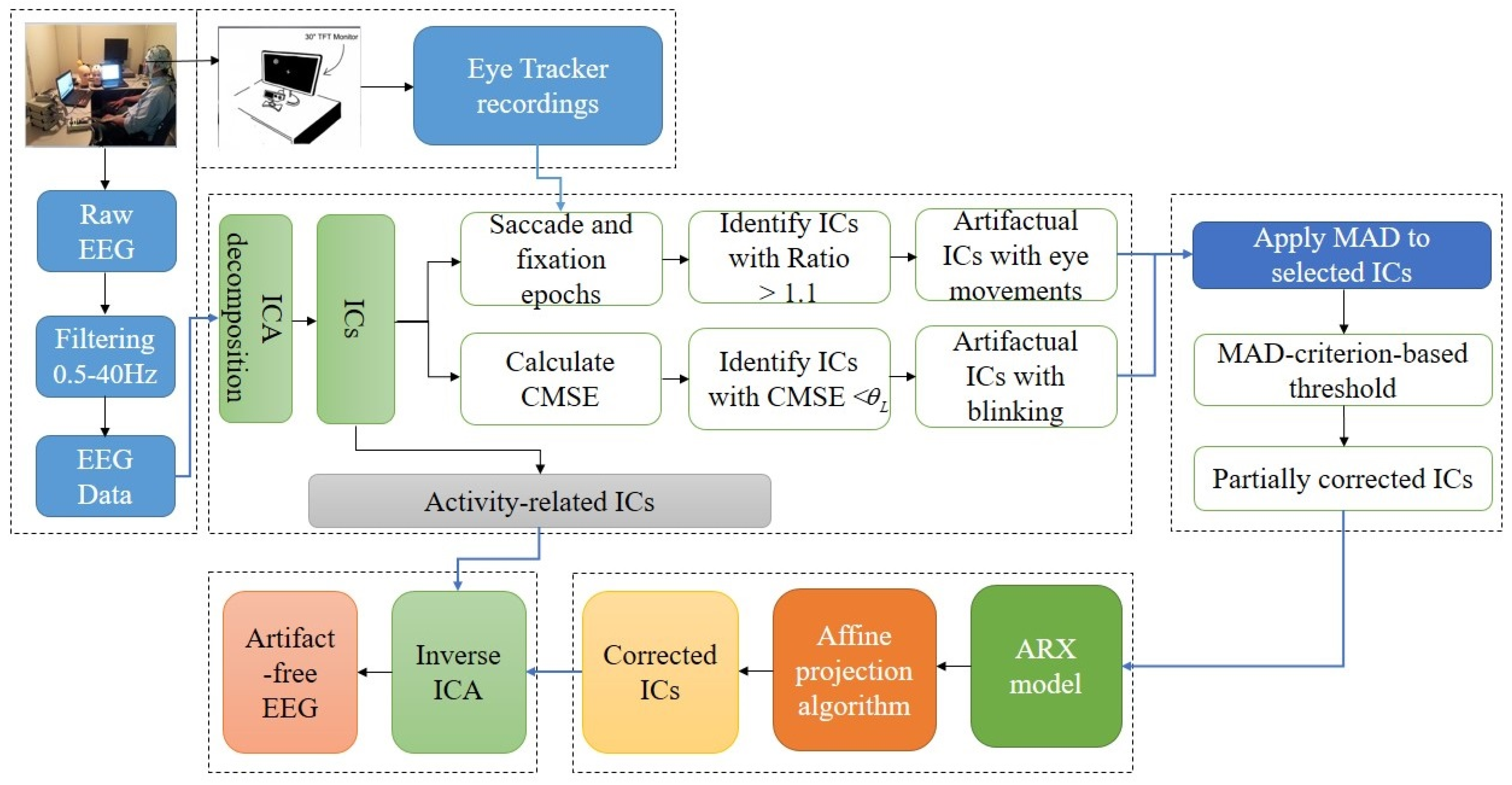
2. Materials and Methods
2.1. Materials
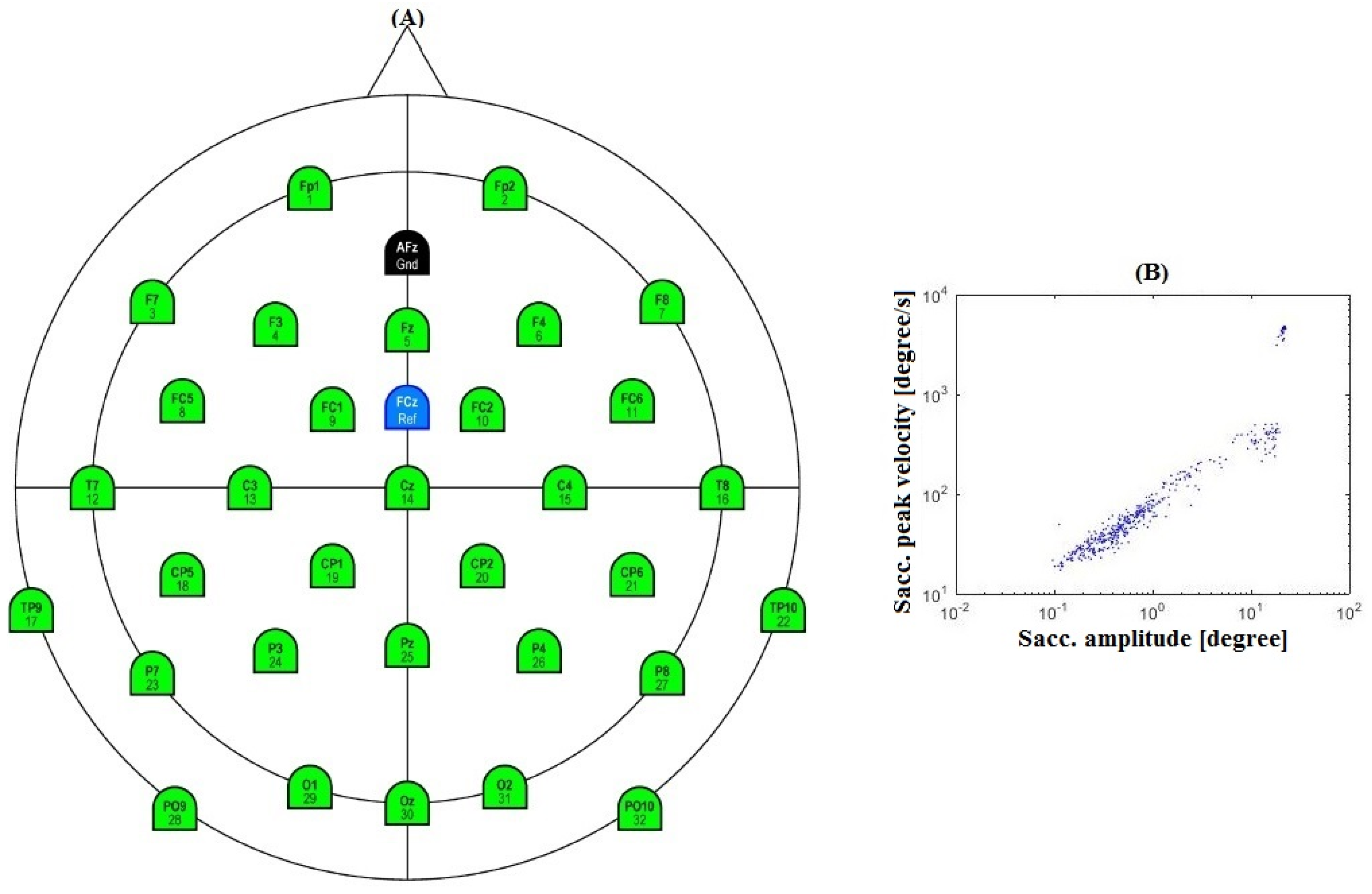
2.1.1. Participants
2.1.2. Experimental Procedure
2.1.3. EEG Recordings
2.1.4. Eye Tracker Recordings
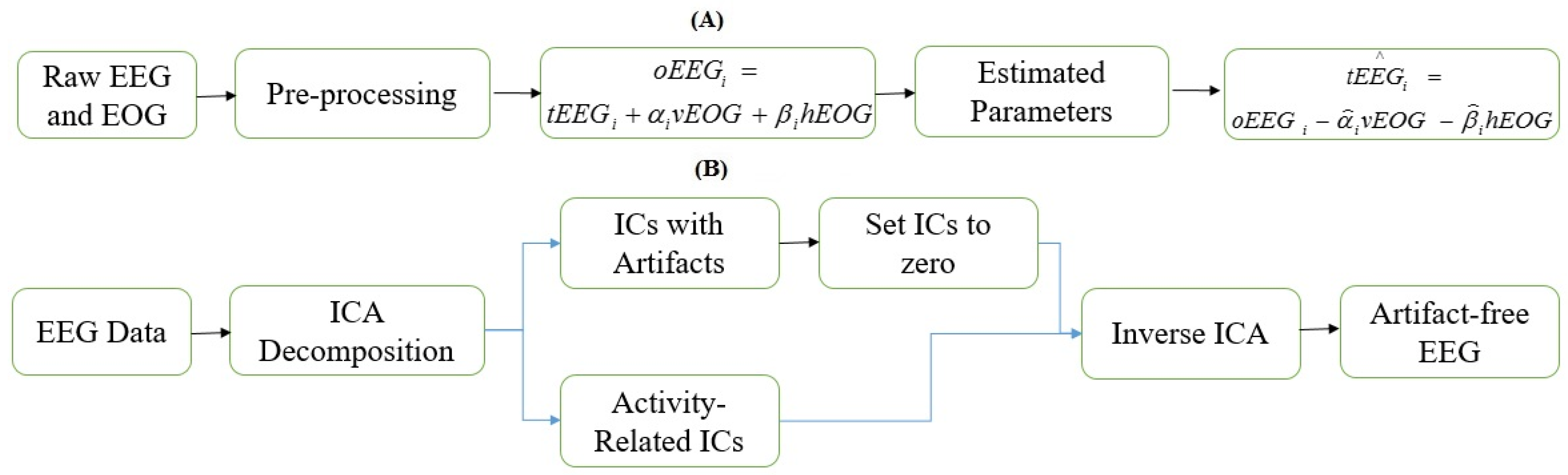
2.1.5. Preprocessing
2.1.6. Standard Dataset
2.2. Methods
2.2.1. Independent Component Analysis
- The number of ICs are less than or equal to the number of observed signals.
- The artifactual and cerebral sources are linearly mixed and statistically independent.
- Propagation delays through the missing medium (brain) are negligible.
2.2.2. Features Computation
Eye Blinks
- (1)
- Let be the ith IC, the lth coarse-grained time series for a scale factor of , can be defined as
- (2)
- In the composite multi-scale entropy algorithm, at a scale factor of , the sample entropies (SampEns) of all coarse-grained time series are calculated and the composite multi-scale entropy value is defined as the mean of entropy values. That iswhere CMSE represents the composite multi-scale entropy. In this study, the composite multi-scale entropy was calculated from , and the sample entropy of each coarse-grained IC was calculated with m = 2 and , where is the standard deviation of the IC [42,44].
Horizontal Eye Movements
Vertical Eye Movements
2.2.3. Median Absolute Deviation
- (1)
- Evaluate the median absolute deviation of the identified ocular activity among the identified artifactual ICs (median absolute deviation is defined as the median of the absolute deviation from the median)where is the median absolute deviation, is the median, is the median of the ith artifactual IC, is a constant;
- (2)
- If exceeds the criteria calculated using Equation (9), it is thresholded to zero:
2.2.4. Auto-Regressive Exogenous Model
2.2.5. Affine Projection Algorithm
3. Evaluation Index
3.1. Relative Error
3.2. Mutual Information
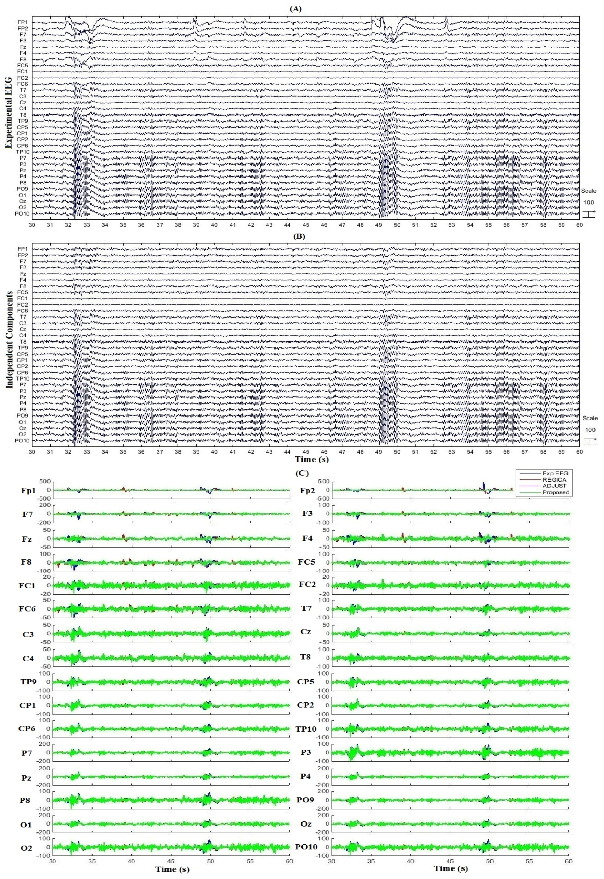
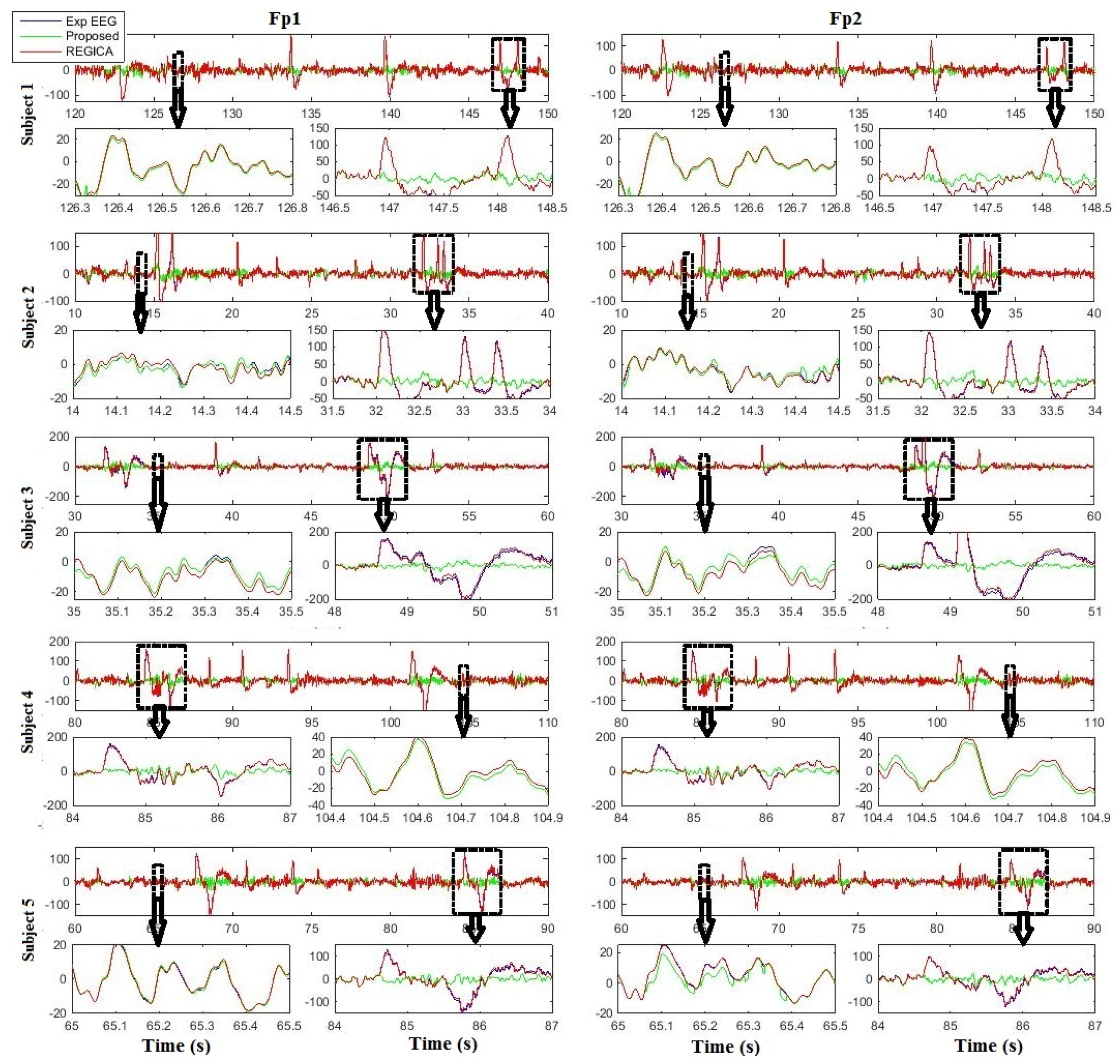
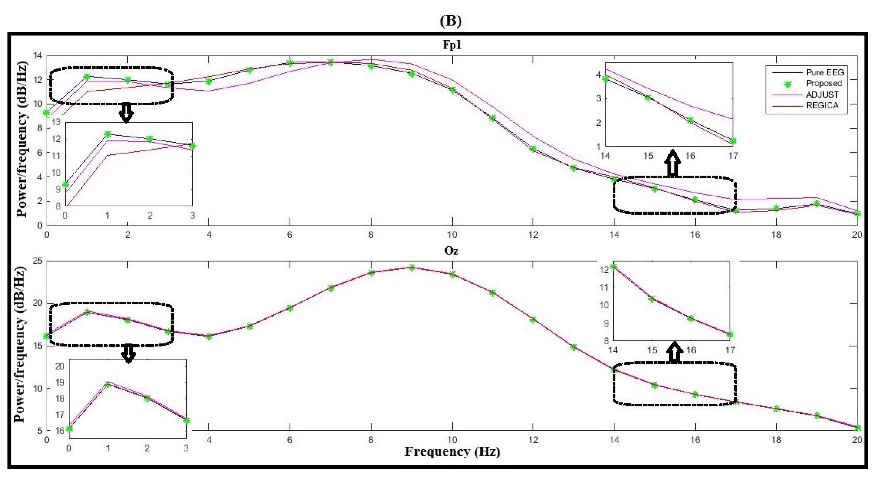
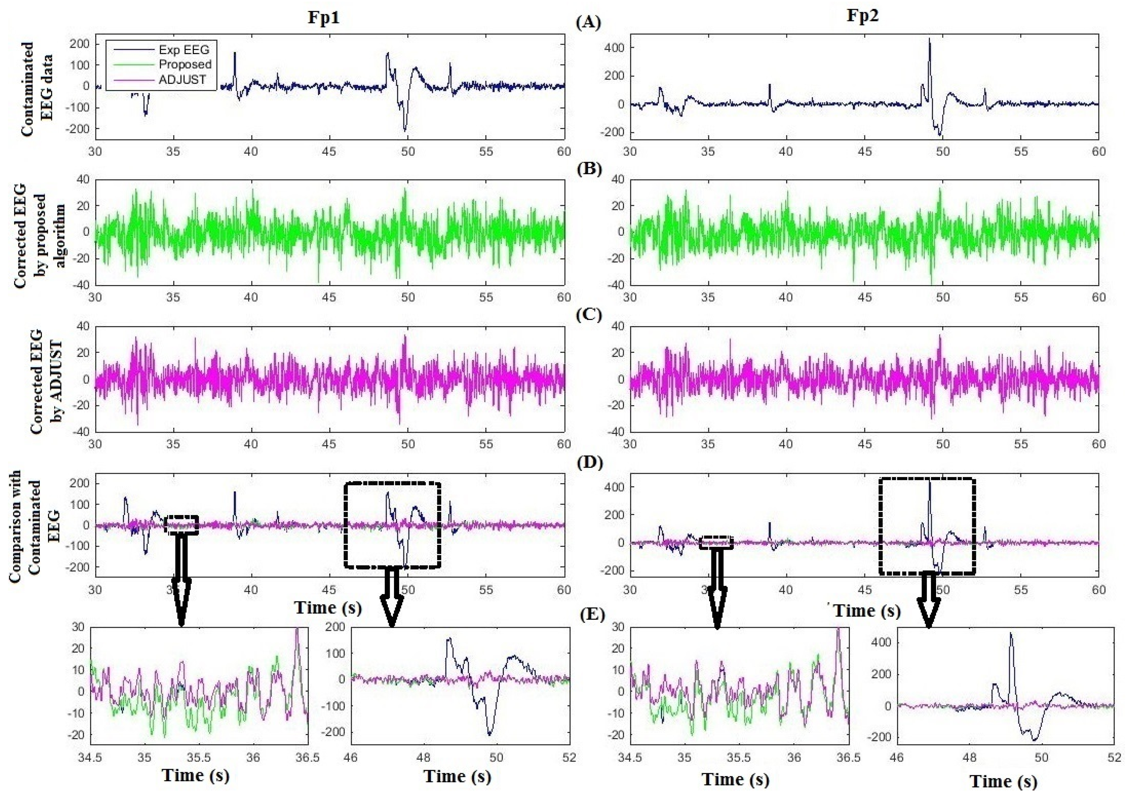
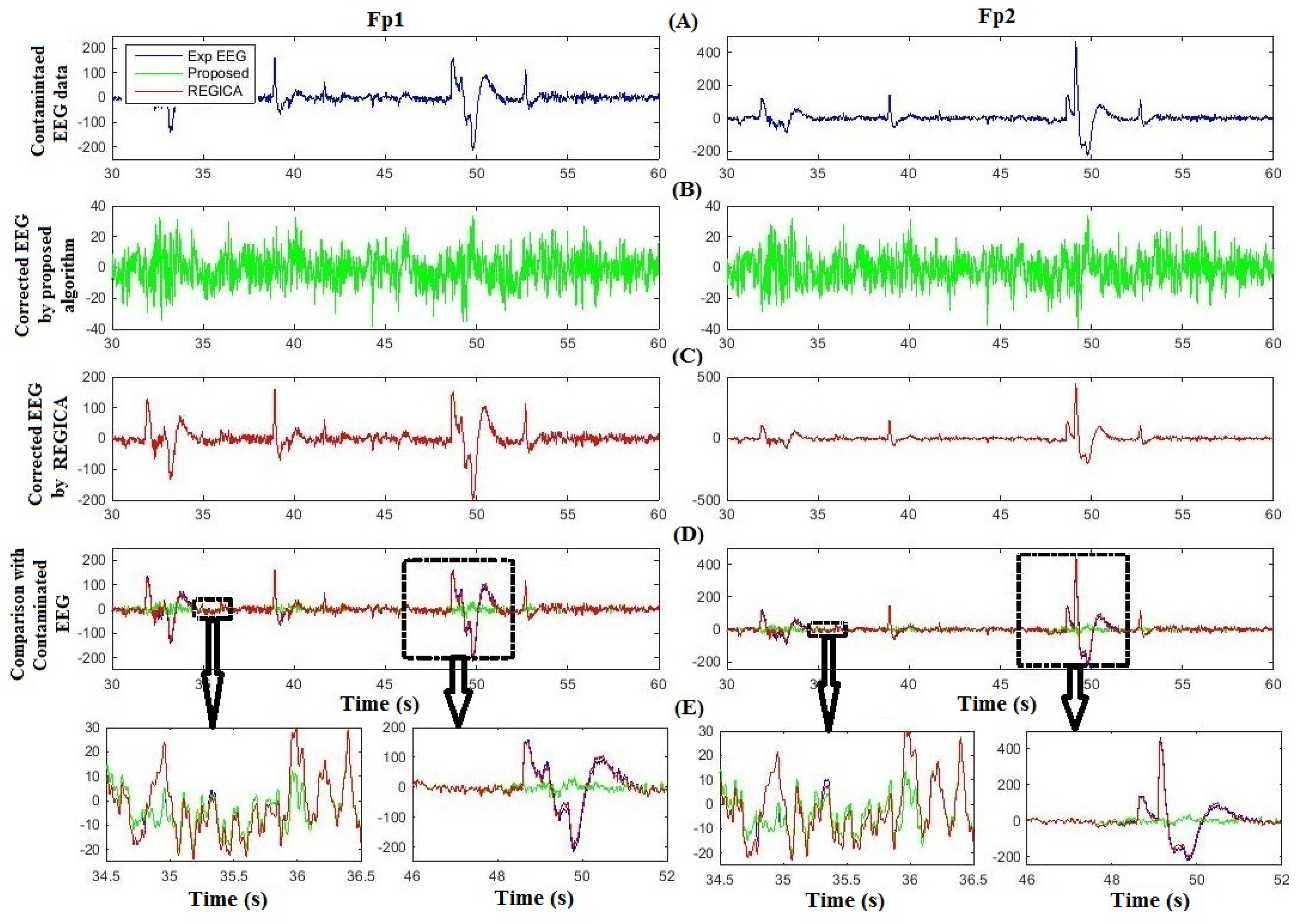
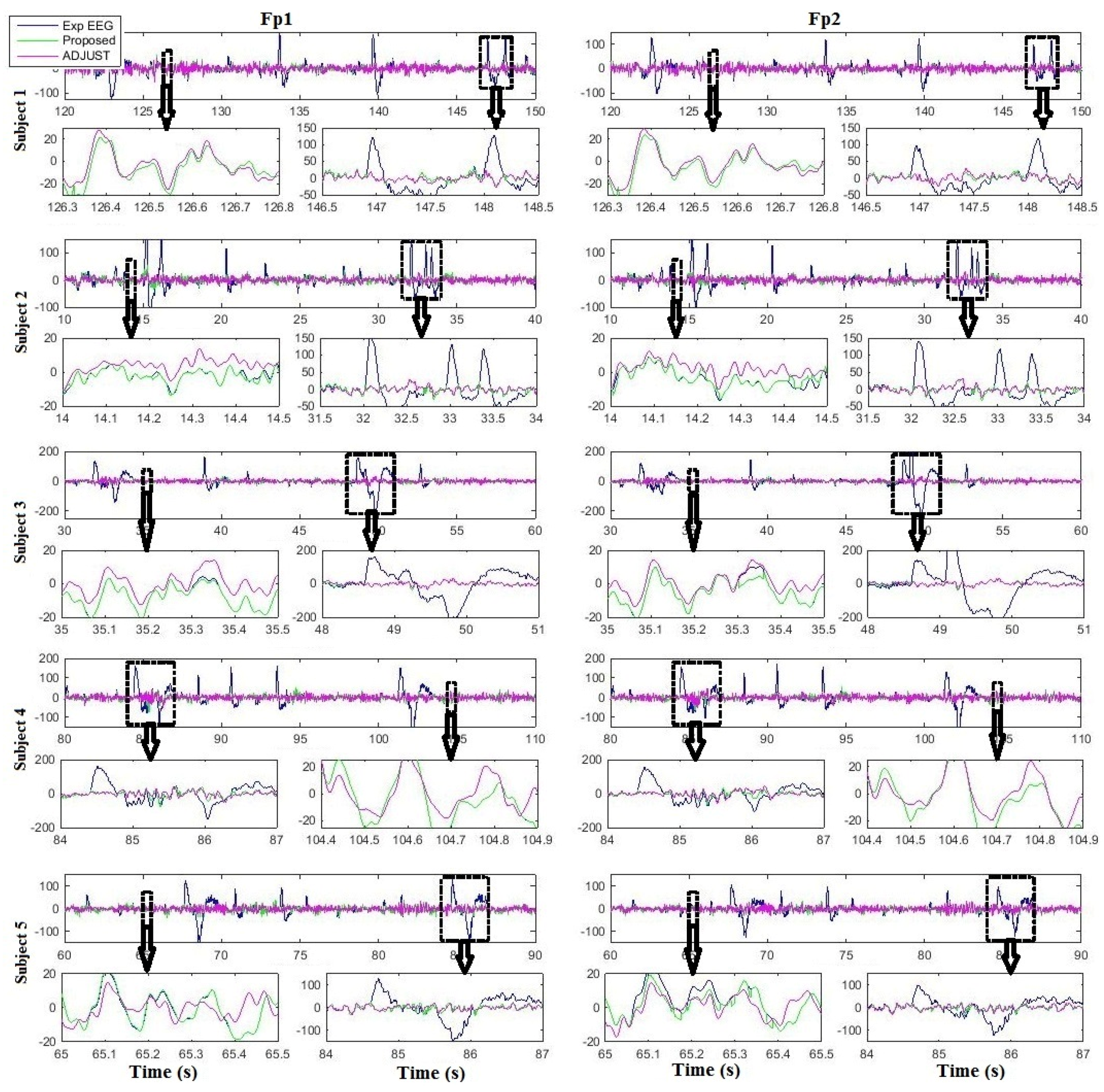
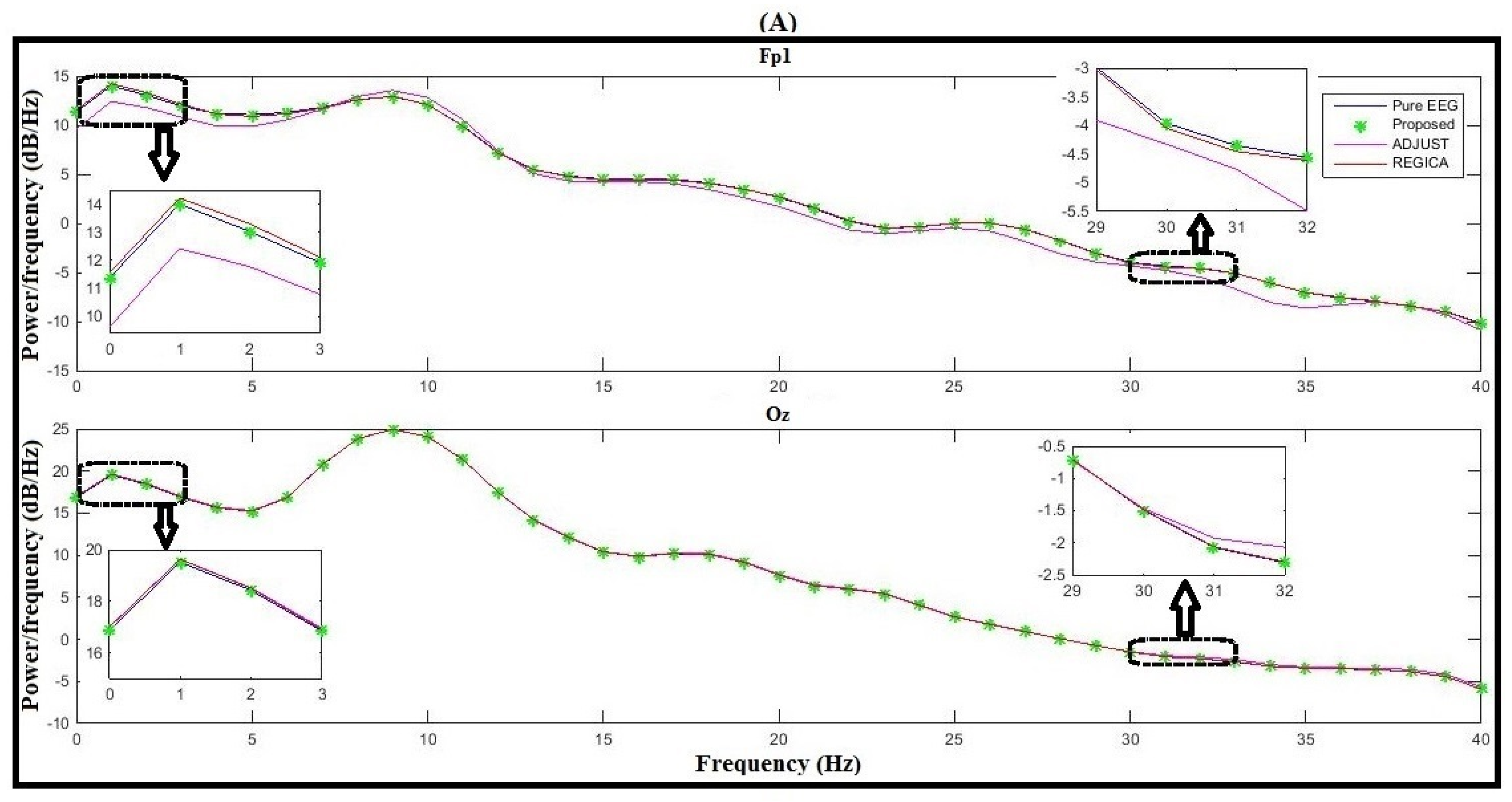
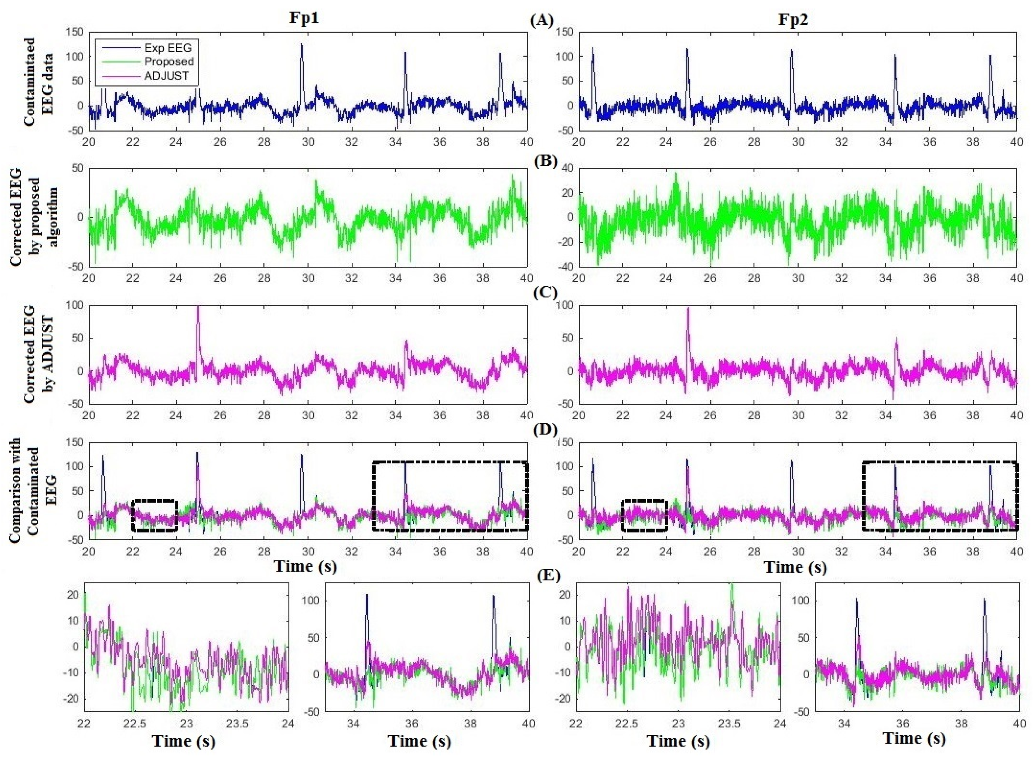
4. Results
5. Discussion
6. Conclusions
Acknowledgments
Author Contributions
Conflicts of Interest
References
- Jöbsis, F.F. Noninvasive infrared monitoring of cerebral and myocardial oxygen sufficiency and circulatory parameters. Science 1977, 198, 1264–1267. [Google Scholar] [CrossRef] [PubMed]
- Friston, K.J.; Jezzard, P.; Turner, R. Analysis of functional MRI time-series. Hum. Brain Mapp. 1994, 1, 153–171. [Google Scholar] [CrossRef]
- Hogervorst, M.A.; Brouwer, A.-M.; van Erp, J.B.F. Combining and comparing EEG, peripheral physiology and eye-related measures for the assessment of mental work load. Front. Neurosci. 2014, 8. [Google Scholar] [CrossRef]
- Kamran, M.A.; Hong, K.-S.; Mannan, M.N.M. Identification of fNIRS based Brain Activity Using Adaptive Algorithm. NUST J. Eng. Sci. 2012, 5, 7–10. [Google Scholar]
- Kamran, M.A.; Jeong, M.Y.; Mannan, M.N.M. Optimal hemodynamic response model for functional near-infrared spectroscopy. Front. Behav. Neurosci. 2015, 9. [Google Scholar] [CrossRef] [PubMed]
- Zeng, H.; Song, A.; Yan, R.; Qin, H. EOG artifact correction from EEG recording using stationary subspace analysis and empirical mode decomposition. Sensors 2013, 13, 14839–14859. [Google Scholar] [CrossRef] [PubMed]
- Sameni, R.; Gouy-Pailler, C. An iterative subspace denoising algorithm for removing electroencephalogram ocular artifacts. J. Neurosci. Methods, 2014, 225, 97–105. [Google Scholar] [CrossRef] [PubMed]
- Berg, P.; Scherg, M. Dipole modelling of eye activity and its application to the removal of eye artefacts from the EEG and MEG. Clin. Phys. Physiol. Meas. 1991, 12, 49–54. [Google Scholar] [CrossRef] [PubMed]
- Wallstrom, G.L.; Kass, R.E.; Miller, A.; Cohn, J.F.; Fox, N.A. Automatic correction of ocular artifacts in the EEG: A comparison of regression-based and component-based methods. Int. J. Psychophysiol. 2004, 53, 105–119. [Google Scholar] [CrossRef] [PubMed]
- Dimigen, O.; Sommer, W.; Hohlfeld, A.; Jacobs, A.; Kliegl, R. Co-registration of eye movements and EEG in natural reading: Analyses and review. J. Exp. Psychol. 2011, 140, 552–572. [Google Scholar] [CrossRef] [PubMed]
- Carl, C.; Hipp, J.F.; König, P.; Engel, A.K. Spectral signatures of saccade target selection. Brain Topogr. 2016, 29, 130–148. [Google Scholar] [CrossRef] [PubMed]
- Carl, C.; Açık, A.; König, P.; Engel, A.K.; Hipp, J.F. The saccade spike artifact in MEG. Neuroimage 2012, 59, 1657–1667. [Google Scholar] [CrossRef] [PubMed]
- Engbert, R.; Mergenthaler, K. Microsaccades are triggered by low retinal image slip. Proc. Natl. Acad. Sci. USA 2006, 103, 7192–7197. [Google Scholar] [CrossRef] [PubMed]
- Fatourechi, M.; Bashashati, A.; Ward, R.K.; Birch, G.E. EMG and EOG artifacts in brain computer interface systems: A survey. Clin. Neurophysiol. 2007, 118, 480–494. [Google Scholar] [CrossRef] [PubMed]
- Croft, R.J.; Barry, R.J. EOG correction: A new perspective. Electroencephalogr. Clin. Neurophysiol. 1998, 107, 387–394. [Google Scholar] [CrossRef]
- Croft, R.J.; Barry, R.J. EOG correction: A new aligned-artifact average solution. Electroencephalogr. Clin. Neurophysiol. 1998, 107, 395–401. [Google Scholar] [CrossRef]
- Croft, R.J.; Barry, R.J. Removal of ocular artifact from the EEG: A review. Clin. Neurophysiol. 2000, 30, 5–19. [Google Scholar] [CrossRef]
- Croft, R.J.; Barry, R.J. EOG correction: Which regression should we use? Psychophysiology 2000, 37, 123–125. [Google Scholar] [CrossRef] [PubMed]
- Croft, R.J.; Chandler, J.S.; Barry, R.J.; Cooper, N.R.; Clarke, A.R. EOG correction: A comparison of four methods. Psychophysiology 2005, 42, 16–24. [Google Scholar] [CrossRef] [PubMed]
- Romero, S.; Mañanas, M.A.; Barbanoj, M.J. Ocular reduction in EEG signals based on adaptive filtering, regression and blind source separation. Ann. Biomed. Eng. 2009, 37, 176–191. [Google Scholar] [CrossRef] [PubMed]
- Barbati, G.; Porcaro, C.; Zappasodi, F.; Rossini, P.M.; Tecchio, F. Optimization of independent component analysis approach for artifact identification and removal in MEG signals. Clin. Neurophysiol. 2004, 115, 1220–1232. [Google Scholar] [CrossRef] [PubMed]
- Joyce, C.A.; Gorodnitsky, I.F.; Kutas, M. Automatic removal of eye movement and blink artifacts from EEG data using blind component separation. Psychophysiology 2004, 41, 313–325. [Google Scholar] [CrossRef] [PubMed]
- Hoffmann, S.; Falkenstein, M. The correction of eye blink artefacts in the EEG: A comparison of two prominent methods. PLoS ONE 2008, 3. [Google Scholar] [CrossRef] [PubMed]
- Ghandeharion, H.; Erfanian, A. A fully automatic ocular artifact suppression from EEG data using higher order statistics: Improved performance by wavelet analysis. Med. Eng. Phys. 2010, 32, 720–729. [Google Scholar] [CrossRef] [PubMed]
- Javidi, S.; Mandic, D.P.; Took, C.C.; Cichocki, A. Kurtosis-based blind source extraction of complex non-circular signals with application in EEG artifact removal in real-time. Front. Neurosci. 2011, 5. [Google Scholar] [CrossRef] [PubMed]
- Winkler, I.; Haufe, S.; Tangermann, M. Automatic classification of artifactual ICA-components for artifact removal in EEG signals. Behav. Brain Funct. 2011, 7. [Google Scholar] [CrossRef] [PubMed]
- Mahajan, R.; Morshed, B.I. Unsupervised Eye Blink Artifact Denoising of EEG Data with Modified Multiscale Sample Entropy, Kurtosis, and Wavelet-ICA. IEEE J. Biomed. Health Inform. 2015, 19, 158–165. [Google Scholar] [CrossRef] [PubMed]
- He, P.; Wilson, G.; Russel, C. Removal of ocular artifacts from electroencephalogram by adaptive filtering. Med. Biol. Eng. Comput. 2004, 42, 407–412. [Google Scholar] [CrossRef] [PubMed]
- Klados, M.A.; Papadelis, C.; Braun, C.; Bamidis, P.D. REG-ICA: A hybrid methodology combining blind source separation and regression techniques for the rejection of ocular artifacts. Biomed. Signal Process. Control 2011, 6, 291–300. [Google Scholar] [CrossRef]
- Peng, H.; Hu, B.; Shi, Q.; Ratcliffe, M.; Zhao, Q.; Qi, Y.; Gao, G. Removal of ocular artifacts in EEG—An improved approach combining DWT and ANC for portable applications. IEEE J. Biomed. Health Inform. 2013, 17, 600–607. [Google Scholar] [CrossRef] [PubMed]
- Iriarte, J.; Urrestarazu, E.; Valencia, M.; Alegre, M.; Malanda, A.; Viteri, C.; Artieda, J. Independent component analysis as a tool to eliminate artifacts in EEG: A quantitative study. J. Clin. Neurophysiol. 2003, 20, 249–257. [Google Scholar] [CrossRef] [PubMed]
- Kong, W.Z.; Zhou, Z.P.; Hu, S.Q.; Zhang, J.H.; Babiloni, F.; Dai, G.J. Automatic and direct identification of blink components from scalp EEG. Sensors 2013, 13, 10783–10801. [Google Scholar] [CrossRef] [PubMed]
- Plöchl, M.; Ossandón, J.P.; König, P. Combining EEG and eye tracking: Identification, characterization, and correction of eye movement artifacts in electroencephalographic data. Front. Hum. Neurosci. 2012, 6. [Google Scholar] [CrossRef] [PubMed]
- Mognon, A.; Jovicich, J.; Bruzzone, L.; Buiatti, M. ADJUST: An automatic EEG artifact detector based on the joint use of spatial and temporal features. Psychophysiology 2010, 48, 229–240. [Google Scholar] [CrossRef] [PubMed]
- Castellanos, N.P.; Makarov, V.A. Recovering EEG brain signals: Artifact suppression with wavelet enhanced independent component analysis. J. Neurosci. Methods 2006, 158, 300–312. [Google Scholar] [CrossRef] [PubMed]
- Ehinger, B.V.; König, P.; Ossandón, J.P. Predictions of visual content across eye movements and their modulation by inferred information. J. Neurosci. 2015, 35, 7403–7413. [Google Scholar] [CrossRef] [PubMed]
- Kierkels, J.; Riani, J.; Bergmans, J. Using an eye tracker for accurate eye movement artifact correction. IEEE Trans. Biomed. Eng. 2007, 54, 1257–1267. [Google Scholar] [CrossRef] [PubMed]
- Noureddin, B.; Lawrence, P.D.; Birch, G.E. Online removal of eye movement and blink EEG artifacts using a high-speed eye tracker. IEEE Trans. Biomed. Eng. 2012, 59, 2103–2110. [Google Scholar] [CrossRef] [PubMed]
- EYE-EEG: Eye tracking & EEG. Available online: http://www2.hu-berlin.de/eyetracking-eeg/testdata.html (accessed on 20 October 2015).
- Delorme, A.; Sejnowski, T.; Makeig, S. Enhanced detection of artifacts in EEG data using higher-order statistics and independent component analysis. Neuroimage 2007, 34, 1443–1449. [Google Scholar] [CrossRef] [PubMed]
- Delorme, A.; Makeig, S. EEGLAB: An open source toolbox for analysis of single-trial EEG dynamics including independent component analysis. J. Neurosci. Methods 2004, 134, 9–21. [Google Scholar] [CrossRef] [PubMed]
- Costa, M.; Goldberger, A.L.; Peng, C.K. Multiscale entropy analysis of biological signals. Phys. Rev. E 2005, 71. [Google Scholar] [CrossRef] [PubMed]
- Bosl, W.; Tierney, A.; Tager-Flusberg, H.; Nelson, H. EEG complexity as a biomarker for autism spectrum disorder risk. BMC Med. 2011, 9. [Google Scholar] [CrossRef] [PubMed]
- Wu, S.-D.; Wu, C.-W.; Lin, S.-G.; Wang, C.-C.; Lee, K.-Y. Time Series Analysis Using Composite Multiscale Entropy. Entropy 2013, 15, 1069–1084. [Google Scholar] [CrossRef]
- Leys, C.; Ley, C.; Klein, O.; Bernard, P.; Licata, L. Detecting outliers: Do not use standard deviation around the mean, use absolute deviation around the median. J. Exp. Soc. Psychol. 2013, 49, 764–766. [Google Scholar] [CrossRef]
- Kamran, M.A.; Hong, K.-S. Linear parameter-varying model and adaptive filtering technique for detecting neuronal activities: An fNIRS study. J. Neural Eng. 2013, 10. [Google Scholar] [CrossRef] [PubMed]
- Wang, Z.; Xu, P.; Liu, T.; Tian, Y.; Lei, X.; Yao, D. Robust removal of ocular artifacts by combining independent component analysis and system identification. Biomed. Signal Process. Control 2014, 10, 250–259. [Google Scholar] [CrossRef]
- Swartz Center for Computational Neuroscience. Available online: http://sccn.ucsd.edu/~jason/minfo.m (accessed on 1 November 2015).
- Stone, J.V. Independent component analysis: An introduction. Trends Cogn. Sci. 2002, 6, 59–64. [Google Scholar] [CrossRef]
- Tran, Y.; Craig, A.; Boord, P.; Craig, D. Using independent component analysis to remove artifact from electroencephalographic measured during stuttered speech. Med. Bio. Eng. Comput. 2004, 42, 627–633. [Google Scholar] [CrossRef]
- Bian, N.Y.; Wang, B.; Cao, Y. Automatic removal of artifacts from EEG data using ICA and nonlinear exponential analysis. Acta Biophys. Sin. 2006, 22, 149–156. [Google Scholar]
- Mammone, N.; Morabito, F.C. Enhanced automatic artifact detection based on independent component analysis and Renyi’s entropy. Neural Netw. 2008, 21, 1029–1040. [Google Scholar] [CrossRef] [PubMed]
- Jung, T.P.; Makeig, S.; Humphries, C.; Lee, T.W.; McKeown, M.J.; Iragui, V.; Sejnowski, T.J. Removing electroencephalographic artifacts by blind source separation. Psychophysiology 2000, 37, 163–178. [Google Scholar] [CrossRef] [PubMed]
- Makeig, S.; Onton, J. ERP Features and EEG Dynamics: An ICA Perspective. In Oxford Handbook of Event-Related Potential Components; Luck, S.J., Kappenman, E.S., Eds.; Oxford University Press: New York, NY, USA, 2009; pp. 51–87. [Google Scholar]
- Urigüen, J.A.; Garcia-Zapirain, B. EEG artifact removal—State-of-the-art and guidelines. J. Neural Eng. 2015, 12. [Google Scholar] [CrossRef] [PubMed]


| Input: Contaminated EEG data, Eye tracker data |
| Output: Artifact-free EEG data |
| Synchronization of EEG and eye tracker Decompose contaminated EEG data using ICA to get ICs Portioning of ICs into saccade and fixation epochs Calculate composite multi-scale entropy and ratio of mean variance to identify ocular artifacts related ICs Apply median absolute deviation to remove high magnitude ocular activities from identified ICs Filter ICs with auto-regressive exogenous model and affine projection algorithm Artifact-free EEG data by back projecting all ICs using inverse ICA |
| Subject | Proposed | ADJUST ICA | p-val | REGICA | p-val |
|---|---|---|---|---|---|
| 1 | 0.0001 ± 0.0001 | 0.0645 ± 0.0944 | <0.001 | 0.0452 ± 0.0613 | <0.001 |
| 2 | 0.0185 ± 0.0305 | 0.1741 ± 0.1417 | <0.001 | 0.0437 ± 0.0393 | <0.011 |
| 3 | 0.0146 ± 0.0230 | 0.1025 ± 0.1191 | <0.001 | 0.0527 ± 0.06– | <0.005 |
| 4 | 0.0134 ± 0.0244 | 0.1893 ± 0.1582 | <0.001 | 0.0779 ± 0.1140 | <0.005 |
| 5 | 0.0273 ± 0.0320 | 0.2727 ± 0.2358 | <0.001 | 0.0366 ± 0.0389 | <0.17 |
| Average | 0.0147 ± 0.0220 | 0.1606 ± 0.1498 | 0.0512 ± 0.0637 |
| Electrode Location | Proposed | ADJUST | REGICA |
|---|---|---|---|
| Fp1 | 2.7148 | 0.6680 | 1.4023 |
| Fp2 | 2.6286 | 0.7578 | 1.4563 |
| F7 | 2.7169 | 1.1796 | 1.9115 |
| F3 | 2.7165 | 1.2789 | 2.0176 |
| Fz | 2.6973 | 1.3932 | 1.9717 |
| F2 | 2.6434 | 1.3789 | 2.0190 |
| F8 | 2.5538 | 1.4237 | 1.8863 |
| FC5 | 2.6946 | 1.2919 | 2.3216 |
| FC3 | 2.5262 | 1.5692 | 2.3181 |
| FC2 | 2.5720 | 1.8742 | 2.3239 |
| FC6 | 2.6034 | 1.8046 | 2.1482 |
| T7 | 2.7158 | 1.4571 | 2.3656 |
| C3 | 2.6685 | 1.4754 | 2.3942 |
| Cz | 2.7435 | 2.0669 | 2.4309 |
| C4 | 2.6075 | 1.9813 | 2.3062 |
| T8 | 2.6611 | 1.8524 | 2.3069 |
| TP9 | 2.6515 | 1.5851 | 2.3234 |
| CP5 | 2.6888 | 1.5566 | 2.4402 |
| CP1 | 2.6669 | 2.0910 | 2.4864 |
| CP2 | 2.7186 | 2.1592 | 2.4384 |
| CP6 | 2.5839 | 2.0236 | 2.3601 |
| TP10 | 2.6307 | 1.9541 | 2.3272 |
| P7 | 2.5600 | 1.8588 | 2.4463 |
| P3 | 2.6275 | 2.1776 | 2.4715 |
| Pz | 2.6076 | 2.1811 | 2.3613 |
| P2 | 2.6230 | 2.1739 | 2.3948 |
| P8 | 2.6290 | 2.0796 | 2.4958 |
| PO9 | 2.6540 | 2.2194 | 2.4626 |
| O1 | 2.6597 | 2.1380 | 2.4375 |
| Oz | 2.6758 | 2.2584 | 2.4655 |
| O2 | 2.5836 | 1.9191 | 2.3257 |
| PO10 | 2.6514 | 2.1337 | 2.4338 |
| Average | 2.6461 | 1.7488 | 2.2578 |
| Subject | EEG Experts | Propsoed Algorithm | ADJUST | |||
|---|---|---|---|---|---|---|
| Vertical and Horizontal | Blink | Vertical and Horizontal | Blink | Vertical and Horizontal | Blink | |
| 1 | 1 | 1 | 1 | 1 | 0 | 1 |
| 2 | 1 | 1 | 1 | 2 | 0 | 2 |
| 3 | 1 | 2 | 2 | 2 | 2 | 2 |
| 4 | 2 | 1 | 2 | 1 | 2 | 1 |
| 5 | 1 | 1 | 1 | 1 | 3 | 1 |
| Method | True Positive (TP) | False Positive (FP) | True Negative (TN) | False Negative (FN) | Average Sensitivity | Average Specificity |
|---|---|---|---|---|---|---|
| Proposed | 12 | 2 | 146 | 0 | 100% | 98.64% |
| ADJUST | 10 | 4 | 144 | 2 | 83.33% | 97.29% |
© 2016 by the authors; licensee MDPI, Basel, Switzerland. This article is an open access article distributed under the terms and conditions of the Creative Commons by Attribution (CC-BY) license (http://creativecommons.org/licenses/by/4.0/).
Share and Cite
Mannan, M.M.N.; Kim, S.; Jeong, M.Y.; Kamran, M.A. Hybrid EEG—Eye Tracker: Automatic Identification and Removal of Eye Movement and Blink Artifacts from Electroencephalographic Signal. Sensors 2016, 16, 241. https://doi.org/10.3390/s16020241
Mannan MMN, Kim S, Jeong MY, Kamran MA. Hybrid EEG—Eye Tracker: Automatic Identification and Removal of Eye Movement and Blink Artifacts from Electroencephalographic Signal. Sensors. 2016; 16(2):241. https://doi.org/10.3390/s16020241
Chicago/Turabian StyleMannan, Malik M. Naeem, Shinjung Kim, Myung Yung Jeong, and M. Ahmad Kamran. 2016. "Hybrid EEG—Eye Tracker: Automatic Identification and Removal of Eye Movement and Blink Artifacts from Electroencephalographic Signal" Sensors 16, no. 2: 241. https://doi.org/10.3390/s16020241
APA StyleMannan, M. M. N., Kim, S., Jeong, M. Y., & Kamran, M. A. (2016). Hybrid EEG—Eye Tracker: Automatic Identification and Removal of Eye Movement and Blink Artifacts from Electroencephalographic Signal. Sensors, 16(2), 241. https://doi.org/10.3390/s16020241