Review

DNA-Based Nanobiosensors as an Emerging Platform for Detection of Disease

by
Khalid M. Abu-Salah
1,2,3,*,
Mohammed M. Zourob
4,
Fouzi Mouffouk
5,
Salman A. Alrokayan
1,2,
Manal A. Alaamery
3 and
Anees A. Ansari
2
1
Research Chair for Medical Applications of Nanomaterials, King Saud University, PO Box 2454, Riyadh 11451, Saudi Arabia
2
King Abdullah Institute for Nanotechnology, King Saud University, PO Box 2454, Riyadh 11451, Saudi Arabia
3
King Abdulla International Medical Research Center, King Abdulaziz Medical City, PO Box 22490, Riyadh 11426, Saudi Arabia
4
Department of Chemistry, Alfaisal University, Al Zahrawi Street, Al Maather, Al Takhassusi Rd, Riyadh 11533, Saudi Arabia
5
Chemistry Department, Faculty of Science, Kuwait University, PO Box 2969, Kuwait
*
Author to whom correspondence should be addressed.
Sensors 2015, 15(6), 14539-14568; https://doi.org/10.3390/s150614539
Submission received: 16 April 2015 / Revised: 19 May 2015 / Accepted: 22 May 2015 / Published: 19 June 2015
(This article belongs to the Section Biosensors)

Abstract

:
Detection of disease at an early stage is one of the biggest challenges in medicine. Different disciplines of science are working together in this regard. The goal of nanodiagnostics is to provide more accurate tools for earlier diagnosis, to reduce cost and to simplify healthcare delivery of effective and personalized medicine, especially with regard to chronic diseases (e.g., diabetes and cardiovascular diseases) that have high healthcare costs. Up-to-date results suggest that DNA-based nanobiosensors could be used effectively to provide simple, fast, cost-effective, sensitive and specific detection of some genetic, cancer, and infectious diseases. In addition, they could potentially be used as a platform to detect immunodeficiency, and neurological and other diseases. This review examines different types of DNA-based nanobiosensors, the basic principles upon which they are based and their advantages and potential in diagnosis of acute and chronic diseases. We discuss recent trends and applications of new strategies for DNA-based nanobiosensors, and emphasize the challenges in translating basic research to the clinical laboratory.

1. Introduction

Deoxyribonucleic acid (DNA) is a molecule found in cells of humans and almost all living organisms. Nearly every cell in the human body contains the same DNA. It comprises information used in our everyday metabolism and physiological activities and influences most of our characteristics. The specific DNA code provides the instructions for each protein to function in a specific manner. Each gene in the DNA encodes a specific protein, which may be involved in different functions and may play a major role in the cell, such as cell signaling and the pathogenesis process of human diseases. DNA is an ideal material for nanofabrication of rigid compositions because its implementation is relatively simple as its assembly can be controlled by base pairing and is comparatively of low cost. The determination of DNA sequences plays an important role in investigating particular diseases. The molecular diagnostics based analyses of genomic sequences have offered a highly sensitive and quantitative method for the detection of infectious diseases, pathogens, and genetic variations. Simple, ultrasensitive detection of sequence-specific DNA has been the aim of many studies and technologies for decades.
Recently, the field of nanotechnology has accelerated the process of integration of various scientific fields, thereby enhancing the breakdown of boundaries between already known disciplines. This eventually led to the emergence of the new interdisciplinary science of nanotechnology and its interrelated biology branch of nanobiotechnology. Advances and developments in this branch culminated in the establishing of nanomedicine, which includes amongst other disciplines, diagnostic materials and devices, molecular imaging, drug delivery systems and regenerative medicine [1]. Remarkably, nanomedicine enables in vitro and in vivo non-invasive diagnosis and targeted therapy by novel discoveries in sensing, processing, and operating processes. Currently, imaging tools based on nanotechnology have been medically applied as non-invasive methods of diagnosis [2,3,4]. The categories of nanodiagnostic technologies, in addition to DNA-based nanobiosensors, include nanoproteomic-based diagnostics, nanoparticle-based immunoassays, nanoparticulate biolabels, nanoscale visualization (e.g., scanning probe microscopy, scanning electron microscopy), biobarcode assays, nanobiotechnology-based, biochips and microarrays, and combinations of multiple diagnostics technologies.
Nanobiosensors have been increasingly used in medical diagnostics for continuous monitoring of human health [1,2,5,6,7], in addition to their applications in the field of food analysis [8], bioterrorism [9,10], and environment [9,10,11,12]. Presently, the most fascinating and prospective nanobiosensors are DNA-based types [13,14,15,16,17,18,19,20].
Unlike enzymes or antibodies, nucleic acid recognition layers in DNA-based nanobiosensors can be readily prepared and regenerated for multiple use. DNA-based nanobiosensors can be constructed by immobilizing single stranded probes on different electrodes using electro active-indicators to measure the hybridization between DNA strands and their complementary DNA probes [21,22,23]. The detection of specific DNA sequences is significant, not only in clinical diagnostics, but it is also of increasing importance in environment and food analysis [3,24,25]. Here, we focus on the potential of DNA-based nanobiosensors that can selectively target markers of acute and chronic diseases and highlight the challenges in translating some of the basic research to the clinical laboratory.

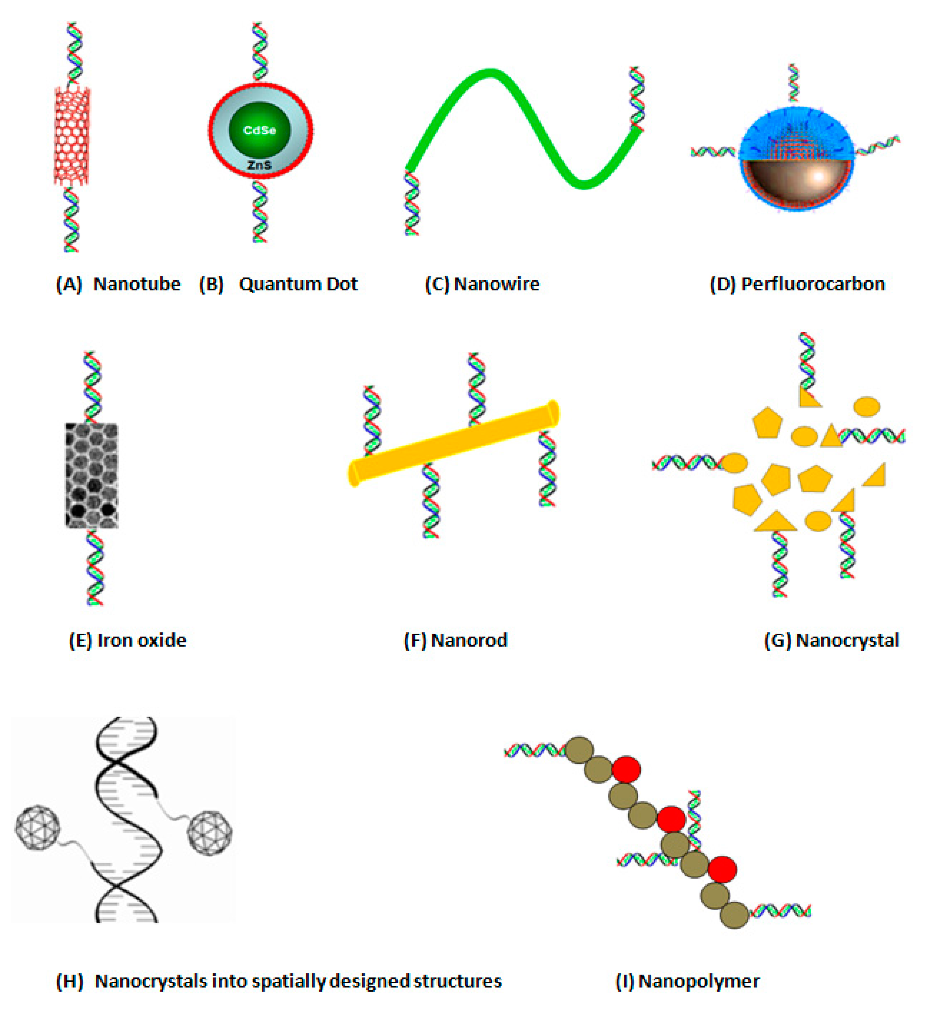
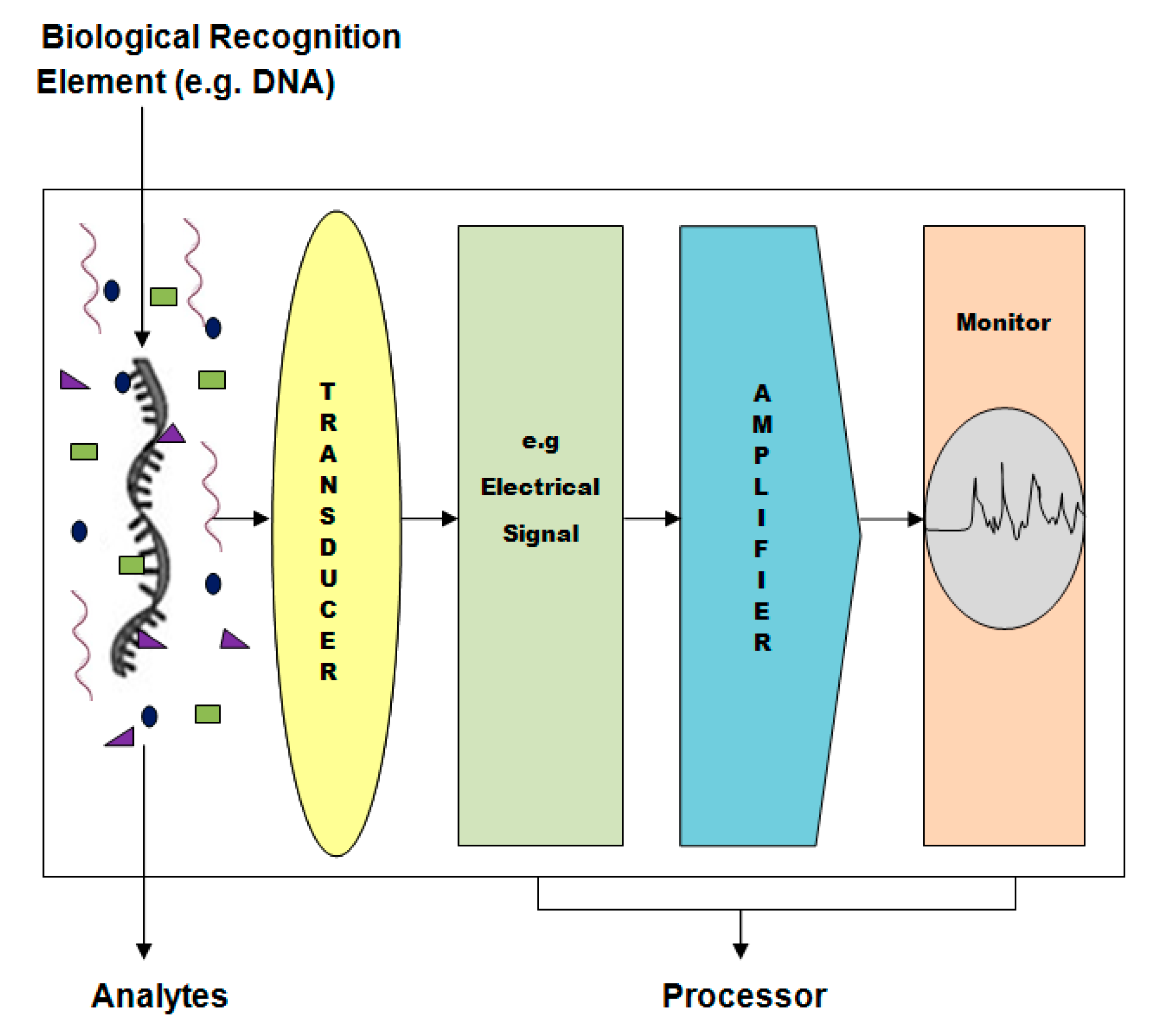
2. What Are DNA-Based Nanobiosensors

A biosensor is a device that detects, transmits and records the information on a biological analyte. Examples of analytes include nucleic acids (DNA, RNA), proteins such as enzymes, antibodies and antigens, or other biological component such as glucose. A basic biosensor assembly includes a biological recognition element, transducer, and processor (Figure 1).
Figure 1. A schematic diagram shows basic biosensor assembly with a biological recognition element, transducer, and processor.
Sensors 15 14539 g001
A biological recognition element can be consolidated to a number of different transducers. The recognition or sensing element, such as nucleic acids, antibodies, enzymes, proteins, or whole cells, can be integrated with a transducer via immobilization by covalent interaction, cross-linking, or adsorption. The transducer converts the molecular biological signal into a digital or electric signal proportional to the concentration of analyte and can be amplified, quantified, displayed, and analyzed through a processor. Nanostructured materials based transducers enhance the sensitivity by more than one order of magnitude compared to that observed at nanomaterials-bare, such as high loading of recognition element and a better electrical communication ability of the nanomaterials conventional electrodes [5,6,26,27]. This improved analytic performance is attributed to factors such as better electrical communication ability of nanomaterials (nanotubes, nanorods, and nanofibers) and high loading of recognition elements. The transducer function depends on the parameter to be measured. It may be optical (measurement of colorimetric, fluorescent, luminescent, and interferometric changes), potentiometric (potential measurement at constant current) [28], amperometric (current measurement at constant potential) [29], piezoelectric and acoustic waves (measurement change in mass) [30], or calorimetric (measurement changes in temperature) [31].
The above functional types of biosensors may make use of nanomaterials as transducing substances. These nanomaterials include: Semionductor nanoparticles, or so called quantum dots (QD), silver, silica, perfluorocarbon and organic polymers, surface enhanced Raman spectroscopy (SERS) nanotags, and fluorescent lanthanide nanorods (Figure 2).
Figure 2. Different types of nanostructures-based transducers to which DNA can be attached.
Sensors 15 14539 g002
A nanobiosensor is a biosensor that acts at the nano-metric level on a nanoscale region. Several types of nanobiosensors have been proposed for use in clinical and laboratory diagnosis. These types include optical, electrochemical, electrical and electronic, nanowire, nanotube-based, viral, nano-shell, and quartz nanobiosensors. DNA-based nanobiosensors, which involve nucleic acid recognition processes, are rapidly being used for assay development towards simple and rapid testing of genetic material and infectious agents. DNA-based nanobiosensors, including gene chips, are of major interest due to their tremendous promise for detection of disease and obtaining sequence—specific information in a faster, simpler and cost-effective manner compared to the traditional hybridization techniques [32,33]. The DNA probe is either chemically or enzymatically labeled with chemiluminiscent or radioactive probes or ligands, such as biotin, since the unlabeled nucleic acid is not able to provide any signal by itself.

3. DNA-Based Nanobiosensors for Detection of Infectious Diseases

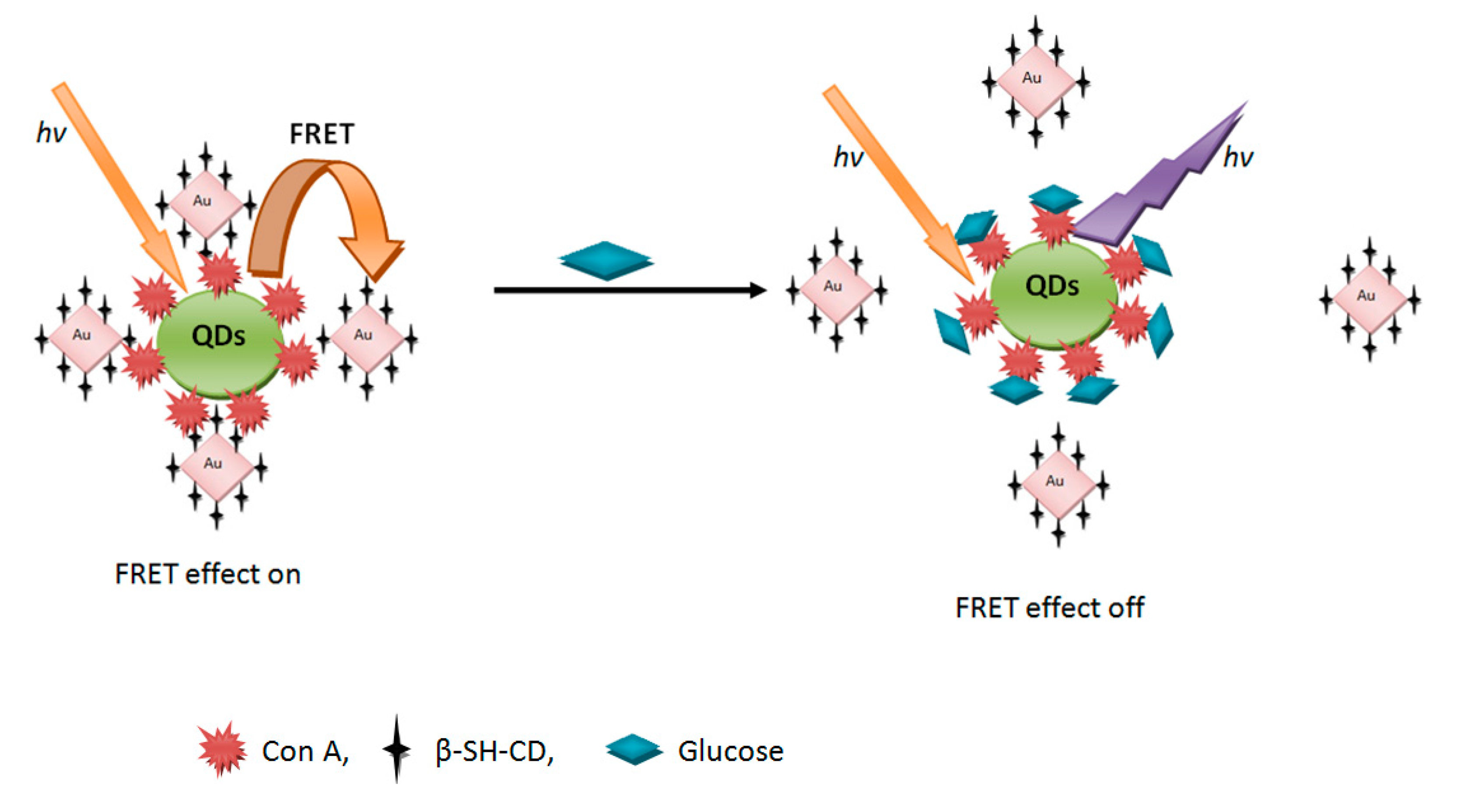
3.1. Optical Nanobiosensors

The rapid and sensitive detection of pathogenic microorganism at the point of care is essential in disease management and in impairing health outcomes. Most of the conventional microbiological diagnostic methods lack ultra-sensitivity and are limited by a delay in getting results. The most common DNA-based nanobiosensors make use of fluorescent nanostructures. Fluorescence occurs when a valence electron is excited from its ground state to an excited state. An electron emits a photon when it returns to its original ground state. Biosensors based on fluorescence resonance energy transfer (FRET) technology, which are used in probe-based assays for quantitative PCR (qPCR), make use of the transfer of energy from a donor fluorophore to an acceptor fluorophore when appreciable overlap exists between the absorption spectrum of the acceptor and the emission spectrum of the donor. This technology has also been used in screening gene mutations, measuring gene expression, and in quantifying viral loads [34,35,36,37].
Significant works have been reported in the literature on the utilization of nanostructured materials for construction of fluorescence sensors. In this context, semiconductors QDs have been used for proteins and DNA sensing. Melvin et al. [36] developed a fluorescence competition assay for DNA detection using gold nanoparticles (NPs) and QD as a FRET donor–acceptor couple. The gold nanoparticles are released from the QDs in the presence of complementary oligonucleotides, thus regenerating the fluorescence of the QD. Figure 3 schematically illustrates how this kind of biosensors works.
Figure 3. Chemical structure of the QDs-ConA-β-CDs-AuNPs nanobiosensor and schematic illustration of its FRET-based operating principles.
Sensors 15 14539 g003
The detection of single-molecule hybridization has been accomplished by a hybridization-detection method using multicolor single stranded DNA-functionalized QDs as a nanobiosensor [38]. Independent hybridization reaction in the presence of various target sequences leads to the generation of discernible sequence-specific spectral codings. Simultaneous detection of multiple relevant sequences can be used for genetic analysis of anthrax bacteria. Moreover, luminal functionalized silver nanoparticles have been utilized by He et al. [39] for ultrasensitive detection of Mycobacterium tuberculosis DNA by calculating chemiluminescence activity. On the other hand, a spectroscopic assay, based on SERS using silver nanorods, has been used for rapid, sensitive, and specific detection of viruses [40]. The technique is based on measuring the change in frequency of near infrared laser as it scatters viral DNA or RNA. Spectral differences, based on frequency, between viruses, viral strains, and viruses with gene deletions can be recorded rapidly (≤60 s) without viral manipulation.

3.2. Label-Based Electrochemical Nanobiosensors

Fluorimetric and other optical detection methods usually require highly transparent sample solution as the color and/or the fluorescence of the solution might interfere with the fluorimetric or optical signal generated from sensors. Electrochemical detection provides another useful technique for non-transparent samples, such as blood and urine, since the signal transduction can be achieved by non-fluorimetric or optical means [41]. Electrochemical devices received considerable attention in the development of sequence-specific DNA hybridization biosensors [42,43]. These devices offer smart routes for relating the DNA recognition element to the signal-transduction processes. Biosensors based on DNA hybridization depend on the generation of an electrical signal from the DNA base-pair recognition event [44,45,46]. Such a hybridization process is commonly detected via increased current signal of an electroactive indicator.

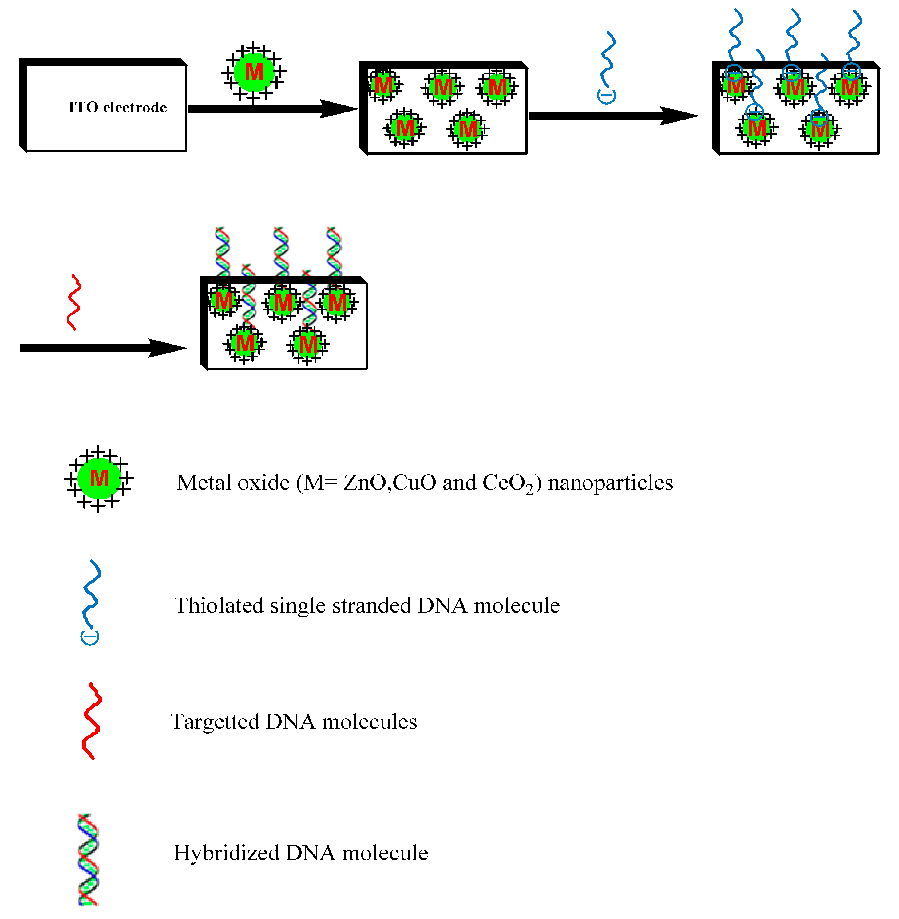
3.3. Label-Free Electrochemical Nanobiosensors

Increased interest has grown recently in direct, label-free electrochemical detection systems. Such direct detection can be accomplished by monitoring electrical changes resulting from the hybridization event. In addition the safety is improved by eliminating the need of using an indicator [47,48,49]. Wang et al. [50] introduced the first indicator-free scheme. Ansari et al. [51] employed a probe specific to Neisseria gonorrhea, which is composed of 20-m thiolated oligonucleotide (th-ssDNA) immobilized onto a sol-gel derived nanostructured zinc oxide (ZnO) film, dip-coated onto a glass substrate of indium-tin-oxide (ITO) (Figure 4).
This fabricated DNA nanobiosensor has been used for the detection of the sexually transmitted disease, gonorrhea. The results of the characterization studies performed on this th-ssDNA-ZnO/ITO bioelectrode using X-ray diffraction, scanning electron microscopy, Fourier-transform infrared, UV-visible revealed the linearity as 0.524 amol–0.524 nmol with a detection limit of 0.704 amol within 60 s. Another novel type of electrochemical DNA sensors has been fabricated by immobilizing thiolated single-stranded oligonucleotide (ss-DNA) probe onto gold (Au) coated glass electrode. Nisseria meningitides detection [52] using this type of DNA biosensor was accomplished by hybridization with complementary DNA (Ctr A) in presence of methylene blue (MB). Complimentary DNA in the range of 4–42 ng/mL (hybridization with response time 60 s) could be detected by this type of DNA/Au electrode. The electrode was found to be stable for about four months when stored at 4 °C. The sensitivity of double-stranded (ds-DNA)/Au electrode is 115.8 µA/ng.
Figure 4. Schematic presentation of immobilization of thiolated single-stranded probe DNA on the surface of ZnO for hybridization detection in double-stranded DNA (target DNA).
Sensors 15 14539 g004
A different approach has been used by Singh et al. [53] to fabricate a nanobiosensor for detection of N. gonorrhea. A nanobiosensor based on a multi-copy gene of N. gonorrhea functionalized nanostructured–polyaniline coated onto ITO-coated-glass plate has been fabricated. Avidin-biotin was used as a cross-linking agent in this type of biosensors. This DNA functionalized electrode can specifically detect up to 0.5 × 10⁻⁵ M of complementary target within 60 s of hybridization time at 25 °C by differential pulsed voltammetry (DPV) using methylene blue as electro-active DNA hybridization indicator. Moreover, the fabricated nanobiosensor can distinguish presence of N. gonorrhea from N. meningitides and Escherichia coli in cultures from urethral swabs of patients. Recently, Singh et al. [54] reported the use of a chitosan/Fe3O4 nanocomposite dispersion deposited on ITO electrode as a new platform for detection of gonorrhea. Chitosan is a biopolymer and has been used as an effective dispersant of nanoparticles for film fabrication [55]. The nanocomposite films deposited on electrode surface were mechanically stable and a further stabilization of the chitosan film containing iron oxide nanoparticles, achieved by chemical cross-linking with free aldehyde groups and avidin-biotin coupling, produced a substrate used for covalent immobilization of ssDNA and rapid and specific (mismatch-discriminating) DNA hybridization detection. The detection of oxygen uptake by chitosan (CH)-iron oxide (Fe3O4) nanocomposite via immobilization of biotinylated probe DNA was robust and applied for assays. A low detection limit (1 × 10⁻¹⁵ M) and very high assay throughput rate (1 × 10⁻¹⁶ M to 1 × 10⁻⁶ M) was achieved. Other analytic applications of chitosan doped nanomaterials have been reported in the literature, such as a biosensor based on chitosan doped with multiwall carbon nanotube (MWCNT), have been successfully used to detect gonorrhea. Methylene blue was employed as a DNA indicator. It was found that carbon nanotubes (CNTs) can enhance the electroactive surface area three fold (0.093 + 0.060 and 0.28 + 0.03 cm2 for chitosan-modified electrodes and chitosan-CNT, respectively). The resulting DNA biosensor shows a wide concentration range from 1 × 10⁻⁶ M to 1 × 10⁻¹⁷ M with a detection limit of 1 × 10⁻¹⁶ M. The amperometric response time is 60 s and extremely stable for about four months when stored at 4 °C [56]. In another approach Singh et al. have applied polyaniline/carbon nanotubes hybrid nanocomposite for immobilization of 5′-amino-labeled N. gonorrhea probe using glutaraldehyde as a cross-linker for sensitive and selective detection of gonorrhoea. The biosensor exhibits excellent response performance to gonorrhea with the linear range from 1 × 10⁻⁶ M to 1 × 10⁻⁷ M and a detection limit of 1.2 × 10⁻¹⁷ M. Furthermore, the biosensor shows high sensitivity, rapid response, long-term stability, good reproducibility and freedom of interference from other coexisting electroactive species [57].
Electrochemical sensors have also proved to be very useful for detecting small DNA damages induced by various enzymatic digestion, chemical agents, or ionizing radiation [58]. Such electrical detection is strongly dependent on the DNA structure. In addition to monitoring changes in tensammetric signals of DNA, such studies examine the response of electroactive damaging agents, such as carcinogens interacting with DNA in connection to the use of DNA-modified electrodes. Anticancer drugs in body fluids could be biosensed with similar electrodes [59]. Another approach which could be of potential applications in detecting viral markers demonstrated the use of DNA as a template for the fabrication of polyaniline nanowires [60]. Oriented DNA strands were formed on a thermally oxidized Si surface, then incubated in a solution of aniline monomer. The aligned aniline monomers were polymerized enzymatically to produce the nanofibers. The sensors were constructed by immobilizing DNA strands across an array of gold electrodes. A variation of electrical conductivity of polyaniline (PANI) nanofiber approach was recently reported by Fan et al. [61] to detect micro RNA (miRNA). To achieve this, capture probes of peptide nucleic acids (PNA, with sequence complementary to the miRNA target, were immobilized in the gaps of microelectrodes array. Addition of horseradish peroxidase, aniline, and H2O2 resulted in the formation of polyaniline nanofibers only when miRNA was hybridized to the PNA target. The amount of miRNA present was found to correlate to the conductance of the deposited PANI. The above technique could be potentially developed to detect miRNA specific for some pathogenic bacteria and viruses.
Malhotra and his coworkers constructed a sensitive amperometric nucleic acid biosensor wherein 21-m oligonucleotide probe (ss-DNA) is immobilized on nano-structured zirconium oxide (ZrO2) film deposited onto gold electrode for specific detection of Mycobacterium tuberculosis [62]. The resulting biosensor based on nanostructured metal oxide films display excellent electrocatalytic responses, which can be used for early and rapid diagnosis of M. tuberculosis with a detection limit of 0.065 ng/μL within 60 s. Zirconia nanoparticles grafted with chitosan (CHIT) (considering that chitosan, with abundant amino groups, exhibits excellent film-forming ability for the solubility in slightly acidic solution due to the protonation and insolubility in solution with pH above pKa (6.3) for deprotonation) has been applied as matrix to covalently immobilize the ss-DNA probe selective for M. tuberculosis detection. Chitosan improved the biocompatibility, toxicity, thermal, and chemical stability of the nanocomposite and enhanced the electron transfer ability efficiently between the analyte and the electrode surface [63]. Cyclic and differential pulse voltammetry measurements were used to investigate the electrochemical response of DNA/CHIT-NanoZrO2/ITO bioelectrode. The sensitivity of the bioelectrode observed was 6.38 × 10⁻⁶ AµM−1 could detect the complementary target DNA up to 0.00078 μM. In another approach similar group employed zirconia nanoparticles grafted MWCNTs based electrochemical impedometric biosensor for M. Tuberculosis detection [64]. The CNTs can amplify protein or DNA recognition and transduction events, which could be utilized as an ultrasensitive method for electrical biosensing of DNA. The carboxylic groups of MWCNTs of the electrode were covalently linked to the oligonucleotides with amine groups at the 5′ end. The hybridization events were monitored by electrochemical impedance spectroscopy measurements of the intercalated DNA of Nisseria gonorrhea. The ability of carbon nanotubes to allow electron-transfer reactions, the high surface area and catalytic activity of zirconia nanoparticles and the sensitivity of developed electrochemical DNA biosensors have been significantly improved. The bioelectrode could selectively detect target DNA concentration ranging from 1 × 10⁻² to 1 × 10⁻⁸ mM with a superior detection limit of 0.01 nM.
Tam et al. [65] have managed to covalently immobilize probe DNA on MWCNTs for label free and direct detection of influenza virus (type A). The investigators used Raman and FTIR spectra for the establishing of covalent bonding in between phosphate groups and amines of the DNA sequence. Target DNA down to 0.5 nM coud be detected by the fabricated DNA biosensor [65]. Recently, semiconductor (quantum dot, QD), e.g., CdSe, CdS, PbS and ZnS nanoparticles have attracted scientists in electrochemical biosensor applications. Owing to their unique (size-tunable fluorescent) properties they cause the labels in the electrochemical biosensors to be very sensitive [66]. Wang’s group was the first to prove the concept by using semiconductor nanoparticle labels for the assay of electrochemical DNA hybridization, and then it was extended to electrochemical DNA biosensors and immunosensors [67,68]. Fan et al. reported an electrochemical DNA biosensor for the measurements of avian influenza virus based on the use of semiconductor quantum dots [66]. A label-free or indicator-free DNA hybridization detection has been achieved by electrochemical signal measurements based on ss-DNA/CdSe modified GCE. The present method has different combining ability of ss-DNA and ds-DNA with CdSe nanostructures as measured by the change of electrochemical signal. The proposed biosensor based on quantum dots reveals excellent sensitivity and selectivity for detecting the Avian Influenza Virus (AIV) DNA sequences.
Potentiometric biosensors were developed by conjugating carbon nanotubes with aptamers [69]. This technique allowed label-free detection of Staphylococcus aureus in real time and promises to allow the detection of other types of bacteria in a similar manner. In this technique anti-S. aureusa aptamers are the recognition element and a network of single-walled carbon nanotubes (SWCNTs) acts as an ion-to-electron potentiometric transducer. Carbon nanotubes were functionalized with aptamers either covalently or non-covalently. The minimum concentration that could be detected with covalent modification was 8 × 10² colony forming units (CFU)/mL and the sensitivity was 0.36 mV/decade. With the non-covalent approach, the minimum concentration detected was significantly affected (107 CFU/mL) but the sensitivity was higher (1.52 mV/decade). Nevertheless, in both cases, the potential demonstrated a linear mode as a function of decade of bacteria.

3.4. DNA-Based Piezoelectric Biosensors

A piezoelectric DNA-based biosensor was developed for the direct detection of Mycobacterium Tuberculosis (MTB) in clinical specimens [70]. The technique involved immobilization of specific synthetic biotinylated probe that was designed from IS6110 gene-specific for MTB [71,72] to the surface of a quartz crystal. Detection of the target DNA was achieved by measuring the frequency change of the quartz crystal occurring upon hybridization of the target DNA with DNA biotinylated probe. Concentrations of non-amplified genomic bacterial DNA-target as low as 0.5 µg/mL could be detected. The sensitivity of this technique could be improved and its applications could be diversified by designing DNA biotinylated probes that suit different DNA targets from different pathogenic microorganisms.

4. Diagnosis of Genetic Diseases with DNA-Based Nanobiosensors

The study of gene polymorphism by analyzing gene sequence play a crucial role in rapid detection of genetic variations allowing for the potential of fairly reliable diagnosis, even before any symptoms of a disease appear. Many detection techniques have been developed that rely upon target hybridization with fluorescent [73,74,75], radioactive, chemiluminescent [71], or other types of labeled probes [76,77]. Additionally, there are indirect detection methods that are based on enzymes, which catalyze the generation of colorimetric, chemiluminescent, or fluorescent signals [22,78].
Maxwell et al. [73] developed a unique method for exploring single base mutations and specific DNA sequences. Their assay relied on labeling oligonucleotide molecules with a thiol group which were attached at one end of a 2.5 nm gold nanoparticle and a fluorophore at the other end. The conformation opens upon target binding and the fluorophore leaves the surface allowing for fluorescence to be consequently restored. Thus, a fluorescent signal is generated as a result of this structural change. The signal was found to be specific and highly sensitive to the target DNA [74].
The molecular beacon (MB), a polynucleotide molecule, is another fairly recent fluorescent probe. It provides exceptional advantages for gene analysis [74]. MBs are designed to form a stem loop structure with a quencher to 3′ end and a fluorophore linked to the 5′ end. The close proximity of the quencher and the fluorophore does not allow for the appearance of any fluorescence. Upon hybridization of the target oligonucleotide with the MB, the quencher and fluorophore are spatially separated, causing the fluorescence signal to be restored. The above two techniques could be adapted to detect changes in specific DNA sequences and single base mutations in a variety of genetic diseases.
Optical fluorometric biosensors have been gaining increasing importance due to their high sensitivity and simplicity [73,74]. Their potential to detect multiple genes on DNA biochips allows for rapid and multiplex analysis of nucleic acid samples, including detection of infectious agents [68], diagnosis of genetic diseases [73], measurements of differential gene expression [79], forensic analysis, and drug screening [80,81,82,83,84].
An obvious challenge in the area of DNA detection is the development of methods that do not rely on target amplification systems such as polymerase chain reaction. One remarkable example is the utilization of nanowires as nanobiosensors, to differentiate between mutant and wild type and genes, for transmembrane receptor protein of cystic fibrosis. The change in chemical potential resulting from a target/analyte binding event, such as DNA hybridization constitute the basis for nanowire operation [85]. A field effect transistor operates by a similar effect. For example, 20-nm-wide silicon nanowires were grown onto catalytic nanoparticles using the technique of vapor deposition [85]. Groups of peptide nucleic acid (PNA) bound by biotin linkers to avidin proteins consisted the molecular elements of recognition on the nanowire. The full sequence of the gene for the cystic fibrosis transmembrane receptor protein was contained in the PNA probes. Thus they were able to bind wild type DNA sequences leading to an abrupt two fold increase in conductance. The authors proposed that this was consistent with an increase in negative surface charge density resulting from binding of negatively charged DNA, at the surface of semiconducting nanowire. Nevertheless, exposure of the device to mutant F 508 DNA also led to a similar change in conductance due to nonspecific binding of the DNA. However, mutant DNA could be easily removed from nanowire surface by washing with DNA free solution, while wild type DNA became firmly bound to the PNA receptors and could not be removed. One main advantage of nanowire sensors is that the density and number of the sensor elements is restricted only by the ability to electronically suit individual nanowires. Thus, measurements of large numbers of different genes and proteins from single cells or very small tissue samples can be achieved upon constructing large scale circuits within very small environment [86]. Park et al. advanced another example, which utilizes conductivity changes in DNA detection without target amplification [77]. They reported a DNA array detection method in which the binding of oligonucleotides functionalized with gold nanoparticles led to conductivity changes associated with target-probe binding events. Silver deposition bridges the electrode gaps in which gold nanoparticles were localized and led to readily measurable conductivity changes. Using this method target DNA at concentrations as low as 500 femtomolar with a point mutation selectivity factor of about 100,000:1 could be detected.
Nanoparticle amplified surface plasmon resonance has been utilized for ultrasensitive detection of DNA hybridization. Use of the Au nanoparticle tags led to a more than 100 fold improvement in sensitivity for the target oligonucleotide as compared to the unamplified binding event [87].
This new approach demonstrated that the oligonucleotide linked quantum dots (semiconductor nanocrystals) could soon become useful biosensors in molecular biology for varied applications, such as detecting mutations and mapping of genes. The specific interaction between complementary nucleic acid strands could be monitored either by the formation of nanocrystal aggregates [88,89] or via the sorting of differently colored nanocrystals in media supporting the matching oligonucleotides [90].
Other types of biosensors made use of conjugates of self-assembled DNA-streptavidin. For example, the conjugates were used as calibration standards of ion switchable nanoparticle networks for scanning probe microscopy [91,92], which could be used for sensitive scanning of a large variety of pathological specimens. The ultimate power of integrating bionanotechnology into complex DNA-based biological systems will emerge as a revolutionary tool for ultrasensitive detection of disease markers and infectious agents. Such detections are necessary for medical diagnosis and fight against bioterrorism.

5. Applications of DNA-Based Nanobiosensors in Management of Cancer

DNA hybridization detection has attracted considerable interest for its wide applications in cancer and genetic diseases diagnosis. Various techniques have been devised for DNA hybridization detection. These include electrochemical [93], fluorescence [94], enzymatic [95], surface plasmon resonance spectroscopy [96], quartz crystal nanobalance and colorimetric [78,97,98] techniques. Electrochemical transducers, for example, are often being used due to their low cost, simplicity, small dimensions, high sensitivity and compatibility with micro fabrication technology [99,100,101].
Many methods including adsorption [102], self-assembly [103,104], covalent binding [105,106], biotin -avidin interactions [107] and entrapment in a polymer matrix [108] have been employed for the surface-immobilization of ss-DNA probes on electrodes. Some of these methods are relatively expensive and complex with sensitivity below pmol·L−1 level. Additionally, the activity of ss-DNA is not retained in some of them.
Ceramic oxides have attracted increasing attention and have been successfully employed in DNA immobilization and its subsequent hybridization with the target. Fabricating DNA hybridization biosensors could be achieved, for example, by applying electro-deposited ZnO2 thin films on gold electrodes [109]. The linear range of detection obtained, however, was very narrow. Other investigators, such as Liu et al. [110], reported DNA immobilization based on ZnO2 gel. The brittleness of such matrix, however, limited its practical application. To overcome some of the limitations of ceramic materials, they have been combined with organic materials to be used as composite for ss-DNA immobilization. For example, Feng et al. [111] reported the fabrication of a nano-porous CeO2/chitosan composite film as the immobilization matrix for colorectal cancer DNA sequence. Such matrix combined the excellent film forming ability of chitosan as a natural cationic polymer and the affinity of nano-CeO2 towards the oxygen of DNA in addition to its good biocompatibility, nontoxicity and electronic conductivity. After hybridization reaction differential pulse voltammetry (DPV) was used to record the signal response of methylene blue (MB) which demonstrates different affinity for ss-DNA and ds-DNA. This response was used to detect and determine the amount of colorectal cancer target DNA sequence. The established electrochemical biosensor has a relatively wide detection range from 1.59 × 10⁻¹¹ to 1.16 × 10⁻⁷ M and the ability to discriminate completely complementary target sequence and four-base-mismatch sequence.
Wang et al. [112] reported a different attractive approach which involves the attachments of biotinylated oligonucleotide probes onto streptavidine-coated magnetic beads, followed by the dissociation of the DNA hybrid from the beads and potentiometric stripping measurement at a renewable graphite pencil electrode. This protocol was applied successfully to the assay and quantitation of DNA sequences related to the breast-cancer BRCA1 gene.
A novel electrochemical biosensor for berberine monitoring based on MWCNTs immobilized with ssDNA on screen-printed carbon electrodes (SPE) has been reported by Ovadecova et al. [113]. The DNA-(GNP–MWNT-SDS)/SPE electrode has been used for detection of effect of berberine on DNA from cancer cells. The electrode was prepared by dispersion of MWCNTs in dimethylformamide (DMF) or a colloidal gold nanoparticles (GNP) in phosphate buffer solution (PBS), solution of sodium dodecyl sulfate (SDS), and a DNA aqueous solution followed by casting of the prepared ssDNA/MWCNTs on the electrode (Figure 5). Ovadecova et al. [113] have also shown the synergy effect between CNTs and redox marker Co[(phen)3]3+ with the significant improvement of redox activity of guanine moiety. The obtained results for DNA-(GNP–MWNT-SDS)/SPE demostrated a 104-fold increase in the sensitivity toward berberine compared with only DNA modified electrodes. Based on this result, the authors contributed the observed remarkable enhancement in the detection sensitivity to the significant increase in the surface area of the electrode caused by the presence of DNA-(GNP–MWNT-SDS)/SPE in the composite, thus allowing a higher density of boronic acid groups to be available for dopamine binding. The resulting biosensor is effective for the detection of berberine effect on human cancer cell line (U937), which had a very strong effect on the structural stability of DNA at relatively low concentrations. Non-cancer cells were affected only at relatively high concentrations of berberine (75 and 50 μg·mL−1) [113].
Figure 5. Schematic illustration of preparation of the nanostructured film of mixed (DNA-nanomaterial/SPE) [113].
Sensors 15 14539 g005
The detection of prostate specific antigen (PSA), as an indicator of breast cancer and prostate cancer were amplified using gold nanoparticles. To achieve this, magnetic particles (MP) were immobilized with a primary antibody targeting PSA, and gold nanoparticles were activated by both a secondary antibody for the PSA and numerous copies of a specific non-human short DNA duplex through end linking on the sense strand of the duplex. After the primary antibody-antigen reaction, MPs were concentrated and washed at a specific location by applying a magnetic field. These MPs were then reacted with activated gold nanoparticles and subjected to several washes under a magnetic field to remove excess nanoparticles. The antisense strands of the specific DNA duplex were released into solution by heat denaturing the MP-nanoparticle duplex. The mount of oligonucleotide released was assayed using a DNA microchip format before the PCR. A detection sensitivity of PSA at a concentration as low as 30 aM could be achieved. By using the same approach, a concentration down to 500 zM (10−21 M) could be obtained [114].
A new bio-barcode amplification (BCA) method was developed that allows detection of molecules at low attomolar levels without the need for PCR amplification [114,115,116]. The target protein to be detected was ‘sandwiched’ between two probes: a targeting probe and a capture probe. Both probes had antibodies that could bind to the protein to be targeted. The resulting complexes were separated from solution using a magnetic field and washed till the tags were denatured from their complements. Geoorganopoulou et al. [117] used scanometric DNA detection for the final product obtained in the BCA technique. In their technique, tag DNA was captured on a glass slide, which was already spotted with DNA strands that were complementary to part of the tag. The nanopaticles were coated with silver followed by measuring the light intensity scattered from each spot. BCA has been successfully used for the detection of PSA [115,116] and could be successfully applied to other types of cancer markers.
Other newly developed DNA-based nanobiosensors could have the potential to detect different cancer markers. One such biosensor takes advantage of the redox state change induced by catalysts immobilized on DNA aptamers [118,119]. This electrochemical biosensor was designed for the detection of thrombin and was based on the redox change induced by platinum nanoparticles (Pt NPs) immobilized on DNA thrombin aptamer. The Pt NPs catalyzed H2O2 reduction to H2O results in cathodic current, enabling thrombin detection with a detection limit as low as 1 nm. To extend the methodology to multiplexed detection, Wang and co-workers [120] used protein labeled quantum dots (QDs) with different chemical compositions. Aptamers for thrombin and lysozyme, for example, were immobilized on Au surface PbS labelled lysozyme and CdS labelled thrombin were then bound on the aptamers, respectively. In the presence of protein analyte, the protein replaced corresponding QD labelled protein on the surface. Electrochemical stripping detection was employed to monitor the remaining QDs and hence the amount of protein in sample solution. The same technology could be extended to other types of proteins especially cancer markers such as Ca 15-3, 27, 125, bladder tumor antigen (BTA), carcinoembryonic antigen (CEA), alpha-fetoprotein (AFP), IL-10, as well as small molecules, as long as the required suitable aptamers are made available. Aptamers-free DNA sensors, however, for detection of bladder cancer biomarkers in urine has been reported recently [121]. In this study, a label-free DNA optical sensor based on silicon microring resonators has been developed. This approach will provide hopefully a highly sensitive and specific platform for genetic analysis in cancer diagnostic and surveillance.

6. DNA-Based Nanobiosensors for Detecting Markers of Immunodeficiency Related Diseases

DNA-based biosensors may assist in identifying different markers of immune deficiency related diseases [122], and may also allow identifying allergens and pollutants [123]. Bio-barcode amplification, for example, has been successfully applied for the detection of the cytokine protein, interleukin that is involved in inflammation in humans [124].
Noorbakhsh et al. applied nickel oxide nanoparticles mixed with [Ru(NH3)5Cl]PF6 complex modified glassy carbon electrode to immobilize ssDNA probe used for determination of the complementary probe, taxon: 32630 (TNF) [125]. The result of electrochemical measurements revealed good sensitivity and selectivity for the complementary probe, taxon: 32630 TNF. It had a linear dynamic range, sensitivity and a detection limit from 4 × 10⁻¹⁰ M to 1 × 10⁻⁸ M, 34.32 nA.nM−1 and 6.8 × 10⁻¹¹ M, respectively. The proposed biosensor exhibited excellent reproducibility and stability with quite simple and relatively low-cost preparation.
A colorimetric bio-barcode method that minimizes the requirements of conventional nonenzymatic cytokine detection assays has been reported by Nam et al. [124]. Their method relied on porous microparticles, which enabled loading of a large number of barcode DNA per particle and gold nanoparticles-based colorimetric barcode detection. The sensitivity of this method goes up to detecting 30 aM concentrations of cytokines (approximately three orders of magnitude more sensitive than other non-enzymic assays). This assay expected to be very useful in diagnosing immunological disorders which involve cytokine(s) as marker(s) [124].
An increase in the sensitivity of TNF-α detection was also reported by using immunopolymerase reaction technique (IPCR) [126]. IPCR is based on chimeric conjugates of nucleic acid molecules and specific antibodies, the former of which are used as markers for signal generation after their PCR amplification. Table 1 summarizes the characterized parameters of some of the published literature reports on DNA biosensors.
Table 1. Characterized parameters of some of the published literature reports on DNA biosensors.
Immobilization MatrixDetected Microorganisms/Protein/VirusLinearity Detection Limit Shelf LifeSensitivityResponseReference
Ag nanoparticlesM. tuberculosis0.1–7.0 fM0.03 f M---He et al. 2011 [39]
ZnO nanoparticlesNeisseria gonorrhea0.000524 fmol–0.524 nmol0.000704 fmol--60 sAnsari et al. 2009 [51]
Au electrodeNisseria meningitides7–42 ng/mL-4 months115.8 µA/ng60 sPatel and Malhotra, 2010 [52]
PANI nanoparticlesNeisseria gonorrhea1 × 1016 to 1 × 10⁶ M0.5 × 10−15 M--60 sSingh et al. 2009 [53]
Chitosan-iron oxide nano-compositeNeisseria gonorrhea1 × 10−16 M to 1 × 10⁻⁶ M1 × 10−15 M4 months-60 sSingh et al. 2011 [54]
Chitosan-MWCNTNeisseria gonorrhea1 × 10−6 M to 1 × 10⁻¹⁷ M1 × 10−16 M4 months-60 sSingh et al. 2010 [56]
Polyaniline/carbon nanotubesNeisseria gonorrhea1 × 10−6 M to 1T10S17 M1.2 × 10−17 M75 days-60 sSingh et al. 2010 [57]
ZrO2Mycobacterium tuberculosis640–0.065 ng/μL0.065 ng/μL4 months7.9 × 10−7μL/ng60 sDas et al. 2010 [62]
Chitosan-ZrO2Mycobacterium tuberculosis-0.00078 μM18 weeks6.38 × 10−6 AμM−160 sDas et al. 2011 [63]
ZrO2-MWCNTMycobacterium tuberculosis1 × 10−2 to 1 × 10⁻⁸ mM0.01 nM---Das et al. 2011 [64]
MWCNTinfluenza virus-0.5 nM--20 minTam et al. 2009 [65]
NiO nanoparticlestaxon: 326304 × 10−10 M to 1 × 10⁻⁸ M68 pM-34.32 nA nM−1-Noorbakhsh et al. 2011 [126]

7. DNA-Based Nanobiosensors for Diagnosis of Neurological Diseases

The ultrasensitive bio-barcode amplification (BCA) mentioned above, has been successfully applied for the detection of amyloid-B-derived diffusible ligands (ADDLs), which are potential soluble pathogenic Alzheimer’s diseases markers. ADDL concentrations for the subjects diagnosed with AD were found to be consistently higher than the levels in the cerebrospinal fluid taken from non-demented age-matched controls [117].

8. DNA-Based Cellular Bioimaging

Early diagnosis and accurate treatment of diseases in general depends on sensitive rapid, and selective detection of diseased cells. Recent diagnosis of cancers such as leukaemia relies on histology and flow cytometry using single dye-labelled antibodies which may not lead to high signal output. To enhance the detection potential of conventional flow cytometry, Esteves et al. [127] combined the selectivity of aptamers with ease of surface functionalization offered by dye-doped silica nanoparticles. By using nanoparticles that trap thousands of dye molecules in silica matrix, one or two orders of magnitude can be achieved. Moreover, the ability to prepare dye-doped nanoparticles with almost any of the currently available fluorophores, or even several dyes inside one nanoparticle [128] permits multiplex detection of several analytes simultaneously.
Aptamers like antibodies can bind with high affinity and specificity to a broad range of targets and when combined with nanostructured material can be used to develop biosensors for various targets such as DNA [129,130,131,132], RNA [133,134], viruses, bacteria, proteins, or small molecules [135]. Some of the molecular probes, such as antibodies and aptamers, can recognize the unique molecular signature of cancer cells; nevertheless, they have relatively weak binding affinities which results in poor signalling and hinders cell targeting. Huang et al. [136] used Au-Ag nanorods as a nanoplatform for multivalent binding of up to 80 fluorophore-labeled aptamer probes. This resulted in a much stronger fluorescence signal. As determined by flow cytometric measurements, an enhancement in fluorescence signal in excess of 300-folds was obtained for the nanorod-aptamer-labeled cells when compared with those labelled by individual aptamer probes. Similarly, binding affinity with cancer cells was improved by at least 26-fold through simultaneous multivalent interactions with the cell membrane receptors. Thus, the ability to perform cellular bioimaging and targeting is greatly improved.
In addition to the above discussed molecular diagnostics, aptamer-nanomaterial conjugates have also been used both as a drug delivery vehicle and fluorescence imager [137]. For example, a QD aptamer conjugate is composed of three components: Prostate cancer cell specific RNA aptamer, QD and the commonly used antitumor drug, doxorubicin (DOX) (Figure 6). Doxorubicin is an anthracycline drug which fluoresces upon intercalating into CG pairs of double stranded oligonucleotide. To form a QD-aptamer-DOX system, aptamers were conjugated to QDs and DOX was intercalated to the aptamer strand [138,139]. This system was initially “off” since the fluorescence of QD was transferred to DOX and the fluorescence of DOX was quenched by the ds RNA aptamer, Figure 6A. Upon injection into cancer cell, DOX is gradually released from the QD-aptamer-DOX system once the target molecule binds onto RNA aptamer. This DOX release recovered the fluorescence of the QD, Figure 6B. Thus the QD-aptamer-DOX system enables monitoring drug release besides allowing targeted drug delivery and bioimaging of the target cells. A similar approach has been applied to super magnetic iron oxide nanoparticles for bioimaging and treatment of prostate cancer [140]. The same methodology could be extended to bioimaging other types of cancer.
Figure 6. QD-aptamer conjugate serving as both a drug delivery vehicle and a fluorescence imaging agent.
Sensors 15 14539 g006
Fluorophores, such as quantum dots, emit fluorescence after at excitation at a certain wavelength. Thus, fluorescence-based sensors require a fluorescence microscope or other types of involved fluorimeter for detection. For on-site use and real time, however, it is more practical if detection is carried out without any equipment. Colorimetric detection provides an advantage in this respect, since it allows detection to be made by simple equipment or even by the naked eye. A more cost effective method requiring simple or no instrumentation with high sensitivity and accuracy would be ideal for point-of-care diagnosis. Medley et al. [141] developed a colorimetric assay for the direct and sensitive detection of cancer cells. The assay uses aptamer-conjugated gold nanoparticles to combine the spectroscopic advantages of gold nanoparticles and the selectivity and affinity of aptamers. The assay was able to differentiate between different types of control and target cells based on the aptamer used in the assay. The assay also showed excellent sensitivity based on both absorbance measurements and observation with naked eye.
A different approach towards cancer detection which shows great potential in the domain of cancer detection, is based on light-addressable label-free, potentiometric sensor (LAPS) that is coupled to a phage recognition element. The phage-LAPS were able to detect the cancer cells (MDA/MB231 breast cancer cell line) and the cancer biomarker hPRL-3 with high sensitivity [142].
A novel approach towards cancer cell bioimaging could be applied by utilizing the DNA-based biosensor developed by Ohmichi et al. [143] for monitoring pH in living cells and in vitro. They reported that a combination of two DNA oligonucleotides, 5′AGAAAGAGAAGA-3′ and 5′TCTTTCTCTTCT-3′ demonstrate a new type of structural transition from a Watson-Crick antiparallel duplex to a parallel Hoogsteen duplex as the pH changes form pH 7.0 to 5.0. By labeling this DNA for fluorescence energy transfer, they were able to develop a sensitive pH sensor that could detect changes between pH 5.0 and 7.0. We expect if the same two DNA nucleotides got attached to polyethylene glycol and a suitable targeting agent, a comparable fluorescence change could be observed in many cancerous tissues due to their characteristic acidic pH, thus, allowing their bioimaging.

9. Nanobiosensing of Toxicity

Aptamer-conjugated gold nanoparticles (Au NPs) have proved sensitive and specific for the determination of heavy metal ions such as Hg2+. Two different Au NPs each functionalized with different thiolated-DNA sequences (5′-thiol-C10A10TA10 and 5′-thiol-C10T10TT10), which are complementary except for a single Thymine-Thymine (T-T) mismatch, have been used. The Au NPs maintained aggregation at higher temperature (T < Tm) in the presence of Hg2+ ions that have been known to coordinate selectively at T-T pair [144,145]. Measurement of the changes in Tm was a relatively simple procedure for determining Hg2+ concentration because each increase in concentration of 1 µM Hg2+ resulted in an increase in Tm by about 5 °C. This biosensing system provided an LOD of 100 nm for Hg2+ ions and promises to be valuable in assessing toxicity with heavy metals.
Metal ions, such as lead, can be monitored by using biosensors that combine the high selectivity of catalytic DNA with the high sensitivity of fluorescent detection [146]. Because DNA is stable, cost effective and easily adaptable to optical fibers and microarray technology for device manufacture, this approach holds great promise for on-site and real time quantitative monitoring of metal ions over a wide concentration range in a variety of fields including clinical toxicology. Table 2 summarizes commercially available DNA sensors and commercially available platforms.
Table 2. Commercially available DNA sensors and microarrays platforms.
Company NamePlatformDetection MechanismWebsite
NanogenNanochipElectrochemicalhttp://www.nanogen.com
Affymetrix, Santa Clara, CA, USAGeneChip® technology either for whole genome or subset gene analysisFluorescencehttp://www.affymetrix.com
ADIAGENE, Bruz, FranceKits for PCR technique (Polymerase Chain Reaction and DNA testing technology for diagnosis of animal diseasesfluorescent probes technologyhttp://www.adiagene.com
Agendia, Amsterdam, NetherlandGenomics platform for tumor gene expression profiling and microarray assay tests that can determine whether an individual patient is at high or low risk for breast or colon cancers recurrence, helping physicians more accurately tailor cancer treatments.Fluorescencehttp://www.agendia.com
Agilent Technologies, Santa Clara, CA, USA1- DNA 500 LabChip® kit provides sizing and quantitation of dsDNA fragments ranging from 25–500 bp.Fluorescencehttp://www.agilent.com
2- Dual-mode gene expressionmicroarray platform providing one- and two-color gene expression capabilities.
3- 2100 Bioanalyzer is a microfluidics-(electrophoresis and flow cytometry) based platform for the analysis of DNA, RNA, proteins and cells.
Beckman Coulter GenomicsDNA variation analysis, whole exome, gene genotyping using next generation sequencing and targeted or individual SNP genotyping using real-time PCR or Sanger sequencing.Fluorescencehttp://www.beckmangenomics.com
Celera Group, Rockville, MD, USAGenetic diagnostic test that are used to detect, characterize, monitor and select treatment for disease,Fluorescencehttp://www.celera.com
CLONDIAG Chip Technologies, Jena, GermanyGenetic in vitro diagnostics at the point-of-care and in the laboratory based using array tube or arraystripOptical http://www.clondiag.com
Roche NimbleGen, Madison, WI, USACGH, ChIP-chip, DNA Methylation, AccuSNP, CGS, and Gene Expression microarraysOpticalhttp://www.nimblegen.com
CombiMatrix Diagnostics, USACombiMatrix 12K ElectraSense® microarray offer DNA-based genomic testing services in the areas of (1) Prenatal and Pediatric developmental disorders and (2) OncologyCMOShttp://www.combimatrix.com
CustomArray, Inc. Bothell, WA, USAElectraSense microarray; Arrays can be synthesized automatically on the instrument using either the 4 × 2 k™, 12 k™, or 90 k™ array chips. In situ synthesis on up to 32 arrays (for 4 × 2 k format) or 8 arrays (for other formats)electrochemicalhttp://customarrayinc.com
Illumina, San Diego, CABar coded microbeadsFluorescencehttp://www.illumina.com
Arrayit Corporation, Sunnyvale, CA, USAArrayit VIP™ (Variation Identification Platform™) technology Universal microarray analysis platform for nucleic acid-based genetic screening, testing, diagnostics, genotyping and single nucleotide polymorphism (SNP) analysis.Fluorescencehttp://www.arrayit.com
Applied Biosystems, Foster City, CA, USAExpression Array System; Microarray assays based on a chemiluminescent detectionchemiluminescentwwwappliedbiosystems.com
DNAmicroarray, Inc.pre-spotted high density DNAmicroarrays http://www.dnamicroarray.com
Eppendorf Biochip Systems, Hamburg, GermanyOffer microarray solutions for routine applications, including DNAmicroarrays for gene expression analysis, detection of infectious agents, GMOs in food and feed and miRNA analysis. http://www.eppendorf-biochip.com
Genisphere LLC, Hatfield, PA, USA3DNA™ microarray detection kits include the Array 350™ Kit—an indirect labeling system for cDNA and oligo arrays, the Array 350RP™Fluorescencehttp://genisphere.com
Infineon Technologies, Munich, GermanyCMOS based platform CMOS-based DNA sensor chips with fully electronic readouthttp://www.infineon.com
DNA Electronics Ltd, London, UKGenalysis®ion-sensitive field effect transistors (ISFETs) basedhttp://dnae.co.uk
Oxford Nanopore Technologies®developed the GridION™ system and miniaturisedMinION™ devices for electronic single molecule sensingnanopores to analyse single molecules including DNA/RNA and proteinshttp://www.nanoporetech.com

10. Conclusions/Outlook

New DNA-based nanobiosensors are being developed using new methods for nanopatterning of new materials. The most important clinical applications of currently available DNA-based nanobiosensors are in the areas of detection of infectious microorganisms, cancer diagnosis and biomarker discovery. The main goal for further development includes development of improved diagnostics that achieve very high sensitivity through the employment of nanomaterials and nanoscale processes that assess markers of specific diseases at points of care. Nevertheless, the development of such platforms is hindered sometimes by the lack of detailed knowledge about specific biochemical interactions, appropriate amplification methods and adequate affinity agents and validated markers. To overcome these deficiencies, it will be necessary to achieve high specificity and sensitivity in nanoscale operations, coupled with a more comprehensive knowledge of the bio-nanointerface affecting nanomaterials target analytes and probes. To improve surface functionality for example, modification processes of nanomaterial should be designed to magnify specific biomolecular interactions and minimize the non-specific ones. One approach is to develop multiple integrated nanobiosensor systems that use besides DNA other nanomaterials such as enzymes, polymers and doped oxides or other components to give the nanobiosensor a very high efficiency and specificity. Such integrated biosensor systems should include all of the sensing components such as software, reagents, and plumbing for nanoscale fluid volumes along with sample processing. Advances in bio nanotechnology and a better understanding of the nano-bio interface will provide robust and multiplexed diagnostic assays where specific signals generated from only a few target molecules can be readily measured with high sensitivity. Advances in bionanotechnology, nanomedicine and nanodiagnostics have largely been the result of persistent collaborations that hybridize the expertise of skillful chemists, biologists, physicians and others with interrelated specialties. Such collaborations should be further encouraged for the sake of achieving more development in the synthesis and characterization of nanomaterial. The value of the advances in nanodiagnostics of the past 10 years and the promise it has for the future of nanomedicine and health care, should not be underestimated. The development of measurement devices based on DNA-based nanobiosensors, which can make hundreds and even thousands of measurements more rapid and cost effective, will become available within the next decade. Further trends in diagnostics will continue in miniaturization of biochip technology to the nanoscale range. Furthermore in the next decade DNA-based nanobiosensors promise to push forward the frontiers of current molecular diagnostics and enable point-of-care diagnosis, integration of nanobiosensors with therapeutics and consequently development of personalized medicine.

Acknowledgments

This work was supported by the Research Chair of “Medical Applications of Nanomaterials” at King Saud University (KSU) and by King Abdulla Institute for Nanotechnology, Nano 6/1429, KSU.

Conflicts of Interest

The authors declare no conflict of interest.

References

  1. Duncan, R.; Gaspar, R. Nanomedicine(s) under the microscope. Mol. Pharm. 2011, 8, 2101–2141. [Google Scholar] [CrossRef] [PubMed]
  2. Malhotra, B.D.; Chaube, A. Biosensors for clinical diagnostics industry. Sens. Actuators B Chem. 2003, 91, 117–126. [Google Scholar] [CrossRef]
  3. Annelies, B.; Dirk, B.; Michel, L. Clinical and analytical performance of the Accu-chek Inform point-of-care glucose water. Point Care 2005, 4, 36–40. [Google Scholar]
  4. Anjum, V.; Pundir, C.S. Biosensors: Future analytical tools. Sens. Transducers 2007, 76, 937–944. [Google Scholar]
  5. Abu-Salah, K.M.; Ansari, A.A.; Alrokayan, S.A. DNA-based applications in nanobiotechnology. J. Biomed. Biotech. 2010. [Google Scholar] [CrossRef] [PubMed]
  6. Abu-Salah, K.M.; Alrokayan, S.A.; Khan, M.N.; Ansari, A.A. Nanomaterials as analytical tools for genosensors. Sensors 2010, 10, 963–993. [Google Scholar] [CrossRef] [PubMed]
  7. El-Sayed, R.; Eita, M.; Barrefelt, A.; Ye, F.; Jain, H.; Fares, M.; Lundin, A.; Crona, M.; Abu-Salah, K.M.; Muhammed, M.; Hassan, M. Thermostable luciferase from luciola cruciate for imaging of carbon nanotubes and carbon nanotubes carrying doxorubicin using in vivo imaging system. Nano Lett. 2013, 13, 1393–1398. [Google Scholar] [PubMed]
  8. Eden-Firstenberg, R.; Schaertel, B.J. Biosensor in the food industry: Present and future. J. Food Prot. 1988, 51, 811–820. [Google Scholar]
  9. Lindner, D. The μchemLab TM project: Micro total analysis system R & D at Sandia National Laboratories. Lab Chip 2001, 1, 15–19. [Google Scholar]
  10. Burkle, F.M. Measures of effectiveness in large scale bioterrorism events. Prehosp. Disaster Med. 2003, 18, 258–262. [Google Scholar] [PubMed]
  11. Maseini, M. Affinity electrochemical biosensors for pollution control. Pure Appl. Chem. 2001, 73, 23–30. [Google Scholar] [CrossRef]
  12. Mouffouk, F.; da Costa, A.M.R.; Martins, J.; Zourob, M.; Abu-Salah, K.M.; Alrokayan, S.A. Micelle Development of a highly sensitive bacteria detection assay using fluorescent pH-responsive polymeric. Biosens. Bioelectron. 2011, 26, 3517–3523. [Google Scholar] [CrossRef] [PubMed]
  13. Prummond, T.G. Electrochemical DNA Sensors. Nat. Biotechnol. 2003, 21, 1192–1199. [Google Scholar] [CrossRef] [PubMed]
  14. Umek, R.M.; Lin, S.W.; Vielmetter, J.; Terbrueggen, R.H.; Irvine, B.; Yu, C.J.; Kayyem, J.F.; Yowanto, H.; Blackburn, G.F.; Farkas, D.H.; et al. Electronic detection of nucleic acids a versatile platform for molecular diagnostics. J. Mol. Diagn. 2001, 3, 74–84. [Google Scholar] [CrossRef]
  15. Junhui, Z.; Hong, C.; Ruifu, Y. DNA based biosensors. Biotechnol. Adv. 1997, 15, 43–58. [Google Scholar]
  16. Arora, K.; Chand, S.; Malhotra, B.D. Recent developments in biomolecular electronics techniques for food pathogens. Anal. Chim Acta 2006, 568, 259–274. [Google Scholar] [CrossRef] [PubMed]
  17. Arora, K.; Prabhakar, N.; Chand, S.; Malhotra, B.D. Ultrasensitive DNA hybridization biosensor based on polyaniline. Biosens. Bioelectron. 2007, 23, 613–620. [Google Scholar] [CrossRef] [PubMed]
  18. Prabhakar, N.; Arora, K.; Singh, S.P.; Pandey, M.K.; Singh, H.; Malhotra, B.D. Polypyrrole-polyvinyl sulphonate film based disposable nucleic acid biosensor. Anal. Chim. Acta 2007, 589, 6–13. [Google Scholar] [CrossRef] [PubMed]
  19. Berdat, D.; Marin, A.; Herrera, F.; Gijs, M.A.M. DNA biosensors using fluorescence microscopy and impedance spectroscopy. Sens. Actuators 2006, 118, 53–59. [Google Scholar] [CrossRef]
  20. Sassolas, A.; Beatrice, D.; Leca-Bouvier; Blum, L.J. DNA Biosensors and Microarrays. Chem. Rev. 2008, 108, 109–139. [Google Scholar] [CrossRef] [PubMed]
  21. Erdem, A.; Kesman, K.; Mesie, B.; Akarea, U.S.; Osoz, M. Novel hybridization indicator methylene blue for the electrochemical detection of short DNA sequence related to Hepatitis B Virus. Anal. Chim. Acta 2000, 423, 139–149. [Google Scholar] [CrossRef]
  22. Campbell, C.N.; Gal, D.; Cristler, N.; Banditrat, C.; Heller, A. Enzyme amplified amperometric sandwich test for RNA and DNA. Anal. Chem. 2002, 74, 158–162. [Google Scholar] [CrossRef] [PubMed]
  23. Millan, K.M.; Mikkelsen, S.R. Sequence selection biosensor for DNA based on electroactive hybridization indicators. Anal. Chem. 1993, 65, 2317–2324. [Google Scholar] [CrossRef] [PubMed]
  24. Ivniski, D.; Hamid, I.A.; Atanasov, P.; Wilkins, E. Biosensors for detection of pathogenic bacteria. Biosens. Bioelectron. 1999, 14, 559–624. [Google Scholar]
  25. Berney, H.; West, J.; Haefele, E.; Alderman, J.; Lane, W.; Collins, J.K. DNA diagnostic biosensor development, characterization and performance. Sens. Actuators B 2000, 68, 100–108. [Google Scholar] [CrossRef]
  26. Ansari, A.A.; Solanki, P.R.; Kaushik, A.; Malhotra, B.D. Recent advances in nanostructured metal oxides based electrochemical biosensors for clinical diagnostics. In Nanostructured Materials for Electrochemical Biosensors; Science Publishers, Inc.: New Delhi, India, 2009. [Google Scholar]
  27. Yogeshwaran, U.; Kumar, S.; Chen, S. Nanostructured Material for Electrochemical Biosensors; Nova Science Publishers: Hauppauge, NY, USA, 2009; ISBN 978-1-60741-706-4. [Google Scholar]
  28. Wang, J.; Xu, D.K.; Kawde, A.N.; Polsky, R. Metal nanoparticle-based electrochemical stripping potentiometric detection of DNA hybridization. Anal. Chem. 2001, 73, 5576–5581. [Google Scholar] [CrossRef] [PubMed]
  29. Ho, K.C.; Cheu, C.Y.; Hsu, H.C.; Cheu, L.C.; Shiesh, S.C.; Lin, X.Z. Amperometric detection of morphine at a prussian blue modified indium tin oxide electrode. Biosens. Bioelectron. 2004, 20, 3–8. [Google Scholar] [CrossRef] [PubMed]
  30. Bunde, R.L.; Jarvi, E.J.; Rosentreter, J.J. Piezoelectric quartz crystal biosensors. Talanta 1998, 46, 1223–1236. [Google Scholar] [CrossRef]
  31. Xie, B.; Ramanathan, K.; Danielsson, B. Mini/micro thermal biosensors and other related devices for biochemical/clinical analysis and monitoring. TRAC-TREND 2000, 19, 340–349. [Google Scholar] [CrossRef]
  32. Motorola Life Sciences Inc. Available online: http://www.motorola.com/Lifesciences (accessed on 15 May 2014).
  33. Toshiba Corporation. Available online: http://www.dna-chip.toshiba.co.jp/eng/ (accessed on 15 May 2014).
  34. Tang, B.; Cao, L.; Xu, K.; Zhuo, L.; Ge, J.; Li, Q.; Yu, L. A new nanobiosensor for glucose with high sensitivity and selectivity in serum based on fluorescence resonance Energy transfer (FRET) between CdTe quantum dots and Au nanoparticles. Chemistry 2008, 14, 3637–3644. [Google Scholar] [CrossRef] [PubMed]
  35. Gill, R.; Bahshi, L.; Freeman, R.; Willner, I. Optical detection of glucose and acetylcholine esterase inhibitors by H2O2-sensitive CdSe/ZnS quantum dots. Angew. Chem. Int. Ed. 2008, 47, 1676–1679. [Google Scholar] [CrossRef] [PubMed]
  36. Dyadyusha, L.; Yin, H.; Jaiswal, S.; Brown, T.; Baumber, J.J.; Booy, F.P.; Melvin, T. Quenching of CdSe quantum dot emission, a new approach for biosensing. Chem. Commun. 2005, 7, 3201–3203. [Google Scholar] [CrossRef] [PubMed]
  37. Wang, Y.M.; Pang, X.F.; Zhang, Y.Y. Recent advances in fiber-optic DNA biosensors. J. Biomed. Sci. Eng. 2009, 2, 312–317. [Google Scholar] [CrossRef]
  38. Ho, Y.P.; Kung, M.C.; Yang, S.; Wang, T.H. Multiplexed hybridization detection with multicolor colocalization of quantum dot nanoprobes. Nano Lett. 2005, 5, 1693–1697. [Google Scholar] [CrossRef] [PubMed]
  39. He, Y.; Liu, D.; He, X.; Cui, H. One-pot synthesis of luminol functionalized silver nanoparticles with chemiluminescence activity for ultrasensitive DNA sensing. Chem. Commun. 2011, 47, 10692–10694. [Google Scholar] [CrossRef] [PubMed]
  40. Shanmukh, S.; Jones, L.; Driskell, J.; Zhao, Y.; Dluhy, R.; Trippa, R.A. Rapid and sensitive detection of respiratory virus molecular signatures using a silver nanorod array SERS substrate. Nano Lett. 2006, 6, 2630–2636. [Google Scholar] [CrossRef] [PubMed]
  41. Katz, E.; Willner, I. Biomolecule-functionalized carbon-nanotubes: Applications in nanobioelectronics. ChemPhysChem 2004, 5, 1084–1104. [Google Scholar] [CrossRef] [PubMed]
  42. Mikkelsen, S.R. Electrochemical biosensors for DNA sequence detection. Electroanalysis 1996, 8, 15–22. [Google Scholar] [CrossRef]
  43. Gooding, J.J. Electrochemical DNA hybridization biosensors. Electroanalysis 2002, 14, 1149–1156. [Google Scholar] [CrossRef]
  44. Bora, U.; Sett, A.; Sing, D. Nucleic Acid Based Biosensors for Clinical Applications. Biosens. J. 2013, 1. [Google Scholar] [CrossRef]
  45. Cagnin, S.; Caraballo, M.; Guiducci, C.; Martini, P.; Ross, M.; Ana, M.S.; Danley, D.; West, T.; Lanfranchi, G. Overview of Electrochemical DNA Biosensors: New Approaches to Detect the Expression of Life. Sensors 2009, 9, 3122–3148. [Google Scholar] [CrossRef] [PubMed]
  46. Ferreira, A.A.P.; Uliana, C.V.; de Souza Castilho, M.; Pesquero, N.C.; Foguel, M.V.; Dos Santos, G.P.; Fugivara, C.S. Amperometric Biosensors for Diagnosis of Disease. INTECH 2013. [Google Scholar] [CrossRef]
  47. Simkova, D.; Beinrohr, E.; Labuda, J. Flow-through electrochemical system with the DNA-based biosensor for the evaluation of deep DNA damage by chemicals and effect of antioxidants. Acta Chim. Slov. 2009, 2, 129–138. [Google Scholar]
  48. Jin, H.; Wei, M.; Wang, J. Electrochemical DNA biosensor based on the BDD nanograss array electrode. Chem. Cent. J. 2013, 7. [Google Scholar] [CrossRef] [PubMed]
  49. Dutse, S.W.; Yusof, N.A.; Ahmad, H.; Hussein, M.Z.; Zainal, Z.; Hushiarian, R. DNA-based Biosensor for Detection of Ganoderma boninense, an Oil Palm Pathogen Utilizing Newly Synthesized Ruthenium Complex (Ru(phen)2(qtpy)]2+ Based on a PEDOT-PSS/Ag Nanoparticles Modified Electrode. Int. J. Electrochem. Sci. 2013, 8, 11048–11057. [Google Scholar]
  50. Wang, J.; Cai, X.; Tian, B.; Shiraishi, H. Microfabricated thick-film electrochemical sensor for nucleic acid determination. Analyst 1996, 121, 965–970. [Google Scholar] [CrossRef]
  51. Ansari, A.A.; Singh, R.; Sumana, G.; Malhotra, B.D. Sol-gel derived nano-structured zinc-oxide film for sexually transmitted disease sensor. Analyst 2009, 134, 997–1002. [Google Scholar] [CrossRef] [PubMed]
  52. Manoj, K.P.; Pratima, R.S.; Ashok, K.; Khare, S.; Gupta, S.; Bansi, D.M. Electrochemical DNA sensor for Neisseria meningitidis detection. Biosens. Bioelectron. 2010, 25, 2586–2591. [Google Scholar]
  53. Singh, R.; Prasad, R.; Sumana, G.; Arora, K.; Sood, S.; Gupta, R.K.; Malhotra, B.D. STD sensor based on nucleic acid functionalized nanostructured polyaniline. Biosens. Bioelectron. 2009, 24, 2232–2238. [Google Scholar] [CrossRef] [PubMed]
  54. Singh, R.; Verma, R.; Kaushik, A.; Sumana, G.; Sood, S.; Gupta, R.K.; Malhotra, B.D. Chitosan-iron oxide nano-composite platform for mismatch-discriminating DNA hybridization for Neisseria gonorrhoeae detection causing sexually transmitted disease. Biosens. Bioelectron. 2011, 26, 2967–2974. [Google Scholar] [CrossRef] [PubMed]
  55. Hlavata, L.; Vyskocil, V.; Benikova, K.; Borbelyova, M.; Labuda, J. DNA-based biosensors with external Nafion and chitosan membranes for the evaluation of the antioxidant activity of beer, coffee and tea. Cent. Eur. J Chem. 2014, 12, 604–611. [Google Scholar] [CrossRef]
  56. Singh, R.; Sumana, G.; Verma, R.; Sood, S.; Sood, K.N.; Gupta, R.K.; Malhotra, B.D. Fabrication of Neisseria gonorrhoeae biosensor based on chitosan-MWCNT platform. Thin Solid Films 2010, 519, 1135–1140. [Google Scholar] [CrossRef]
  57. Singh, R.; Dhand, C.; Sumana, G.; Verma, R.; Sood, S.; Gupta, R.K.; Malhotra, B.D. Polyaniline/carbon nanotubes platform for sexually transmitted disease detection. J. Mol. Recognit. 2010, 23, 472–479. [Google Scholar] [CrossRef] [PubMed]
  58. Fojta, M. Electrochemical sensors for DNA interactions and damage. Electroanalysis 2002, 14, 1449–1463. [Google Scholar] [CrossRef]
  59. Brett, A.M.; Serrano, S.; Macedo, T.; Raimundo, T.D.; Marquez, D.; LaScalea, M. Electrochemical determination of carboplatin in serum using a DNA-modified glassy carbon electrode. Electroanalysis 1996, 8, 992–995. [Google Scholar] [CrossRef]
  60. Ma, Y.; Zhang, J.; Zhang, G.; He, H. Polyaniline nanowires on Si surfaces fabricated with DNA templates. J. Am. Chem. Soc. 2000, 126, 7097–7101. [Google Scholar] [CrossRef] [PubMed]
  61. Fan, Y.; Chen, X.; Trigg, A.D.; Tung, C.H.; Kong, J.; Gao, Z. Detection of microRNAs using target-guided formation of conducting polymer nanowires in nanogaps. J. Am. Chem. Soc. 2007, 129, 5437–5443. [Google Scholar] [CrossRef] [PubMed]
  62. Das, M.; Sumana, G.; Nagarajan, R.; Malhotra, B.D. Zirconia based nucleic acid sensor for Mycobacterium tuberculosis detection. Appl. Phys. Lett. 2010, 96. [Google Scholar] [CrossRef] [Green Version]
  63. Das, M.; Dhand, C.; Sumana, G.; Srivastava, A.K.; Nagarajan, R.; Nain, L.; Iwamoto, M.; Manaka, T.; Malhotra, B.D. Electrophoretic fabrication of chitosan-zirconium-oxide nanobiocomposite platform for nucleic acid detection. Biomacromolecules 2011, 12, 540–547. [Google Scholar] [CrossRef] [PubMed]
  64. Das, M.; Dhand, C.; Sumana, G.; Srivastava, A.K.; Vijayan, N.; Nagarajan, R.; Malhotra, B.D. Zirconia grafted carbon nanotubes based biosensor for M. Tuberculosis detection. Appl. Phys. Lett. 2011, 99. [Google Scholar] [CrossRef]
  65. Tam, P.D.; Hieu, N.V.; Chien, N.D.; Le, A.T.; Tuan, M.A. DNA sensor development based on multi-wall carbon nanotubes for label-free influenza virus (type A) detection. J. Immunol. Methods 2009, 350, 118–124. [Google Scholar] [CrossRef] [PubMed]
  66. Fan, H.; Ju, P.; Ai, S. Controllable synthesis of CdSe nanostructures with tunable morphology and their application in DNA biosensor of Avian Influenza Virus. Sens. Actuators B Chem. 2010, 149, 98–104. [Google Scholar] [CrossRef]
  67. Liu, Q.; Lu, X.; Li, J.; Yao, X.; Li, J. Direct electrochemistry of glucose oxidase and electrochemical biosensing of glucose on quantum dots/carbon nanotubes electrodes. Biosens. Bioelectron. 2007, 22, 3203–3209. [Google Scholar] [CrossRef] [PubMed]
  68. Wang, J.; Liu, G.; Polsky, R.; Merkoci, A. Electrochemical stripping detection of DNA hybridization based on cadmium sulfide nanoparticle tags. Electrochem. Commun. 2002, 4, 722–726. [Google Scholar] [CrossRef]
  69. Gustavo, A.; Guillen, Z.; Sebastian-Avila, J.L.; Blondeau, P.; Riu, J.F.; Rius, X. Label-free detection of Staphylococcus aureus in skin using real-time potentiometric biosensors based on carbon nanotubes and aptamers. Biosens. Bioelectron. 2012, 31, 226–232. [Google Scholar]
  70. Kaewphinit, T.; Santiwatanakul, S.; Promptmas, C.; Chansiri, K. Development of Piezoelectric DNA-BASED Biosensor for Direct Detection of Mycobaterium Tuberculosis in Clinical Specimens. Sens. Transducers 2010, 113, 115–126. [Google Scholar]
  71. Pathak, S.; Choi, S.K.; Arnheim, N.; Thompson, M.E. Hydroxylated quantum dots as luminescent probes for in situ hybridization. Am. Chem. Soc. 2001, 123, 4103–4104. [Google Scholar] [CrossRef]
  72. Negi, S.S.; Anand, R.; Pasha, S.T.; Basir, S.F.; Gupta, S.; Khare, S.; Lal, S. Detection of M. tuberculosis in clinical samples of diversified nature by IS6110 based PCR. J. Commun. Dis. 2006, 38, 325–332. [Google Scholar] [PubMed]
  73. Maxwell, D.J.; Taylor, J.R.; Nie, S. Self-assembled nanoparticle probes for recognition and detection of biomolecules. Am. Chem. Soc. 2002, 124, 9606–9612. [Google Scholar] [CrossRef]
  74. Fang, X.; Li, J.J.; Perlette, J.; Tan, W.; Wang, K. Molecular beacons: Novel fluorescent probes. Anal. Chem. 2001, 72, 747A–753A. [Google Scholar]
  75. Zhang, C.Y.; Yeh, H.C.; Kuroki, M.T.; Wang, T.H. Single-quantum-dot-based DNA nanosensor. Letters 2005, 4, 826–831. [Google Scholar] [CrossRef]
  76. Su, M.; Li, S.; Dravid, V.P. Microcantilever resonance based DNA detection with nanoparticle probes. Appl. Phys. Lett. 2003, 82, 3562–3564. [Google Scholar] [CrossRef]
  77. Park, S.J.; Taton, T.A.; Mirkin, C.A. Array-based electrical detection of DNA with nanoparticle probes. Science 2002, 295, 1503–1506. [Google Scholar] [PubMed]
  78. Liu, J.; Lu, Y. Calorimetric biosensor based on DNAzyme-assembled gold nanoparticles. J. Fluoresc. 2004, 14, 343–354. [Google Scholar] [CrossRef] [PubMed]
  79. Ferguson, J.A.; Boles, T.C.; Adams, C.P.; Walt, D.R. A fiber-optic DNA biosensor microarray for the analysis of gene expression. Nat. Biotechnol. 1996, 14, 1681–1684. [Google Scholar] [CrossRef] [PubMed]
  80. Marks, R.S.; Lowe, C.R.; Cullen, D.C.; Weetal, H.H.; Karube, I.; et al. Handbook of Biosensors Biochips; John Willey & Sons: Chechister, UK, 2007. [Google Scholar]
  81. Wang, J. From DNA biosensors to gene chips. Nucleic Acid Res. 2000, 28, 3011–3016. [Google Scholar] [CrossRef] [PubMed]
  82. Taton, T.A.; Mirkin, C.A.; Letsinger, R.L. Scanometric DNA array detection with nanoparticle probes. Science 2000, 289, 1757–1760. [Google Scholar] [CrossRef] [PubMed]
  83. Ramsay, G. DNA chip: State-of-the art. Nat. Biotechnol. 1998, 16, 40–44. [Google Scholar] [CrossRef] [PubMed]
  84. Peter, C.; Meusel, M.; Grawe, F.; Katerkamp, A.; Cammann, K.; Borchers, T. Optical DNA-sensor chip for real time detection of hybridization events. Fresenius J. Anal. Chem. 2001, 371, 120–127. [Google Scholar] [CrossRef] [PubMed]
  85. Hahm, J.I.; Lieber, C.M. Direct ultrasensitive electrical detection of DNA and DNA sequence variations using nanowire nanosensors. Nano Lett. 2004, 4, 51–54. [Google Scholar] [CrossRef]
  86. Hood, L.; Heath, J.R.; Phelps, M.E.; Lin, B. Systems biology and new technologies enable predictive and preventative medicine. Science 2004, 306, 640–643. [Google Scholar] [CrossRef] [PubMed]
  87. He, L.; Musick, M.D.; Nicewarner, S.R. Detection of DNA hybridization with Au nanoparticles and Plasmon resonance. Am. Chem. Soc. 2007, 122, 907. [Google Scholar]
  88. Dubertret, B.; Skourides, P.; Norris, D.J.; Noireaux, V.; Brivanlou, A.H.; Libchaber, A. In vivo imaging of quantum dots encapsulated in phospholipid micelles. Science 2002, 298, 1759–1762. [Google Scholar] [CrossRef] [PubMed]
  89. Mitchell, G.P.; Mirkin, C.A.; Letsinger, R.L. Programmed assembly of DNA functionalized quantum dots. Am. Chem. Soc. 1999, 121, 8122–8123. [Google Scholar] [CrossRef]
  90. Klerreich, E. Biologists join the dots. Nature 2001, 413, 450–452. [Google Scholar] [CrossRef] [PubMed]
  91. Gao, S.; Chi, L.; Lenhert, S. High-quality mapping of DNA protein complexes by dynamic scanning force microscopy. ChemPhysChem 2001, 2, 384–388. [Google Scholar] [CrossRef]
  92. Pignataro, B.; Chi, L.F.; Gao, S. Dynamic scanning force microscopy study of self-assembled DNA-protein oligomers. Appl. Phys. A 2002, 74, 447–452. [Google Scholar] [CrossRef]
  93. Wang, J.; Liu, G.D.; Merkoci, A. Electrochemical coding technology for simultaneous detection of multiple DNA targets. Am. Chem. Soc. 2003, 125, 3214–3215. [Google Scholar] [CrossRef] [PubMed]
  94. Zhao, X.J.; Dytioco, R.T.; Tan, W.H. Ultrasensitive DNA detection using highly fluorescent bioconjugated nanoparticles. J. Am. Chem. Soc. 2003, 125, 11474–11475. [Google Scholar] [CrossRef] [PubMed]
  95. De Lumley, T.; Campbell, C.; Heller, A. Direct enzyme-amplified electrical recognition of a 30-base model oligonucleotide. J. Am. Chem. Soc. 1996, 118, 5504–5505. [Google Scholar] [CrossRef]
  96. Peterlinz, K.A.; Georgiadis, R.M.; Herne, T.M.; Tarlov, M.J. Observation of hybridization and dehybridization of thiol-tethered DNA using two-color surface plasmon resonance spectroscopy. J. Am. Chem. Soc. 1997, 119, 3401–3402. [Google Scholar] [CrossRef]
  97. Storhoff, J.J.; Elghanian, R.; Mucic, R.C.; Mirkin, C.A.; Letsinger, R.L. One-pot colorimetric differentiation of polynucleotides with single base imperfections using gold nanoparticle probes. J. Am. Chem. Soc. 1998, 120, 1959–1964. [Google Scholar] [CrossRef]
  98. Sato, K.; Hosokawa, K.; Maeda, M. Colorimetric biosensor based DNA nanoparticle conjugates. Anal. Sci. 2007, 23, 17–20. [Google Scholar] [CrossRef] [PubMed]
  99. Xie, H.; Zhang, C.Y.; Gao, Z.Q. Amperometric detection of nucleic acid at femtomolar levels with a nucleic acid/electrochemical activator bilayer on gold electrode. Anal. Chem. 2004, 76, 1611–1617. [Google Scholar] [CrossRef] [PubMed]
  100. Cai, H.; Cao, X.; Jiang, Y.; He, P.G.; Fang, Y.Z. Carbon nanotube-enhanced electrochemical DNA biosensor for DNA hybridization detection. Anal. Bioanal. Chem. 2003, 375, 287–293. [Google Scholar] [PubMed]
  101. Wang, J. Electroanalysis and biosensors. Anal. Chem. 1993, 65, 450–453. [Google Scholar] [CrossRef]
  102. Abbaspour, A.; Mehrgardi, M.A. Electrocatalytic oxidation of guanine and DNA on a carbon paste electrode modified by cobalt hexacyanoferrate films. Anal. Chem. 2004, 76, 5690–5696. [Google Scholar] [CrossRef] [PubMed]
  103. Tonya, M.H.; Tarlov, M.J. Characterization of DNA Probes Immobilized on Gold Surfaces. J. Am. Chem. Soc. 1997, 119, 8916–8920. [Google Scholar]
  104. Ozkan, D.; Erdem, A.; Kara, P.; Kerman, K.; Gooding, J.J.; Nielsen, P.E.; Ozsoz, M. Electrochemical detection of hybridization using peptide nucleic acids and methylene blue an self-assembled alkanethiol monolayer. Electrochem. Commun. 2002, 4, 796–802. [Google Scholar] [CrossRef]
  105. Tlili, C.; Korri-Youssoufi, H.; Ponsonnet, L.; Martelet, C.; Jaffrezic-Renault, N.J. Electrochemical impedance probing of DNA hybridisation on oligonucleotide-functionalised polypyrrole. Talanta 2005, 68, 131–137. [Google Scholar] [CrossRef] [PubMed]
  106. ODonnell, M.J.; Tang, K.; Koster, H.; Smith, C.L.; Cantor, C.R. High-density, covalent attachment of DNA to silicon wafers for analysis by maldi-tof mass spectrometry. Anal. Chem. 1997, 69, 2438–2443. [Google Scholar] [CrossRef] [PubMed]
  107. Ebersole, R.C.; Moran, J.R.; Ward, M.D. Spontaneously Formed Functionally Active Avidin Monolayers on Metal Surfaces: A Strategy for Immobilizing Biological Reagents and Design of Piezoelectric Biosensors. J. Am. Chem. Soc. 1990, 112, 3239–3241. [Google Scholar] [CrossRef]
  108. Wang, J.; Jiang, M. Toward genoelectronics: Nucleic acid doped conducting polymers. Langmuir 2000, 16, 2269–2274. [Google Scholar] [CrossRef]
  109. Zhu, N.N.; Zhang, A.P.; Wang, Q.J.; He, P.G.; Fang, Y.Z. Lead sulfide nanoparticle as oligonucleotides labels for electrochemical stripping detection of DNA hybridization. Electroanalysis 2004, 16, 577–582. [Google Scholar] [CrossRef]
  110. Liu, S.Q.; Xu, J.J; Chen, H.Y. ZrO(2) gel-derived DNA-modified electrode and the effect of lanthanide on its electron transfer behavior. Bioelectrochemistry 2002, 57, 149–154. [Google Scholar] [CrossRef]
  111. Feng, K.J.; Yang, Y.H.; Wang, Z.J.; Jiang, J.H.; Shen, G.L.; Yu, R.Q. A nano-porous CeO2/Chitosan composite film as the immobilization matrix for colorectal cancer DNA sequence-selective electrochemical biosensor. Talanta 2006, 70, 561–565. [Google Scholar] [CrossRef] [PubMed]
  112. Wang, J.; Kawde, A.; Erdem, A.; Salaza, M. Magnetic bead-based label-free electrochemical detection of DNA hybridization. Analyst 2001, 126, 2020–2024. [Google Scholar] [CrossRef] [PubMed]
  113. Ovadekova, R.; Jantova, S.; Letasiova, S.; Stepanek, I.; Labuda, J. Nanostructured electrochemical DNA biosensors for detection of the effect of berberine on DNA from cancer cells. Anal. Bioanal. Chem. 2006, 386, 2055–2062. [Google Scholar] [CrossRef] [PubMed]
  114. Sano, T.; Smith, C.L.; Cantor, C.R. Immuno-PCR: Very sensitive antigen detection by means of specific antibody-DNA conjugate. Science 1992, 58, 120–122. [Google Scholar] [CrossRef]
  115. Nam, J.M.; Stoeva, S.I.; Mirkin, C.A. Bio-bar-code-based DNA detection with PCR-like sensitivity. J. Am. Chem. Soc. 2004, 126, 5932–5933. [Google Scholar] [CrossRef] [PubMed]
  116. Nam, J.M.; Thaxton, C.S.; Mirkin, C.A. Nanoparticle-based bio-bar codes for the ultrasensitive detection of proteins. Science 2003, 301, 1884–1886. [Google Scholar] [CrossRef] [PubMed]
  117. Geoorganopoulou, D.G.; Chang, L.; Nam, J.M.; Thaxton, C.S.; Mufson, E.J.; Klein, W.L.; Mirkin, C.A. Nanoparticle-based detection in cerebrospinal fluid of a soluble pathogenic biomarker for Alzheimer’s disease. Proc. Natl. Acad. Sci. USA 2005, 102, 2273–2275. [Google Scholar] [CrossRef] [PubMed]
  118. Patolsky, F.; Lichtenstein, A.; Willner, I. Detection of single-base DNA mutations by enzyme-amplified electronic transduction. Nat. Biotechnol. 2001, 19, 253–257. [Google Scholar] [CrossRef] [PubMed]
  119. Polsky, R.; Gill, R.; Kaganovsky, L.; Willner, I. Nucleic acid-functionalized Pt nanoparticles: Catalytic labels for the amplified electrochemical detection of biomolecules. Anal. Chem. 2006, 78, 2268–2271. [Google Scholar] [CrossRef] [PubMed]
  120. Hansen, J.A.; Wang, J.; Kawde, A.N.; Xiang, Y.; Gothelf, K.V.; Collins, G. Quantum-dot/aptamer-based ultrasensitive multi-analyte electrochemical biosensor. J. Am. Chem. Soc. 2006, 128, 2228–2229. [Google Scholar] [CrossRef] [PubMed]
  121. Shin, Y.; Perera, A.P.; Park, M.K. Label-free DNA sensor for detection of bladder cancer biomarkers in urine. Sens. Actuators B Chem. 2013, 178, 200–206. [Google Scholar] [CrossRef]
  122. Janeway, C.A.; Travers, P.; Walport, M.; Capra, J.D. Immunobiology: The Immune System in Health and Disease, 6th ed.; Garland Science Publishing: New York, NY, USA, 2005. [Google Scholar]
  123. Niemeyer, C.M.; Alder, M.; Wacker, R. Immuno-PCR: High sensitivity detection of proteins by nucleic acid amplification. Trends Biotechnol. 2005, 23, 208–216. [Google Scholar] [CrossRef] [PubMed]
  124. Nam, J.M.; Wise, A.R.; Groves, J.T. Bio-barcode amplification assay for cytokines. Anal. Chem. 2005, 77, 6985–6988. [Google Scholar] [CrossRef] [PubMed]
  125. Noorbakhsh, A.; Salimi, A. Development of DNA electrochemical biosensor based on immobilization of ssDNA on the surface of nickel oxide nanoparticles modified glassy carbon electrode. Biosens. Bioelectron. 2011, 30, 188–196. [Google Scholar] [CrossRef] [PubMed]
  126. Saito, K.; Kobayashi, D.; Sasaki, M.; Araake, H.; Kida, T.; Yagihashi, A.; Yajima, T.; Kameshima, H.; Watanabe, N. Detection of Human Serum Tumor Necrosis Factor-alpha in Healthy Donors, Using a Highly Sensitive Immuno-PCR Assay. Clin. Chem. 1999, 45, 665–669. [Google Scholar] [PubMed]
  127. Estévez, M.C.; O’Donoghue, M.B.; Chen, X.; Tan, W. Enhancement of flow cytometry sensitivity with highly fluorescent dye-doped silica nanoparticles. Small 2009, 2, 448–461. [Google Scholar]
  128. Wang, L.; Tan, W. Multicolor FRET silica nanoparticles by single wavelength excitation. Nano Lett. 2006, 6, 84–88. [Google Scholar] [CrossRef] [PubMed]
  129. Du, D.; Yang, Y.; Lin, Y. Graphene-based materials for biosensing and bioimaging. Mater. Res. Soc. 2012, 37, 1290–1296. [Google Scholar] [CrossRef]
  130. Elghanian, R.; Storhoff, J.J.; Mucic, R.C.; Letsinger, R.L.; Mirkin, C.A. Selective Colorimetric Detection of Polynucleotides Based on the Distance-Dependent Optical Properties of Gold Nanoparticles. Science 1997, 277, 1078–1081. [Google Scholar] [CrossRef] [PubMed]
  131. Hazarika, P.; Ceyhan, B.; Niemeyer, C.M. Reversible Switching of DNA-Gold Nanoparticle Aggregation. Angew. Chem. 2004, 116, 6631–6633. [Google Scholar] [CrossRef]
  132. Li, H.; Rothberg, L. Colorimetric Detection of DNA Sequences Based on Electrostatic Interactions with Unmodified Gold Nanoparticles. Proc. Natl. Acad. Sci. USA 2004, 101, 14036–14039. [Google Scholar] [CrossRef] [PubMed]
  133. Kang, W.J.; Cho, Y.L.; Chae, J.R.; Lee, J.D.; Choi, K.J.; Kim, S. Molecular beacon-based bioimaging of multiple microRNAs during myogenesis. Biomaterials 2011, 32, 1915–1922. [Google Scholar] [CrossRef] [PubMed]
  134. Li, H.; Rothberg, L. Detection of Specific Sequences in RNA Using Differential Adsorption of Single-Stranded Oligonucleotides on Gold Nanoparticles. Anal. Chem. 2005, 77, 6229–6233. [Google Scholar] [CrossRef] [PubMed]
  135. Li, Y.; Lu, Y. Functional Nucleic Acids for Analytical Applications; Springer: New York, NY, USA, 2009; Volume 8, p. 396. [Google Scholar]
  136. Huang, Y.F.; Chang, H.T.; Tan, W. Cancer cell targeting using multiple aptamers conjugated on nanorods. Anal. Chem. 2008, 80, 567–572. [Google Scholar] [CrossRef] [PubMed]
  137. Bagalkot, V.; Zhang, L.; Levy-Nissenbaum, E.; Jon, S.; Kantoff, P.W.; Langer, R.; Farokhzad, O.C. Quantum Dot-Aptamer Conjugates for Synchronous Cancer Imaging, Therapy, Sensing of Drug Delivery Based on Bi-Fluorescence Resonance Energy Transfer. Nano Lett. 2007, 7, 3065–3070. [Google Scholar] [CrossRef] [PubMed]
  138. Bagalkot, V.; Farokhzad, O.C.; Langer, R.; Jon, S. An Aptamer-Doxorubicin Physical Conjugate as a Novel Targeted Drug-Delivery Platform. Angew. Chem. Int. Ed. 2006, 45, 8149–8152. [Google Scholar] [CrossRef] [PubMed]
  139. Lee, J.H.; Yigit, M.V.; Mazumdar, D.; Lu, Y. Molecular diagnostic and drug delivery agents based on aptamer-nanomaterial conjugates. Adv. Drug Deliv. Rev. 2010, 30, 592–605. [Google Scholar] [CrossRef] [PubMed]
  140. Wang, A.Z.; Bagalkot, V.; Vasilliou, C.C.; Gu, F.; Alexis, F.; Zhang, L.; Shaikh, M.; Yuet, K.; Cima, M.J.; Langer, R.; et al. Superparamagnetic Iron Oxide Nanoparticle-Aptamer Bioconjugates for Combined Prostate Cancer Imaging and Therapy. Chem Med Chem 2008, 3, 1311–1315. [Google Scholar] [CrossRef] [PubMed]
  141. Medley, C.D.; Smith, J.E.; Tang, Z.; Wu, Y.; Tan, W. Gold Nanoparticle Based Colorimetric Assay for the Direct Detection of Cancerous Cells. Anal. Chem. 2008, 80, 1067–1072. [Google Scholar] [CrossRef] [PubMed]
  142. Jia, Y.; Qin, M. Zhang, Label-free biosensor: A novel phage-modified Light-Addressable Potentiometric Sensor system for cancer cells monitoring. Biosens. Bioelectron. 2007, 22, 3261–3266. [Google Scholar] [CrossRef] [PubMed]
  143. Ohmichi, T.; Kawamoto, Y.; Wu, P.; Miyoshi, D.; Karimata, H.; Sugimoto, N. DNA-Based Biosensor for Monitoring pH in Vitro and in Living Cells. Biochemistry 2005, 44, 7125–7130. [Google Scholar] [CrossRef] [PubMed]
  144. Miyake, Y.; Togashi, H.; Tashiro, M.; Yamaguchi, H.; Oda, S.; Kudo, M.; Tanaka, Y.; Kondo, Y.; Sawa, R.; Fujimoto, T.; et al. MercuryII - mediated Formation of Thyamine-HgII-Thyamine Base Pairs in DNA Duplexes. J. Am. Chem. Soc. 2006, 128, 2172–2173. [Google Scholar] [CrossRef] [PubMed]
  145. Tanaka, Y.; Oda, S.; Yamaguchi, H.; Kondo, Y.; Kojima, C.; Ono, A. 15N-15NJ-Coupling Across HgII: Direct Observation of HgII-Mediated T-T Base Pairs in a DNA Duplex. J. Am. Chem. Soc. 2007, 129, 244–245. [Google Scholar] [CrossRef] [PubMed]
  146. Lu, Y.; Liu, J.; Li, J.; Bruesehoff, P.J.; Pavot, C.M.B.; Brown, A.K. New highly sensitive, selective catalytic DNA biosensors for metal ions. Biosens. Bioelectron. 2003, 18, 529–540. [Google Scholar] [CrossRef]

