A Pyridine-Containing Cu2+-Selective Probe Based on Naphthalimide Derivative


Abstract
: A new fluorescent probe P derived from naphthalimide bearing a pyridine group has been synthesized and characterized. The proposed probe P shows high selectivity and sensitivity to Cu2+ in aqueous media. Under optimized conditions, the linear response of P (2 μM) toward Cu2+ was 0.05–0.9 μM in ethanol-water solution (3:2, v:v, 50 mM HEPES, pH 7.4), and the detection limit was 0.03 μM.1. Introduction
Development of optical sensors for the detection of environmental targets has been an actively research topic recently. Because of its simplicity and high sensitivity, the fluorescence technique has become a powerful tool among the methods available for chemical sensors [1]. Most metal ions play important roles in living systems and have an extreme eco-toxicological impact on the environment and humans. Among these various metal ions, Cu2+ is both a significant environment pollutant and an essential trace element in biological systems [2], so detecting the presence of Cu2+ has received considerable attention. So far, many Cu2+-selective fluorescent probes have been successfully devised [2–24]. It is unfortunate that only a few examples of “off-on” type probes are available due to the fluorescence quenching nature of paramagnetic Cu2+ [2–20]. In most practical applications, fluorescence quenching changes in fluorescence intensity can be interrupted by many other poorly quantified or variable factors such as photobleaching, probe molecule concentration, the environment around the probe molecule (pH, polarity, temperature, and so on), and stability under illumination, etc. [21–23]. To increase the selectivity and sensitivity of a measurement for analytical purposes, probes in which the binding of Cu2+ leads to a fluorescence enhancement are desirable. Therefore, there is still room to develop highly sensitive and selective “off-on” probes for Cu2+ in neutral aqueous media.
For the construction of a highly efficient probe for a target, it is necessary to choose an efficient fluorophore and consider the geometry of the coordination sites of the target [2,3,23]. Naphthalimide derivatives, which are widely used as fluorescent dyes, have excellent photophysical properties, such as high fluorescence quantum yields, large Stokes shifts, strong absorption band and stability. Furthermore, the recognition moiety should be preliminarily considered in designing probes because they are responsible for the selectivity and binding efficiency of the whole probe. According to Hard-Soft-Acid-Base theory, O and N donor atoms established the high affinity for Cu2+ [14]. With this intention, a Cu2+-specific “off-on” type fluorescent probe P derived from naphthalimide with N and O as coordination sites was designed and synthesized (Scheme 1).
2. Experimental Section
2.1. Reagents and Instruments
All of the materials were analytical reagent grade and used without further purification. The metal ions and anions salts employed are NaCl, MgCl2·6H2O, CdCl2, HgCl2, CaCl2·2H2O, FeCl3·6H2O, CrCl3·6H2O, Zn(NO3)2·6H2O, AgNO3, CoCl2·6H2O, MnCl2·4H2O, CuCl2·2H2O, NiCl2·6H2O, PbCl2, NaClO, NaNO3, Na2CO3, NaCl, NaAc, NaClO4, KBr and Na2HPO4, respectively. NMR spectra were recorded in DMSO-d6 at 25 °C on a Bruker WM-300 spectrometer (Fällanden, Switzerland). Electrospray ionization (ESI) analyses were performed on a Thermo TSQ Quantum Mass Spectrometer (Waltham, MA, USA). UV-Vis spectra were obtained on a Beckman DU-800 spectrophotometer (Bremen, Germany) with 1 cm quartz cell at 25 °C. Fluorescence measurements were carried out on a HORIBA Fluoromax-4 luminescence spectrometer (Paris, France). Fluorescence imaging was performed by confocal fluorescence microscopy on an Olympus FluoView Fv1000 laser scanning microscope (Osaka, Japan). pH values were measured with a PBS-3C pH-meter (Shanghai, China).
2.2. Synthesis
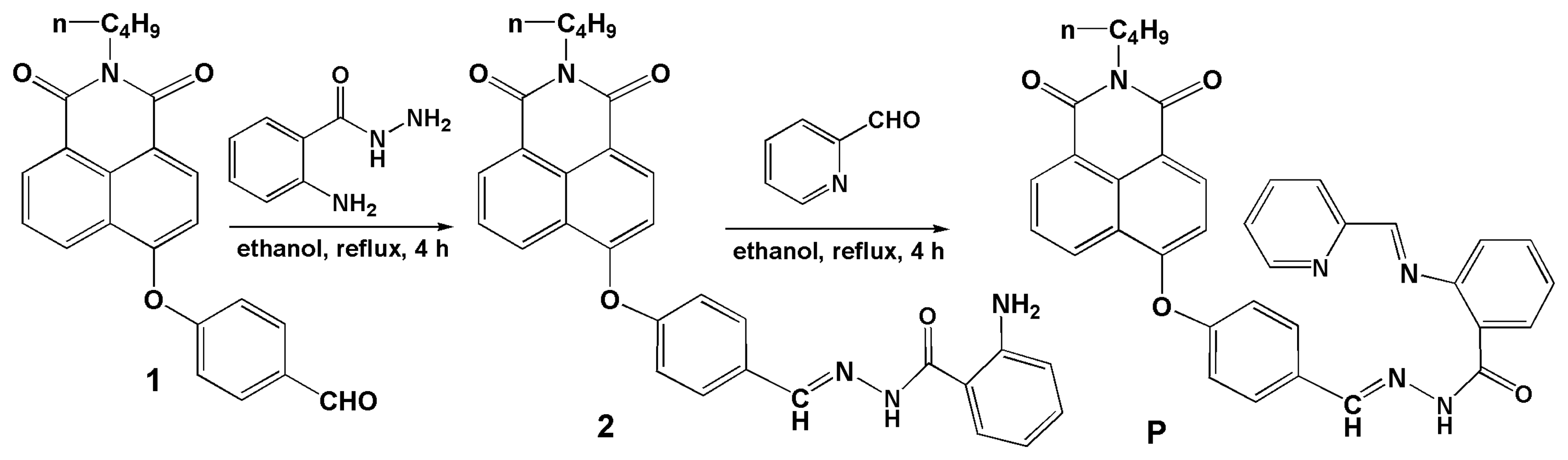
Compounds 1 and 2 were obtained according to our previous work [25]. Briefly, under N2, compound 1 (373.1 mg, 1.0 mmol) and anthraniloyl hydrazine (181.3 mg, 1.2 mmol) were combined in ethanol (50 mL). The reaction solution was refluxed for 4 h and stirred. The precipitate so obtained was filtered and washed three times with ethanol. The crude product was purified by recrystallization from ethanol to give light yellow crystals of 2. Yield: 75%. MS: m/z 507.10 [M + H]+. 1H-NMR (DMSO-d6, δ ppm): 11.65 (s, 1 H), 8.64 (d, 1 H, J = 8.35), 8.55 (d, 1 H, J = 8.15), 8.43 (t, 2 H, J = 7.42), 7.90 (t, 1 H, J = 7.82), 7.85 (d, 1 H, J = 8.20), 7.58 (d, 1 H, J = 7.85), 7.36 (d, 2 H, J = 8.35), 7.21 (t, 1 H, J = 7.65), 7.13 (d, 1 H, J = 8.25), 6.77 (d, 1 H, J = 8.30), 6.59 (t, 1 H, J = 7.47), 6.40 (b, 2 H), 4.04 (t, 2 H, J = 7.32), 1.62 (m, 2 H, J = 7.41), 1.36 (m, 2 H, J = 7.37), 0.93 (t, 3 H, J = 7.35). 13C-NMR (DMSO-d6, δ ppm): 163.86, 163.22 (C=O), 158.71, 156.54, 150.54, 133.07, 132.73, 132.21, 131.96, 129.59, 129.44, 128.60, 127.72, 123.96, 122.68, 121.04, 117.22, 116.85, 115.06, 112.63, 36.25, 30.14, 20.27, 14.19.
Compound P: Compound 2 (506.2 mg, 1.0 mmol) and 2-pyridinecarboxaldehyde (128 μL, 1.2 mM) were reacted in refluxing ethanol (50 mL) for 4 h, and then cooled to room temperature, the precipitate so obtained was purified by recrystallization from ethanol to give light yellow crystals of P. Yield: 72.5%. MS: m/z 596.15 [M + H]+. 1H-NMR (DMSO-d6, δ ppm): 8.98 (s, 1 H), 8.62 (d, 1 H, J = 8.35), 8.55 (d, 1 H, J = 7.25), 8.52 (d, 1 H, J = 7.25), 8.20 (d, 1 H, J = 8.25), 7.91 (s, 1 H), 7.88 (d, 1 H, J = 7.85), 7.84 (s, 1 H), 7.83 (s, 1 H), 7.80 (d, 1 H, J = 9.50), 7.73 (d, 1 H, J = 9.20), 7.43 (d, 1 H, J = 7.95), 7.34 (s, 1 H), 7.32 (s, 2 H), 7.27 (t, 1 H, J = 8.45), 7.10 (d, 1 H, J = 8.25), 6.79 (d, 1 H, J = 8.00), 6.73 (t, 1 H, J = 7.92), 6.49 (d, 1 H, J = 3.30), 4.03 (t, 2 H, J = 7.40), 1.61 (m, 2 H, J = 7.46), 1.35 (m, 2 H, J = 7.43), 0.92 (t, 3 H, J = 7.35). 13C-NMR (DMSO-d6, δ ppm): 163.86, 163.21, 161.25 (C=O), 158.78, 158.72, 156.89 (ArC), 150.03, 149.68 (C=N), 146.30, 137.57, 134.30, 133.03, 132.28, 131.95, 129.96, 129.42, 128.58, 128.46, 127.70, 124.04, 123.90, 122.67, 121.31, 121.07, 118.22, 117.20, 115.49, 115.15, 112.58, 30.14, 20.26, 14.18 (see Supplementary Material, Figures S1–S3).
2.3. General Procedure for Spectroscopic Measurements
A stock solution of P (1 mM) was prepared in DMSO. To 5 mL glass tubes, P (10 μL, 1 mmol) and a proper amount of Cu2+ stock solution (1.0 mmol) were added succesively and then diluted with ethanol-water solution (3:2, v:v, 50 mM HEPES, pH 7.4). The resulting solution was thoroughly mixed. For all measurements, excitation and emission slit widths were 2 nm, excitation wavelength was 360 nm.
3. Results and Discussion
3.1. pH Effects on P and P with Cu2+
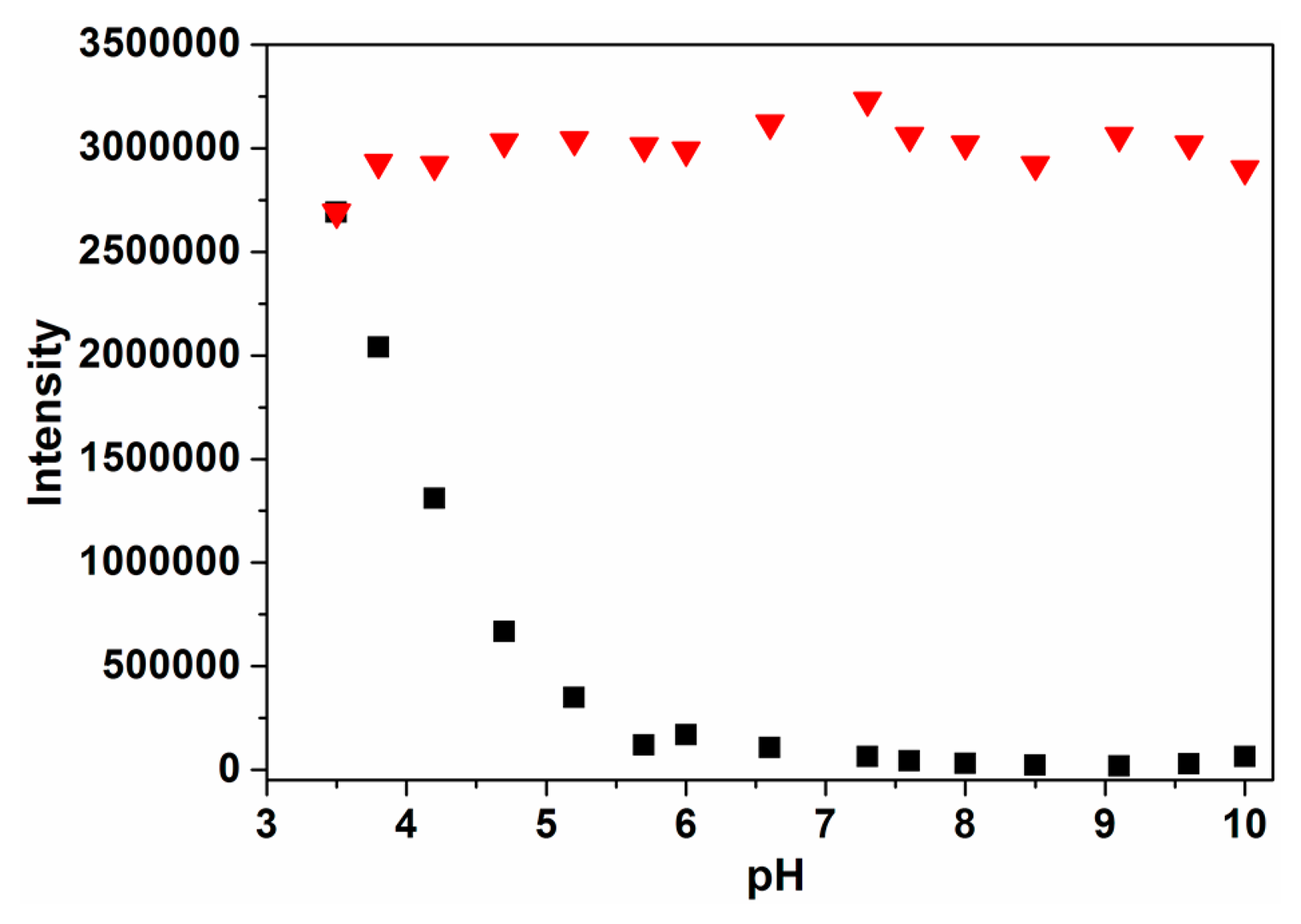
The influence of pH on the fluorescence response of probe P was determined first (Figure 1). At pH below 5.7, the fluorescence response of P was affected by pH to some extent. With the increase of pH from 5.7 and 10.0, “off-on” fluorescence signals at 432 nm were mainly caused by the addition of Cu2+. This indicated that the receptor gradually captured Cu2+ and formed the P-Cu2+ complex. In this work, pH 7.4 was chosen as an optimum experimental condition in that P could work with very low background fluorescence.
3.2. Fluorescence Spectral Response of P
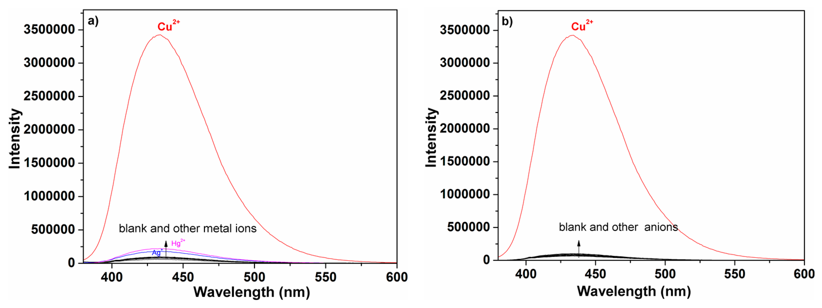
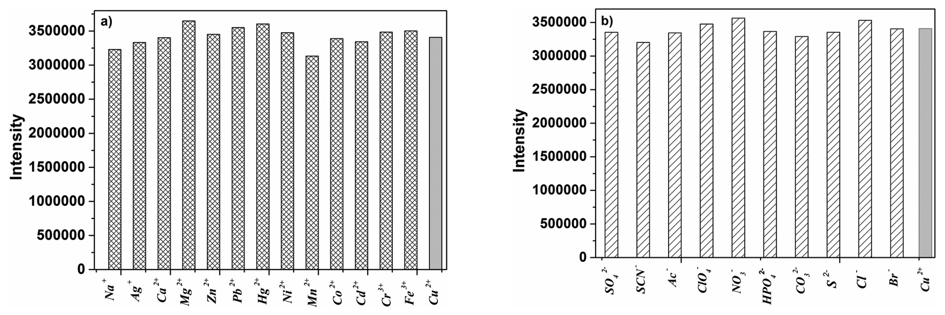
An important feature of P was its selectivity toward Cu2+ over other competitive species, and the selectivity experiments for probe P were conducted as shown in Figure 2. Fluorescence spectral changes of P were examined with addition of metal ions and anions including Na+, K+, Ag+, Mg2+, Ca2+, Zn2+, Pb2+, Cd2+, Co2+, Ni2+, Mn2+, Hg2+, Cu2+, Cr3+, Fe3+, Al3+, S2−, SO42−, SCN−, NO3−, CO32−, Cl−, Ac−, ClO4−, Br− and HPO42−. An obvious enhancement of fluorescence intensity at 432 nm was observed only upon addition of Cu2+, which was attributable to the complexation between P and Cu2+. In contrast, no obvious changes were observed in the case of other metal ions and anions. Moreover, to check the interferences from other metal ions and anions on the fluorescence signal of Cu2+, competition experiments were performed between Cu2+ and selected metal ions and anions (Figure 3). When selected metal ions and anions (50 μM) were added into ethanol-water solution (3:2, v:v, 50 mM HEPES, pH 7.4) of P (2 μM) containing Cu2+ (10 μM), the emission spectra displayed a similar pattern to that with Cu2+ alone. This experiment clearly demonstrated that selected metal ions and anions even in higher concentrations did not interfere the Cu2+ detection, which made it applicable for Cu2+ sensing in the real sample.
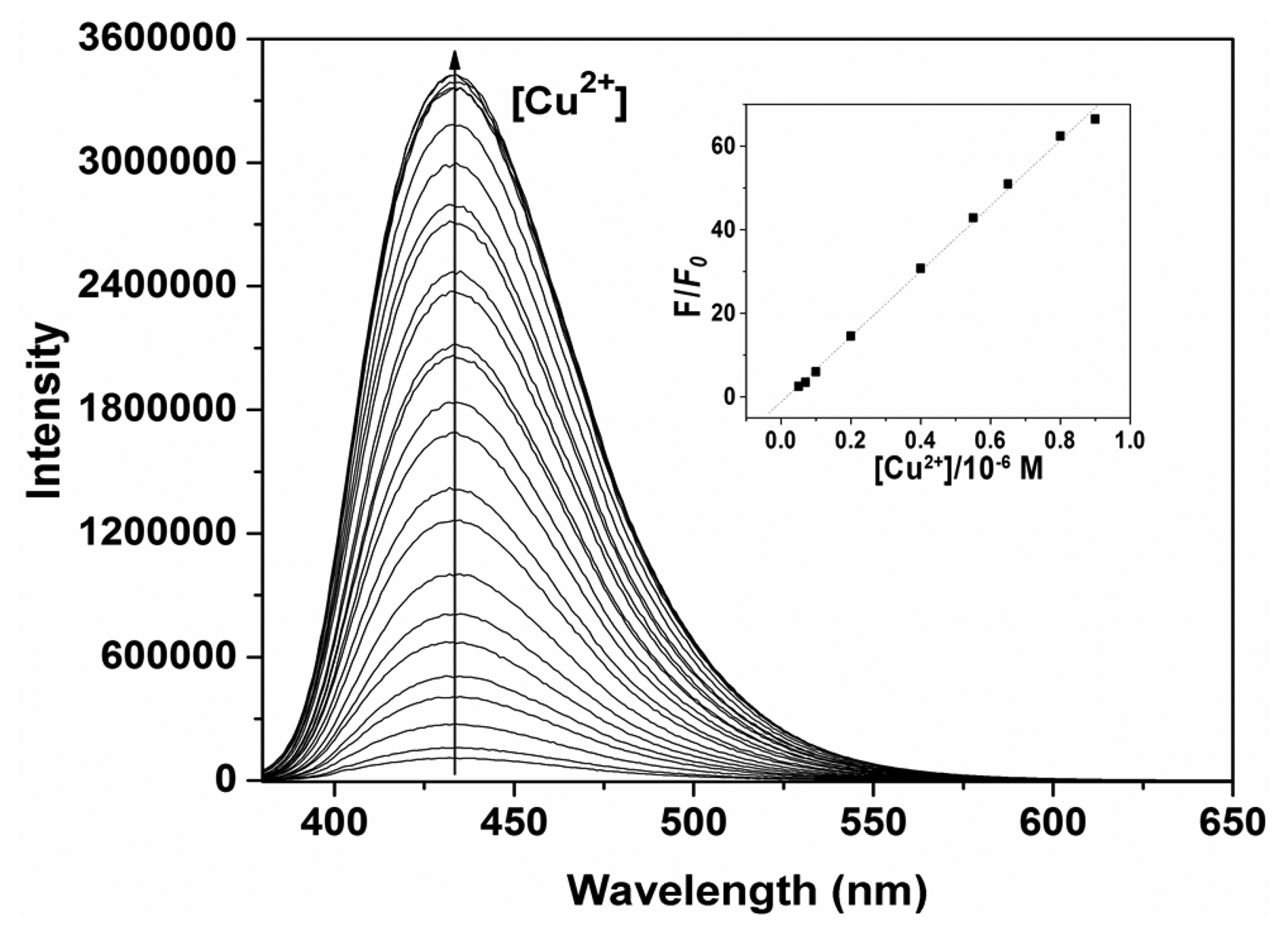
In addition, the titration of P with various amounts of Cu2+ in ethanol-water solution (3:2, v:v, 50 mM HEPES, pH 7.4) were studied (Figure 4).
As the Cu2+ concentration increased, the fluorescence emission intensity at 432 nm gradually increased accordingly. The linear fluorescence enhancement of P (2 μM) to Cu2+ was obtained in the range of 0.05–0.9 μM (R = 0.999; inset of Figure 4). The limit of detection (LOD) was 0.03 μM, based on 3 × δblank/k (where δblank is the standard deviation of the blank solution and k is the slope of the calibration plot).
3.3. The Binding Mechanism
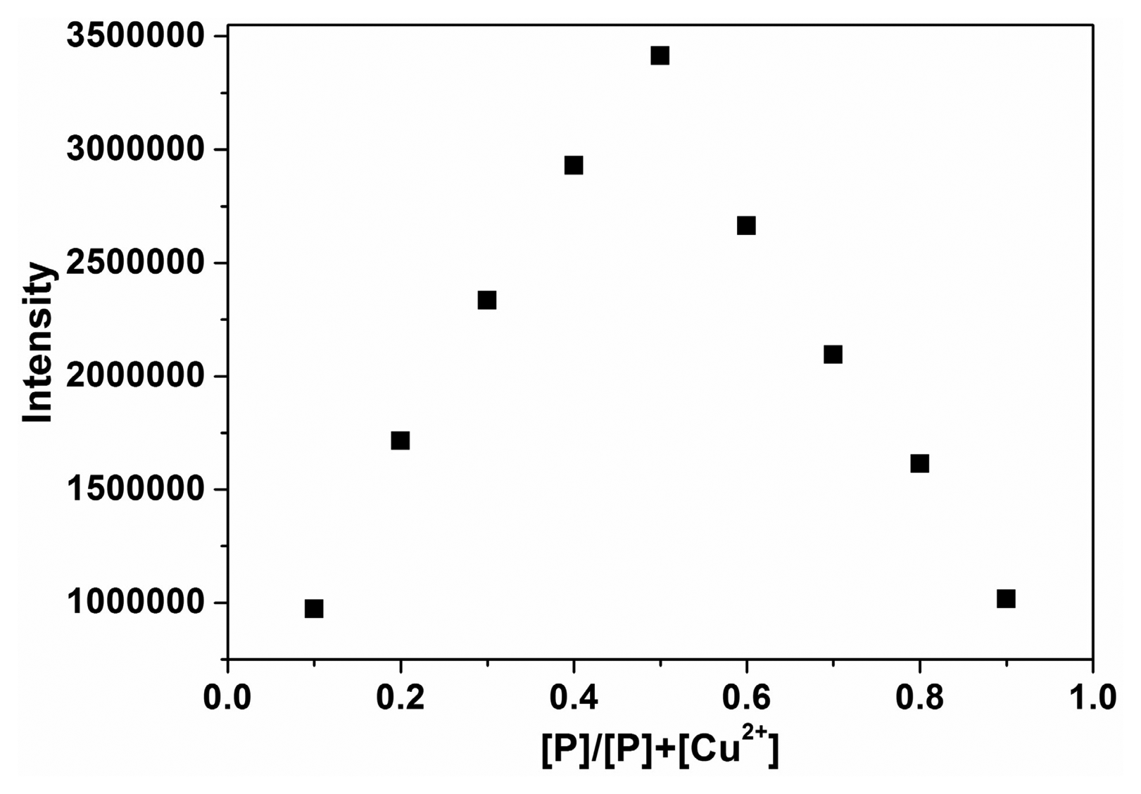
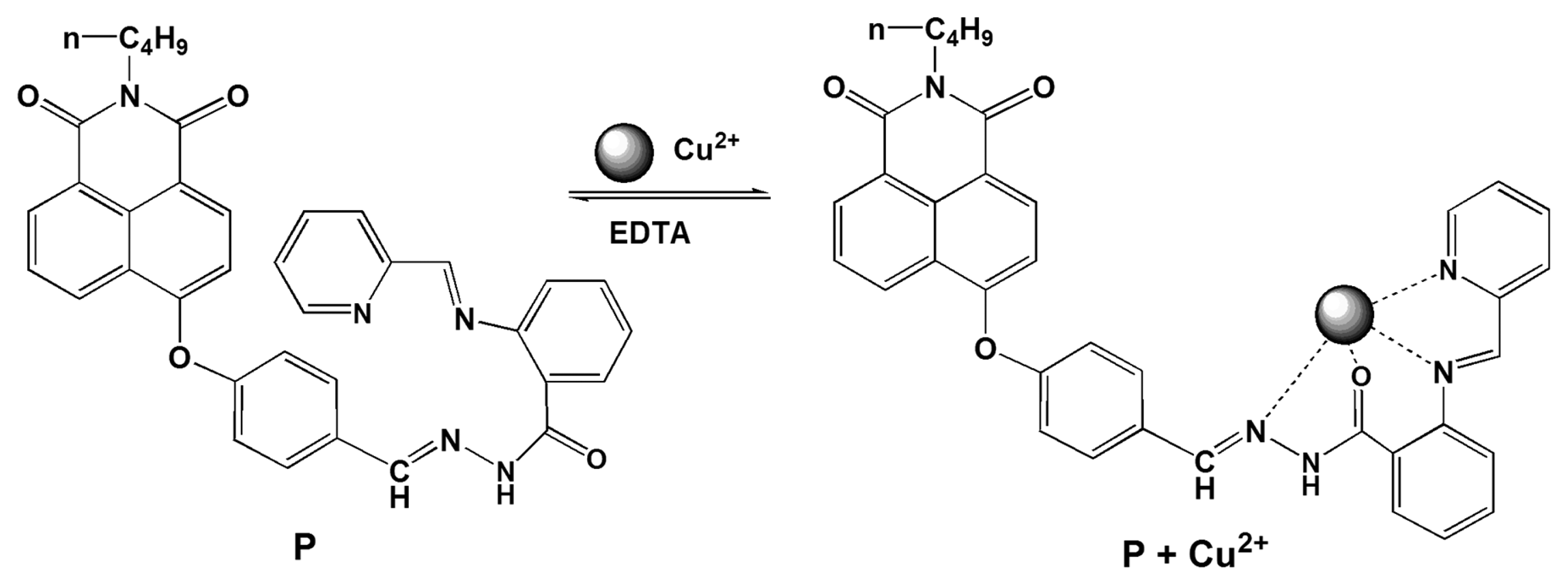
To quantify the complexation ration between P and Cu2+, a Job plot experiment was carried out by keeping the total concentration of P and Cu2+ at 10 μM (Figure 5). The results suggested that a 1:1 complex of P with Cu2+ was formed, which was supported by the presence of a peak at m/z 659.2 corresponding to P-Cu2+ in the ESI-MS spectrum of the components of the mixture of P and 1 equivalent Cu2+ in ethanol (Supplementary Material, Figure S4). The 1H-NMR spectra also indicated the binding of P with Cu2+ (Supplementary Material, Figures S5 and S6). The association constant K was determined from the slope to be 6.2 × 10⁵ M−1, by plotting the fluorescence intensity 1/(F − F0) against 1/[Cu2+].
From Figure 2, no significant changes in fluorescence spectra were observed when probe P was exposed to other metal ions and anions. We believe that this is due to a rapid isomerization of the C=N double bond in the excited state [26,27], though other mechanisms such as photoinduced electron transfer (PET) may also contribute to it. Notably, by adding Cu2+, the fluorescence character of P was different from free P and other metal ions and anions, its fluorescence at λem 432 nm was turned from “off” to “on”. The enhancement of fluorescence was likely due to restriction of acyclic C=N isomerization in the Schiff base upon addition of Cu2+ [25–27]. Accordingly, the proposed binding mode of P with Cu2+ can be illustrated as in Scheme 2. It is believed that this process is reversible, which has been proved by a test using EDTA-Cu2+ (Figure 6). As seen, in absence of Cu2+, probe P had a weak fluorescence. Addition of Cu2+ led to a reversible coordination with the ligand, resulting in an appearance of fluorescence enhancement at λem 432 nm. Thus, an “off-on” based fluorescent probe for Cu2+ was implemented.
4. Conclusions
In summary, an efficient “off-on” probe P for Cu2+ was proposed. Our studies showed that P was a highly selective and sensitive probe for Cu2+, which could work in neutral aqueous solution media and has great potential use in environmental sensing applications. These results open up new possibilities for the construction of “off-on” probes for other metal ions.
Acknowledgments
This work was financially supported by the National Natural Science Foundation of China (Nos. 81260268, 81360266), the Natural Science Foundation of Hainan Province (Nos. 812188, 413131), the Colleges and Universities Scientific Research Projects of the Education Department of Hainan Province (Hjkj2013-29).
Author Contributions
In this paper, Zhang is on the duty of the design and synthesis of probe P. Wu mainly focuses on the testing experiments of P. Yu mainly focused on the optimization of the testing conditions of probe P. Yu is responsible for organizing the experimental data and the writing of the paper.
Conflicts of Interest
The authors declare no conflict of interest.
References
- Que, E.L.; Domaille, D.W.; Chang, C.J. Metals in neurobiology: Probing their chemistry and biology with molecular imaging. Chem. Rev. 2008, 108, 1517–1549. [Google Scholar]
- Yu, C.W.; Zhang, J.; Wang, R.; Chen, L.X. Highly sensitive and selective colorimetric and off-on fluorescent probe for Cu2+ based on rhodamine derivative. Org. Biomol. Chem. 2010, 8, 5277–5279. [Google Scholar]
- Chen, Z.J.; Wang, L.M.; Zou, G.; Tang, J.; Cai, X.F.; Teng, M.S.; Chen, L. Highly selective fluorescence turn-on chemosensor based on naphthalimide derivatives for detection of copper(II) ions. Spectrochim. Acta Part A 2013, 105, 57–61. [Google Scholar]
- Zhang, J.; Yu, C.W.; Qian, S.Y.; Lu, G.; Chen, J.L. A selective fluorescent chemosensor with 1, 2, 4-triazole as subunit for Cu(II) and its application in imaging Cu(II) in living cells. Dyes Pigm. 2012, 92, 1370–1375. [Google Scholar]
- Zhang, J.; Zhao, B.J.; Li, C.; Zhu, X.F.; Qiao, R.Z. A BODIPY-based “turn-on” fluorescent and colorimetric sensor for selective detection of Cu2+ in aqueous media and its application in cell imaging. Sens. Actuators B Chem. 2014, 196, 117–122. [Google Scholar]
- Wang, H.X.; Yang, L.; Zhang, W.B.; Zhou, Y.; Zhao, B.; Li, X.Y. A colorimetric probe for copper(II) ion based on 4-amino-1, 8-naphthalimide. Inorg. Chim. Acta 2012, 381, 111–116. [Google Scholar]
- Xu, Z.C.; Pan, J.; Spring, D.R.; Cui, J.N.; Yoon, J. Ratiometric fluorescent and colorimetric sensors for Cu2+ based on 4,5-disubstituted-1,8-naphthalimide and sensing cyanide via Cu2+ displacement approach. Tetrahedron 2010, 66, 1678–1683. [Google Scholar]
- Zhang, Y.; Zeng, X.; Mu, L.; Chen, Y.; Zhang, J.X.; Redshaw, C.; Wei, G. Rhodamine-triazine based probes for Cu2+ in aqueous media and living cells. Sens. Actuators B Chem. 2014, 204, 24–30. [Google Scholar]
- Huang, C.B.; Li, H.R.; Luo, Y.Y.; Xu, L. A naphthalimide-based bifunctional fluorescent probe for the differential detection of Hg2+ and Cu2+ in aqueous solution. Dalton Trans. 2014, 43, 8102–8108. [Google Scholar]
- Chen, X.Q.; Jou, M.J.; Lee, H.Y.; Kou, S.Z.; Lim, J.S.; Nam, S.W.; Park, S.S.; Kim, K.M.; Yoon, J.Y. New fluorescent and colorimetric chemosensors bearing rhodamine and binaphthyl groups for the detection of Cu2+. Sens. Actuators B Chem. 2009, 137, 597–602. [Google Scholar]
- Fan, J.L.; Zhan, P.; Hu, M.M.; Sun, W.; Tang, J.Z.; Wang, J.Y.; Sun, S.G.; Song, F.L.; Peng, X.J. A fluorescent ratiometric chemodosimeter for Cu2+ based on TBET and its application in living cells. Org. Lett. 2013, 15, 492–495. [Google Scholar]
- Hu, Z.Q.; Wang, X.M.; Feng, Y.C.; Ding, L.; Lu, H.Y. Sulfonyl rhodamine hydrazide: A sensitive and selective chromogenic and fluorescent chemodosimeter for copper ion in aqueous media. Dyes Pigm. 2011, 88, 257–261. [Google Scholar]
- Zhao, Y.; Zhang, X.B.; Han, Z.X.; Qiao, L.; Li, C.Y.; Jian, L.X.; Shen, G.L.; Yu, R.Q. Highly sensitive and selective colorimetric and off-on fluorescent probe for Cu2+ in aqueous solution and living cells. Anal. Chem. 2009, 81, 7022–7030. [Google Scholar]
- Yu, C.W.; Zhang, J.; Li, J.H.; Liu, P.; Wei, P.H.; Chen, L.X. Fluorescent probe for copper(II) ion based on a rhodamine spirolactame derivative and its application to fluorescent imaging in living cells. Microchim. Acta 2011, 174, 247–255. [Google Scholar]
- Huang, L.; Wang, X.; Xie, G.Q.; Xi, P.X.; Li, Z.P.; Xu, M.; Wu, Y.J.; Bai, D.C.; Zeng, Z.Z. A new rhodamine-based chemosensor for Cu2+ and the study of its behaviour in living cells. Dalton Trans. 2010, 39, 7894–7896. [Google Scholar]
- Liu, Y.L.; Sun, Y.; Du, J.; Lv, X.; Zhao, Y.; Chen, M.L.; Wang, P.; Guo, W. Highly sensitive and selective turn-on fluorescent and chromogenic probe for Cu2+ and ClO− based on a N-picolinyl rhodamine B-hydrazide derivative. Org. Biomol. Chem. 2011, 9, 432–437. [Google Scholar]
- Lin, W.; Yuan, L.; Tan, W.; Feng, J.; Long, L. Construction of fluorescent probes via protection/deprotection of functional groups: a ratiometric fluorescent probe for Cu2+. Chem. Eur. J. 2009, 15, 1030–1035. [Google Scholar]
- Yu, C.W.; Chen, L.X.; Zhang, J.; Li, J.H.; Liu, P.; Wang, W.H.; Yan, B. “Off-On” based fluorescent fluorescent probe for Cu2+ in aqueous media and living cells. Talanta 2011, 85, 1627–1633. [Google Scholar]
- Kim, H.J.; Park, S.Y.; Yoon, S.; Kim, J.S. FRET-derived ratiometric fluorescence for Cu2+. Tetrahedron 2008, 64, 1294–1300. [Google Scholar]
- Yu, C.W.; Wen, Y.Y.; Zhang, J. Synthesis of a Cu2+-selective probe derived from rhodamine and its application in cell imaging. Sensors 2014, 14, 21375–21384. [Google Scholar]
- Xu, Z.C.; Kim, S.; Kim, H.N.; Su, S.J.; Lee, C.; Kim, J.S.; Qian, X.H.; Yoon, J. A naphthalimide–calixarene as a two-faced and highly selective fluorescent chemosensor for Cu2+ or F−. Tetrahedron Lett. 2007, 48, 9151–9154. [Google Scholar]
- Kar, C.; Adhikari, M.D.; Ramesh, A.; Das, G. NIR- and FRET-based sensing of Cu2+ and S2− in physiological conditions and in live cells. Inorg. Chem. 2013, 52, 743–752. [Google Scholar]
- Kim, M.H.; Noh, J.H.; Kim, S.; Ahn, S.; Chang, S.K. The synthesis of crown ether-appended dichlorofluoresceins and their selective Cu2+ chemosensing. Dyes Pigm. 2009, 82, 341–346. [Google Scholar]
- Shao, N.; Zhang, Y.; Cheung, S.M.; Yang, R.H.; Chan, W.H.; Mo, T.; Li, K.A.; Liu, F. Copper ion-selective fluorescent sensor based on the inner filter effect using a spiropyran derivative. Anal. Chem. 2005, 77, 7294–7303. [Google Scholar]
- Yu, C.W.; Zhang, J. Copper(II)-responsive “off-on” chemosensor based on a naphthalimide derivative. Asian J. Org. Chem. 2014, 3, 1312–1316. [Google Scholar]
- Wu, J.S.; Liu, W.M.; Zhuang, X.Q.; Wang, F.; Wang, P.F.; Tao, S.L.; Zhang, X.H.; Wu, S.K.; Lee, S.T. Fluorescence turn on of coumarin derivatives by metal cations: A new signaling mechanism based on C=N isomerization. Org. Lett. 2007, 9, 33–36. [Google Scholar]
- Sheng, J.R.; Feng, F.; Qiang, Y.; Liang, F.G.; Sen, L.; Wei, F.H. A coumarin-derived fluorescence chemosensors selective for copper(II). Anal. Lett. 2008, 41, 2203–2213. [Google Scholar]


© 2014 by the authors; licensee MDPI, Basel, Switzerland. This article is an open access article distributed under the terms and conditions of the Creative Commons Attribution license ( http://creativecommons.org/licenses/by/4.0/).
Share and Cite
Zhang, J.; Wu, Q.; Yu, B.; Yu, C. A Pyridine-Containing Cu2+-Selective Probe Based on Naphthalimide Derivative. Sensors 2014, 14, 24146-24155. https://doi.org/10.3390/s141224146
Zhang J, Wu Q, Yu B, Yu C. A Pyridine-Containing Cu2+-Selective Probe Based on Naphthalimide Derivative. Sensors. 2014; 14(12):24146-24155. https://doi.org/10.3390/s141224146
Chicago/Turabian StyleZhang, Jun, Qiang Wu, Bangliang Yu, and Chunwei Yu. 2014. "A Pyridine-Containing Cu2+-Selective Probe Based on Naphthalimide Derivative" Sensors 14, no. 12: 24146-24155. https://doi.org/10.3390/s141224146
APA StyleZhang, J., Wu, Q., Yu, B., & Yu, C. (2014). A Pyridine-Containing Cu2+-Selective Probe Based on Naphthalimide Derivative. Sensors, 14(12), 24146-24155. https://doi.org/10.3390/s141224146