Review

Electrochemical Metal Ion Sensors. Exploiting Amino Acids and Peptides as Recognition Elements

by
J. Justin Gooding
*,
D. Brynn Hibbert
* and
Wenrong Yang
School of Chemistry, The University of New South Wales, Sydney 2052, Australia
*
Authors to whom correspondence should be addressed.
Sensors 2001, 1(3), 75-90; https://doi.org/10.3390/s10300075
Submission received: 27 July 2001 / Accepted: 27 July 2001 / Published: 8 August 2001

Abstract

:
Amino acids and peptides are known to bind metal ions, in some cases very strongly. There are only a few examples of exploiting this binding in sensors. The review covers the current literature on the interaction of peptides and metals and the electrochemistry of bound metal ions. Peptides may be covalently attached to surfaces. Of particular interest is the attachment to gold via sulfur linkages. Sulfur-containing peptides (eg cysteine) may be adsorbed directly, while any amino group can be covalently attached to a carboxylic acid-terminated thiol. Once at a surface, the possibility for using the attached peptide as a sensor for metal ions becomes realised. Results from the authors’ laboratory and elsewhere have shown the potential for selective monitoring of metal ions at ppt levels. Examples of the use of poly-aspartic acid and the copper binding peptide Gly-Gly-His for detecting copper ions are given.

Introduction

The development of practical sensors for the detection and quantification of metal ions in environmental samples is the subject of considerable research. The majority of this research involves the synthesis and testing of macrocyclic ligands with selectivity for a target metal ion. In nature however, metal binding is achieved with a high degree of selectivity using peptide motifs rather than macrocyclic ligands. With the advent of biosensors exploiting Nature’s methods of selective recognition, it is surprising how little research has been conducted into the development of solid state metal ion sensors based on peptide ligands. This lack of research is even more surprising considering the significant amount of literature on complexing metals using amino acids, oligopeptides and peptides.
The aim of this review is to highlight the potential for using oligopeptides for the fabrication of solid-state metal-ion biosensors. The complexation of metals by oligopeptides will be briefly outlined (although the reader is directed to more comprehensive reviews on this subject by Sigel and Martin 1 and Kozlowski et al 2). Subsequently the application of peptides and amino acids for solution based detection of metals will be addressed, followed by early examples of using oligopeptides and amino acids for solid state devices and finally future prospects will be discussed.

Complexes of Metal Ions with Peptides
Peptides can be very effective, and often specific, ligands for a variety of metal ions. They contain a great number of potential donor atoms through the peptide backbone and amino acid side chains. The complexes formed exist in a variety of conformations that are sensitive to the pH environment of the complex 1,2. With at least 20 amino acid combinations available, some with coordinating side chains, in any particular order and length, the number of ligands that can be synthesized using simple amino acids is practically infinite. To appreciate the diversity of ligands and their selectivities for different metals requires an introduction to the complexing of metals with peptides. More detailed treatises of metal-peptide complexation can be found in reviews by Sigel and Martin 1 and Kozlowski et al 2.
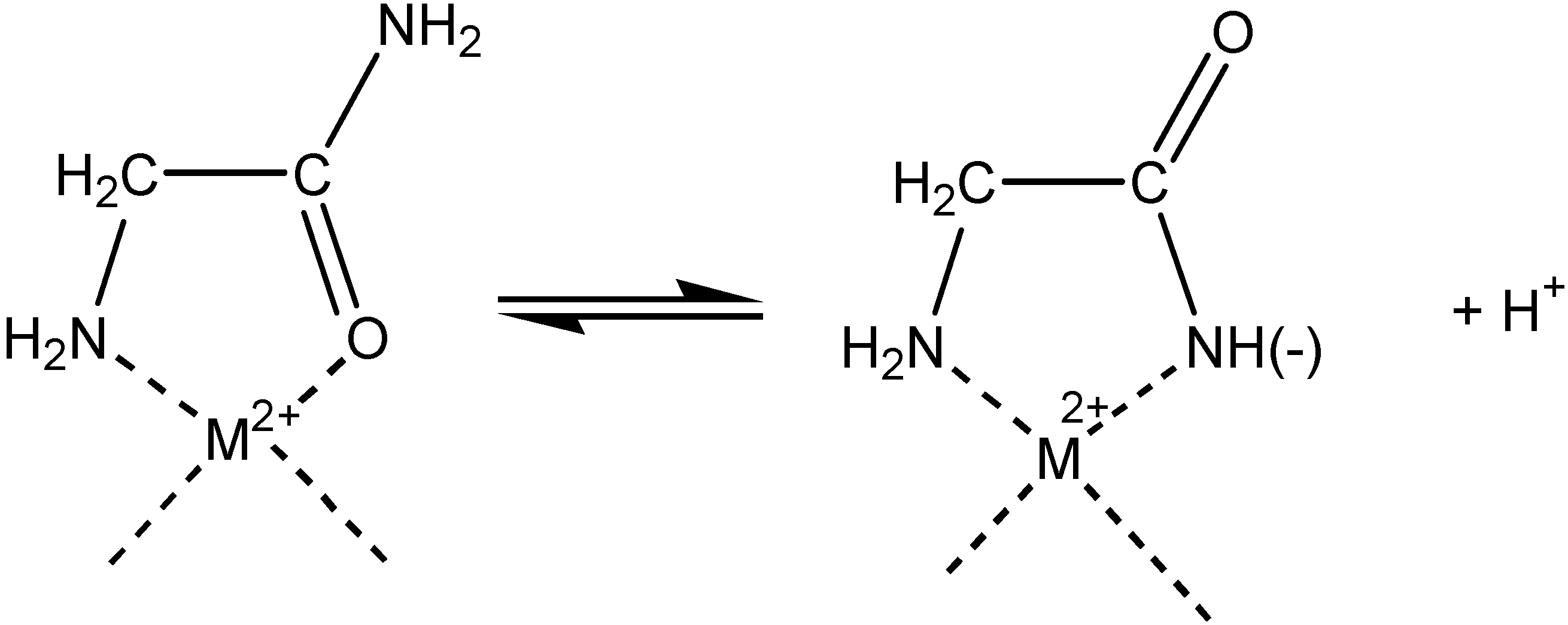
In a single amino acid, with a non-coordinating side chain, there are two donor atoms that complex the metal, the terminal amine and carbonyl oxygen or amide nitrogen as shown in scheme 1.
Scheme 1.
Scheme 1.
Sensors 01 00075 g006
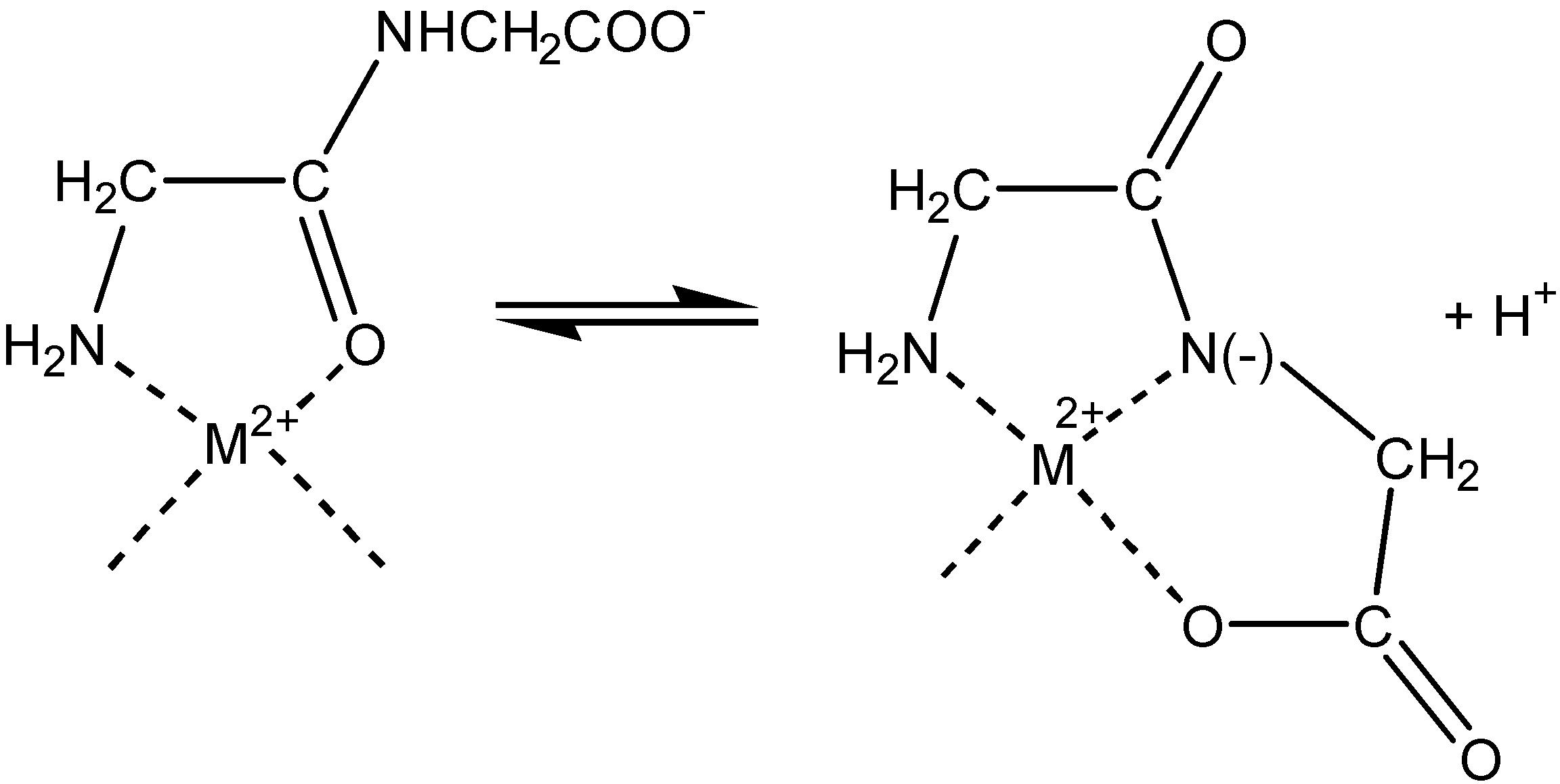
With a peptide the potential donor atoms is extended to the amide in the peptide backbone. Binding to metal ions involving an amide group also presents two possibilities, either the carbonyl oxygen or the amide nitrogen can be involved as shown in scheme 2 for diglycine.
Scheme 2.
Scheme 2.
Sensors 01 00075 g007
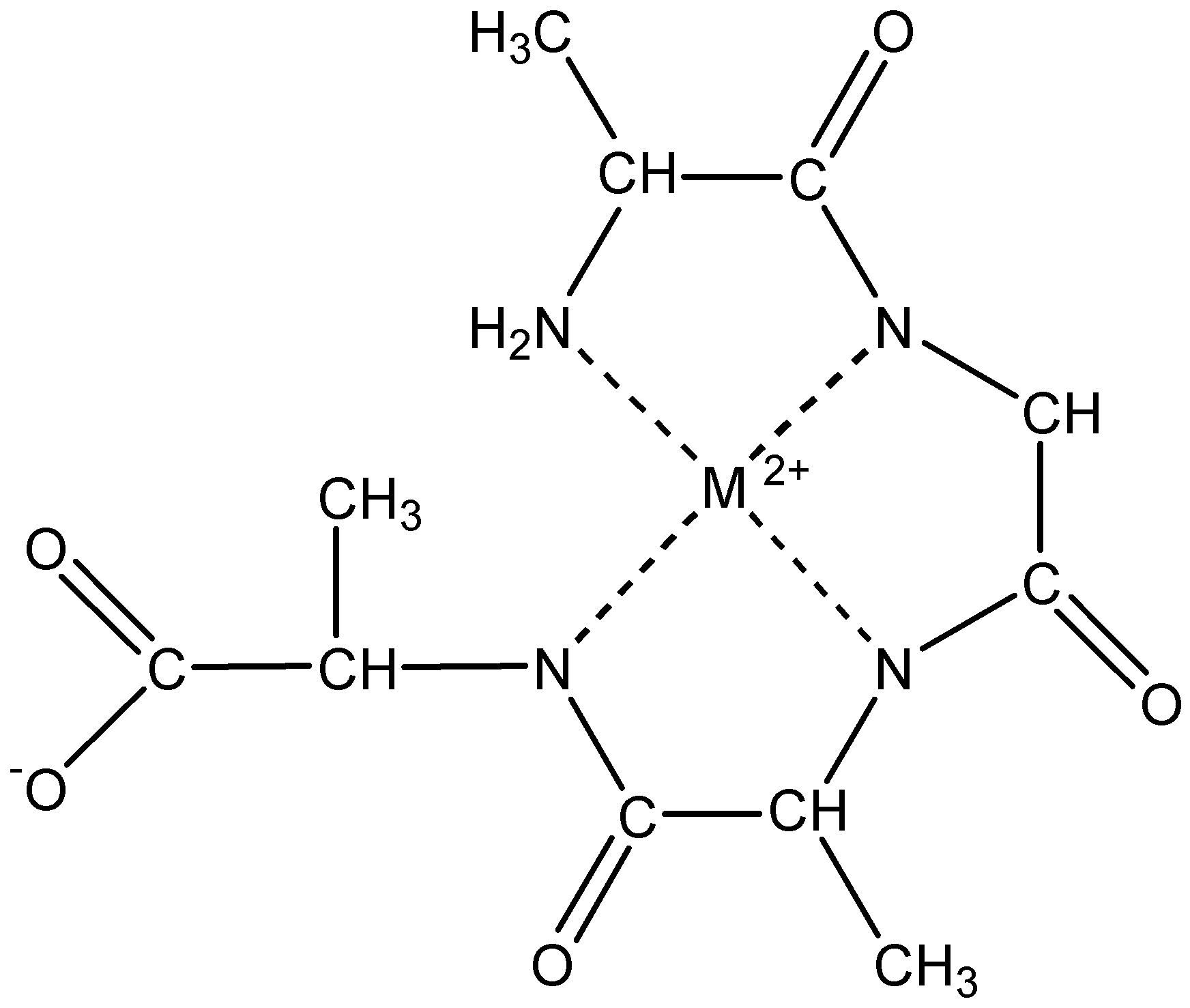
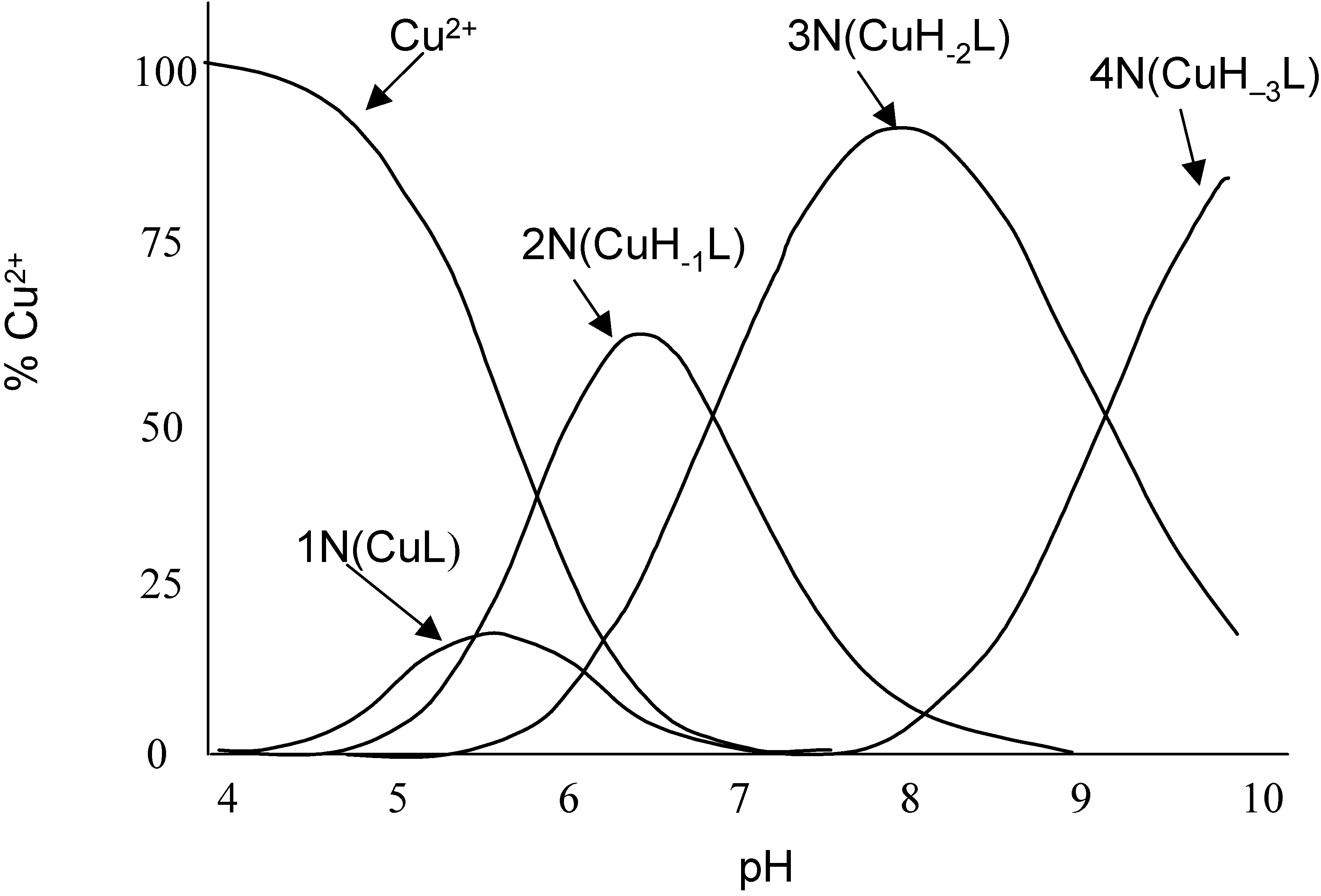
Significantly stronger binding is achieved when the amide nitrogen is involved1. Extending this Gly-Gly dipeptide to Gly-Gly-Gly and Gly-Gly-Gly-Gly results in tetradentate ligands with the potential to produce more stable complexes. In view of these binding modes it is clear that to coordinate strongly to the amide group the metal ions must be capable of substituting for the nitrogen bound amide hydrogen. Therefore, the number of peptide nitrogens involved in the bonding is dependent on the pH. Coordination of metal ions such as Cu2+ and Ni2+ starts at the N-terminal amino nitrogen. The adjacent carbonyl oxygen is the second donor to complete the chelate ring. By raising the pH the metal ions are able to deprotonate successive peptide nitrogens forming M-N- bonds until eventually a 4N complex is formed around pH 9-10 (see scheme 3 and figure 1). The well separated pKs for the deprotonation of the peptide nitrogens indicates that there is no cooperation in the binding process.
Scheme 3.
Scheme 3.
Sensors 01 00075 g008
The stability constant of the metal-ligand complex defines the selectivity of a ligand for a particular metal. It has been shown that the higher the pKa of the terminal amino group, the greater is the stability constant of peptide ligands for Cu2+ and Ni2+ 1. A consequence of this is that electron-withdrawing groups on the side chains of the terminal amino acid promote stronger binding. Non-coordinating side chains can also influence complex stability is less easily predicted ways. Examples include the ‘superstability’ afforded to Cu(II) by the sequence Asn-Ser-Phe-Arg-Tyr-NH2 (NSFRY-NH2) where metal assisted peptide organization accounts for this ligand being the most effective Cu(II) ligand among oligopeptides with non-coordinating side chains. The superstability of this complex arises from the side chains of Asn and Phe and also from Arg and Tyr forming ‘fences’ around the Cu(II). A simple substitution of Ala for Asn results in the loss of this enhanced stability. Other stabilising contributions of a non-coordinating nature come from aromatic rings in the side chains, such as with Tyr and Phe, can also contribute to the stability through hydrophobic interactions or ring stacking. These same aromatic rings can enhance complex stability in a coordinating role through direct electronic interactions with the metal.
Figure 1. Comparison of species distribution curves for Cu(II) complexes with Gly-Gly-Gly as a function of pH. Initial metal ion concentrations are 1 mM, and metal-to-ligand molar ratios 1:1 (Redrawn from 2).
Sensors 01 00075 g001
The ability of some side chains to coordinate with the metal can have an even more dramatic influence on increasing the stability constant of the ligand-metal complex. The classic and most dramatic example is Gly-Gly-His, also known as copper binding peptide. The side chain imidazole ring of His has a very efficient nitrogen donor (the imidazole N), which can form a six-membered chelate ring for coordination as in Gly-Gly-His in scheme 4 below.
The coordination properties of a His residue within a peptide sequence depends greatly on the position of the His residue in the peptide chain. In the coordinated Cu(II) complex shown in scheme 4 above, complexation proceeds cooperatively with the formation of three fused chelate rings and thus saturation of the coordination space. The saturation of the coordination space leads to the most stable of the 4N complexes formed by peptides. However, if the His residue is the first or second amino acid in the tripeptide (His-Gly-Gly or Gly-His-Gly) there is a greater than 10 fold reduction in complex stability. In the case of His-Gly-Gly, the imidazole nitrogen wins the competition with the amide nitrogen for Ni(II) and hinders deprotonation of the amide nitrogens when complexing Cu(II).
Scheme 4.
Scheme 4.
Sensors 01 00075 g009
Factors that influence the affinity of a ligand to different metals are numerous. These factors include the hardness/softness of the metal ion, the coordination geometry of the complex, ligand field stabilisation effects and the hardness/softness of any coordinating side chains of the amino acid sequence. The ability of the metal ion to cause deprotonation of the amide nitrogens plays a key role in the final stability of the resultant complex. Metal-induced deprotonation of the amide nitrogens is important because it increases the number of coordination points between the metal and the ligand. The order of peptide hydrogen displacement by metal ions, with a representative pKa value in brackets for short peptides is given by Pd2+ (2) > Cu2+ (4) > Ni2+ (8) > Co2+ (10) 1. Note that the order relates to reasonably hard metal ions as they interact with the hard amide ligands 3.
The importance of the peptide ligand having soft ligands for soft metals and hard ligands for hard metals is illustrated by the tripeptide glutathione. Glutathione has the amino acid sequence γ-Glu-Cys-Gly. Glutathione has eight possible coordination sites, which fall into three classes. There are two carboxyl groups and one amino acid at the glutamic acid end, the soft thiol side-chain of the cysteine and the two pairs of carbonyl and amide nitrogen donors associated with the peptide bonds. Hard metal donors interact primarily with the glutamic acid moiety while the primary anchor for soft metals is through the thiol 4.
The combination of ligand field stabilisation, changes in complex geometry during coordination, the ability of the metal ion to deprotonate the amide nitrogen, steric effects in the peptide and the presence of soft coordination points on amino acid side chains makes it difficult to precisely predict the relative affinity of a given ligand for a given metal ion. However, what is clear from the above discussion is that the amino acid building blocks provide a myriad of peptide ligands that will have a broad spectrum of affinities for different metal ions. The simple, generic chemistry involved in synthesizing different ligands renders peptide ligands a highly attractive and under-exploited class of ligands for the development of solid-state metal ion sensors.

Electrochemistry of peptides and amino acids in the presence of complexing metal ions

The interaction between amino acids and peptides and metal ions has been explored using electrochemistry in one of three ways. First, there has been considerable research by the groups of Esteban and Rodriguez both independently and in collaboration using electrochemistry at mercury electrodes coupled with multivariate analysis to explore speciation of metal ions such as Cd(II) 5 and/or Zn(II) 6 and peptides such as glutathione and metallothionen fragments (such as Lys-Cys-Thr-Cys-Cys-Ala) 7,8,9,10. These studies show that in solution the speciation chemistry is rich and complex with the thiol groups from the cysteines playing a crucial role in metal binding.
The second key application of electrochemistry in the investigation of metal-peptide binding is for the detection of amino acids and peptides where the metal ion is used as an electroactive label. Complications associated with the overlapping electrochemistry of different amino acids themselves can thus be obviated 11,12,13,14 as complexation can stabilise a metal oxidation state, thus shifting reduction to more cathodic potentials 15 or separating overlapping reduction waves of different amino acids based on their relative affinities to a metal ion 16. The detection of amino acids using complexation with metals dates back to the 1950s where the electrochemistry of cysteine and cystine was investigated using polarography 17,18. The dropping mercury electrode plays a direct role in the electrochemistry. For example, the first polarographic reduction wave for cystine at mercury results from the reduction of mercuric cysteinate. More recently stripping voltametric methods for the determination of cysteine, cystine and N-acetylcysteine in one solution have been reported 19. The measurements are performed by accumulating the amino acids at a mercury electrode in the presence of Ni2+ at potentials ranging between 0.0 and –0.4 V vs Ag-AgCl reference electrode. The catalytic reduction of Ni2+ gives a peak at –0.6 V. An additional effect of the Ni2+ is the suppression of the reduction peak of mercury cysteinate. Cysteine or cystine and N-acetylcysteine can be determined simultaneously or independently in the same sample. In a similar way the same workers also use Ni2+ to detect glutathione and oxidised glutathione down to nM levels. More complex peptides which have been detected via the interaction of the thiol groups of some amino acids with soft metals include the detection of felypressin (H-Cys-Phe-Phe-Gln-Asn-Cys-Pro-Lys-Gly-NH2) by cathodic stripping voltammetry at a mercury electrode 21 and phytochelatins 22. Metal complexation has however not been restricted to thiol-containing peptides 23,24. Histidine has been detected in the presence of Cu(II) using differential-pulse adsorptive stripping voltammetry at a hanging mercury drop electrode 24. Similarly Gly-His-Gly has been detected down to 10 nM in the presence of Cu(II) using cathodic stripping voltammetry with adsorptive accumulation 23. An interesting development in using metal ions in the analysis of peptides is in the detection of toxic and tumour promoting peptides such as microcystins 25,26. Yan et al 26 reported pM detection limits for microcystin-LR using several different metals coupled with differential pulse voltammetry at gold electrodes.
The plethora of studies above in which the electrochemistry of different metal ions are used to detect many types of amino acids and peptides, implies that peptides could be used as selective ligands for the detection of metal ions. The complexation of metal ions by even simple amino acids has produced remarkably low detection limits. Tanaka and Yoshida 27 have reported detection limits of 0.6 nM Cu2+ with relative standard deviations at 10 nM of 4% for the anodic stripping of copper at a mercury electrode in the presence of cysteine. The presence of cysteine results in a more anodic peak than when only copper is present which is attributed to the transformation of a cupric cysteinate complex to a mercuric cysteinate complex. Bai et al 28 also observed a positive shift in peak potential which they attributed to the formation of copper(I)-cysteine complex.
Fogg and coworkers modified a hanging mercury drop electrode (HDME) with poly-L-histidine. Cu(II) could be detected in the range of 5 nM – 400 nM after 2 minutes accumulation with the electrode poised at –0.4V vs Ag/AgCl at a pH of 4.5. No significant interference was observed from micro molar levels of Cr(II), Pb(II), Ni(II), Cd(II), Mg(II) or EDTA 29. The same workers used a poly-lysine modified dropping mercury electrode to monitor nitroprusside Fe(CN)5NO2- with a detection limit of 0.5 nM and a relative standard deviation of <2% 30. A similar approach was used to detect hexacyanoferrate (III) down to 20 nM with a poly-L-lysine modified HDME 31. This latter example suffered from interference from zinc and surfactants which highlighted the relatively non-selective complexation of the analytes with the polypeptide.
Synthesizing peptide ligands with high selectivity for a given metal ions can be achieved with the appropriate selection of amino acid building blocks. Such a strategy has been demonstrated using fluorescence-based sensing for Zn(II) by Imperiali and co-workers 32 and Cu(II) 33. Changing the ligand sequence from Baa(Dns)-βGly-His-Ser-Ser to Baa(Dns)-βAla-His-Ser-Ser (where Baa refers to l-β−aminoalanine and Dns to the fluorophore 5-(dimethylamino)naphthalene-1-sulfonamide) significantly improved the ligand’s selectivity for Cu(II) in the presence of Fe(II) as an interfering species.
The solution based electroanalytical studies discussed above demonstrate there is considerable potential for using peptides to detects metals. However, ease of use for in field analysis would be greatly enhanced if the metal ions were immobilised onto electrode surfaces. This is the subject of the next section of this review.

Peptides on surfaces

To create an oriented layer of peptide molecules at a surface requires suitable attachment chemistry with control over the conformation and spacing of the molecules to give a packing density that allows the analyte to access the binding sites. Transduction of the recognition event is also an issue. Self-assembly is always an attractive option, as once the surface and molecules are properly chosen the resulting structure is created as a consequence of the self-assembly. Immobilization on a surface then opens up possibilities for sensing, nanoscale devices and surfaces with engineered biocompatibility. Most published work has focussed on self-assembly of sulfur-containing molecules such as thiols, cysteine and lipids on gold or silanes on hydroxylated surfaces such as silica or glass.
Miura et al. 34 used ion pairing to attach helical peptides to a carboxylate-terminated alkanethiol self assembled monolayer (SAM) on gold. By forming an ammonium group at one end of the peptide and a carboxylate at the other, there is preferential orientation to couple the ammonium group to the carboxyl of the SAM giving nearly vertical orientation on the substrate. In particular the peptide H(NH-CH(CH3)CONHC(CH3)2CO)8OH was well oriented on 11-mercaptoundecanoic acid.
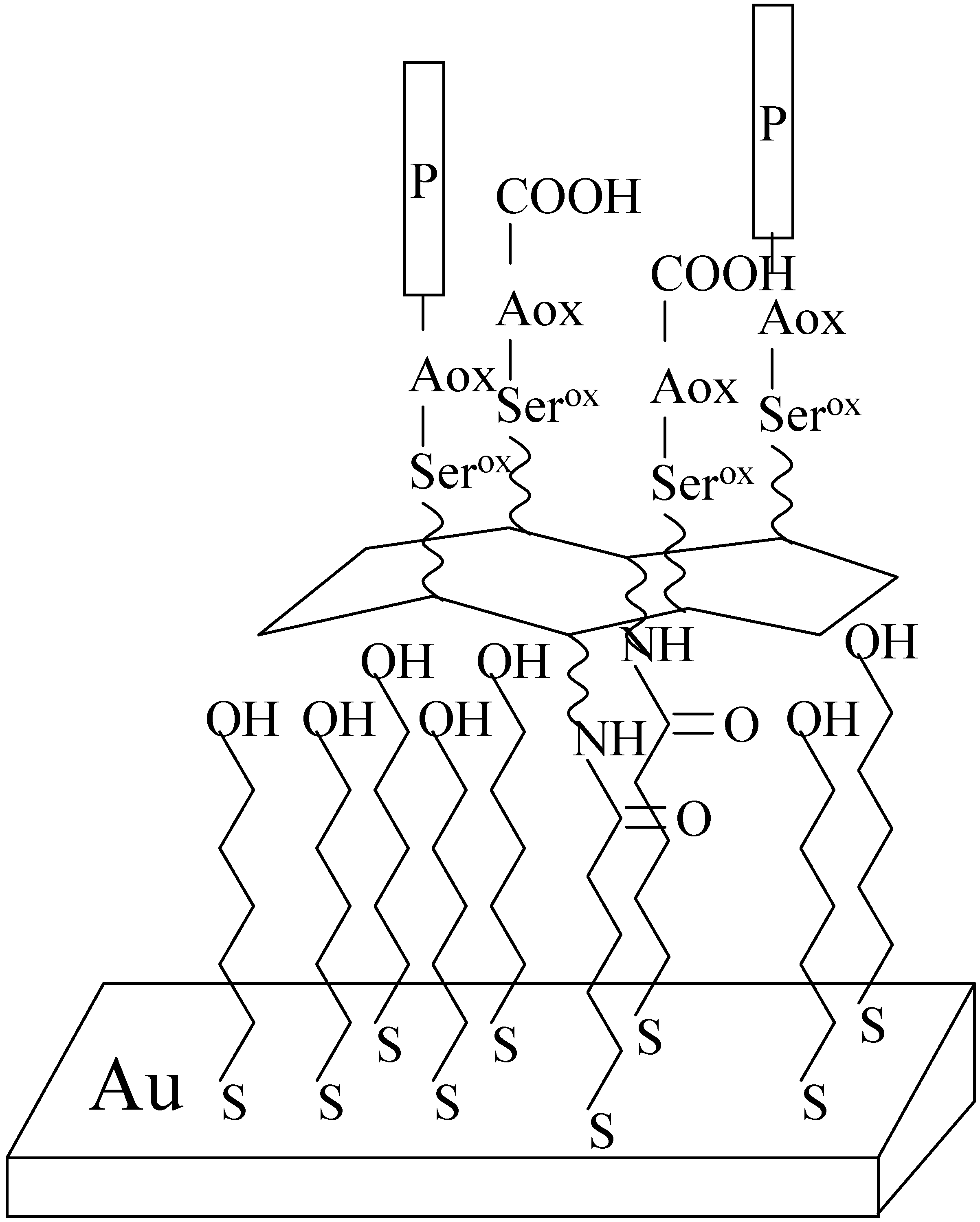
Vogel’s group 35,36 has shown that template assembled synthetic proteins (TASP) that expose binding sites to metals or antigens to SAMs `give` highly sensitive and selective surfaces. Further work36 has led to the development of regio-selective addressable functional templates (RAFT) that have different reactive, spatially distinguishable domains. As an example, a derivative of a peptide that has shown response to malaria parasites, (NANP)3 has been attached to a SAM of topological templates. The RAFT molecule was a derivative of the cyclic peptide c[(K(Boc)K(Boc)PGK(Alloc))2], which had carboxythioalkanes attached to the K(Alloc) side chains, and serines to the remaining deprotected K(Boc) sites of the serine groups. The hydroxyl groups of the serines were oxidised to aldehydes before the aminooxyacetyl-containing antigenic peptide was attached by the formation of oxime bonds. The resulting monolayers contained functionalities for the selective and reversible binding of the monoclonal antibody Sp3E9 which is directed against the NANP peptide. As is required with most SAMs, the remaining surface was blocked with a 11-mercaptoundecanol. The final assembly is shown in Fig 2.
Figure 2. Functionalized gold surface with receptor sites for selective binding of a monoclonal antibody through a peptide (4) attached to a RAFT (regioselectively addressable functional template) (Redrawn from 36).
Sensors 01 00075 g002
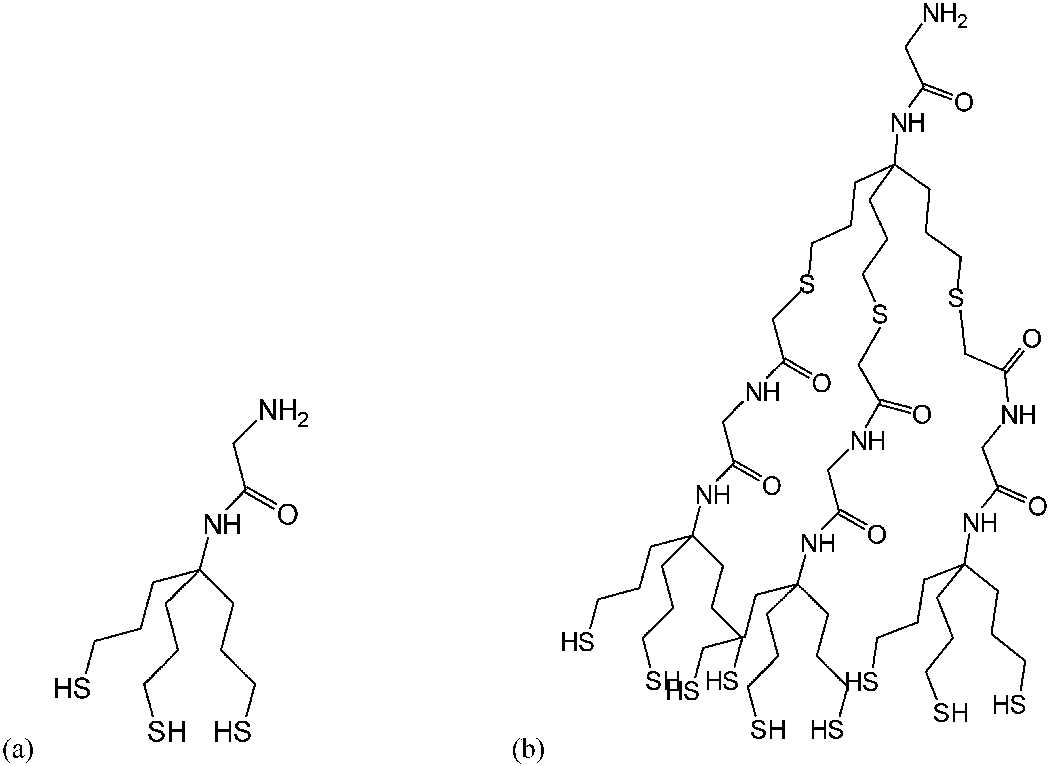
Whitesell and Chang 37 constructed directionally oriented peptide layers on gold and indium-tin-oxide glass, by using functionalised surfaces on which the initiation sites for polymerization were spaced at distances consistent with the helical diameter of the peptide. The interest of this group is in making optically switching materials. An amino acid such as alanine will form a helix with diameter 0.9 nm if more than 10 residues are in the polymer. Gold [100] has an interatomic spacing about half this value. To provide a larger footprint, an aminotrithiol was prepared (Fig 3a) and a trio of these molecules provided an even greater area (Fig 3b). Polyalanine grew readily on these substrates from a solution of the N-carboxyanhydride of alanine and was shown to be helical from FTIR.
Strong and Moore 38,39 have also reported the formation of self-assembled helical oligopeptides with three methionine residues or two cysteine residues. Unlike other workers who use a strategy of terminal connection to the underlying surface, Moore created a helix by including sulfur-containing residues (Met or Cys) along the oligopeptide to promote helical arrangement when these atoms interact with a gold surface. The first oligopeptide that was shown to be helical when adsorbed on a gold surface was Fcb-Ala-Aib-Ala-Met-Aib-Ala-Ala-Met-Ala-Aib-Met-Ala-Ala-NH2 where Fcb is ferrocene butyric acid and Aib is α-aminoisobutyric acid. Ala and Aib are known to form helices in solution. This oligopeptide self assembles on gold as evidenced by a well-formed CV of ferrocene (compared to a similar oligopeptide that did not have Met residues), and the RAIRS spectrum confirmed it to be helical with the axis parallel to the surface. The longer oligopeptide Ala-Ala-Aib-Ala-Met-Ala-Phe-Ala-Met-Aib-Phe-Met-Aib-Ala-Ala-Ala-NH2 was also prepared and shown to be helical. The cysteine containing oligopeptide Ala-Ala-Aib-Ala-Phe-Ala-Cys-Phe-Leu-Cys-Aib-(NO2)Phe-Ala-Aib-Leu-Ala-OH adsorbed on gold in a mixed α-helix and 310-helix (three residues form one complete turn with ten atoms within the intramolecular ring formed by the hydrogen bond between NH of residue i+3 and the CO of residue i). A problem encountered with Cys as the binding residue was the formation of intermolecular disulfide bonds, particularly in protic solvents.
Figure 3. (a) Structure of an aminotrithiol for assembling helical peptides on a gold surface. (b) Three aminotrithiol units supporting a fourth aminotrithiol (Redrawn from 37).
Sensors 01 00075 g003
A non-terminal strategy for the immobilisation of polypeptides was used by Corn and co-workers40,41,42. In the first example 42 poly(l-lysine) was covalently attached to a gold surface modified with 11-mercaptoundecanoic acid (MUA) by activating the carboxylic acid terminated SAM to the N-hydroxysulfosuccinimide ester. The presence of amino groups on the side chains of the amino acids resulted in the polypeptide forming multiple attachment points to the underlying SAM. In later work40,41, rather than covalently attach the poly(l-lysine) it was electrostatically adsorbed to the MUA modified gold surface by exploiting the net positive charge of the polypeptide. Repeated exposure to poly(l-glutamic acid) and poly(l-lysine) allowed multilayer structures to be fabricated which were used to bind electroactive anions.
Holcombe’s group have also investigated binding to polypeptides, in this case metal binding to poly(L-cysteine) and poly(L-aspartate) 43,44,45,46,47. The polypeptides were immobilized in silanized controlled pore glass using gluteraldehyde. Although the thrust of this work has been towards binding environmentally sensitive metals, it did show the possibilities of using these polypeptides for analysis.

Methods for investigation peptides on surfaces

Following binding events of redox active species such as metal ions is usually most easily accomplished electrochemically. This requires immobilization on a conducting electrode, and some way of avoiding direct oxidation or reduction on the bare electrode. In the case of attachment to gold by alkanethiols to achieve measurable currents the chains tethering the ionophore to the surface are kept short. For example mercaptopropionic acid (MPA) with three carbon atoms is often used. The quality of the SAMs is poorer than if a long alkyl chain was used, but this is of no consequence for many applications 48. Cyclic voltammetry of complexed ions on a modified electrode gives oxidation and reduction peaks whose heights increase linearly with scan rate with equal slope. Integration of peaks also allows calculation of the coverage of ions. The uptake of peptide and then metal can also be studied by a quartz crystal microbalance or surface plasmon resonance.
Once formed, secondary ion mass spectrometry (SIMS) and X-ray photoelectron spectroscopy can probe the composition of a surface layer, although the spectra require some interpretation. Infra red (FTIR) or surface attenuated total reflection (ATR) can also be used to pick out functional groups.

Peptides on surfaces as metal sensors

An approach described by Takehara 49 used self-assembled glutathione (Glu-Cys-Gly) on gold to detect lanthanides by a secondary ‘gate’ effect on the electrochemistry of ferrocyanide in the solution. In the absence of metal ion and at a pH greater than 5.7, glutathione, which is adsorbed through the sulfur atom of the Cys residue fill the surface creating a barrier of negatively charged carboxylate groups to the movement of the redox active species to the electrode (Fig 4a). When a metal ion is bound, the bringing together of the arms of the glutathione causes a channel to open and redox-active ions have access to the surface (Fig 4b). The response to lanthanides was La3+ > Eu3+ > Lu3+ with a detection limit of about 1 μM La3+. The sensitivity to lanthanides was about three orders of magnitude greater than to alkaline earth metals.
Metallothioneins (MTs) are a group of proteins that are rich in cysteine residues. They bind a number of divalent metal ions strongly including zinc, copper, cadmium and mercury. Self-assembled on gold, a MT containing 6.7% cadmium and 0.5% zinc rapidly took up Hg2+ from solution, binding four mercury ions per MT molecule 50. Cathodic stripping voltammetry with a two minute accumulation time gave a reduction peak for 0.08 μM Hg2+, the height of which saturated at 3 μM. The stripping peak current followed a Langmuir-type adsorption isotherm.
Figure 4(a). Schematic of glutathione molecules on a gold surface with complexed lanthanide ion.
Sensors 01 00075 g004a
Figure 4(b). Schematic of glutathione molecules on a gold surface without complexed lanthanide ion (Redrawn from 49).
Sensors 01 00075 g004b
Recently we and others 51,52 have investigated self-assembled layers of L-cysteine on gold for the detection of copper 53. The combination of molecular recognition processes and the two-dimensional structural order of monomolecular layers formed by self-assembly techniques offers a very powerful route to the development of sensing devices. The adsorption of L-cysteine on gold electrodes was studied by electrochemical reductive desorption in 0.5 M KOH, and the interaction of L-cysteine with copper ion was investigated by cyclic voltammetry, chronoamperometry and X-ray Photoelectron Spectroscopy. At low concentrations the ratio of L-cysteine to bound Cu(II) is 2:1. At higher concentrations (0.1 M) Cu2+ oxidized adsorbed cysteine forming copper sulfide on the electrode surface. On a modified L-cysteine gold electrode, Osteryoung square wave voltammetric determination of Cu(II) at ppb level concentrations was successfully demonstrated.
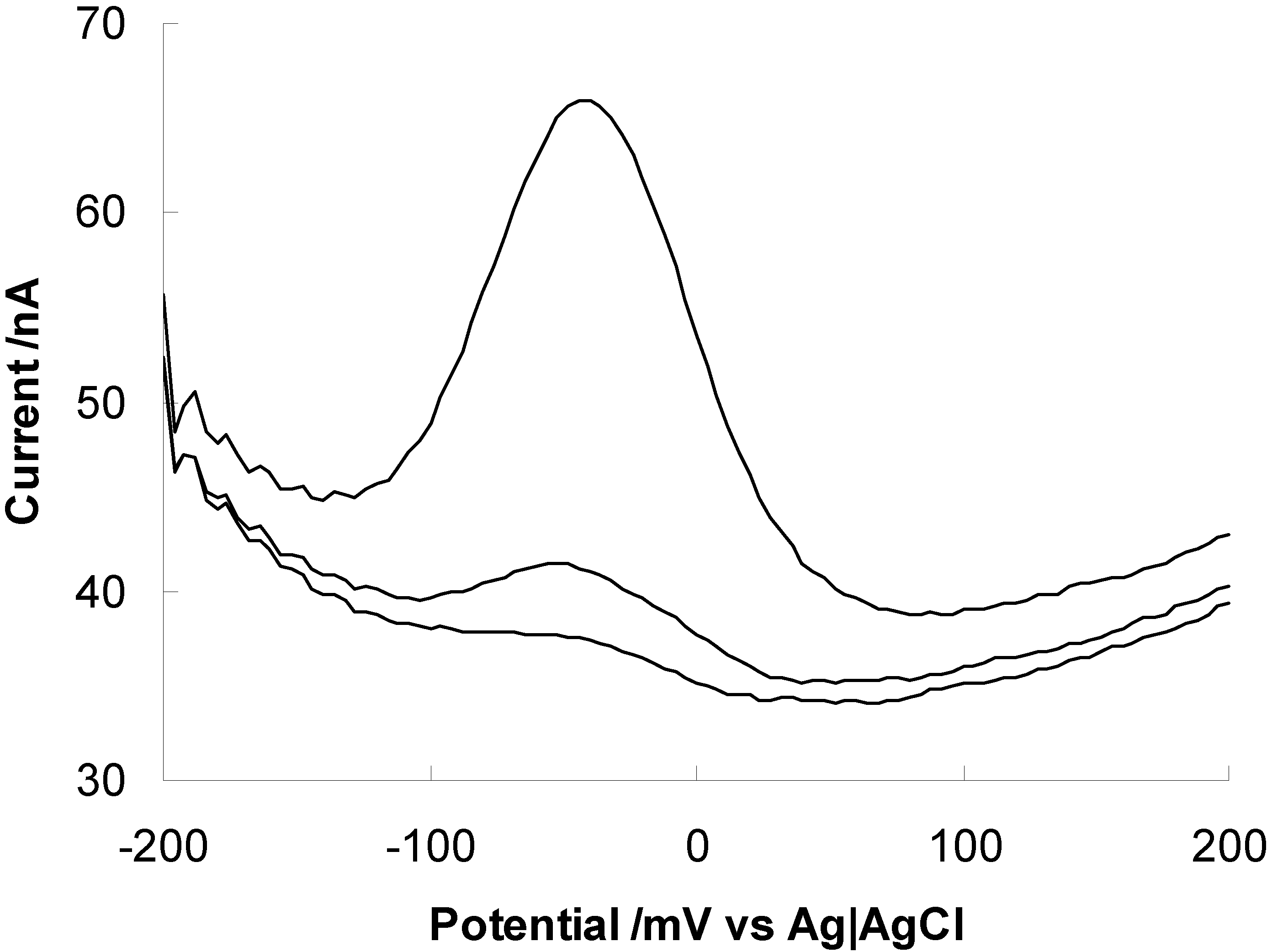
Adsorption of a peptide on gold through the sulfur atoms of a cysteine or methionine residue gives immediate self-assembly. If it is desired to attach an amino acid residue that does not include a sulfur atom an efficient method is via carbodiimide coupling to a self-assembled alkane thiol with a carboxyl terminating group. The self-assembled monolayer is activated using a mixture of N-hydroxysuccinimide (NHS) and 1-ethyl-3 (3-dimethylaminopropyl) carbodiimide hydrochloride (EDC) in pH 5.5 buffer for 30 minutes 54. The resulting succinimide ester-activated monolayer then reacts with a terminal amino group on the peptide to give a peptide bond. An example recently described was the immobilization of the so-called copper binding peptide Gly-Gly-His discussed above (see scheme 4 and Figure 5).
Figure 5. Osteryoung square wave voltammogram of Cu(II) bound to Gly-Gly-His immobilized on a gold electrode. Voltammograms with increasing current for accumulation of copper from solutions containing 0, 1.28 and 25.3 pM. Cu(II) was accumulated at the Gly-Gly-His modified electrode at open circuit for 10 minutes in a 0.05 M ammonia acetate buffer solution (pH 7.0) containing copper nitrate, removed, rinsed and then analysed in a copper-free ammonia acetate buffer solution.
Sensors 01 00075 g005
Copper was accumulated at the Gly-Gly-His modified electrode (GGHME) at open circuit for 10 minutes in a 0.05M ammonia acetate buffer solution (pH 7.0) containing copper nitrate. The copper was detected by square wave voltammetry with a linear range of calibration to 10 pM and detection limit less than 3 pM (0.2 ppt).

Future Prospects

The demonstration by us and others that amino acids and peptides can be attached to surfaces by forming peptide bonds using carbodiimide coupling indicates any peptide could be attached to an electrode surface for the detection of metal ions. Furthermore, the synthesis of peptides from component amino acids directly on the electrode surface can also be achieved is a similar manner to solid phase peptide synthesis 55. With the plethora of peptide motifs already known to have high selectivity for particular metal ions a simple generic strategy for the fabrication of metal ion sensors exists. As Walkup and Imperiali 32,33,56 have demonstrated with their fluorescence sensors substitution of a peptide ligand for a metal can be altered dramatically by changing even a single amino acid.
We plan to employ this ability to tune peptide ligand selectivity not for making metal ion sensors specific for a particular metal ion but to fabricate electrode arrays with semi-selective ligands for the detection of several metal ions on a single electrochemical chip surface. Such a strategy relies on the array of modified electrodes giving a characteristic response pattern for each metal ion to be analysed. Alternatively, decreased detection limit but reduced linear range we have observed when modifying gold electrodes with cysteine, polyaspartic acid and Gly-Gly-His respectively indicates electrochemical sensor arrays could be fabricated which would respond to a single metal ion, for example copper, concentrations from pM to mM.

References

  1. Sigel, H.; Martin, R.B. Coordinating Properties of the Amide Bond. Stability and Structure of Metal Ion Complexes of Peptides and Related Ligands. Chem. Rev. 1982, 82, 385–426. [Google Scholar] [CrossRef]
  2. Kozlowski, H.; Bal, W.; Dyba, M.; Kowalik-Jankowska, T. Specific structure-stability relations in metallopeptides. Coord. Chem. Rev. 1999, 184, 319–346. [Google Scholar] [CrossRef]
  3. Mackay, K.M.; Mackay, R.A. Introduction to Modern Inorganic Chemistry, 3rd ed.; International Textbook Company: London, 1981. [Google Scholar]
  4. Krezel, A.; Bal, W. Coordination chemistry of glutathione. Acta Biochim. Pol. 1999, 46, 567–580. [Google Scholar] [PubMed]
  5. Mendieta, J.; Diaz-Cruz, M.S.; Tauler, R.; Esteban, M. Application of multivariate curve resolution to voltammetric data. II. Study of metal-binding properties of the peptides. Anal. Biochem. 1996, 240, 134–141. [Google Scholar] [CrossRef] [PubMed]
  6. Diaz-Cruz, M.S.; Diaz-Cruz, J.M.; Mendieta, J.; Tauler, R.; Esteban, M. Soft- and hard-modeling approaches for the determination of stability constants of metal-peptide systems by voltammetry. Anal. Biochem. 2000, 279, 189–201. [Google Scholar] [CrossRef] [PubMed]
  7. Harlyk, C.; Nieto, O.; Bordin, G.; Rodriguez, A.R. Electrochemical study of a metallothionein related peptide in the presence of zinc using cyclic voltammetry. J. Electroanal. Chem. 1998, 451, 267–272. [Google Scholar] [CrossRef]
  8. Harlyk, C.; Bordin, G.; Nieto, O.; Rodriguez, A.R. Electrochemical behavior of a metallothionein related peptide in the presence of cadmium using cyclic voltammetry. J. Electroanal. Chem. 1998, 446, 139–150. [Google Scholar] [CrossRef]
  9. Mendieta, J.; Diaz-Cruz, M.S.; Monjonell, A.; Tauler, R.; Esteban, M. Complexation of cadmium by the C-terminal hexapeptide Lys-Cys-Thr-Cys-Cys-Ala from mouse metallothionein: study by differential pulse polarography and circular dichroism spectroscopy with multivariate curve resolution analysis. Anal. Chim. Acta 1999, 390, 15–25. [Google Scholar] [CrossRef]
  10. Diaz-Cruz, M.S.; Esteban, M.; Rodriguez, A.R. Square wave voltammetry data analysis by multivariate curve resolution: application to the mixed-metal system Cd-Zn-{Lys-Cys-Thr-Cys-Cys-Ala}. Anal. Chim. Acta 2001, 428, 285–299. [Google Scholar] [CrossRef]
  11. Hibbert, D.B.; Weitzner, K.; Carter, P. Voltammetry of platinum in artificial perilymph solution. J. Electrochem. Soc. 2001, 148, E1–E7. [Google Scholar] [CrossRef]
  12. Saurina, J.; Hernandez-Cassou, S.; Fabregas, E.; Alegret, S. Cyclic voltammetric simultaneous determination of oxidizable amino acids using multivariate calibration methods. Anal. Chim. Acta 2000, 405, 153–160. [Google Scholar] [CrossRef]
  13. Heyrovsky, M.; Vavricka, S. Adsorption effects of electroactive species in dc polarography demonstrated in the case of the anodic waves of cysteine. J. Electroanal. Chem. 1997, 423, 125–130. [Google Scholar] [CrossRef]
  14. Heyrovsky, M.; Vavricka, S. Electrochemical reactivity of homocysteine at mercury electrodes as compared with cysteine. Bioelectrochem. Bioenerg. 1999, 48, 43–51. [Google Scholar] [CrossRef]
  15. Bilewicz, R. The reduction of copper(II) complexes of histidine and histidyl peptides at mercury electrodes. J. Electroanal. Chem. 1989, 267, 231–41. [Google Scholar] [CrossRef]
  16. Banica, F.G.; Fogg, A.G.; Moreira, J.C. Catalytic cathodic stripping voltammetry at a hanging mercury drop electrode of glutathione in the presence of nickel ion. Analyst 1994, 119, 2343–9. [Google Scholar] [CrossRef]
  17. Kolthoff, I.M.; Stricks, W.; Kapoor, R.C. Equilibrium constants of exchange reactions of cystine with glutathione and with thioglycolic acid both in the oxidized and reduced state. J. Am. Chem. Soc. 1955, 77, 4733–4739. [Google Scholar] [CrossRef]
  18. Witwicki, J. Polarographic studies of blood using mercury drop electrodes. I. Polarographic study of individual components of blood. Postepy Biochem. 1955, 1, 63–83. [Google Scholar]
  19. Banica, F.G.; Moreira, J.C.; Fogg, A.G. Application of catalytic stripping voltammetry for the determination of organic sulfur compounds at a hanging mercury drop electrode. Behavior of cysteine, cystine and N-acetylcysteine in the presence of nickel ion. Analyst (Cambridge, U. K.) 1994, 119, 309–318. [Google Scholar] [CrossRef]
  20. Banica, F.G.; Fogg, A.G.; Moreira, J.C. Catalytic cathodic stripping voltammetry of oxidized glutathione at a hanging mercury drop electrode in the presence of nickel ion. Talanta 1995, 42, 227–234. [Google Scholar] [CrossRef]
  21. Forsman, U. Cathodic stripping voltammetry of the peptide felypressin in trace amounts. Anal. Chim. Acta 1984, 156, 43–49. [Google Scholar] [CrossRef]
  22. Scarano, G.; Morelli, E. Polarographic behavior of metal phytochelatin complexes. Electroanalysis 1998, 10, 39–43. [Google Scholar] [CrossRef]
  23. Ertas, F.N.; Fogg, A.G.; Moreira, J.C.; Barek, J. Differential pulse cathodic stripping voltammetry of the copper complexes of glycyl-L-histidyl-glycine at a hanging mercury drop electrode. Talanta 1993, 40, 1481–1488. [Google Scholar] [CrossRef]
  24. Moreira, J.C.; Fogg, A.G. Determination of nanomolar levels of histidine by differential-pulse adsorptive-cathodic stripping voltammetry of its copper(II) complex. Analyst 1990, 115, 41–43. [Google Scholar] [CrossRef]
  25. Humble, A.V.; Gadd, G.M.; Codd, G.A. Binding of copper and zinc to three cyanobacterial microcystins quantified by differential pulse polarography. Water Res. 1997, 31, 1679–1686. [Google Scholar] [CrossRef]
  26. Yan, F.; Ozsoz, M.; Sadik, O.A. Electrochemical and conformational studies of microcystin-LR. Anal. Chim. Acta 2000, 409, 247–255. [Google Scholar] [CrossRef]
  27. Tanaka, S.; Yoshida, H. Enhancement Effect of Cysteine on Anodic Stripping Voltammetry of Copper. J. Electroanal. Chem. 1982, 137, 261–270. [Google Scholar] [CrossRef]
  28. Bai, Y.; Ruan, X.Y.; Mo, J.Y.; Xie, Y.Q. Potentiometric stripping analysis of copper using cysteine modified mercury film electrode. Anal. Chim. Acta 1998, 373, 39–46. [Google Scholar] [CrossRef]
  29. Moreira, J.C.; Zhao, R.; Fogg, A.G. Modification of electrodes with adsorbed polyamino acids. Part 1. Cathodic stripping voltammetric determination of copper(II) at a hanging mercury drop electrode using adsorptive accumulation on an adsorbed layer of poly-L-histidine. Analyst 1990, 115, 1561–1564. [Google Scholar] [CrossRef]
  30. Pirzad, R.; Moreira, J.C.; Rangel, A.O.S.S.; Alonso, R.M.; Edmonds, T.E.; Fogg, A.G. Differential-pulse cathodic stripping voltammetric determination of sodium nitroprusside at a hanging mercury drop electrode aided by copper(II) and poly-L-lysine modification. Analyst 1994, 119, 963–968. [Google Scholar] [CrossRef]
  31. Moreira, J.C.; Fogg, A.G. Modification of electrodes with adsorbed polyamino acids. Part 2. Adsorptive stripping voltammetric determination of hexacyanoferrate(III) at a hanging mercury drop electrode in the presence of an adsorbed layer of copper-modified poly-L-lysine. Analyst 1990, 115, 1565–1568. [Google Scholar] [CrossRef]
  32. Walkup, G.K.; Imperiali, B. Fluorescent chemosensors for divalent zinc based on zinc finger domains. Enhanced oxidative stability, metal binding affinity, and structural and functional characterization. J. Am. Chem. Soc. 1997, 119, 3443–3450. [Google Scholar] [CrossRef]
  33. Torrado, A.; Walkup, G.K.; Imperiali, B. Exploiting polypeptide motifs for the design of selective Cu(II) ion chemosensors. J. Am. Chem. Soc. 1998, 120, 609–610. [Google Scholar] [CrossRef]
  34. Miura, Y.; Kimura, S.; Imanishi, Y.; Umemura, J. Oriented helical peptide layer on the carboxylate-terminated alkanethiol immobilized on a gold surface. Langmuir 1999, 15, 1155–1160. [Google Scholar] [CrossRef]
  35. Scheibler, L.; Dumy, P.; Stamou, D.; Duschl, C.; Vogel, H.; Mutter, M. Functionalization of gold surfaces via topological templates. Tetrahedron 1998, 54, 3725–3734. [Google Scholar] [CrossRef]
  36. Scheibler, L.; Dumy, P.; Boncheva, M.; Leufgen, K.; Mathieu, H.-J.; Mutter, M.; Vogel, H. Functional molecular thin films: topological templates for the chemoselective ligation of antigenic peptides to self-assembled monolayers. Angew. Chem., Int. Ed. 1999, 38, 696–699. [Google Scholar] [CrossRef]
  37. Whitesell, J.K.; Chang, H.K. Directionally aligned helical peptides on surfaces. Science (Washington, D. C., 1883-) 1993, 261, 73–76. [Google Scholar] [CrossRef] [PubMed]
  38. Strong, A.E.; Moore, B.D. Self-assembling chiral monolayers of helical peptides bound to gold via side-chain thioethers. Chem. Commun. 1998, 473, 473–474. [Google Scholar] [CrossRef]
  39. Strong, A.E.; Moore, B.D. Self-assembling monolayers of helical oligopeptides on gold with applications in molecular electronics. J. Mater. Chem. 1999, 9, 1097–1105. [Google Scholar] [CrossRef]
  40. Cheng, Y.F.; Corn, R.M. Ultrathin polypeptide multilayer films for the fabrication of model liquid/liquid electrochemical interfaces. J. Phys. Chem. B 1999, 103, 8726–8731. [Google Scholar] [CrossRef]
  41. Cheng, Y.F.; Murtomaki, L.; Corn, R.M. Electrochemical characterization of the ultrathin polypeptide film/1,2-dichloroethane liquid vertical bar liquid interface. J. Electroanal. Chem. 2000, 483, 88–94. [Google Scholar] [CrossRef]
  42. Frey, B.L.; Corn, R.M. Covalent Attachment and Derivatization of Poly(L-lysine) Monolayers on Gold Surfaces as Characterized by Polarisation-Modulation FT-IR Spectroscopy. Anal. Chem. 1996, 68, 3187–3193. [Google Scholar] [CrossRef]
  43. Autry, H.A.; Holcombe, J.A. Cadmium, Copper and Zinc-Complexes of Poly-L-Cysteine. Analyst 1995, 120, 2643–2647. [Google Scholar] [CrossRef] [PubMed]
  44. Gutierrez, E.; Miller, T.C.; Gonzalez-Redondo, J.R.; Holcombe, J.A. Characterization of immobilized poly-L-aspartate as a metal chelator. Environ. Sci. Technol. 1999, 33, 1664–1670. [Google Scholar] [CrossRef]
  45. Howard, M.; Jurbergs, H.A.; Holcombe, J.A. Effects of oxidation of immobilized poly(L-cysteine) on trace metal chelation and preconcentration. Anal. Chem. 1998, 70, 1604–1609. [Google Scholar] [CrossRef]
  46. Jurbergs, H.A.; Holcombe, J.A. Characterization of immobilized poly(L-cysteine) for cadmium chelation and preconcentration. Anal. Chem. 1997, 69, 1893–1898. [Google Scholar] [CrossRef]
  47. Miller, T.C.; Holcombe, J.A. An ion-exchange microcolumn employing gold minigrids as supports for the on line immobilization of poly(L-aspartate). Anal. Chem. 1999, 71, 2667–2671. [Google Scholar] [CrossRef]
  48. Gooding, J.J.; Hibbert, D.B. The Application of Alkanethiol Self-Assembled Monolayers to Enzyme Electrodes. TrAC 1999, 18, 525–533. [Google Scholar] [CrossRef]
  49. Takehara, K.; Aihara, M.; Ueda, N. An Ion-Gate Response of a Glutathione Monolayer Assembly Highly Sensitive to Lanthanide Ions. Electroanalysis 1994, 6, 1083–1086. [Google Scholar] [CrossRef]
  50. Ju, H.X.; Leech, D. Electrochemical study of a metallothionein modified gold disk electrode and its action on Hg2+ cations. J. Electroanal. Chem. 2000, 484, 150–156. [Google Scholar] [CrossRef]
  51. Arrigan, D.W.M.; Le Bihan, L. A study of L-cysteine adsorption on gold via electrochemical desorption and copper(II) ion complexation. Analyst 1999, 124, 1645–1649. [Google Scholar] [CrossRef]
  52. Liu, A.-C.; Chen, D.-c.; Lin, C.-C.; Chou, H.-H.; Chen, C.-h. Application of Cysteine Monolayers for Electrochemical Determination of Sub-ppb Copper(II). Anal. Chem. 1999, 71, 1549–1552. [Google Scholar] [CrossRef]
  53. Yang, W.; Gooding, J.J.; Hibbert, D.B. Characterisation of Gold Electrodes Modified with Self Assembled Monolayers of L-Cysteine for the Adsorptive Stripping Analysis of Copper. J. Electroanal. Chem. 2001. submitted. [Google Scholar] [CrossRef]
  54. Gooding, J.J.; Praig, V.; Hall, E.A.H. Platinum Mediated Enzyme Electrodes Immobilised on Gold using Self-Assembled Monolayers. Anal. Chem. 1998, 70, 2396–2402. [Google Scholar] [CrossRef] [PubMed]
  55. Gaus, K. Ph. D., Cambridge University, 1999.
  56. Imperiali, B.; Walkup, G.K. U.S.; (California Institute of Technology, USA); US 6083758; 2000; p. 19. [Google Scholar]
  • Sample Availability: Not available.