Article

Gonadal Steroid and Transcriptome Profiles Reveal Sexual Dimorphism in the Freshwater Bivalve Sinanodonta woodiana


1
Wuxi Fisheries College, Nanjing Agricultural University, Wuxi 214081, China
2
College of Marine Science and Technology and Environment, Dalian Ocean University, Dalian 116023, China
3
College of Fisheries and Life Science, Dalian Ocean University, Dalian 116023, China
4
Ministry of Fisheries and Blue Economy Federal Government of Somalia, Mogadishu, Somalia
5
Freshwater Fisheries Research Center, Chinese Academy of Fishery Sciences, Wuxi 214081, China
*
Author to whom correspondence should be addressed.
Diversity 2026, 18(9), 578; https://doi.org/10.3390/d18090578
Submission received: 30 July 2026 / Revised: 19 September 2026 / Accepted: 20 September 2026 / Published: 21 September 2026
(This article belongs to the Special Issue Ecology and Conservation of Freshwater Bivalves)

Abstract

Sinanodonta woodiana is one of the most widely distributed freshwater bivalves in the world. However, the sexual dimorphism of this species remains poorly understood. In this study, gonads of clearly sexed S. woodiana were used to analyze steroid hormones, including estrone (E1), estradiol-17β (E2), estriol (E3), and testosterone (T), by liquid chromatography-mass spectrometry, followed by comparative transcriptome analysis. Compared with males, females showed no significant differences in E1 (p = 0.251), E3 (p = 0.136), or T (p = 0.917), whereas E2 concentration in females (15.2 ± 1.4 ng/g wet weight) was significantly higher than that in males (3.2 ± 1.7 ng/g wet weight) (p = 0.009). In addition, a total of 6716 differentially expressed genes (DEGs) were identified, including several sex-related genes such as forkhead box L2 (FOXL2), upregulated in females, and splicing factor 1 (SF1), upregulated in males. All DEGs were mainly enriched in biological processes such as metabolic process, cellular process, and single-organism process, as well as signaling pathways including neuroactive ligand–receptor interaction, glycosphingolipid biosynthesis—lacto and neolacto series, and oocyte meiosis. Our findings provide valuable information for sex identification of adult S. woodiana and offer candidate markers for future testing in juveniles, while also enhancing our understanding of its sex-specific gene-expression diversity.

1. Introduction

The freshwater bivalve Sinanodonta woodiana originated in the Yangtze River basin of China [1] and has since been widely distributed across Asia, Europe, the Americas, and Africa [2]. This species has been exploited for various purposes, including as a protein source, for pearl culture, in biopharmaceutical applications, as a fishmeal substitute in feed, as a bioindicator organism, and for water purification [3,4,5,6,7,8,9,10,11,12]. Despite its diverse uses, sex identification remains a challenge in S. woodiana [3,4,5,6,7,8,9,10,11,12]. Although the species is dioecious, there is currently no effective method to determine sex at the juvenile stage; sex can typically only be identified by examining the outer gills and/or gonads upon reaching sexual maturity (approximately 750 days) [13]. This poses practical problems for applications. For example, when fast-growing juveniles are used for bioremediation of aquaculture water, farmers are often concerned that once the mussels mature, they may release glochidia that parasitize fish and impair their growth [1]. Thus, there is a clear need to investigate sexual dimorphism in S. woodiana.
Steroids have been suggested as potential indicators for discriminating sex in bivalves. Studies have shown that in the hard-shelled mussel Mytilus coruscus, the ratio of testosterone (T) to estradiol-17β (E2) in the male gonad is significantly higher than that in females [14]. In the blue mussel Mytilus trossulus, females exhibit significantly higher concentrations of estrone (E1), E2, and estriol (E3) than males [15]; conversely, T levels are higher in male gonads [15]. Furthermore, the concentrations of these steroid hormones show seasonal variation, with higher levels during the reproductive season than in the non-reproductive season [14,15]. Notably, whether steroid hormones exhibit sex differences in freshwater bivalves has not yet been reported. Nevertheless, E1, E2, E3, and T represent important references for discriminating sexual dimorphism in freshwater bivalves.
In addition to steroids, sex-related genes exist in bivalves. Comparative transcriptome analysis is a key approach for identifying sex-related genes in bivalves [16,17,18]. In marine bivalves, transcriptomic analysis of gonads from male and female blood clam Tegillarca granosa has identified sex-related genes, including FOXL2 (forkhead box L2), SOX (sex-determining region Y-box), β-catenin, CBX (chromobox homolog), and SXL (Sex-lethal) [16]. Transcriptomic analysis of gonads from the yesso scallop Patinopecten yessoensis revealed that DMRT1 (doublesex and mab-3 related transcription factor 1) is the most important gene for sex determination and differentiation, determining the male sex phenotype [19]. In freshwater bivalves, transcriptomic analysis of gonads from the triangle sail mussel Hyriopsis cumingii has also identified sex-related genes, such as the male-associated DMRT1, SOX9, and SF1 (splicing factor 1), and the female-associated FOXL2 [17]. Therefore, transcriptome analysis holds promise for distinguishing sex differences in S. woodiana at the genetic level.
In this study, we used adult S. woodiana of clearly distinguishable sex to compare gonadal steroid profiles (including E1, E2, E3, and T) and to perform transcriptomic analysis. As a preliminary and pioneering investigation, we hypothesized that sexually dimorphic differences in steroids and transcriptomes exist in adult S. woodiana; whether these differences also occur in juveniles remains to be determined. Our findings will provide valuable information for sex identification in S. woodiana and improve our understanding of the sex-specific gene-expression diversity of this species.

2. Materials and Methods

2.1. Sample Collection

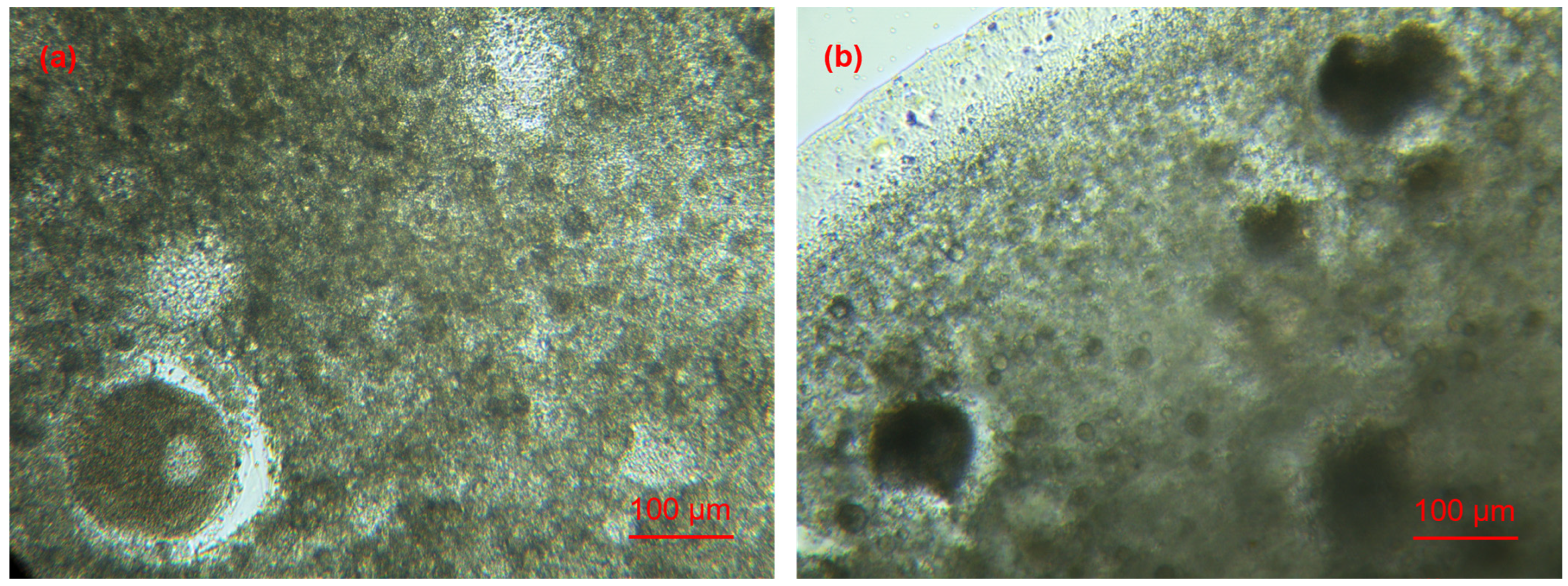
Adult S. woodiana were collected during winter (non-reproductive season) from the Freshwater Fisheries Research Center, Chinese Academy of Fishery Sciences (Wuxi, China). Initial sex determination was performed by visual inspection of the number of filaments on the outer gills [20]: females were identified by 112–167 filaments, whereas males by 62–73 filaments. This was further confirmed by microscopic examination of gonadal subsamples, with the presence of ovaries (oocytes) indicating females (Figure 1a) and the presence of testes (spermatocytes) indicating males (Figure 1b). Through this two-step process, any ambiguous individuals were excluded. Ultimately, eleven females (3 years old; shell length 10.9 ± 0.8 cm) and eleven males (3 years old; shell length 10.4 ± 0.7 cm) were used for the experiment. Of these, five females and five males were used for steroid detection, three females and three males for transcriptome analysis, and the remaining three females and three males for real-time quantitative PCR (RT-qPCR) validation. Gonads were dissected using a stainless steel scalpel. To avoid cross-contamination, the scalpel was rinsed with sterile water after each dissection. The dissected gonads were placed in cryotubes, immediately frozen in liquid nitrogen for 24 h, and then transferred to −80 °C until further analysis.

2.2. Steroid Determination

Steroid analysis was performed following previously described methods [21,22] with minor modifications. Briefly, liquid chromatography-mass spectrometry (LC-MS) was employed. The gonads were ground to a powder in liquid nitrogen, after which 5 mL of acetonitrile was added, vortexed for 30 s, and ultrasonicated at low temperature for 30 min. After centrifugation at 12,000× g for 5 min at 4 °C, the supernatant was collected. The supernatant was then purified by adding 20 mg of C18 (octadecylsilane) and 20 mg of PSA (N-propyl ethylenediamine), followed by vortexing and centrifugation. The supernatant was dried under nitrogen, and the residue was redissolved in 200 μL of methanol, filtered through a 0.22-μm membrane, and analyzed by LC-MS. The LC system (Agilent Infinity 1260, Agilent Technologies, Santa Clara, CA, USA) was equipped with a binary pump degasser, an autosampler, and a variable wavelength UV–Vis detector. Chromatographic separation was performed on a Poroshell 120 EC-C18 column (2.1 mm × 150 mm, 2.7 μm; Agilent Technologies, Santa Clara, CA, USA). The mobile phase consisted of aqueous 0.1% formic acid (phase A) and methanol (phase B). The injection volume was 2 µL, the flow rate was 0.3 mL/min, and the column temperature was maintained at 35 °C. For steroid analysis, a mass spectrometer equipped with a quadrupole mass analyzer (Agilent 6420, Agilent Technologies, Santa Clara, CA, USA) was operated in positive electrospray ionization mode. The system was run in single-ion monitoring mode, with the [M + H]+ pseudomolecular ions for E1, E2, E3, and T at m/z 271, 273, 289 and 289, respectively. To ensure unambiguous identification, E3 and T, which share the same precursor ion at m/z 289, were distinguished by confirmatory product ions (E1: m/z 253.1/107; T: m/z 109/97). Optimized fragmentor voltages were selected individually for each compound. Steroid concentrations were expressed as ng per gram of tissue wet weight. The LC-MS method was validated for the four target steroids. Calibration curves were constructed using authentic standards at seven concentration levels, with correlation coefficients (R2) > 0.99 for all compounds. Recoveries ranged from 79% to 91%. The detection limits were 0.1 ng/g for E1, E2, and E3, and 0.05 ng/g for T.

2.3. Transcriptome Analysis

Transcriptome analysis was performed as previously described [23,24]. Briefly, total RNA was extracted using RNAiso Plus (Takara Bio Inc., Kusatsu, Shiga, Japan). The RNA integrity and quantity of each sample were determined using an Agilent 2100 Bioanalyzer (Agilent Technologies, Santa Clara, CA, USA) and a NanoDrop 1000 Spectrophotometer (Thermo Scientific, Waltham, MA, USA). cDNA libraries were constructed by PCR enrichment and sequenced on the Illumina NovaSeq 6000 platform (Illumina Inc., San Diego, CA, USA). After trimming and filtering, clean reads were reassembled using Trinity software (v2.4.0). The Q30 and GC content of clean reads were calculated. Gene annotation was performed using the NCBI non-redundant protein sequence database (Nr), Clusters of Orthologous Groups (COG), Gene Ontology (GO), Kyoto Encyclopedia of Genes and Genomes (KEGG), euKaryotic Orthologous Groups (KOG), Protein family (Pfam), and UniProtKB/Swiss-Prot (Swiss-Prot) databases. The Pearson correlation analysis and principal components analysis (PCA) were performed to evaluate biological replicates. Differentially expressed genes (DEGs) were identified using DESeq2 software (v1.22.2) based on raw read counts, with thresholds of |log2(fold change)| > 1 and false discovery rate (FDR) < 0.05. Finally, GO enrichment and KEGG pathway analyses were performed on the DEGs.

2.4. Real-Time Quantitative PCR Validation

To verify the reliability of the transcriptome data, RT-qPCR was performed. Five DEGs involved in sex-related pathways were selected for RT-qPCR analysis: hypothetical protein LOTGIDRAFT_141290, partial (LOTGIDRAFT), Speedy protein 1-B (Speedy protein), mitotic spindle assembly checkpoint protein MAD2A-like isoform X1 (MAD2A), dual specificity mitogen-activated protein kinase kinase 1-like isoform X1 (kinase 1), and hypothetical protein CAPTEDRAFT_83962, partial (CAPTEDRAFT). The primers used for these genes are listed in Table 1. RT-qPCR was performed with β-actin as the internal reference gene, following the method described previously [23]. Relative gene expression was calculated using the 2−ΔΔCT method [25].

2.5. Statistical Analysis

Statistical analyses were performed using R software (version 4.5.2). For steroid concentrations, given the small sample size, non-parametric Mann–Whitney U tests were used for comparisons among E1, E2, E3, and T within each sex, as well as between females and males, following Zabrzańska et al. [26]. Independent-sample t-tests were used to compare the expression levels of selected DEGs between sexes. No multiple testing correction was applied, as only two groups were compared for each gene. All data are presented as mean ± standard deviation.

3. Results

3.1. Steroid Concentrations

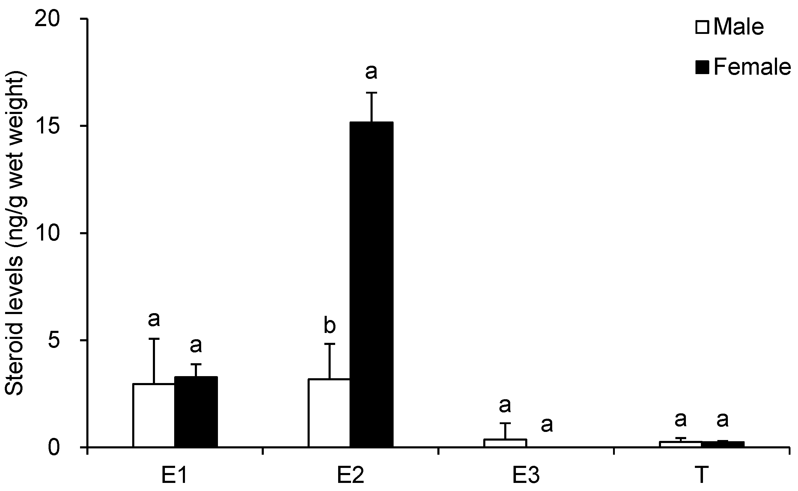
In female gonads, E3 was below the detection limit, whereas E1, E2, and T were clearly detected. Significant differences were observed among E1, E2, and T (p = 0.005–0.009), with concentrations ranked as E2 > E1 > T. In male gonads, E1, E2, E3, and T were all detected, with E1 and E2 being significantly higher than E3 and T (p = 0.009–0.015). Compared with males, females showed no significant differences in E1 (p = 0.251), E3 (p = 0.136), or T (p = 0.917) concentrations (Figure 2), whereas E2 concentration in females (15.2 ± 1.4 ng/g wet weight) was significantly higher than that in males (3.2 ± 1.7 ng/g wet weight) (p = 0.009), with the female average being 4.8 times that of males (Figure 2).

3.2. Differentially Expressed Genes

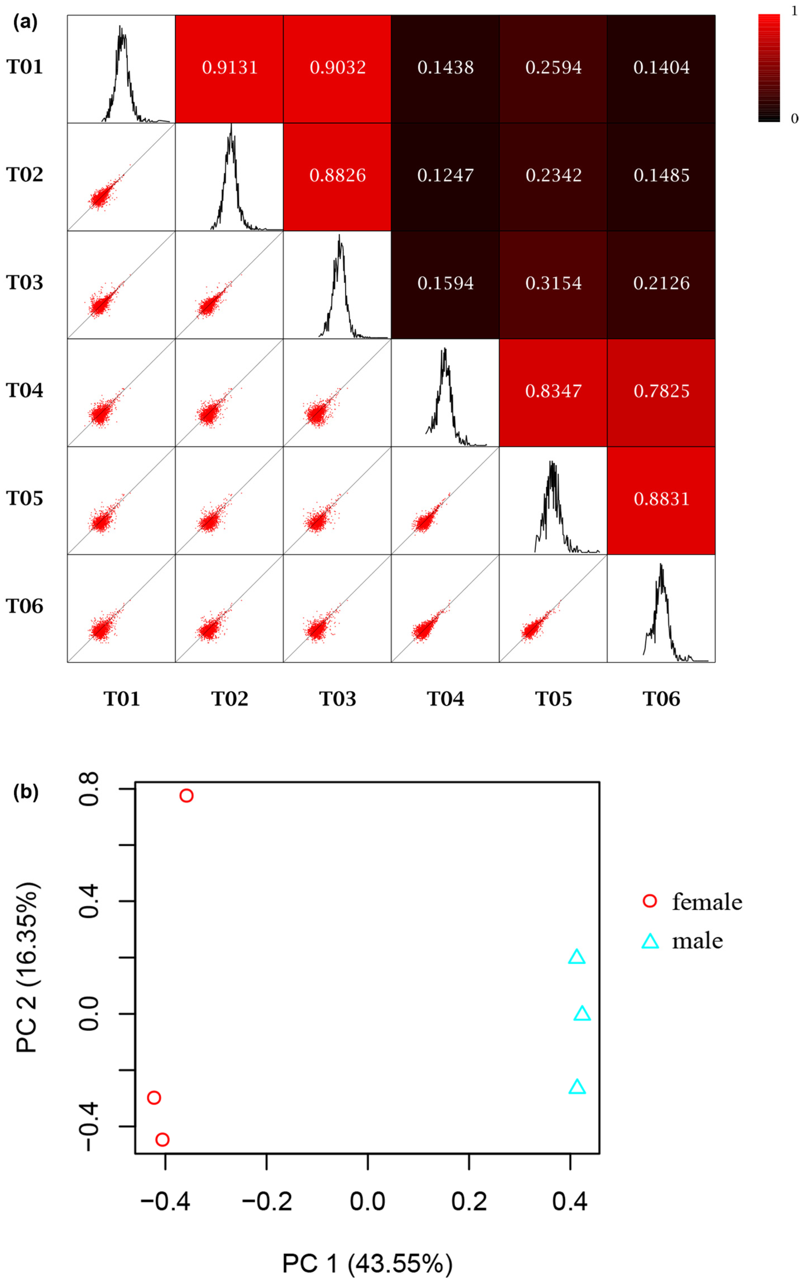
The summary statistics of S. woodiana gonad transcriptome sequencing data are shown in Table 2. In total, 45.48 Gb clean reads were obtained. The Q30 values of females and males exceeded 90.49% and 91.37%, respectively. The GC content of females and males exceeded 39.61% and 38.62%, respectively. Raw sequence data have been deposited in NCBI under BioProject PRJNA760975. The assembly yielded 211,841 transcripts and 106,115 unigenes, with N50 values of 1242 bp for transcripts and 1040 bp for unigenes. A total of 36,247 unigenes were successfully annotated. As shown in Figure 3, the reproducibility among biological replicates within each group was good.
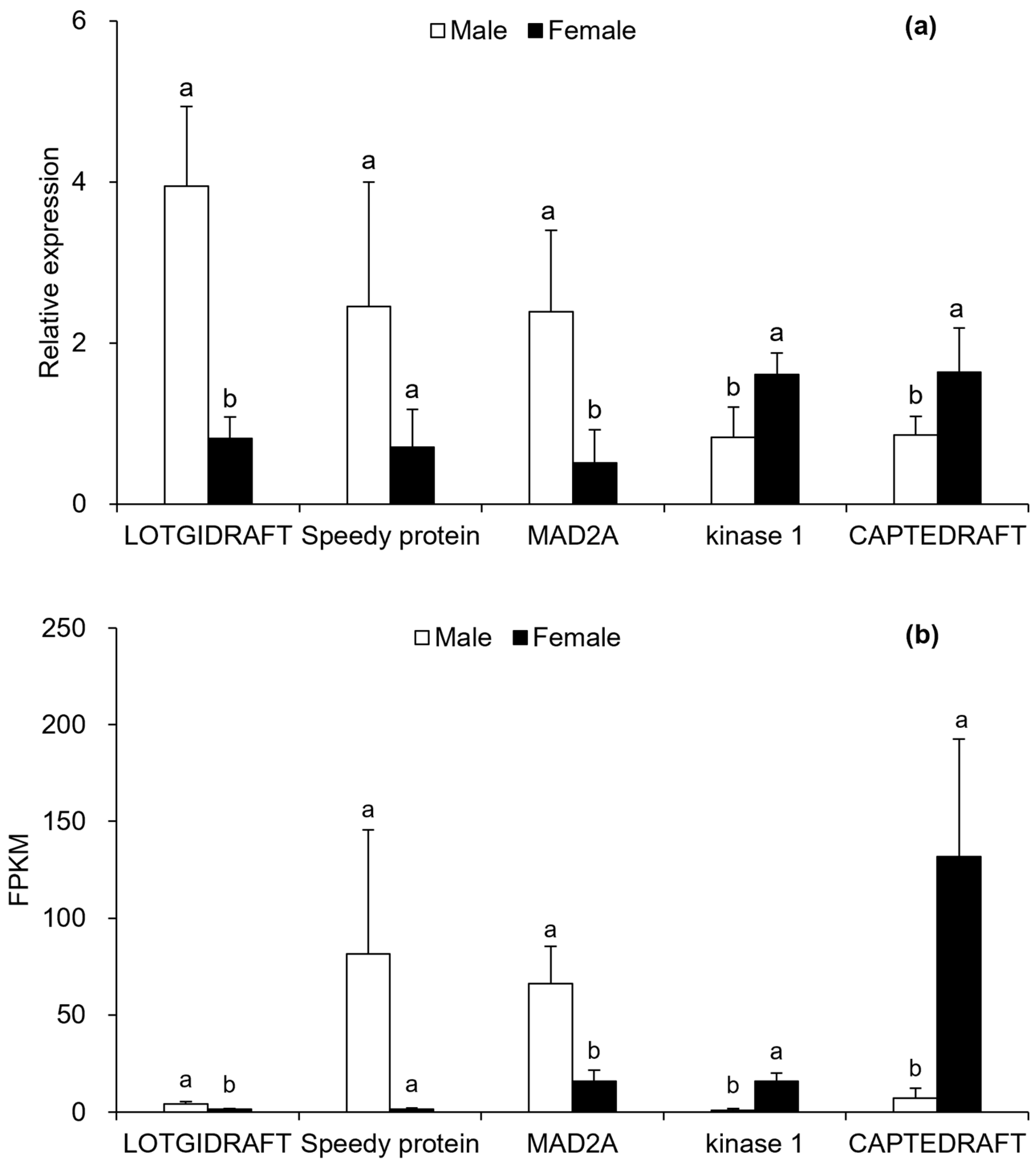
Comparing females with males, a total of 6716 DEGs were identified, of which 3358 were significantly upregulated and 3358 downregulated. These DEGs included several sex-related genes. For example, FOXL2 expression in females (201.7 ± 36.0) was significantly higher than in males (0.1 ± 0.2) (p = 0.0006), while SF1 expression in males (116.1 ± 3.7) was significantly higher than in females (31.8 ± 12.1) (p = 0.0003). Furthermore, the relative expression patterns of the five selected DEGs determined by RT-qPCR (Figure 4a) were consistent with the FPKM values from the transcriptome analysis (Figure 4b), confirming the reliability of the transcriptome data.

3.3. Pathway Enrichment Analysis

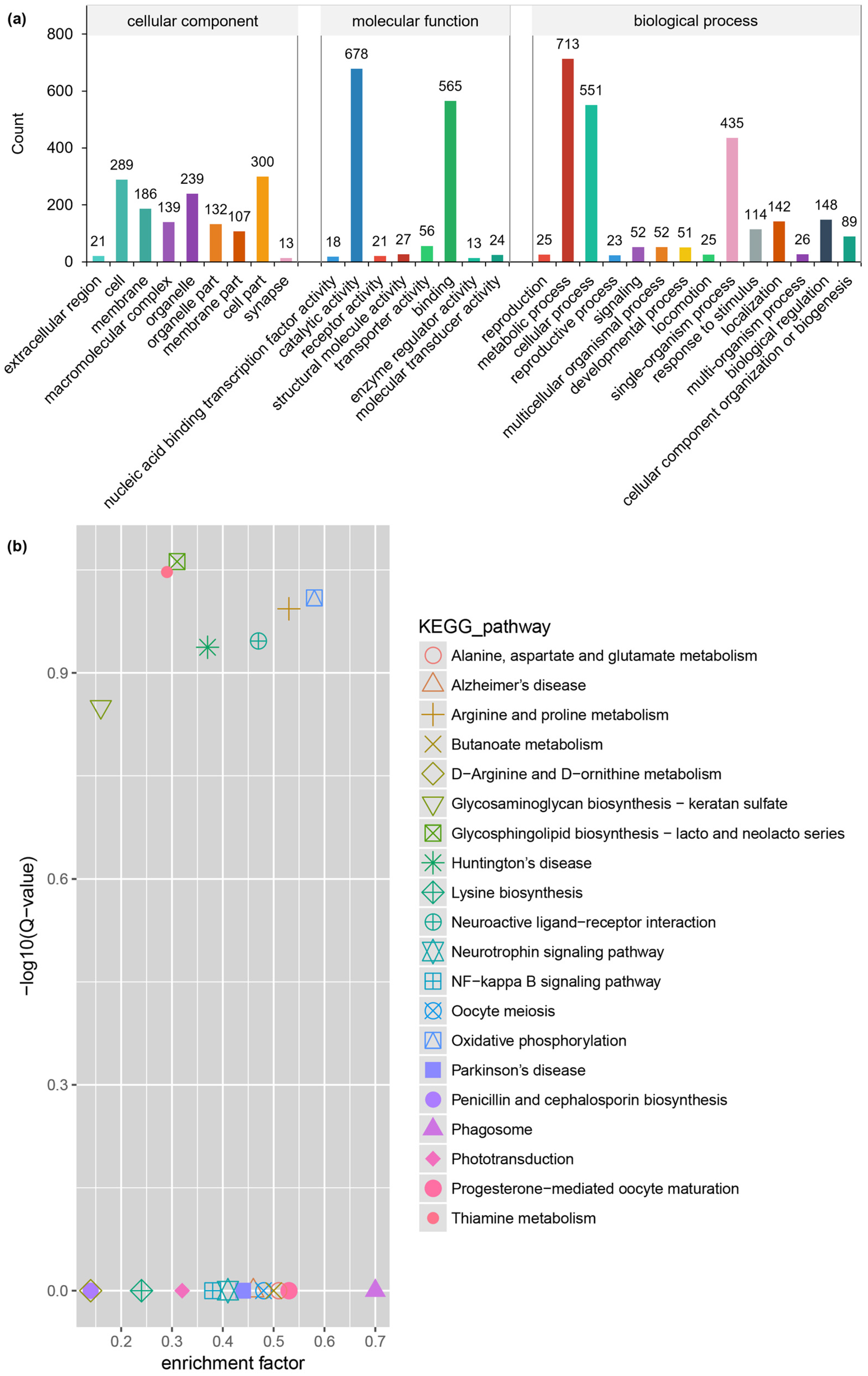
GO and KEGG analyses were performed to functionally annotate the DEGs. GO analysis (Figure 5a) showed that DEGs were mainly enriched in biological processes such as metabolic process (GO: 0008152), cellular process (GO: 0009987), and single-organism process (GO: 0044699). KEGG analysis (Figure 5b) revealed that DEGs were mainly enriched in pathways such as neuroactive ligand–receptor interaction (ko04080), glycosphingolipid biosynthesis—lacto and neolacto series (ko00603), and oocyte meiosis (ko04114). In the oocyte meiosis pathway, 13 DEGs were enriched, including the upregulated cytoplasmic polyadenylation element-binding protein 1 (CPEB1). In the steroid hormone biosynthesis pathway (ko00140), however, only one DEG (cytochrome P450 3A19) was enriched.

4. Discussion

4.1. Sex Differences in Steroid Concentrations in S. woodiana

In bivalves, steroid hormones generally originate from two sources [15,27,28]: (1) uptake from the aquatic environment (exogenous), and (2) endogenous synthesis. For example, exposure of the Manila clam (Ruditapes philippinarum) to 10 μg/L E2 for two months increased the female-to-male ratio by 50% compared to the control group (without exogenous E2) and induced a 20-fold increase in hermaphroditism, confirming exogenous uptake of steroids [27]. However, in M. trossulus, microsomal and mitochondrial fractions of gills and gonads contain enzymatic complexes capable of aromatization (estrogen synthesis), and mussels can uptake testosterone and androstenedione from the water and metabolize them into E1 and E2, confirming endogenous estrogen synthesis [28]. In the present study, all specimens were collected from the same site, so environmental variation was reduced, although individual differences in steroid uptake and metabolism cannot be excluded.
The steroids E1, E2, and E3 are estrogens, while T is an androgen. In M. trossulus, females have significantly higher E1, E2, and E3 concentrations and significantly lower T concentrations than males [15]. Interestingly, in S. woodiana gonads, unlike M. trossulus, E1, E3, and T showed no sex differences, whereas E2 was significantly higher in females, representing a novel pattern. In bivalves such as M. coruscus [14,29] and M. trossulus [15], E2 may be involved in sex determination and play a regulatory role in female ovarian development. Furthermore, E2 levels show seasonal variation correlated with the reproductive cycle [14,15,29], with higher concentrations during the reproductive season than in the non-reproductive season. Although the adult S. woodiana used in this study were in the non-reproductive season, E2 levels in females were still significantly higher than in males (more than 4-fold), suggesting that E2 is likely associated with sex differentiation/determination in S. woodiana during the non-reproductive season. Since juveniles have not yet reached sexual maturity and are also in a non-reproductive state, E2 may provide candidates for future testing in juvenile S. woodiana.
The steroid data were obtained from five individuals per sex; this sample size remains relatively limited for broad statistical generalization. Nevertheless, the observed sex difference in E2 concentration was large (>4-fold) and consistent across individuals, supporting its biological relevance. Validation in larger cohorts is needed to confirm this pattern. It should be noted that steroid and transcriptome data were obtained from different individuals. Therefore, direct correlations between E2 levels and gene expression patterns at the individual level cannot be inferred from the current data.

4.2. Sex Differences in Gene Expression in S. woodiana

Comparative transcriptome analysis has been used to investigate sex-related genes in various bivalves, including T. granosa [16], H. cumingii [17], R. philippinarum [18], P. yessoensis [19], and Cyclina sinensis [30]. In this study, we successfully applied transcriptome analysis to discover sex-differentially expressed genes in the genus Sinanodonta, comprehensively revealing the diversity and differences in gene expression between male and female S. woodiana.
Among the 106,115 assembled unigenes, 6716 DEGs were identified between females and males, with comparable numbers of up- and downregulated genes. Although the RT-qPCR validation was based on a limited sample size (n = 3 per sex) and may constrain statistical generalizability, the expression trends were consistent with RNA-seq, supporting the reliability of the transcriptome data. Notably, sex-related genes such as FOXL2 and SF1 were identified. FOXL2 is a member of the forkhead box transcription factor family [16,17]. It plays a key role not only in vertebrate sex differentiation, determination, and ovarian development but has recently also been found to have similar functions in invertebrates, such as the bivalves Chlamys farreri and H. cumingii [17,31,32]. SF1 is an important transcription factor involved in steroidogenesis, reproduction, and male sex differentiation [17]. In H. cumingii, SF1 is also male-associated [17]. In this study, the expression levels of FOXL2 and SF1 differed significantly between female and male S. woodiana. The average expression level of FOXL2 in females was more than 2000 times that in males; conversely, the average expression level of SF1 in males was nearly 4 times that in females. This suggests that FOXL2 and SF1 are likely candidate sex-associated genes in S. woodiana and may serve as candidate markers for sex determination in this species. However, RT-qPCR validation of FOXL2 and SF1 in future studies would further support their roles as sex markers.
All DEGs in S. woodiana were mainly enriched in biological processes such as metabolic process, cellular process, and single-organism process, as well as signaling pathways including neuroactive ligand–receptor interaction, glycosphingolipid biosynthesis—lacto and neolacto series, and oocyte meiosis. Neuroactive ligand–receptor interaction not only involves all receptors and ligands of intracellular and extracellular signaling pathways on the plasma membrane but also functions to stimulate gonadal development [33]. Oocyte meiosis directly regulates gonadal development in female bivalves [18,30]. In the oocyte meiosis pathway, CPEB1 is likely a downstream responsive gene of FOXL2 [34], and the two genes show co-expression. CPEB1 may influence ovarian development by regulating oocyte meiosis [35], suggesting that oocyte meiosis-related genes are female-biased. Although sex-related DEGs in other bivalves, such as T. granosa [16], H. cumingii [17], R. philippinarum [18], P. yessoensis [19], and C. sinensis [30], also involve some of the above biological processes and/or signaling pathways, marked differences exist among species. This indicates that the gene regulation of sexual dimorphism in bivalves is species-specific, highlighting the necessity of species-level investigations rather than generalization across taxa.

5. Conclusions

This study reveals sex-specific differences in adult S. woodiana with respect to estrogen E2 and sex-related genes including FOXL2 and SF1. E2 concentrations in female gonads were significantly higher than in males, with the female average being more than four times that of males. The female-associated FOXL2 was expressed at significantly higher levels in female gonads than in males, with the female average being more than 2000 times higher; conversely, the male-associated SF1 was expressed at significantly higher levels in male gonads than in females, with the male average being nearly four times higher. Therefore, E2, FOXL2, and SF1 may be considered candidate markers for adults, but their applicability to juvenile sex identification remains to be demonstrated. Given the limited sample size (n = 3–5 per sex), these findings should be considered preliminary, and validation in larger cohorts across multiple reproductive stages is needed.

Author Contributions

Conceptualization, X.C.; investigation, X.D. and X.C.; methodology, T.J. and J.X.; formal analysis, M.G., M.W. and Y.C.; writing—original draft preparation, X.D.; writing—review and editing, X.C. and M.A.M.; funding acquisition, X.C. All authors have read and agreed to the published version of the manuscript.

Funding

This research was funded by the National Natural Science Foundation of China (32373140); the Basic Research Program of Jiangsu (BK20231137); and the Central Public-interest Scientific Institution Basal Research Fund, CAFS (2026SJZD0103; 2023TD18).

Institutional Review Board Statement

The study protocol was approved by the Ethics Committee of the Freshwater Fisheries Research Center, Chinese Academy of Fisheries Sciences (protocol code LAECFFRC-2022-06-13).

Data Availability Statement

The original contributions presented in this study are included in the article. Further inquiries can be directed to the corresponding author.

Conflicts of Interest

The authors declare no conflicts of interest.

References

  1. Douda, K.; Zieritz, A.; Vodáková, B.; Urbańska, M.; Bolotov, I.N.; Marková, J.; Froufe, E.; Bogan, A.E.; Lopes-Lima, M. Review of the globally invasive freshwater mussels in the genus Sinanodonta Modell, 1945. Hydrobiologia 2025, 852, 1243–1273. [Google Scholar] [CrossRef] [Scilit]
  2. Chen, X.B.; Jiang, T.; Xue, J.R.; Gu, M.Y.; Wang, M.Y.; Liu, K. Chromosome-level genome assembly of the freshwater bivalve Anodonta woodiana. Sci. Data 2025, 12, 731. [Google Scholar] [CrossRef] [Scilit]
  3. Liu, J.; Gu, B.; Bian, J.; Hu, S.G.; Cheng, X.; Ke, Q.K.; Yan, H.F. Antitumor activities of liposome-incorporated aqueous extracts of Anodonta woodiana (Lea, 1834). Eur. Food Res. Technol. 2008, 227, 919–924. [Google Scholar] [CrossRef] [Scilit]
  4. Xie, L.C.; Mei, X.Y.; Jeppesen, E.; Rudstam, L.G.; Naselli-Flores, L.; Dumont, H.J.; Gal, G.; Liu, Z.W.; Tong, C.F.; Zhang, X.F. Freshwater mussels can enhance zooplankton size and their grazing potential on phytoplankton with implications for water clarity in tropical eutrophic shallow lakes. Hydrobiologia 2025, 852, 4373–4386. [Google Scholar] [CrossRef] [Scilit]
  5. Zhou, Y.J.; Sun, F.; Wang, H.; Zha, Z.M.; Li, Z.P.; Dong, C.H. Purification, structural analysis, and hepatoprotective activity of a polysaccharide from pearl mussel (Anodonta woodiana) meat by-product against alcoholic liver injury. Int. J. Biol. Macromol. 2026, 363, 152151. [Google Scholar] [CrossRef] [Scilit]
  6. Liu, Y.X.; Hao, A.M.; Iseri, Y.; Li, C.J.; Zhang, Z.J.; Kuba, T. The evaluation of Sinanodonta woodiana application feasibility as a Microcystis-blooming removal tool in microcosm experiments. J. Jpn. Soc. Civ. Eng. G 2013, 69, 45–53. [Google Scholar] [CrossRef] [Scilit]
  7. Seo, Y.J.; Kim, M.S. Control of cyanobacteria (Microcystis aeruginosa) blooms by filter-feeder bivalves (Unio douglasiae, Anodonta woodiana): An in situ mesocosm experiment using stable isotope tracers. Korean J. Ecol. Environ. 2018, 51, 245–252. [Google Scholar] [CrossRef] [Scilit]
  8. Rahayu, S.Y.S.; Solihin, D.D.; Manalu, W.; Affandi, R. Nucleus pearl coating process of freshwater mussel Anodonta woodiana (Unionidae). HAYATI J. Biosci. 2013, 20, 24–30. [Google Scholar] [CrossRef] [Scilit]
  9. Konieczny, P.; Tomaszewska-Gras, J.; Andrzejewski, W.; Mikołajczak, B.; Urbańska, M.; Mazurkiewicz, J.; Stangierski, J. DSC and electrophoretic studies on protein denaturation of Anodonta woodiana (Lea, 1834). J. Therm. Anal. Calorim. 2016, 126, 69–75. [Google Scholar] [CrossRef] [Scilit]
  10. Gecheva, G.; Yancheva, V.; Velcheva, I.; Georgieva, E.; Stoyanova, S.; Arnaudova, D.; Stefanova, V.; Georgieva, D.; Genina, V.; Todorova, B.; et al. Integrated monitoring with moss-bag and mussel transplants in reservoirs. Water 2020, 12, 1800. [Google Scholar] [CrossRef] [Scilit]
  11. Georgieva, E.; Antal, L.; Stoyanova, S.; Arnaudova, D.; Velcheva, I.; Iliev, I.; Vasileva, T.; Bivolarski, V.; Mitkovska, V.; Chassovnikarova, T.; et al. Biomarkers for pollution in caged mussels from three reservoirs in Bulgaria: A pilot study. Heliyon 2022, 8, e09069. [Google Scholar] [CrossRef] [Scilit]
  12. Sicuro, B.; Castelar, B.; Bergamino, C.; Mioletti, S.; Squadrone, S.; Griglione, A.; Falzone, M.; Colombino, E.; Capucchio, M.T. Freshwater mussel meal as new alternative ingredient for rainbow trout (Oncorhynchus mykiss) feeds: Growth performance and histomorphological analyses. Aquac. Int. 2024, 32, 431–445. [Google Scholar] [CrossRef] [Scilit]
  13. Chen, X.B.; Liu, H.B.; Su, Y.P.; Yang, J. Morphological development and growth of the freshwater mussel Anodonta woodiana from early juvenile to adult. Invertebr. Reprod. Dev. 2015, 59, 131–140. [Google Scholar] [CrossRef] [Scilit]
  14. Zhu, X.J.; Guo, C.Y.; Lin, C.Y.; Wang, D.L.; Wang, C.L.; Xu, S. Estradiol-17β and testosterone levels during the annual reproductive cycle of in Mytilus coruscus. Anim. Reprod. Sci. 2018, 196, 35–42. [Google Scholar] [CrossRef] [Scilit]
  15. Smolarz, K.; Zabrzańska, S.; Konieczna, L.; Hallmann, A. Changes in steroid profiles of the blue mussel Mytilus trossulus as a function of season, stage of gametogenesis, sex, tissue and mussel bed depth. Gen. Comp. Endocrinol. 2018, 259, 231–239. [Google Scholar] [CrossRef] [Scilit]
  16. Chen, H.; Xiao, G.Q.; Chai, X.L.; Lin, X.G.; Fang, J.; Teng, S.S. Transcriptome analysis of sex-related genes in the blood clam Tegillarca granosa. PLoS ONE 2017, 12, e0184584. [Google Scholar] [CrossRef] [Scilit]
  17. Wang, Y.Y.; Duan, S.H.; Wang, G.L.; Li, J.L. Integrated mRNA and miRNA expression profile analysis of female and male gonads in Hyriopsis cumingii. Sci. Rep. 2021, 11, 665. [Google Scholar] [CrossRef] [Scilit]
  18. Huang, K.Y.; Chen, Y.M.; Gao, Y.J.; Liu, H.C.; Gao, Z.Y.; Liu, Y.; Huo, Z.M.; Qin, Y.J. An integrated transcriptome, proteome and targeted metabolome analysis reveals genes and steroids related to gonadal development in Ruditapes philippinarum. Comp. Biochem. Phys. D 2025, 56, 101587. [Google Scholar] [CrossRef] [Scilit]
  19. Zhou, L.Q.; Liu, Z.H.; Dong, Y.H.; Sun, X.J.; Wu, B.; Yu, T.; Zheng, Y.X.; Yang, A.G.; Zhao, Q.; Zhao, D. Transcriptomics analysis revealing candidate genes and networks for sex differentiation of yesso scallop (Patinopecten yessoensis). BMC Genom. 2019, 20, 671. [Google Scholar] [CrossRef] [Scilit]
  20. Chen, X.B.; Yang, J.; Wen, H.B.; Liu, H.B.; Zhao, Y.; Su, Y.P. Studies on the morphology of Anodonta woodiana elliptica at several important developmental stages and its effective temperature sum during parasitism. J. Nanjing Agric. Univ. 2010, 33, 100–104. [Google Scholar]
  21. Hallmann, A.; Smolarz, K.; Konieczna, L.; Zabrzańska, S.; Belka, M.; Bączek, T. LC–MS measurement of free steroids in mussels (Mytilus trossulus) from the southern Baltic Sea. J. Pharm. Biomed. Anal. 2016, 117, 311–315. [Google Scholar] [CrossRef] [Scilit]
  22. Yu, J.F.; Suo, D.C.; Lei, X.Y.; Li, Y.; Lin, G.Y.; Bi, S.M. Multiresidue determination of 19 anabolic steroids in animal oil using enhanced matrix removal lipid cleanup and ultrahigh performance liquid chromatography-tandem mass spectrometry. Anal. Methods 2021, 13, 2374–2383. [Google Scholar] [CrossRef] [Scilit]
  23. Love, M.I.; Huber, W.; Anders, S. Moderated estimation of fold change and dispersion for RNA-seq data with DESeq2. Genome Biol. 2014, 15, 550. [Google Scholar] [CrossRef] [Scilit]
  24. Chen, X.B.; Liu, H.B.; Liber, K.; Jiang, T.; Yang, J. Copper-induced ionoregulatory disturbance, histopathology, and transcriptome responses in freshwater mussel (Anodonta woodiana) gills. Fishes 2023, 8, 368. [Google Scholar] [CrossRef] [Scilit]
  25. Livak, K.J.; Schmittgen, T.D. Analysis of relative gene expression data using real-time quantitative PCR and the 2−ΔΔCT method. Methods 2001, 25, 402–408. [Google Scholar] [CrossRef] [Scilit]
  26. Zabrzańska, S.; Smolarz, K.; Hallmann, A.; Konieczna, L.; Bączek, T.; Wołowicz, M. Sex-related differences in steroid concentrations in the blue mussel (Mytilus edulis trossulus) from the southern Baltic Sea. Comp. Biochem. Phys. A 2015, 183, 14–19. [Google Scholar] [CrossRef] [Scilit]
  27. Huo, Z.M.; Xiao, J.X.; Liu, Y.; Fang, L.; Li, J.X.; Li, D.D.; Yang, W.W.; Wu, Q.D.; Li, Z.; Gu, J.; et al. Long-term exposure to 17β-estradiol of the Manila clam (Ruditapes philippinarum): Shifts of gender ratio and changes of gene expression. Genomics 2025, 117, 111052. [Google Scholar] [CrossRef] [Scilit]
  28. Hallmann, A.; Konieczna, L.; Swiezak, J.; Milczarek, R.; Smolarz, K. Aromatisation of steroids in the bivalve Mytilus trossulus. PeerJ 2019, 7, e6953. [Google Scholar] [CrossRef] [Scilit]
  29. Guo, L.F.; Cui, Q.H.; Li, R.H.; Gu, Z.Q.; Huang, J.; Mu, C.K.; Song, W.W.; Wang, C.L. Annual variation in gonadal parameters and the effects of estradiol (E2) on ovarian development in the thick-shelled mussel, Mytilus coruscus. Aquaculture 2026, 611, 743039. [Google Scholar] [CrossRef] [Scilit]
  30. Liu, M.M.; Ni, H.W.; Rong, Z.C.; Wang, Z.; Yan, S.S.; Liao, X.T.; Dong, Z.G. Gonad transcriptome analysis reveals the differences in gene expression related to sex-biased and reproduction of clam Cyclina sinensis. Front. Mar. Sci. 2023, 9, 1110587. [Google Scholar] [CrossRef] [Scilit]
  31. Liu, X.L.; Liu, J.; Wang, Z.D.; Wang, Z.H.; Li, Y.S.; Cui, L.B. Necessity of Foxl2 in the oogenesis of the scallop Chlamys farreri. Mar. Sci. 2018, 42, 127–131. [Google Scholar]
  32. Wang, G.L.; Dong, S.S.; Guo, P.F.; Cui, X.Y.; Duan, S.H.; Li, J.L. Identification of Foxl2 in freshwater mussel Hyriopsis cumingii and its involvement in sex differentiation. Gene 2020, 754, 144853. [Google Scholar] [CrossRef] [Scilit]
  33. Ma, Y.W.; Ye, Y.Y.; Yao, R.H.; Qi, P.Z.; Li, J.J. Transcriptome sequencing analysis of sex-related genes in the gonads of Mytilus unguiculatus. Fishes 2023, 8, 456. [Google Scholar] [CrossRef] [Scilit]
  34. Fan, J.; Ma, D.; Zhu, H.; Lin, M.; Zhong, Z.; Tian, Y. Full-length transcriptome sequencing and comparative transcriptomics reveal the molecular mechanisms underlying gonadal development in sleepy cod (Oxyeleotris lineolata). Biology 2025, 14, 232. [Google Scholar] [CrossRef] [Scilit]
  35. Wang, P.; Ren, Y.; Mao, Y.; Jin, X.; Miao, Y.; Wang, G.; Li, J. Expression of CPEB1 gene and its function in male and female gonadal development of Hyriopsis cumingii. J. Fish. China 2026. [Google Scholar] [CrossRef]
Figure 1. Microscopic examination of gonads of female (a) and male (b) Sinanodonta woodiana.
Diversity 18 00578 g001
Figure 2. Steroid concentrations in the gonads of male and female Sinanodonta woodiana (n = 5). E1, estrone; E2, estradiol-17β; E3, estriol; T, testosterone. Different superscript letters indicate significant differences (p < 0.05).
Diversity 18 00578 g002
Figure 3. Pearson correlation analysis (a) and principal component analysis (b) of biological replicates.
Diversity 18 00578 g003
Figure 4. Comparison of gene expression profiles by RT-qPCR (a) and RNA-Seq (b) (n = 3). LOTGIDRAFT: hypothetical protein LOTGIDRAFT_141290, partial; Speedy protein: Speedy protein 1-B; MAD2A: mitotic spindle assembly checkpoint protein MAD2A-like isoform X1; kinase 1: dual specificity mitogen-activated protein kinase kinase 1-like isoform X1; CAPTEDRAFT: hypothetical protein CAPTEDRAFT_83962, partial. mRNA expression levels in the RNA-Seq analysis were calculated as fragments per kilobase per million (FPKM). Different superscript letters indicate significant differences (p < 0.05).
Diversity 18 00578 g004
Figure 5. Major Gene Ontology (a) and Kyoto Encyclopedia of Genes and Genomes (b) enrichment analyses of differentially expressed genes.
Diversity 18 00578 g005
Table 1. Primers used for RT-qPCR verification.
GenePrimer Sequence
LOTGIDRAFTF: GGCGTCTTTCAAGCACTGTTA
R: TAGGCCGTGAGAAGCGAAC
Speedy proteinF: CGCCGACTGACAACTGCTCT
R: CATACTTCAAACGGGCTGGATA
MAD2AF: GTACGCAACCGCACTTCCT
R: CAGCCTCAGTGACATTTCTTCC
kinase 1F: TGAGCCACCACCAACATTACC
R: CACCCAGTTTCCTATGTCAACG
CAPTEDRAFTF: TGTCCTTTCATTTGCGTCTTG
R: TGGCTGGGTTTCAAACTTCA
Table 2. Summary statistics of Sinanodonta woodiana gonad transcriptome sequencing data. F for female; M for male.
SampleClean ReadsClean DataGC (%)Q30 (%)
F121,308,9676,392,690,10039.6190.49
F223,042,6546,912,796,20040.5893.77
F321,721,8386,516,551,40040.891.75
M131,912,7649,573,829,20039.5891.54
M223,124,3416,937,302,30041.4891.96
M330,477,4979,143,249,10038.6291.37
Note: Clean data: After quality control, the high-quality reads are called clean data. Clean Reads: Total number of pair-end reads in clean data.
Disclaimer/Publisher’s Note: The statements, opinions and data contained in all publications are solely those of the individual author(s) and contributor(s) and not of MDPI and/or the editor(s). MDPI and/or the editor(s) disclaim responsibility for any injury to people or property resulting from any ideas, methods, instructions or products referred to in the content.