Article

A New Species of Gammanema (Nematoda: Chromadorida: Selachinematidae) from Jeju Island, South Korea †

1
Department of Environmental Science, Hanyang University, Seoul 04763, Republic of Korea
2
Center for Creative Convergence Education, Hanyang University, Seoul 04763, Republic of Korea
3
Research Institute of Natural Science, Hanyang University, Seoul 04763, Republic of Korea
*
Author to whom correspondence should be addressed.
urn:lsid:zoobank.org:pub:28231FAA-D7C7-4A2A-B06E-C7F781367EA5.
Diversity 2025, 17(9), 639; https://doi.org/10.3390/d17090639
Submission received: 21 August 2025 / Revised: 9 September 2025 / Accepted: 9 September 2025 / Published: 11 September 2025
(This article belongs to the Special Issue Distribution, Biodiversity, and Ecology of Nematodes)

Abstract

During a survey of the intertidal zone on the eastern coast of Jeju Island, Korea, a new species of free-living marine nematode belonging to the Selachinematidae (Chromadorida) family was discovered and described. Gammanema papillatum sp. nov. is morphologically most similar to G. lunatum and G. agglutinans, both recorded from New Zealand, by sharing a loop-shaped amphid in males, unlike the multispiral amphid typical of most congeners, and the presence of cuticular spines. It differs from G. lunatum in body length (1122–1366 µm vs. 754–1196 µm), a-ratio (21–23 vs. 13–15), shape of the supplementary organs (papilla-shaped vs. cup-shaped), and distance from the posterior-most supplement to the cloacal opening (58–63 µm vs. 18–32 µm). In terms of precloacal supplementary organ morphology, the new species also resembles Gammanema conicauda, as both are the only congeners with papilla-shaped precloacal supplementary organs. However, G. papillatum sp. nov. differs from G. conicauda by the number of supplementary organs (7–8 vs. 22), amphidial shape (loop-shaped vs. unispiral), and the presence of cuticular spines (absent in G. conicauda). Near full-length SSU and D2–D3 region LSU rDNA sequences were obtained for the new species. Molecular analyses revealed the lowest divergence from G. lunatum (SSU: 1.7%; LSU: 19.8%), with greater divergence from other congeners (SSU: 4.0–4.6%; LSU: 30.5–37.1%). This represents the second record of Gammanema from Korean waters and provides new insights into trait combinations that may help define a subgroup within the genus.

1. Introduction

Selachinematidae Cobb, 1915 is a widely distributed nematode family reported from coastal shallows to deep-sea habitats across the world’s oceans [1,2,3,4]. Its members are distinguished by their robust, cylindrical bodies, truncate anterior ends, and heavily armored buccal cavities equipped with rhabdions, denticles, or mandible-like structures—adaptations for capturing and ingesting other nematodes [1,5,6]. Within this family, the genus Gammanema Cobb, 1920 currently comprises 15 valid species [3], with records spanning from tropical to subarctic latitudes [6]. Their morphology is diverse, and recent findings suggest that certain combinations of morphological traits have so far been observed only in specific regions [3].
In 2020, Gammanema okhlopkovi was discovered at the sandy intertidal zone at Shinyang Seopjikoji Beach, on the eastern coast of Jeju Island, South Korea [6]. Recently, a second Gammanema species was discovered only a few kilometers from the type locality of G. okhlopkovi. While morphologically distinct from G. okhlopkovi, this new species shares multiple features with Gammanema species described from New Zealand, including a loop-shaped amphid in males, combined with cuticular spines [3], a combination of traits previously unrecorded from the Asian continent.
Among the 15 valid species, only 2 exhibit this amphid–cuticular spines combination, which until now was known only from the New Zealand fauna. Therefore, the present discovery represents a notable range extension of this morphological character set and adds to the global inventory of the genus.
The type species of the genus, Gammanema ferox Cobb, 1920, was later placed in synonymy with G. rapax (Cobb, 1920) [7]. The status of G. rapax has long been contentious: considered dubious by Murphy (1965) [8], but continued to be treated as valid by several subsequent authors (Platt & Warwick, 1988; Okhlopkov, 2002; Ahmed et al., 2020) [1,9,10,11], and then regarded as invalid in recent revisions by Tchesunov et al. (2020) and Leduc (2025) [3,6]. As a result, online databases such as Nemys currently list 16 valid species in the genus, including G. rapax. In the present study, however, we follow Tchesunov et al. (2020) and Leduc (2025) in treating G. rapax as invalid [3,6,11]. This raises the possibility that the genus is anchored to a type species that is not only a synonym but also taxonomically unrecognizable. Resolution of this problem will require either topotypic recollection and redescription of G. rapax, or formal neotype designation under the ICZN to stabilize the taxonomy of the genus.
In this study, we describe and illustrate a new Gammanema species from Jeju Island, South Korea, providing light-microscopy and SEM photographs, detailed line drawings, and ribosomal DNA SSU and LSU D2–D3 sequences. Our results further demonstrate that the LSU marker is highly effective for delimiting closely related nematode species, a finding that is congruent with previous studies [12,13,14]. By combining morphological and molecular evidence, we aim to establish a robust diagnosis of the new species and to clarify its phylogenetic position and taxonomic relationships within the genus. In doing so, this work not only represents the second record of Gammanema from Korean waters, but also provides new insights into trait combinations, particularly the amphid-cuticular spines character set, that correlates with molecular divergence and may help define subgroups within the genus.

2. Materials and Methods

2.1. Sample Collection and Morphological Analysis

Sampling was conducted in August 2025 at Gwangchigi Beach, situated on the easternmost shoreline of Jeju Island, South Korea (33°27′24.35″ N, 126°55′31.34″ E). The site is a wide intertidal flat exposed during a low tide, with substrates composed of dark sand mixed with pebbles and muddy sediments, interspersed with scattered cobbles and patches of green algae. Samples were collected qualitatively by scraping surface sediments into a bucket filled with sieved freshwater, followed by repeated decantation and filtration until sufficient specimens were concentrated. A total of two replicate samples were obtained. One replicate was immediately preserved in 5% neutral buffered formalin for morphological studies, and the latter was fixed in 99% ethanol for molecular analysis.
Back in the laboratory, samples preserved in ethanol were stored at −24 °C, whereas formalin-fixed specimens were maintained at ambient temperature. For morphological examination, the formalin samples were transferred to Petri dishes, where individual nematodes were handpicked separately under a dissecting microscope (Olympus SZX7, Tokyo, Japan). Sorted specimens were placed into a 10% glycerol–distilled water mixture and placed in a drying oven set at 40 °C for 3–4 days. This gradual evaporation method allowed the nematodes to transition safely into pure glycerin without distortion.
Processed specimens were mounted on permanent slides using the wax-ring mounting technique following standard protocols [15]. Observations and imaging were performed using an Olympus BX51 compound microscope (Olympus, Tokyo, Japan) equipped with a Canon EOS 90D digital camera (Canon, Tokyo, Japan). Morphometric data were acquired using IC Measure (v.2.0.0.286), and extended depth-of-field images were generated with Helicon Focus (v.8.3.3). All light micrographs were taken using a Nomarski differential interference contrast (DIC) microscopy. All anatomical illustrations were initially sketched using a camera lucida attached to the microscope and later finalized digitally. For scanning electron microscopy (SEM), specimens were removed from permanent slides and placed in wells containing 0.1 M cacodylate buffer to remove glycerin for 10 min; this step was repeated three times. They were then subjected to primary fixation in 2.5% glutaraldehyde prepared in 0.1 M cacodylate buffer for 2 h, followed by secondary fixation in 1% osmium tetroxide (OsO4) in 0.1 M cacodylate buffer for 4 h. After fixation, the specimens were dehydrated through a graded ethanol series (30% to 100%, 10 min each step). Critical point drying was performed using a Leica EM CPD300 Automated Critical Point Dryer (Leica, Wetzlar, Germany), following the manufacturer’s protocol. The dried specimens were mounted on stubs and sputter coated with a gold–palladium mixture using a COXEM SPT-20 Ion Coater (COXEM, Daejeon, Republic of Korea). Prepared specimens were then examined and photographed with a COXEM EM-30 Scanning Electron Microscope (COXEM, Daejeon, Republic of Korea).

2.2. DNA Extraction and Amplification

To ensure accurate linkage between molecular sequences and morphological identification, nematodes preserved in ethanol were individually handpicked and temporarily mounted on glass slides for rapid examination under a compound microscope. Digital images were taken to serve as photographic vouchers for each specimen.
Each nematode specimen was transferred to an individual well of a 12-well plate filled with ultrapure water to be rinsed for 20 min. Genomic DNA was extracted using the HotSHOT method described by Meeker et al. (2007) [16]. Each specimen was transferred into a microcentrifuge tube containing 95 µL ultrapure water and 5 µL 1 M NaOH and heated in a thermocycler at 95 °C for 20 min, then cooled at 4 °C for 5 min. After brief centrifugation, the lysate was neutralized by adding 10 µL of 1 M Tris-HCl, yielding approximately 110 µL of DNA extract per specimen. The DNA extracts were either used immediately for PCR amplification or stored at −24 °C until further processing.
Polymerase chain reaction (PCR) was performed using IP-Taq PCR premix (COSMOgenetech, Seoul, Republic of Korea). Each 20 µL reaction included 6 µL of DNA template, 2 µL of ultrapure water, and 1 µL each of forward and reverse primers.
PCR amplification targeted the 18S and 28S ribosomal RNA genes using established primer sets and thermal profiles. For 18S rRNA, two primer pairs were used: 988F (5′-CTCAAAGATTAAGCCATGC-3′) with 1912R (5′-TTTACGGTCAGAACTAGGG-3′), and 1813F (5′-CTGCGTGAGAGGTGAAAAT-3′) with 2646R (5′-GCTACCTGTTTACGACTTTT-3′), following the protocol of Holterman et al. (2006) [17]. The thermal cycling consisted of an initial denaturation at 94 °C for 5 min, followed by 5 cycles of 94 °C for 30 s, 45 °C for 30 s, and 72 °C for 70 s, then 35 cycles at 94 °C for 30 s, 54 °C for 30 s, and 72 °C for 70 s, with a final extension at 72 °C for 5 min. For 28S rRNA, the D2A (5′-ACAAGTACCGTGAGGGAAAGTTG-3′) and D3B (5′-TCGGAAGGAACCAGCTACTA-3′) primers were used according to De Ley et al. (2005) [18]. PCR conditions included an initial denaturation at 95 °C for 5 min, followed by 37 cycles of 95 °C for 30 s, annealing at 56 °C for 1 min, and extension at 72 °C for 1 min 30 s, with a final extension step at 72 °C for 5 min.
PCR success was verified by running products on a 1% agarose gel. Successfully amplified samples were sent to Bioneer (Daejeon, Republic of Korea) to proceed with purification and sequencing.

2.3. Phylogenetic Analysis

All sequence chromatograms were examined using FinchTV v1.4.0 (Geospiza, Seattle, WA, USA; https://digitalworldbiology.com/FinchTV, accessed on 1 August 2025), and quality control was performed by comparing peak quality between forward and reverse strands. Sequence alignment of forward and reverse strands was carried out in MEGA v11.0.13 using the ClustalW algorithm [19,20]. For 18S rRNA (SSU), which was amplified in two overlapping fragments, the sequences were assembled into near full-length contigs using the online tool EMBOSS Merger (https://www.bioinformatics.nl/cgi-bin/emboss/merger, accessed on 1 August 2025).
The assembled sequences were submitted to BLAST (NCBI webserver, accessed June 2025) searches against the GenBank database to identify close matches. Pairwise genetic distances for 18S and 28S rDNA sequences, were calculated under the Kimura 2-parameter (K2P) model using MEGA v11 [21].
Recently, Leduc & Zhao (2025) conducted phylogenetic analysis of the Selachinematidae family along with all available sequences of Gammanema [3]. For the purposes of simply checking the topology of the new species, several sequences used in their phylogenetic study were retrieved and used for the analysis. The 18S and 28S datasets were aligned with sequences retrieved from GenBank for phylogenetic inference. Bayesian Inference (BI) was performed with MrBayes v3.2.6 [22], with the best-fit model parameters estimated using jModelTest v2.1.7 [23]. For the 18S dataset, the BI analysis was run with the following parameters: nst = 6, base = (0.2693, 0.2069, 0.2581, 0.2657), rmat = (1.3551, 3.2364, 2.2237, 0.6456, 6.0610, 1), rates = gamma, shape= 0.5330, pinvar= 0.3040, ncat = 4. For the 28S dataset, the BI analysis was run with the following parameters: nst = 6, base = (0.2249, 0.2003, 0.2864, 0.2884), rmat = (0.9216, 3.6809, 1.5356, 0.6837, 4.9295, 1), rates = gamma, shape= 0.2030, ncat = 4. The MCMC was set to run for 1,000,000 generations with four chains, sampling every 100 generations. Burn-in was set to 25% (250 samples), with branch lengths saved and summarized accordingly. Resulting phylogenetic trees were visualized and edited using FigTree v1.4.4 [24].

3. Results

3.1. Morphological Analysis

Systematics:
Class Chromadorea Inglis, 1983 [25];
Order Chromadorida Chitwood, 1933 [26];
Family Selachinematidae Cobb, 1915 [27];
Genus Gammanema Cobb, 1920 [28].
Diagnosis: from Tchesunov et al., 2020 [6].
Cuticle with homogenous annulations, without longitudinal ridges or lateral differentiation. Six inner and six outer labial sensilla, either setose or papillose; four cephalic setae, often slender and longer; outer labial and cephalic sensilla often combined in common circle of 10, with dorso- and ventrosublateral sensilla arranged in four pairs with cephalic sensilla. Amphidial fovea spiral or loop-shaped, usually noticeably larger in males than in females. Somatic setae in irregular longitudinal rows; anterior cervical setae may be as long as cephalic setae. Mouth opening surrounded by partly fused lips shaping a circumoral membrane with fine longitudinal striation. Twelve projections, from small and inconspicuous to prominent and elaborate, at rim of mouth opening. Buccal cavity (pharyngostome) with two chambers, anterior cup-shaped and posterior cylindrical; walls of each chamber strengthened with three cuticularized rhabdions; rhabdions of anterior chamber terminate posteriorly in minute denticles. Pharynx cylindroids, evenly muscular and devoid of terminal bulb. Alimentary tract terminates by rectum and anus. Precloacal midventral supplementary organs sucker-like, cup-shaped, tubular, or absent. Tail short, conical, cuticle of its terminal cone levigated or smooth.
Type species: Gammanema ferox Cobb, 1920 (junior synonym of Gammanema rapax (Ssaweljev, 1912))
List of valid species
  • Gammanema agglutinans Leduc, 2013 (Leduc 2013: 21–25, Figures 11–13, Table 2; Continental slope of New Zealand, Chatham Rise, 350–1238 m depth) [29].
  • Gammanema anthostoma Okhlopkov, 2002 (Okhlopkov 2002: 42–45, Figures 2 and 3; Russia, White Sea, Kandalaksha Bay) [1].
  • Gammanema cancellatum Gerlach, 1955 (Gerlach 1955: 271–272, Figure 10; Libertad, coastal groundwater, San Salvador, El Salvador) [30].
  • Gammanema conicauda Gerlach, 1953 (Gerlach, 1953: 553–555, Figure 17; Tyrrhenian Sea, area of Naples, surf zone; North Sea coast) [31].
  • Gammanema curvata Gagarin & Klerman, 2007: 780, Figure 2, Table 2; Mediterranean coast of Israel) [32].
  • Gammanema fennicum Gerlach, 1953 (Gerlach 1953: 22–23, Figure 6; Baltic Sea, as Halichoanolaimus fennicus; Okhlopkov 2002: 45–47, Figures 4 and 5; Russia, White Sea, Kandalaksha Bay) [1,33].
  • Gammanema kosswigi Gerlach, 1964 (Gerlach 1964: 38–39, Figure 11; Maldive Islands) [7].
  • Gammanema lunatum Leduc & Zhao, 2025 (Leduc & Zhao 2025: 133–138, Figures 6–9; New Zealand, Upper continental slope, 680 m depth) [3].
  • Gammanema magnum Shi & Xu, 2018 (Shi & Xu 2018: 3–9, Figures 1–4, Table 1; East China Sea, Nanji Islands, Dasha’ao Beach) [34].
  • Gammanema mediterraneum Vitiello, 1970 (Vitiello 1970: 491–493, Figure XV 30 a–e; West Mediterranean) [35].
  • Gammanema okhlopkovi Tchesunov, Jeong & Lee, 2020 (Tchesunov, Jeong & Lee 2020: 8–13, Figure 4–7, Table 3; Jeju Island, South Korea) [6].
  • Gammanema polydonta Murphy, 1965 (Murphy 1965: 176–170, Figures 3A–C, 4A–H; Chilean coast) [8].
  • Gammanema smithi Murphy, 1964 (Murphy 1964: 194–198, Figures 3A,B,D,E, 4, 5B; Puget Sound [36].
  • Gammanema tchesunovi Gagarin & Klerman, 2007 (Gagarin & Klerman 2007: 782–785, Figure 3, Table 3; Mediterranean coast of Israel) [32].
  • Gammanema uniformis (Cobb, 1920) Tchesunov & Okhlopkov, 2006 (Cobb 1920: 293–294, Figure 75; Atlantic coast, New Hampshire, United States. Tchesunov & Okhlopkov 2006: 40–43, Figure 12, Table 6; Atlantic coast, Maine, United States) [2,28].
Description
Gammanema papillatum sp. nov. (Figure 1, Figure 2, Figure 3 and Figure 4, Table 1).
Etymology
The species name is derived from the Latin ‘papilla’ (“nipple, small projection”) and the suffix-atus (“provided with, possessing”), to refer to the presence of a prominent papilla-like supplementary organ in males.
Zoobank registration: urn:lsid:zoobank.org:act:9CD95DE6-1ECF-4B72-877C-2390F538E9AB
  • Locality: collected from a sandy subtidal zone of Gwangchigi Beach (33°27′24.35″ N 126°55′31.34″ E), Goseong-ri, Seongsan-eup, Seogwipo-si, Jeju Island, Republic of Korea.
  • Materials examined: Holotype (NIBRIV0000927420), allotype (NIBRIV0000927421), two paratype males (one sequenced after digital micrograph for measurements, NIBRIV0000927422) and two paratype females (NIBRIV0000927423–NIBRIV0000927424) deposited to the National Institute of Biological Resources (Republic of Korea). All specimens were collected on 29 April 2025.
  • Description:
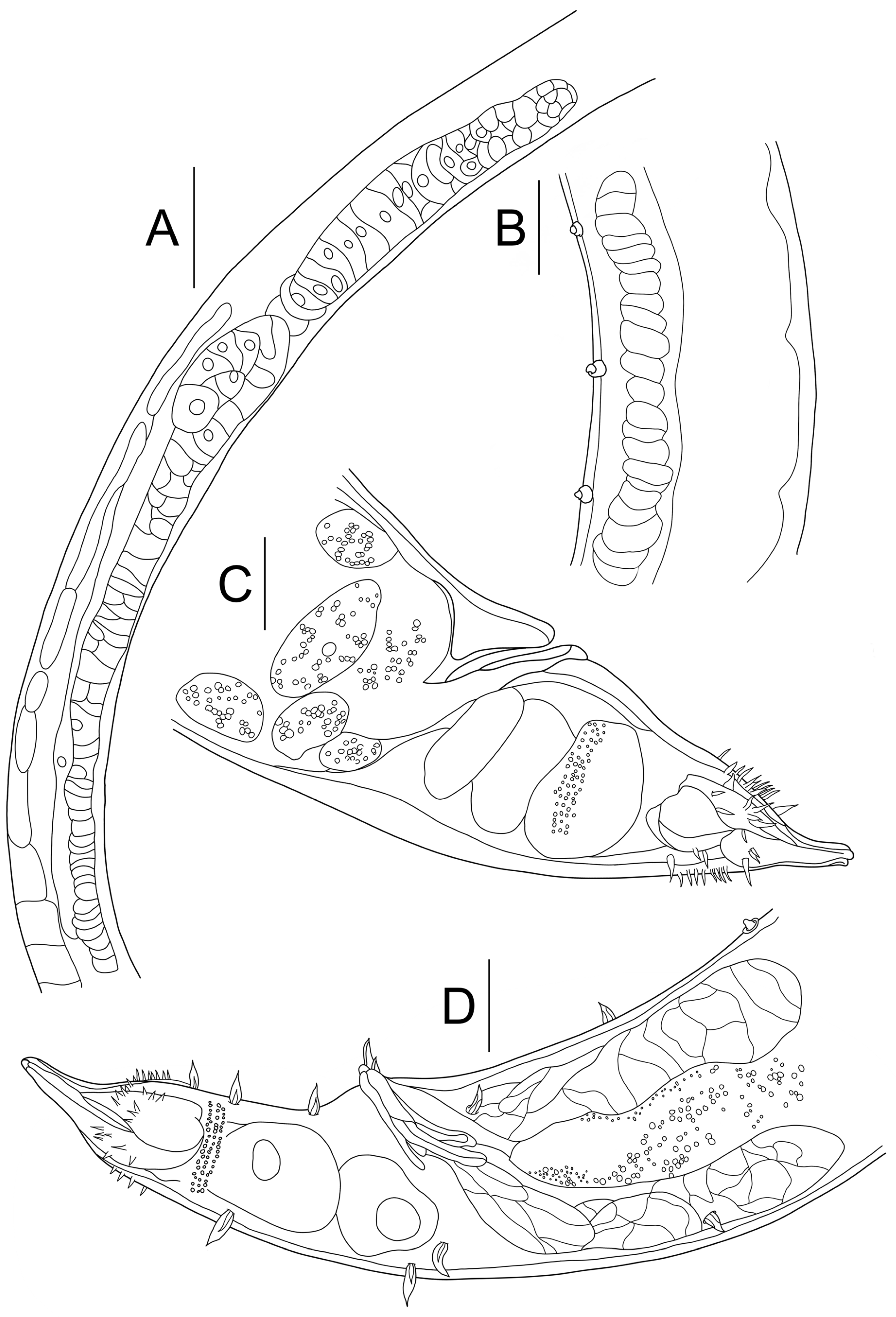
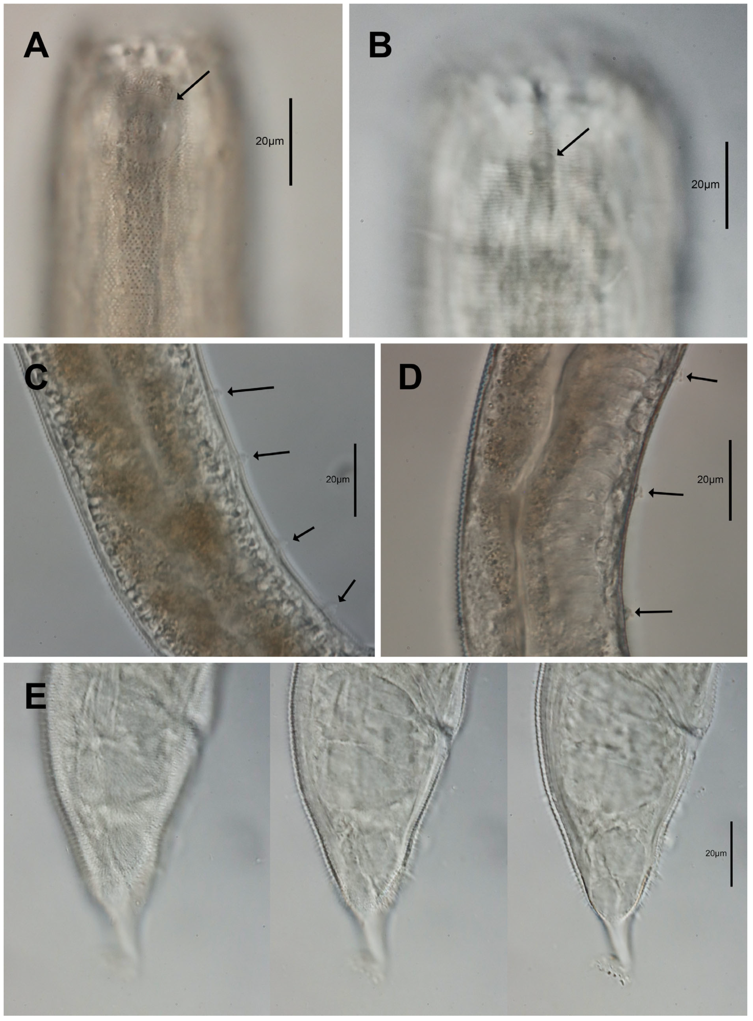
Male: Body cylindrical, relatively stout. Cuticle homogeneous with small punctations arranged in transverse rows, without lateral differentiation. Somatic setae are distributed along the whole body, with cervical setae that are comparably longer than somatic setae in the tail region, with somatic setae in the tail region are slightly thicker in width. Lip region with six extended leaf-shaped cuticular structures, each with a short inner labial seta near the base. Six outer labial setae are located at the outer margin, and four cephalic setae are located below the ring of outer labial setae. Amphideal fovea loop-shaped in males (3/4 turns), situated slightly posterior to cephalic setae (Figure 3A and Figure 4A). The buccal cavity is distinguished by a cup-shaped anterior buccal cavity (gymnostome) and a cylindrical posterior buccal cavity (stegostome). Anteriorly, three broad cuticularized rhabdions, each terminating posteriorly in three small branches (denticles) (Figure 4C,D). The posterior buccal cavity is narrower and cylindrical with six Y-shaped cuticular rhabdions. The pharynx is broad and muscular, without pharyngeal bulbs. Cardia small; intestine densely packed with large cells, with a brownish pigment. The male reproductive system is diorchic, with two testes situated ventrally relative to the intestine. The anterior testis is outstretched, while the posterior testis is reflexed. Spicules paired, arcuate (slightly curved), tapering distally. A gubernaculum is present, consisting of two parallel, slightly arcuate pieces partially surrounding the spicules. The posterior-most, precloacal supplementary papilla (Figure 3C,D and Figure 4E,F) closest to the cloacal opening, is located approximately 60 µm anterior to the cloacal opening. Between the cloaca and this supplementary papilla, one or two somatic setae are aligned uniformly, but positioned on a different longitudinal row, slightly offset from that of the supplementary papillae (Figure 4E). A total of 7–8 papilla-shaped supplements organ present anterior to the cloaca. The tail is short and conical, narrowing cylindrically near the tip, with minute, short hair-like, cuticular spines (Figure 3E and Figure 4H). It has three caudal glands and a spinneret are present.
Female: Similarly to the male except with a stouter body (a ratio = 16.7–20.6 vs. ~22 in males), smaller unispiral amphideal fovea (Figure 3B and Figure 4B), and tail shape (shorter and broader tail tip). Sexual dimorphism is evident in amphidial morphology (¾-turn loop in males vs. unispiral in females, with female amphid sometimes cryptospiral, comma-like shape; Figure 3B). The female reproductive system is didelphic–amphidelphic with two opposed, reflexed ovaries, positioned on the right side of the intestine. The vulva is located approximately 60% of the body region, with a slight protrusion of the vulva lip (Figure 1A and Figure 4G). Anus and rectum are developed and present. The female tail has a shorter, broader tail tip.
  • Diagnosis and relationships:
Gammanema papillatum sp. nov. is most similar to G. lunatum, sharing several key morphological traits. Both species possess a loop-shaped amphidial fovea in males with approximately ¾ turn, as well as unispiral amphids in females with one full turn. Minute cuticular spines are present in both species (near the posterior tail region), and they overlap in male amphidial fovea width as a percentage of the corresponding body diameter (23–38% vs. 27–46%), and exhibit similar lengths of cephalic and outer labial setae. However, G. papillatum sp. nov. differs from G. lunatum in the shape of the precloacal (papilla- to cup-shaped). Additionally, they differ in having a significantly higher ratio (21–23 vs. 13–15), indicating a more slender body, and slightly longer body length (1122–1366 µm vs. 754–1196 µm) (Table 2). This distinction in body proportions constitutes the main diagnostic feature separating the two species.
Compared to G. agglutinans, the new species is clearly differentiated by both the shape and number of turns of the amphidial fovea: males of G. agglutinans have a ½ turn loop and females a 1.5 turn loop, whereas the new species has ¾ and 1.0 turns, respectively. The precloacal supplements are also distinctly different in form (papilla-shaped in G. papillatum sp. nov. compared with tubular in G. agglutinans), and the new species exhibits a larger body size (1122–1366 µm vs. 544–696 µm) and more slender proportions (a = 21–23 vs. 10–11).
When compared to G. conicauda, they share the papilla-shaped precloacal supplementary organ and show partially comparable amphid morphology (¾-turn loop vs. unispiral in males). Nevertheless, they are readily distinguished by the number of precloacal supplements (22 in G. conicauda vs. 7–8 in G. papillatum sp. nov.), by body length (shorter in the new species), and by much shorter outer labial and cephalic setae (2–3 and 11–15 µm vs. 16 and 30–33 µm, respectively)]. Taken together, these characters clearly support G. papillatum sp. nov. as a distinct species within the genus.

3.2. Molecular Analysis

A total of six specimens of Gammanema papillatum sp. nov. were sequenced. A near full-length sequence of the 18S rRNA gene (~1600 bp) and a partial sequence of the 28S rRNA gene corresponding to the D2-D3 expansion domain were successfully obtained for the new species (Table 3). To assess genetic divergence among congeners and closely related genera, pairwise Kimura 2-parameter (K2P) distances were calculated using available Gammanema sequences from GenBank.
Several 18S and 28S rRNA sequences of Gammanema were publicly available, including those of Gammanema lunatum and Gammanema rapax. Despite Gammanema rapax being taxonomically ambiguous, as discussed by Tchesunov et al. (2020) [6], its species designation/annotation is maintained in the molecular analyses solely for the purpose of molecular comparison.
Morphologically similar genera such as Latronema Wieser, 1954 [37], Halichoanolaimus de Man, 1886 [38], and Choanolaimus de Man, 1880 [39] were also included for genus-level K2P distance comparisons. All six individuals of the new species were genetically identical, showing a K2P distance of 0% among themselves for both 18S and 28S rRNA sequences.
For the 18S rRNA gene, pairwise genetic distances between the new species and other Gammanema species ranged from 1.7% to 4.5%. The smallest divergence (1.7%) was observed with Gammanema lunatum (PQ286529), indicating the closest relationship (Table S1). Other Gammanema species showed divergences of approximately 3.9–4.5%. Comparisons with other selachinematid genera revealed intergeneric divergences ranging from 6.3% to 10.8%, with Halichoanolaimus funestus (PQ286530) showing the lowest divergence (6.3%) and Latronema showing higher divergence (9.4–10.7%).
For the 28S rRNA gene, molecular divergence between the new species and other Gammanema species ranged from 19.8% to 37.1%, with the smallest value again observed with Gammanema lunatum (19.8%), though still substantial (Table S2). Divergence from other Gammanema species ranged between 30.5 and 37.1%. In contrast, species of other selachinematid genera exhibited markedly higher divergence, ranging from 49.8% to 63.0%.
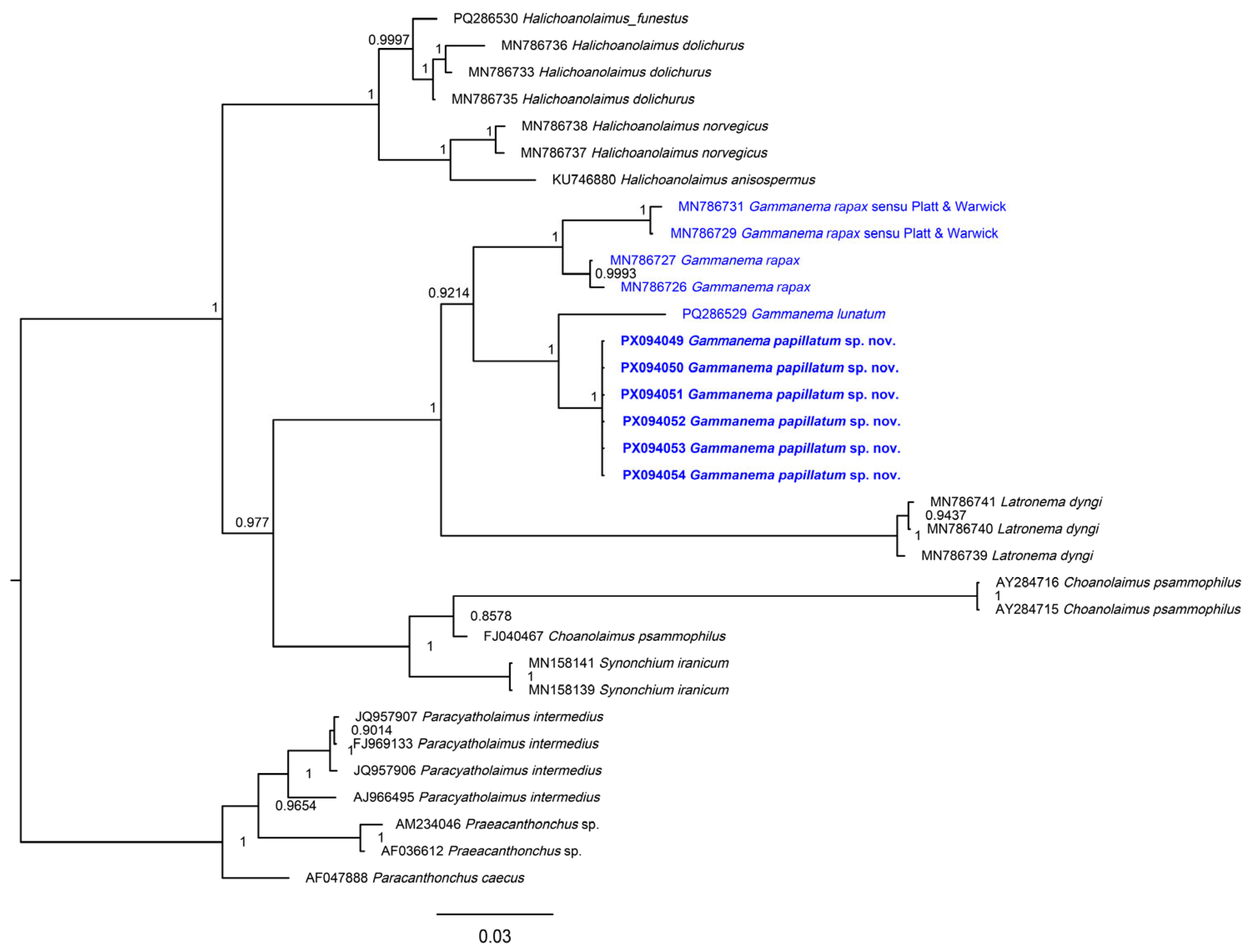
In the 18S BI tree (Figure 5), the six sequences of Gammanema papillatum sp. nov. formed a strongly supported clade (PP = 1.00) with G. lunatum (PQ268529), indicating a close phylogenetic relationship. This grouping was nested within a larger, well-supported assemblage (PP = 0.9993) that also included other Gammanema spp. from GenBank (MN786727, MN786726, MN786731, MN786729), suggesting monophyly of the genus. No sequences of G. agglutinans or G. conicauda were available for comparison, preventing direct molecular assessment of their relationships to the new species. In the 28S BI tree (Figure 5), the topology seen from Leduc & Zhao (2025) [3] was retained. The new species again grouped closest to G. lunatum, with strong support (PP = 0.96). The genus once more formed a monophyletic group, albeit with moderate support (PP = 0.87), and clustered with a group of Latronema with strong support (PP = 1.00).

4. Discussion

Morphologically, three characteristics are of particular importance in the new species: amphid shape, the presence of cuticle spines, and the shape of the supplementary organ. The amphid + cuticular spines combination had, until now, been reported from New Zealand congeners and is now also observed in the Korean material described here. Among taxa with this character set, only Gammanema lunatum has publicly available molecular sequences. When compared with the new species, which shares this combination, our analyses recover them as each other’s most closely related congeners, with K2P distances reported per marker (SSU = 1.7%; LSU = 19.8%). The shared morphological trait set and genetic affinity indicate congruence between morphology and molecular phylogenetics and suggest that the amphid + cuticle-spines combination may be phylogenetically informative within Gammanema. If molecular sequences for G. agglutinans can be obtained, this would provide stronger evidence for evaluating whether these traits consistently reflect phylogenetic relationships. If such a correlation is observed in the future, it may be an indication that species with these traits may represent a monophyletic subgroup within the genus. It remains uncertain, however, whether the shape of the supplementary organ correlates more closely with molecular divergence than the amphid + cuticle spines combination. Testing this possibility would require obtaining sequence data for G. conicauda and comparing its genetic divergence with that of other congeners.
Beyond intrageneric comparisons, phylogenetic analyses consistently place Latronema as the sister group to Gammanema (Figure 5 and Figure 6). This relationship is supported by shared morphological features, including a two-chambered buccal cavity reinforced by cuticularized rhabdions or rugae, a cylindrical pharynx without a bulb, and the presence of sucker-like or cup-shaped precloacal supplements. They also possess conical tails, and both lack lateral differentiation. The distinction between Gammanema and Latronema lies in their cuticle annulation (homogeneous without ridges vs. 12–50 longitudinal ridges), amphids (large loop/spiral vs. small spiral/round, often obscure), and buccal armature (three rhabdions vs. 12 rugae). Together, these patterns underline the close evolutionary link between the two genera and highlight traits of potential phylogenetic significance within Selachinematidae.
The combined usage of SSU and LSU rDNA sequences has been useful in confirming species identity and for assessing how certain morphological traits correlate with molecular divergence. The LSU region was especially informative for distinguishing species at the interspecific level, supporting earlier studies that showed its utility in resolving cryptic taxa where substantial LSU divergence (24.5–34.4%) may occur between species with nearly identical morphology [12]. The SSU region shows much less molecular divergence between closely related species, but it remains important to resolve deeper phylogenetic relationships and to provide broader evolutionary context. Together, the two markers are a great tool for substantiating and strengthening taxonomic decisions made through morphological comparative analysis. Such integrative work combining morphology with multilocus molecular evidence will continue to be essential for species delimitation and advancing nematode systematics and biodiversity research.

Supplementary Materials

The following supporting information can be downloaded at https://www.mdpi.com/article/10.3390/d17090639/s1, Table S1: Kimura 2-parameter distances among Gammanema species and related genera based on 18S rRNA alignments with 1000 bootstrap replicates. Standard deviations are indicated in blue. Table S2: Kimura 2-parameter distances among Gammanema species and related genera based on 28S rRNA alignments with 1000 bootstrap replicates. Standard deviations are indicated in blue.

Author Contributions

Conceptualization, K.S. and R.J.; methodology, K.S. and R.J.; software, R.J.; validation, K.S. and R.J.; formal analysis, K.S. and R.J.; investigation, K.S. and R.J.; resources, R.J.; data curation, K.S. and R.J.; writing—original draft preparation, K.S. and R.J.; writing—review and editing, K.S. and R.J.; visualization, K.S. and R.J.; supervision, R.J.; project administration, R.J.; funding acquisition, R.J. All authors have read and agreed to the published version of the manuscript.

Funding

This research was funded by the National Institute of Biological Resources (NIBR), Ministry of Environment (MOE) of the Republic of Korea (NIBR202402107), and Basic Science Research Program through the National Research Foundation of Korea (NRF), funded by the Ministry of Education (2021R1I1A2043807).

Institutional Review Board Statement

Not applicable.

Data Availability Statement

All specimens from this study were deposited in the National Institute of Biological Resources (NIBR), Republic of Korea. The corresponding molecular sequences have been submitted to GenBank, with accession numbers provided in Table 3.

Conflicts of Interest

The authors declare no conflicts of interest. The funders had no role in the design of the study; in the collection, analyses, or interpretation of data; in the writing of the manuscript; or in the decision to publish the results.

Abbreviations

The following abbreviations are used in this manuscript:
LWhole body length
aBody length divided by maximum body diameter
bBody length divided by pharynx length
cBody length divided by tail length
c’Tail length divided by anal body diameter
cbdCorresponding body diameter

References

  1. Okhlopkov, J.R. Free-living nematodes of the families Selachinematidae and Richtersiidae in the White Sea (Nematoda, Chromadoria). Zoosystematica Rossa 2002, 11, 41–55. [Google Scholar] [CrossRef]
  2. Tchesunov, A.V.; Okhlopkov, J.R. On some selachinematid nematodes (Chromadorida: Selachinematidae) deposited in the collection of the Smithsonian National Museum of Natural History. Nematology 2006, 8, 21–44. [Google Scholar] [CrossRef]
  3. Leduc, D.; Zhao, Z.Q. Three new nematode species (Chromadorida: Selachinematidae) from the continental slope of New Zealand. Eur. J. Taxon. 2025, 989, 119–143. [Google Scholar] [CrossRef]
  4. Miljutin, D.M.; Gad, G.; Miljutina, M.M.; Mokievsky, V.; Fonseca-Genevois, V.; Esteves, A.M. The state of knowledge on deep-sea nematode taxonomy: How many valid species are known down there? Mar. Biodivers. 2010, 40, 143–159. [Google Scholar] [CrossRef]
  5. Moens, T.; Braeckman, U.; Derycke, S.; Fonseca, G.; Gallucci, F.; Gingold, R.; Guilini, K.; Ingels, J.; Leduc, D.; Vanaverbeke, J. Ecology of free-living marine nematodes. In Handbook of Zoology: Gastrotricha, Cycloneuralia and Gnathifera; De Gruyter: Berlin, Germany, 2013; Volume 2, pp. 109–152. [Google Scholar]
  6. Tchesunov, A.; Jeong, R.; Lee, W. Two New Marine Free-Living Nematodes from Jeju Island Together with a Review of the Genus Gammanema Cobb 1920 (Nematoda, Chromadorida, Selachinematidae). Diversity 2020, 12, 19. [Google Scholar] [CrossRef]
  7. Gerlach, S.A. Revision der Choniolaiminae und Selachinematinae (freilebende Meeres-Nematoden). In Mitteilungen aus dem Hamburgischen Zoologischen Museum und Institut (Kosswig-Festschrift); Lütcke & Wulff: Hamburg, Germany, 1964; pp. 23–49. [Google Scholar]
  8. Murphy, D.G. Chilean marine nematodes. Veröffentlichungen Inst. Meeresforsch. Bremerhav. 1965, 9, 173–203. [Google Scholar]
  9. Platt, H.; Warwick, R. Free-Living Marine Nematodes; Part 2: British Chromadorids; Brill, E.J., Backhuys, W., Eds.; Linnean Society of London: London, UK, 1988; p. 502. [Google Scholar]
  10. Ahmed, M.; Boström, S.; Holovachov, O. Revision of the genus Cobbionema Filipjev, 1922 (Nematoda, Chromadorida, Selachinematidae. Eur. J. Taxon. 2020, 702, 1–34. [Google Scholar] [CrossRef]
  11. Nemys (Ed.) Nemys: World Database of Nematodes. Available online: https://nemys.ugent.be (accessed on 1 August 2025).
  12. Son, K.; Jeong, R. The Discovery and Delimitation of a New Cryptic Species of Spirinia (Nematoda: Desmodoridae) Using SSU and LSU rDNA Divergence. J. Mar. Sci. Eng. 2025, 13, 1251. [Google Scholar] [CrossRef]
  13. Pereira, T.J.; Fonseca, G.; Mundo-Ocampo, M.; Guilherme, B.C.; Rocha-Olivares, A. Diversity of free-living marine nematodes (Enoplida) from Baja California assessed by integrative taxonomy. Mar. Biol. 2010, 157, 1665–1678. [Google Scholar] [CrossRef]
  14. Leduc, D.; Zhao, Z.Q. Morphological and molecular characterisation of Spirinia antipodea Leduc n. sp.(Nematoda: Desmodoridae), a cryptic species related to S. parasitifera, from the coast of New Zealand. Nematology 2019, 21, 91–105. [Google Scholar] [CrossRef]
  15. Hopper, D. Drawing and measuring nematodes. In Ministry of Agriculture, Fisheries and Food; Her Majesty’s Stationery Office: London, UK, 1970. [Google Scholar]
  16. Meeker, N.D.; Hutchinson, S.A.; Ho, L.; Trede, N.S. Method for isolation of PCR-ready genomic DNA from zebrafish tissues. Biotechniques 2007, 43, 610–614. [Google Scholar] [CrossRef]
  17. Holterman, M.; van der Wurff, A.; van den Elsen, S.; van Megen, H.; Bongers, T.; Holovachov, O.; Bakker, J.; Helder, J. Phylum-wide analysis of SSU rDNA reveals deep phylogenetic relationships among nematodes and accelerated evolution toward crown clades. Mol. Biol. Evol. 2006, 23, 1792–1800. [Google Scholar] [CrossRef]
  18. De Ley, P.; De Ley, I.T.; Morris, K.; Abebe, E.; Mundo-Ocampo, M.; Yoder, M.; Heras, J.; Waumann, D.; Rocha-Olivares, A.; Jay Burr, A.H.; et al. An integrated approach to fast and informative morphological vouchering of nematodes for applications in molecular barcoding. Phil. Trans. R. Soc. B 2005, 360, 1945–1958. [Google Scholar] [CrossRef]
  19. Tamura, K.; Stecher, G.; Kumar, S. MEGA11: Molecular Evolutionary Genetics Analysis Version 11. Mol. Biol. Evol. 2021, 38, 3022–3027. [Google Scholar] [CrossRef] [PubMed]
  20. Thompson, J.D.; Higgins, D.G.; Gibson, T.J. Clustal-W—Improving the Sensitivity of Progressive Multiple Sequence Alignment through Sequence Weighting, Position-Specific Gap Penalties and Weight Matrix Choice. Nucleic Acids Res. 1994, 22, 4673–4680. [Google Scholar] [CrossRef] [PubMed]
  21. Kimura, M. A Simple Method for Estimating Evolutionary Rates of Base Substitutions through Comparative Studies of Nucleotide-Sequences. J. Mol. Evol. 1980, 16, 111–120. [Google Scholar] [CrossRef] [PubMed]
  22. Huelsenbeck, J.P.; Ronquist, F. Mrbayes: Bayesian inference of phylogenetic trees. Bioinformatics 2001, 17, 754–755. [Google Scholar] [CrossRef]
  23. Darriba, D.; Taboada, G.L.; Doallo, R.; Posada, D. jModelTest 2: More models, new heuristics and high-performance computing. Nat. Methods 2012, 9, 772. [Google Scholar] [CrossRef]
  24. Rambaut, A. Figtree, Version 1.4.4; Institute of Evolutionary Biology, University of Edinburgh: Edinburgh, UK, 2018. [Google Scholar]
  25. Inglis, W.G. An Outline Classification of the Phylum Nematoda. Aust. J. Zool. 1983, 31, 243–255. [Google Scholar] [CrossRef]
  26. Chitwood, B.G. A revised classification of the Nematoda. J. Parasitol. Pap. Helminthol. Ninth Annu. Meet. 1933, 20, 115–148. [Google Scholar]
  27. Cobb, N.A. Selachinema, a new nematode genus with remarkable mandibles. In Contributions to a Science of Nematology; University of Nebraska–Lincoln: Lincoln, NE, USA, 1914; Volume 4, pp. 113–116. [Google Scholar]
  28. Cobb, N.A. One hundred new nemas (type species of 100 new genera). In Contributions to a Science of Nematology; University of Nebraska–Lincoln: Lincoln, NE, USA, 1914; Volume 9, pp. 217–343. [Google Scholar]
  29. Leduc, D. Two new genera and five new species of Selachinematidae (Nematoda, Chromadorida) from the continental slope of New Zealand. Eur. J. Taxon. 2013, 63, 1–32. [Google Scholar] [CrossRef]
  30. Gerlach, S.A. Zur Kenntnis der freilebenden marinen Nematoden von San Salvador. Z. Wiss. Zool. 1955, 158, 249–303. [Google Scholar]
  31. Gerlach, S.A. Die Nematodenbesiedlung des Sandstrandes und des Küstengrundwassers an der italienischen Küste I. Systematischer Teil. Arch. Zool. Ital. 1953, 37, 417–640. [Google Scholar]
  32. Gagarin, V.G.; Klerman, A.K. New species of predatory chromadorids (Nematoda, Chromadorida) from the Mediterranean Sea. Zool. Zhurnal 2007, 86, 778–786. [Google Scholar]
  33. Gerlach, S.A. Die Nematodenfauna der Uferzonen und des Küstengrundwassers am finnischen Meeresbusen. Acta Zool. Fenn. 1953, 73, 1–32. [Google Scholar]
  34. Shi, B.Z.; Xu, K.D. Two new rapacious nematodes from intertidal sediments, Gammanema magnum sp. nov. and Synonchium caudatubatum sp. nov. (Nematoda, Selachinematidae). Eur. J. Taxon. 2018, 405, 1–17. [Google Scholar] [CrossRef]
  35. Vitiello, P. Nématodes libres marins des vases profondes du Golfe du Lion. II. Chromadorida. Téthys 1970, 2, 449–500. [Google Scholar]
  36. Murphy, D.G. Free-living marine nematodes, I. Southerniella youngi, Dagda phinneyi, and Gammanema smithi, new species. Proc. Helminthol. Soc. Wash. 1964, 31, 190–198. [Google Scholar]
  37. Wieser, W. Free-living marine nematodes II. Chromadoroidea. Acta Univ. Lund. NF 1954, 50, 1–148. [Google Scholar]
  38. de Man, J.G. Sur quelques nématodes libres de la mer du Nord, nouveaux ou peu connus. Mémoires Société Zool. Fr. 1888, 1, 1–51. [Google Scholar]
  39. de Man, J.G. Die Einheimischen, Frei In Der Reinen Erde Und Im Süssen Wasser Lebenden Nematoden. Monographisch Bearbeitet. Vorläufiger Bericht und descriptiv-systematischer Theil. Tijdschr. Ned. Dierkd. Ver. 1879, 5, 1–104. [Google Scholar]
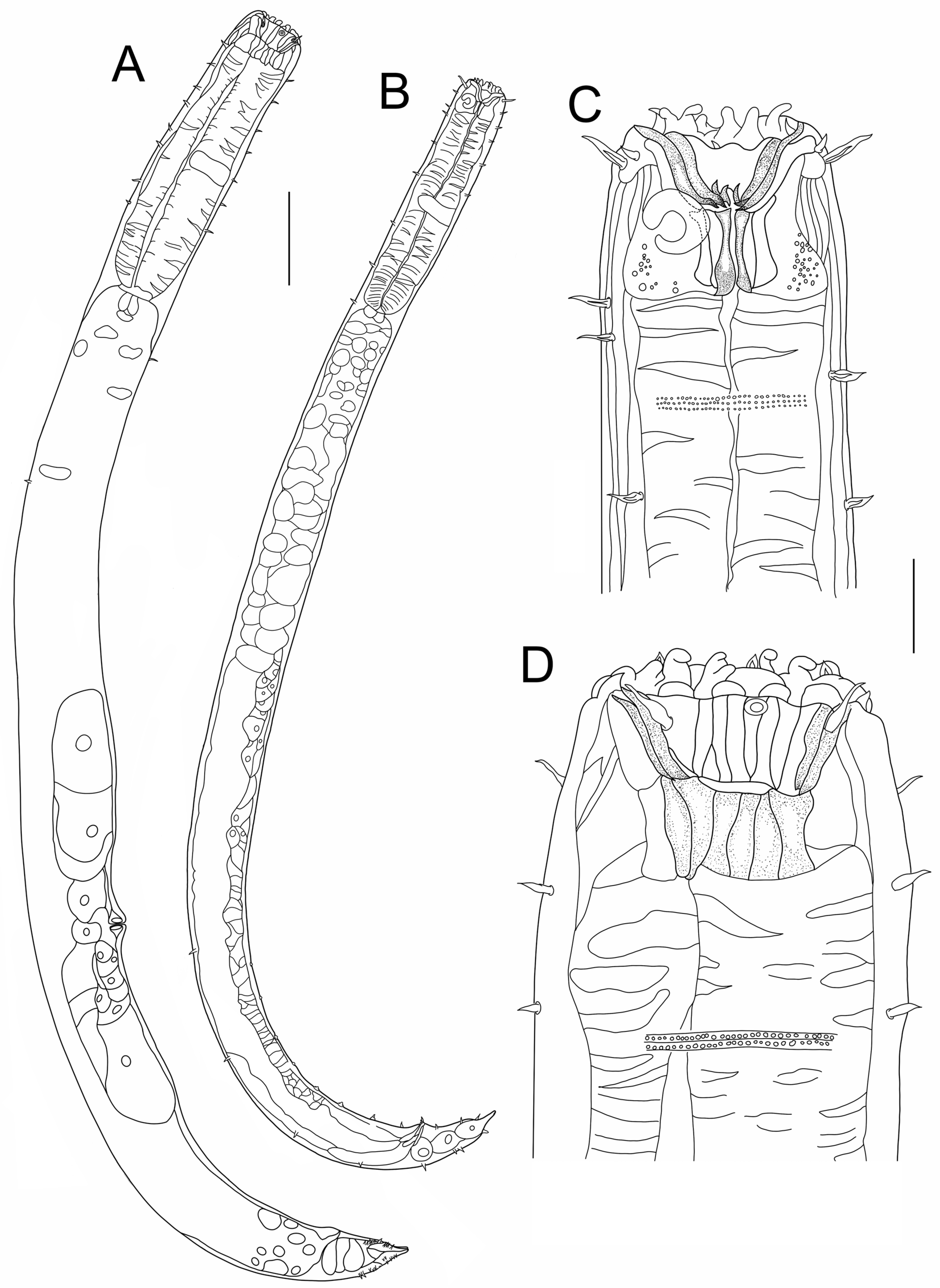
Figure 1. Gammanema papillatum sp. nov. (A) allotype female entire body; (B) holotype male entire body; (C) holotype male cephalic region; (D) allotype female cephalic region (scale bar: (A,B) = 100 μm; (C,D) = 20 μm).
Diversity 17 00639 g001
Figure 2. Gammanema papillatum sp. nov. (A) paratype male testis; (B) paratype male precloacal supplement region; (C) allotype female tail region; (D) holotype male tail region (scale bar: (A) = 50 μm; (BD) = 20 μm).
Diversity 17 00639 g002
Figure 3. Gammanema papillatum sp. nov. light micrographs (A) ♂ loop-shaped amphid (arrow); (B) ♀ unispiral amphid (arrow); (C,D) ♂ different paratypes with papilla-shaped precloacal supplementary organs (arrows); (E) ♀ tail region at different z-plain, showing cuticular spines (scale bar: (AE) = 20 μm).
Diversity 17 00639 g003
Figure 4. SEM micrographs of Gammanema papillatum sp. nov. (A) ♂, dorsolateral view of cephalic region with loop-shaped amphid; (B) ♀, dorsolateral view of cephalic region with unispiral amphid; (C,D) ♀, apical view of the mouth opening of different specimens showing three rhabdions terminating in three branches (denticles); (E) ♂, lateral view of cloacal region, with protruding spicules (spic) followed by two somatic setae (s.s.) positioned between the posterior-most precloacal supplementary papilla (supp); (F) ♂, ventral view above precloacal region showing vertically arranged supplementary papillae; (G) ♀, ventral view of vulva lip; (H) ♀, tail region with numerous minute cuticular spines (arrows) (scale bar: (AH) = 10 μm).
Diversity 17 00639 g004
Figure 5. Rooted Bayesian inference phylogenetic tree of the 18S rRNA region, with species of Cyatholaimidae used as the outgroup. Sequences generated in this study are shown in bold, and Gammanema species are highlighted in blue. Posterior probability values are given at each node.
Diversity 17 00639 g005
Figure 6. Rooted Bayesian inference phylogenetic tree of the 28S rRNA region, with species of Cyatholaimidae used as the outgroup. Sequences generated in this study are shown in bold, and Gammanema species are highlighted in blue. Posterior probability values are given at each node.
Diversity 17 00639 g006
Table 1. Measurement of Gammanema papillatum sp. nov. (All measurements are in µm; “n/a” indicates not applicable. Holotype (male) and allotype (female) are listed separately. Summary columns include them in the counts (males n = 3; females n = 3); remaining specimens are paratypes.)
CharactersMaleFemale
HolotypeMales (n = 3) Mean ± sd (Range)AllotypeFemales (n = 3) Mean ± sd (Range)
L13661247 ± 99.7 (1122–1366)15741539 ± 35.5 (1503–1574)
a (L/gbd)23.622 ± 1 (21.2–23.6)16.720 ± 0.5 (19.7–20.6)
b (L/pharynx length)5.25 ± 0.2 (4.8–5.2)4.75 ± 0.1 (4.8–5)
c (L/tail length)16.117 ± 1 (16.1–18.4)19.719 ± 1.1 (18.3–20.6)
c’ (tail length/anal body diameter)2.12 ± 0.2 (1.7–2.1)1.52 ± 0.2 (1.6–2)
Inner labial/Outer labial setae length32 ± 0.5 (2–3)33 ± 0.5 (2–3)
Cephalic setae length1112 ± 1.9 (11–15)1319 ± 0.5 (18–19)
Cephalic setae cbd4239 ± 2.1 (37–42)6657 ± 0.5 (56–57)
Amphid (at center) cbd4744 ± 2.9 (40–47)7061 ± 1.5 (59–62)
Amphid height1314 ± 1.9 (13–17)76 ± 0 (6–6)
Amphid width1113 ± 3.1 (10–17)76 ± 0 (6–6)
Amphid width/body width (%)0.230 ± 0.1 (0.2–0.4)0.10.1 ± 0 (0.1–0.1)
Amphid (at center) distance to anterior1715 ± 1.7 (13–17)2226 ± 1 (25–27)
Buccal cavity (pharyngostome) distance to the anterior3735 ± 2.2 (32–37)4751 ± 2 (49–53)
Anterior (gymnostome) depth1918 ± 1.9 (15–19)2726 ± 1 (25–27)
Anterior rhabdion length2120 ± 0.8 (19–21)2626 ± 1 (25–27)
Posterior rhabdion length1716 ± 1.2 (14–17)2221 ± 2 (19–23)
Nerve ring distance to the anterior125121 ± 5.2 (114–125)186163 ± 2 (161–165)
Nerve ring cbd (corresponding body diameter)5350 ± 5 (43–54)8366 ± 2 (64–68)
Pharynx length263249 ± 11 (236–263)335313 ± 2 (311–315)
Pharyngeal base width3734 ± 2.5 (31–37)6957 ± 6 (51–63)
Pharynx cbd5049 ± 0.9 (48–50)7656 ± 4.5 (51–60)
Max body diameter5855 ± 2.1 (53–58)9477 ± 3.5 (73–80)
Spicule length (as arc)4338 ± 4.1 (33–43)n/an/a
Gubernaculum length2425 ± 2.9 (22–29)n/an/a
Supplementary organs78 ± 0.5 (7–8)n/an/a
Posterior-most supplement distance to the anterior to the cloacal opening5861 ± 2.2 (58–63)n/an/a
Distance between supplementary organs28–4227–36, 20–26n/an/a
Cloacal/anal body diameter4139 ± 2.8 (35–41)5344 ± 1 (43–45)
Tail length8572 ± 9.9 (61–85)8080 ± 6.5 (73–86)
Terminal cone portion (smooth)1716 ± 0.9 (15–17)1619 ± 6 (13–25)
Vulva distance to anteriorn/an/a1018965 ± 27 (938–992)
Vulva body diam.n/an/a9281 ± 1.5 (79–82)
Vulva distance to anterior/total body lengthn/an/a0.650.6 ± 0 (0.6–0.6)
Table 2. Comparison of morphometric measurements of closely related congeners. “calc” indicates values calculated directly using the figure provided in the original reference.
CharacteristicG. papillatum sp. nov.G. lunatumG. agglutinansG. conicaudaOther Gammanema spp.
Male amphidial fovea shapeLoop-shaped (¾ turn)Loop-shaped (¾ turn)Loop-shaped (½ turn)UnispiralMultispiral
Female amphidial fovea shapeUnispiral (1.0 turn)Unispiral (1.0 turn)Spiral (1.5 turns)UnispiralMultispiral
Male amphid width/cbd (%)23–3827–4628–2936 (calc)
Female amphid width/cbd (%)1030 (error?) 10 (calc)10–1211 (calc)
Cuticular spinesPresent (minute)Present (minute)Present (minute)Not specifiedAbsent
Precloacal supplements (number and shape)7–8 papillae6–10 cup-shaped<6 tubular22 papillae
Posterior-most supplement distance to the anterior to the cloacal opening58–6318–32
Distance between precloacal supplements18–355–17
Body length (µm)1122–1366754–1196544–6961985–2419
a ratio (L/gbd)21–2313–1510–1141–48
b55–64–57.3–7.7
c16–1813–2012–1333–38
c’1.7–21.3–1.61.2–1.31.1–1.4
Outer labial setae length (µm)2–32–4216
Cephalic setae length (µm)11–158–2010–1130–33
Table 3. GenBank accession numbers corresponding to sequences obtained from this study.
#Species NameIsolate NumberGenBank Accession Number
18S28S
988F, 1813FD2A
/1912R, 2646R/D3B
(~1600 bp)(~750 bp)
1Gammanema papillatum sp. nov.K1PX094049PX094055
2Gammanema papillatum sp. nov.K2PX094050PX094056
3Gammanema papillatum sp. nov.K3PX094051PX094057
4Gammanema papillatum sp. nov.K6PX094052PX094058
5Gammanema papillatum sp. nov.K8PX094053PX094059
6Gammanema papillatum sp. nov.K9PX094054PX094060
Disclaimer/Publisher’s Note: The statements, opinions and data contained in all publications are solely those of the individual author(s) and contributor(s) and not of MDPI and/or the editor(s). MDPI and/or the editor(s) disclaim responsibility for any injury to people or property resulting from any ideas, methods, instructions or products referred to in the content.