Interesting Images

Evidence for Initial Infestation by Mothocya parvostis (Isopoda: Cymothoidae) on Body Surface of Juvenile Japanese Halfbeak, Hyporhamphus sajori (Beloniformes: Hemiramphidae)

1
Seto Marine Biological Laboratory, Field Science Education and Research Center, Kyoto University, 459 Shirahama, Nishimuro, Wakayama 649-2211, Japan
2
Graduate School of Integrated Sciences for Life, Hiroshima University, 1-4-4 Kagamiyama, Higashi-Hiroshima 739-8528, Japan
*
Author to whom correspondence should be addressed.
Diversity 2025, 17(9), 613; https://doi.org/10.3390/d17090613
Submission received: 15 July 2025 / Revised: 27 August 2025 / Accepted: 28 August 2025 / Published: 30 August 2025
(This article belongs to the Section Marine Diversity)

Abstract

Mothocya parvostis Bruce, 1986 (Isopoda: Cymothoidae) is a parasite that mainly infests the branchial cavity of Japanese halfbeak, Hyporhamphus sajori (Temminck and Schlegel, 1846) (Beloniformes: Hemiramphidae) (pelagic marine fish). Although it has generally been considered to parasitize the branchial cavity throughout its life, early studies suggested that mancae (first free swimming stage) initially attach to the body surface of the host. In this study, H. sajori juveniles were collected off Ohkurokamishima Island, Hiroshima, Japan, and examined for cymothoid infestation. Of the 57 individuals that were examined, 5 were parasitized, all on the ventral surface of the fish bodies. One manca was genetically identified as M. parvostis based on the sequence of cytochrome c oxidase subunit I (100% or 99.81% identity with known sequences). These findings support the earlier claim that mancae infest the body surface of H. sajori. Due to the host’s slender body and small branchial cavity, early-stage parasites may remain external in the initial phase of infestation.

Graphical Abstract
Mothocya parvostis Bruce, 1986 (Isopoda: Cymothoidae) infests the branchial cavity of fish [1], including the Japanese halfbeak, Hyporhamphus sajori (Temminck and Schlegel, 1846) (pelagic marine fish), as its definitive final host. Cymothoids feed on fish tissue and blood and are dependent on their hosts for nutrition. An increase in juvenile mortality rate due to cymothoids was reported [2]. Elucidating the cymothoid life cycle is important to understand this potential threat to fish. The life cycle of M. parvostis was reported earlier than that of many other cymothoid species [3]. Inoue [3] reported that mancae (first free swimming stage) of these parasites were released from the brood pouch of female M. parvostis and infested the body surface of the host H. sajori, where they become young males. Some of these males then moved into the opercular cavity of the fish and changed sex to female. In addition, M. parvostis has been reported to use juveniles of some fish species as optional intermediate hosts when it cannot parasitize juvenile H. sajori [4,5].
However, in many cymothoid species, it has been thought that the infestation site remains almost unchanged as their life cycle progresses [6,7,8,9,10,11,12]. Among cymothoids, reports of cases such as Mothocya lineata (Miers, 1876), where parasites change their attachment sites depending on their life cycle stage, are extremely rare [1,13]. Therefore, the relocation of the infestation site proposed by Inoue [3] has been largely ignored in subsequent studies (e.g., Smit et al. [14]), and M. parvostis has been treated as a species that parasitizes the branchial cavity throughout its life [1,15,16].
In this study, H. sajori juveniles were collected from the surface waters around oyster-culturing rafts off Ohkurokamishima Island, Etajima, Hiroshima Prefecture, Japan (see Kawai et al. [17]), on 30 May 2019, using paper cups. The presence or absence of cymothoids was confirmed and photographed. The cytochrome c oxidase subunit I (COI) sequence of the parasite was analyzed for species identification because most cymothoid species are morphologically difficult to identify, except in the case of adult females. DNA extraction and PCR amplification were performed following the protocol outlined by Fujita et al. [18]. The PCR product was sent to Eurofins Genomics (Tokyo, Japan) for sequencing using the dye terminator method. The sequence was submitted to GenBank (accession number: PX108758).
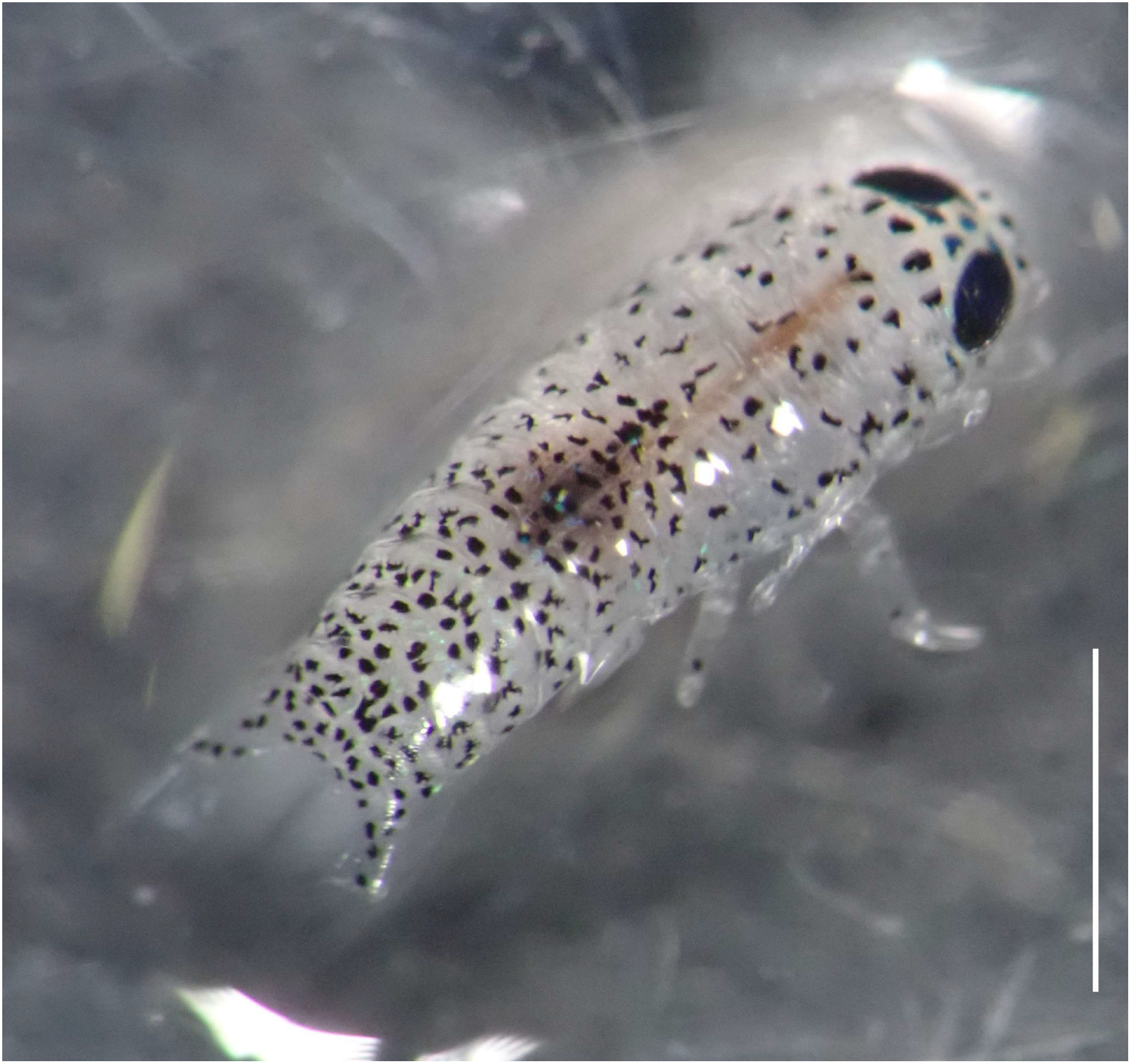
Of the 57 individuals checked, 5 were found to be infested by cymothoid mancae or juveniles (Figure 1). No cases of multiple parasites infesting a single fish were observed. The infestation site was the ventral surface of the body in all individuals (Figure 1). The bodies of the live cymothoids were translucent, and the digestive tract appeared to contain fish blood (Figure 2). One cymothoid parasite was removed from an H. sajori juvenile and fixed in 99.5% ethanol for observation and molecular analyses. The fixed cymothoid was identified as a manca because it lacked pereopod 7 (Figure 3). After observation, the fish were released.
Comparison of the COI sequence of the specimen with the sequences of M. parvostis used for redescription by Fujita et al. [15] (LC763834–LC764100) showed 100% or 99.81% similarity, with an E-value of 0.0, supporting its identification as M. parvostis.
Fujita et al. [19] reported that when M. parvostis parasitizes juveniles of black sea bream, Acanthopagrus schlegelii (Bleeker, 1854), it first attaches to the surface of the host around the mouth, taking advantage of the host’s feeding behavior, and then moves to the branchial cavity. However, all five individuals obtained in this study were parasitizing the same site (the ventral surface of the body), and what appeared to be fish blood was visible in their digestive tract (Figure 2). Therefore, it is unlikely that they were newly attached and had not yet entered the branchial cavity, or that they accidentally adhered to the body surface due to handling during collection. At least in the case of mancae, the claim by Inoue [3] that M. parvostis infests the body surface was proven partly. Hyporhamphus sajori has a slender body shape, and the branchial cavities of their juveniles are small (Figure 1), so parasites may remain on the body surface because they cannot enter the branchial cavity.
Similarly, there are cases in which mancae of Cymothoa pulchrum Lanchester, 1902, which normally parasitizes the buccal cavity of fish, grow into juveniles on the body surface of fish under captivity [20]. Many aspects of the behavior of Cymothoidae in the early stages remain unclear, and further observations of many species are necessary.

Author Contributions

H.F.: Formal Analysis, Investigation, Resources, Writing—Original Draft. K.K.: Investigation, Resources, Writing—Review and Editing. All authors have read and agreed to the published version of the manuscript.

Funding

This work was partially supported by grants-in-aid from the Japan Society for the Promotion of Science (KAKENHI No. 23KJ1170).

Institutional Review Board Statement

Ethical review and approval were waived following Notice No. 71 of the Japanese Ministry of Education, Culture, Sports, Science and Technology, as well as Regulations on Animal Experimentation at Kyoto University, because the research target species is an invertebrate, and the fish were released.

Data Availability Statement

The DNA sequences in this study were deposited in NCBI GenBank (accession number: PX108758).

Conflicts of Interest

The authors declare no conflicts of interest.

References

  1. Bruce, N.L. Revision of the isopod crustacean genus Mothocya Costa, in Hope, 1851 (Cymothoidae, Flabellifera), parasitic on marine fishes. J. Nat. Hist. 1986, 20, 1089–1192. [Google Scholar] [CrossRef]
  2. Rajkumar, M.; Perumal, P.; Trilles, J.P. Cymothoa indica (Crustacea, Isopoda, Cymothoidae) parasitizes the cultured larvae of the Asian seabass Lates calcarifer under laboratory conditions. Dis. Aquat. Organ. 2005, 66, 87–90. [Google Scholar] [CrossRef] [PubMed]
  3. Inoue, M. On sexuality in Cymothoidae, Isopoda II Irona melanosticta Schoedte & Meinert parasitic in the branchial cavity of the halfbeak, Hyporhamphus sajori (Temminck & Schlegel). J. Sci. Hiroshima Univ. Ser. B 1941, 1, 219–238. [Google Scholar]
  4. Fujita, H.; Kawai, K.; Deville, D.; Umino, T. Molecular and morphological characterizations of the fish parasitic isopod Mothocya parvostis (Crustacea: Cymothoidae) parasitizing optional intermediate hosts: Juveniles of the cobaltcap silverside Hypoatherina tsurugae and yellowfin seabream Acanthopagrus latus. Zool. Stud. 2023, 62, e21. [Google Scholar] [CrossRef]
  5. Fujita, H.; Kawai, K.; Taniguchi, R.; Tomano, S.; Sanchez, G.; Kuramochi, T.; Umino, T. Infestation of the parasitic Isopod Mothocya parvostis on juveniles of the black sea bream Acanthopagrus schlegelii as an optional intermediate host in Hiroshima Bay. Zool. Sci. 2020, 37, 544–553. [Google Scholar] [CrossRef] [PubMed]
  6. Fujita, H.; Okumura, Y.; Shinoda, H. Ceratothoa arimae (Isopoda: Cymothoidae) infesting buccal cavity of largescale blackfish, Girella punctata (Centrarchiformes: Kyphosidae), in Seto Inland Sea, Japan. Fishes 2025, 10, e126. [Google Scholar] [CrossRef]
  7. Aneesh, P.T.; Kappalli, S.; Kottarathil, H.A.; Gopinathan, A.; Paul, T.J. Cymothoa frontalis, a cymothoid isopod parasitizing the belonid fish Strongylura strongylura from the Malabar Coast (Kerala, India): Redescription, description, prevalence and life cycle. Zool. Stud. 2015, 54, e42. [Google Scholar] [CrossRef]
  8. Aneesh, P.T.; Kappalli, S.; Kottarathil, H.A.; Gopinathan, A. Mothocya renardi (Bleeker, 1857) (Crustacea: Isopoda: Cymothoidae) parasitising Strongylura leiura (Bleeker) (Belonidae) off the Malabar coast of India: Redescription, occurrence and life-cycle. Syst. Parasitol. 2016, 93, 583–599. [Google Scholar] [CrossRef] [PubMed]
  9. Aneesh, P.T.; Sudha, K.; Helna, A.K.; Anilkumar, G. Agarna malayi Tiwari 1952 (Crustacea: Isopoda: Cymothoidae) parasitising the marine fish, Tenualosa toli (Clupeidae) from India: Re-description/description of parasite life cycle and patterns of occurrence. Zool. Stud. 2018, 57, e25. [Google Scholar] [CrossRef]
  10. Brusca, R.C. Studies on the cymothoid fish symbionts of the eastern Pacific (Isopoda, Cymothoidae) I. Biology of Nerocila californica. Crustaceana 1978, 34, 141–154. [Google Scholar] [CrossRef]
  11. Brusca, R.C. Studies on the cymothoid fish symbionts of the eastern Pacific (Crustacea: Isopoda: Cymothoidae). II Systematics and biology of Lironeca vulgaris Stimpson. Allan Hancock Found. Spec. Publ. New Ser. 1978, 2, 1–19. [Google Scholar]
  12. Brusca, R.C. A monograph on the Isopoda, Cymothoidae (Crustacea) of the eastern Pacific. Zool. J. Linn. Soc. 1981, 73, 117–199. [Google Scholar] [CrossRef]
  13. Hadfield, K.A.; Bruce, N.L.; Smit, N.J. Redescription of poorly known species of Ceratothoa Dana, 1852 (Crustacea, Isopoda, Cymothoidae), based on original type material. Zookeys 2016, 592, 39–91. [Google Scholar] [CrossRef] [PubMed]
  14. Smit, N.J.; Bruce, N.L.; Hadfield, K.A. Global diversity of fish parasitic isopod crustaceans of the family Cymothoidae. Int. J. Parasitol. Parasite Wildl. 2014, 3, 188–197. [Google Scholar] [CrossRef] [PubMed]
  15. Fujita, H.; Aneesh, P.T.; Kawai, K.; Kitamura, S.I.; Shimomura, M.; Umino, T.; Ohtsuka, S. Redescription and molecular characterization of Mothocya parvostis Bruce, 1986 (Crustacea: Isopoda: Cymothoidae) parasitic on Japanese halfbeak, Hyporhamphus sajori (Temminck & Schlegel, 1846) (Hemiramphidae) with Mothocya sajori Bruce, 1986 placed into synonymy. Zootaxa 2023, 5277, 259–286. [Google Scholar] [CrossRef] [PubMed]
  16. Kawanishi, R.; Sogabe, A.; Nishimoto, R.; Hata, H. Spatial variation in the parasitic isopod load of the Japanese halfbeak in western Japan. Dis. Aquat. Organ. 2016, 122, 13–19. [Google Scholar] [CrossRef] [PubMed]
  17. Kawai, K.; Fujita, H.; Sanchez, G.; Umino, T. Oyster farms are the main spawning grounds of the black sea bream Acanthopagrus schlegelii in Hiroshima Bay, Japan. PeerJ 2021, 9, e11475. [Google Scholar] [CrossRef] [PubMed]
  18. Fujita, H.; Kawai, K.; Deville, D.; Umino, T. Quatrefoil light traps for free-swimming stages of cymothoid parasitic isopods and seasonal variation in their species compositions in the Seto Inland Sea, Japan. Int. J. Parasitol. Parasite Wildl. 2023, 20, 12–19. [Google Scholar] [CrossRef] [PubMed]
  19. Fujita, H.; Kawai, K.; Shimomura, M.; Umino, T. “Lure fishing” strategies by Mothocya parvostis (Isopoda: Cymothoidae): Feeding behavior-mediated infestation on juveniles of black sea bream, Acanthopagrus schlegelii. Int. J. Parasitol. Parasite Wildl. 2025, 26, e101057. [Google Scholar] [CrossRef] [PubMed]
  20. Fujita, H.; Shinoda, H.; Okumura, Y. Description of life cycle stages of fish parasite Cymothoa pulchrum (Isopoda: Cymothoidae), with DNA barcode linked to morphological details. Fishes 2025, 10, e155. [Google Scholar] [CrossRef]
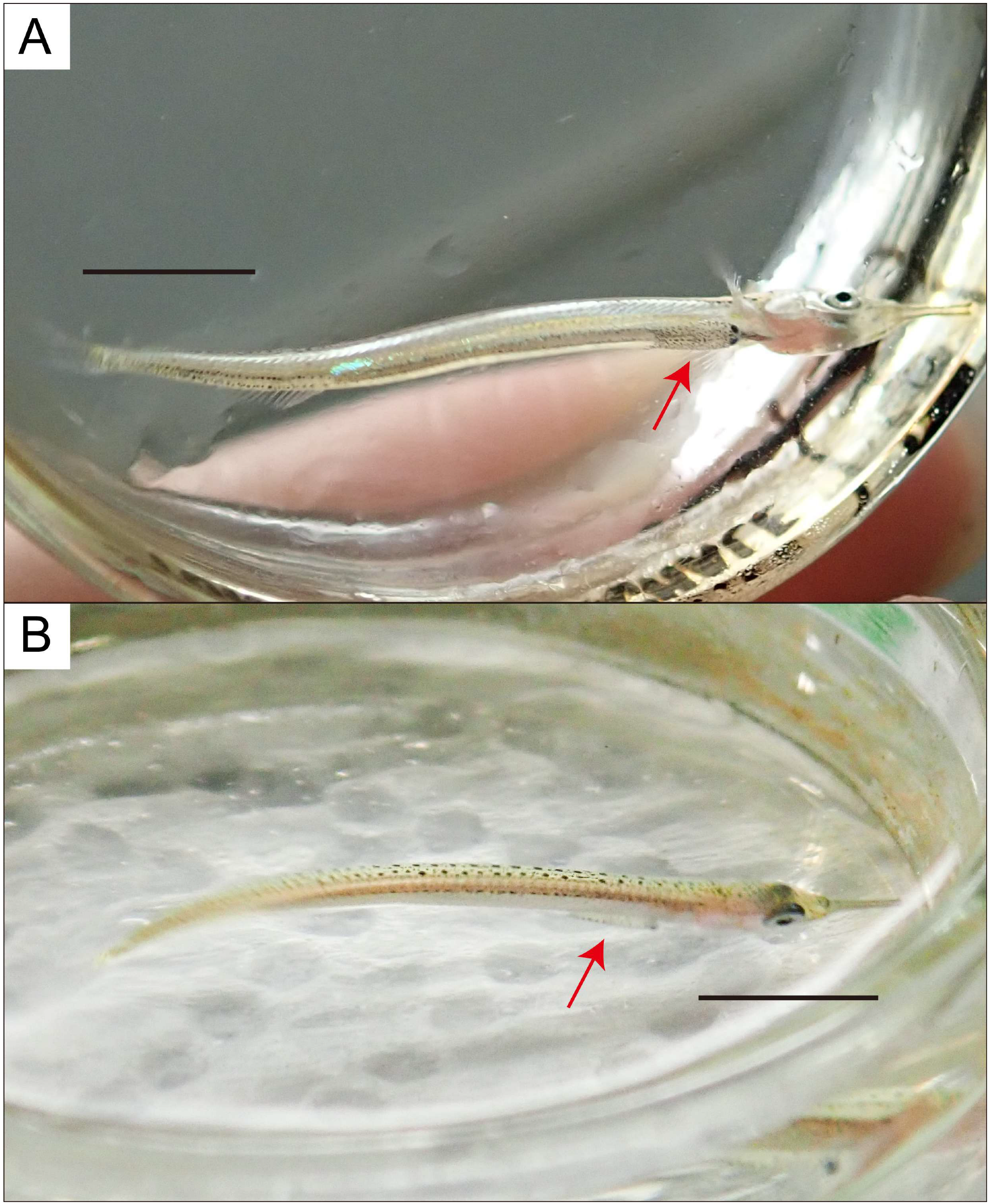
Figure 1. Live Mothocya parvostis Bruce, 1986 infesting the ventral surface of a juvenile Japanese halfbeak, Hyporhamphus sajori (Temminck and Schlegel, 1846) collected from the surface waters around oyster-culturing rafts off Ohkurokamishima Island, Etajima, Hiroshima Prefecture, Japan, on 30 May 2019. (A) Ventrolateral view; (B) dorsolateral view. Arrows indicate the infestation sites of M. parvostis. Scale bars: 5 mm.
Diversity 17 00613 g001
Figure 2. Dorsolateral view of live Mothocya parvostis Bruce, 1986 removed from a juvenile Japanese halfbeak, Hyporhamphus sajori (Temminck and Schlegel, 1846) collected from the surface waters around oyster-culturing rafts off Ohkurokamishima Island, Etajima, Hiroshima Prefecture, Japan, on 30 May 2019. The tissue of fish consumed by parasites can be seen inside the digestive tract. Scale bar: 1 mm.
Diversity 17 00613 g002
Figure 3. Manca of Mothocya parvostis Bruce, 1986 (ethanol preserved) removed from a juvenile Japanese halfbeak, Hyporhamphus sajori (Temminck and Schlegel, 1846) collected from the surface waters around oyster-culturing rafts off Ohkurokamishima Island, Etajima, Hiroshima Prefecture, Japan, on 30 May 2019. (A) Dorsal view; (B) ventral view. Scale bars: 1 mm.
Diversity 17 00613 g003
Disclaimer/Publisher’s Note: The statements, opinions and data contained in all publications are solely those of the individual author(s) and contributor(s) and not of MDPI and/or the editor(s). MDPI and/or the editor(s) disclaim responsibility for any injury to people or property resulting from any ideas, methods, instructions or products referred to in the content.