Review

Pore Characteristics of Deep-Sea Benthic Foraminifera

by
Bruce H. Corliss
1,2,* and
Anthony E. Rathburn
3
1
Nicholas School of the Environment, Duke University, Durham, NC 27708, USA
2
Graduate School of Oceanography, University of Rhode Island, Narragansett, RI 02882, USA
3
Department of Geological Sciences, California State University Bakersfield, Bakersfield, CA 93311, USA
*
Author to whom correspondence should be addressed.
Diversity 2025, 17(5), 343; https://doi.org/10.3390/d17050343
Submission received: 5 April 2025 / Revised: 2 May 2025 / Accepted: 5 May 2025 / Published: 13 May 2025
(This article belongs to the Special Issue Foraminiferal Research: Modern Approaches and Emerging Trends)

Abstract

:
A review of the pore patterns of deep-sea benthic foraminifera is presented with a discussion of their characteristics, function and relationship with dissolved oxygen levels. Pore characteristics of deep-sea benthic foraminifera are of timely interest due to their potential for reconstructing dissolved oxygen conditions from the sedimentary record. Scanning electron micrographs of 20 epifaunal and infaunal deep-sea taxa from the Sulu Sea, Monterey Bay, California Bight and northwest Atlantic Ocean are presented to illustrate the wide range of pore patterns found in deep-sea taxa. New SEM observations of three taxa with biconvex test shapes, Oridorsalis umbonatus, Hoeglundina elegans, and Epistominella umbonifera, suggest that these taxa have an infaunal habitat for at least part of their life span.

1. Introduction

Benthic foraminiferal morphology is highly variable and is related to the physical nature of habitats found in the deep sea. Pores are found in many taxa and are among the most prominent characteristics of test morphology. Early work on the nature and function of pores in foraminiferal tests was summarized by Boltovskoy and Wright [1].
A number of functions for the pores of benthic foraminiferal tests have been suggested, including conduits for pseudopodia [2,3] and food acquisition and osmoregulation [4]. Pores have also been suggested to be important in the gas exchange of the cell (e.g., [5,6,7]. A cytoplasmic study of living species from low-oxygen locations in the San Pedro Basin revealed clusters of mitochondria close to the inner end of the pores on seven species [5]. The proximity of the mitochondria to pore openings was suggested to reflect the importance of gas exchange (O2 in and CO2 out) through the pores. However, all taxa from low-oxygen environments might not have this mitochondria–pore association observed by Leutenegger and Hansen [5]. In an ultrastructural study of the infaunal species, Stainforthia fusiformis (Williamson, 1848), mitochondria were most abundant nearest the aperture and concentrated at the periphery of chambers, but not associated with pores [8], suggesting that mitochondria in pseudopodia could also be important for some infaunal taxa [9].
An association with rod-shaped microbes, suggested to be ectosymbionts, and pores of Bolivina pacifica (Cushman and McCulloch, 1942) was found in specimens from low-oxygen waters (0.7 µM) [10]. Mitochondria in this species were concentrated beneath pores, similar to observations made earlier [5]. In contrast, an examination of specimens of Bolivina cf. B. lanceolata (Parker, 1954) from well-aerated sediments from the Florida Keys (Little Duck Key, Florida) found that specimens did not have ectosymbionts or mitochondria associated with pores [10]. This difference in the distribution of mitochondria between well-oxygenated and oxygen-poor environments supports suggestions by Leutenegger and Hansen [5] and others that the pores are sites for gas exchange.
Benthic foraminiferal taxa have evolved a number of mechanisms to live in oxygen-stressed conditions, including bacterial symbionts [10], engulfing functional chloroplasts [11,12], cellular adaptations [5,10], dormancy [13,14], and nitrate respiration [7,15,16,17,18,19,20].
Variability of pore characteristics between individuals of a given species [21,22] supports the idea that surface pores are ecophenotypic aspects of the test that can change in response to local conditions. For example, Cibicidoides bradyi (Trauth, 1918) increased the surface area (and thereby the number of test pores) of their test in sediments of the Sulu Sea in low-oxygen levels [23]. Glock et al. [15] noted that an increase in length of Bolivina spissa within the sediments appeared to be associated with the need for greater surface area for increased gas exchange via test pores.
Observations on pore densities of Ammonia beccarii (Linnaeus, 1758) maintained in the laboratory show that chambers formed in dysoxic conditions (<12.5 µM) have higher porosities (i.e., larger pores) than chambers formed in well-oxygenated waters (~225 µM) [24]. Compared to calcareous foraminiferal assemblages living in well-oxygenated environments, species from oxygen-poor habitats tend to have larger and more numerous pores on their tests [15,24,25]. In addition to the influence of oxygen, Glock et al. [15] suggested that there is a relationship between bottom water O2 and NO3 and pore density in tests of the infaunal species Bolivina spissa (Cushman, 1926). More recently, the relationship between pore characteristics and oxygen availability has been examined in deep-water infaunal taxa [26,27], and a detailed quantitative analysis of epifaunal taxa is presented in Rathburn et al. [23].
The number and size of pores that a test can have are limited by the size, shape and robustness of the test [28]. The key measure for oxygen accessibility is the percentage of umbilical surface area of the test occupied by pores [23] Higher pore surface area can result from an increase in pore density (number of pores per surface area), an increase in pore size, or a combination of both [20,28,29]. Structural constraints of the test likely means that pore size and pore density in foraminiferal tests are interdependent, and these constraints may limit pore characteristics of the test [28]. Using the shallow water taxon, Ammonia, as a template, Richirt et al. [28] suggested that due to mechanical constraints, the upper limit of porosity (surface area percentage) is 30%. However, porosities of C. wuellerstorfi can reach up to 50% in oxygen-poor conditions [23,30]. Clearly, mechanical limitations for porosity are different for different taxa, and likely depend on test thickness, shape, size, and perhaps mobility (C. wuellerstorfi specimens that had 50% porosity were attached).
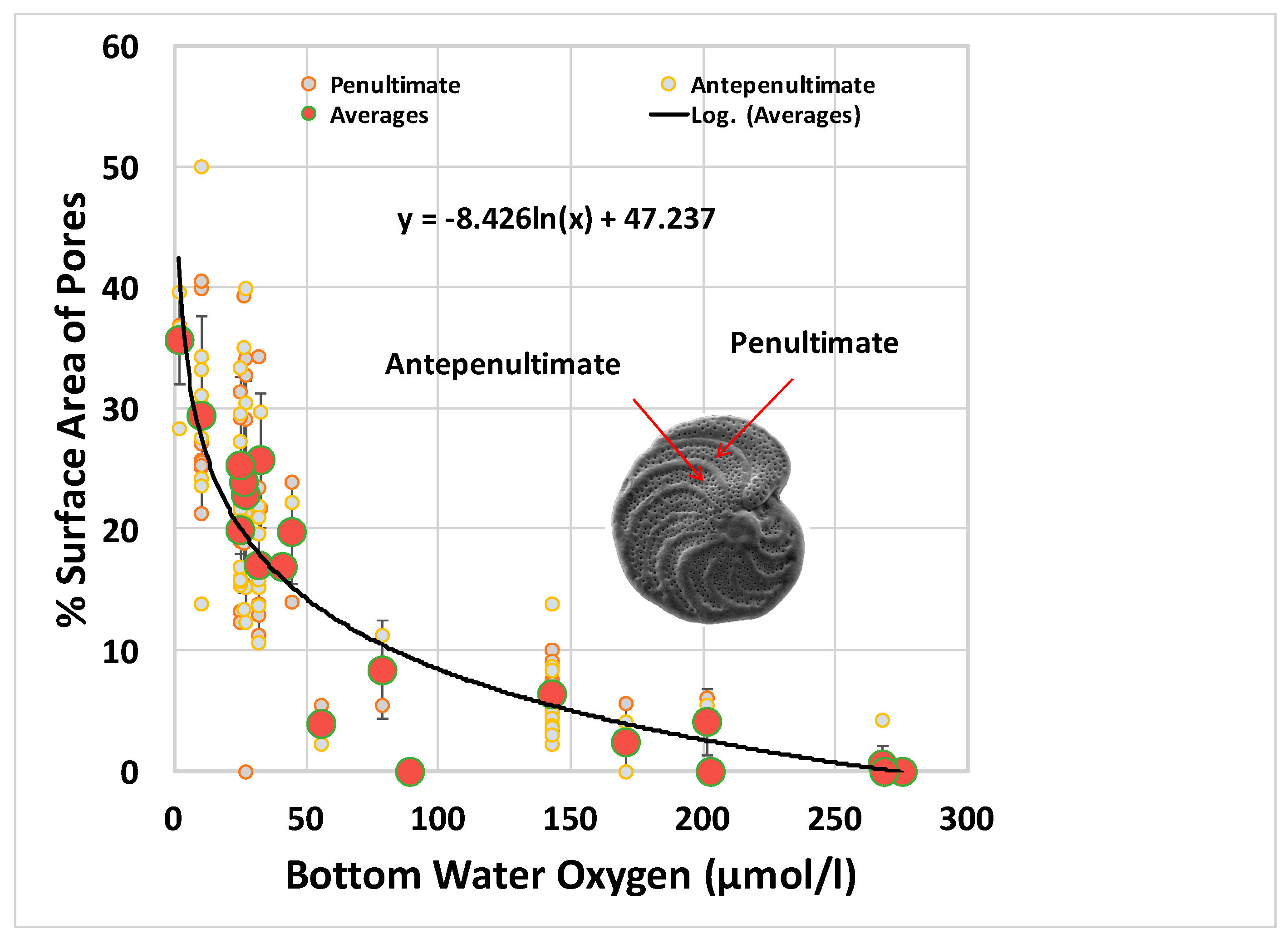
The presence or absence of pores was linked to bottom water oxygen levels and microhabitat preferences of deep-sea benthic foraminifera in well oxygenated regions of the oceans (e.g., [31,32]. Epifaunal taxa lack pores over the entire test or, in the case of plano-convex species such as Cibicidoides wuellerstorfi (Schwager, 1866), have large pores on the spiral side from which protoplasm is extruded. Infaunal species found in lower oxygen conditions within the upper ~10 cm of the sediments have pores over a portion or the entire surface of the test. However, this pore pattern of epifaunal species is not found in low-oxygen regions. A review by Jorissen et al. [33] identified 64 taxa previously considered as indicators of well-oxygenated conditions that were found living (Rose Bengal-stained) in dysoxic to suboxic habitats, and this list included epifaunal taxa. Recent studies confirm that epifaunal taxa occur in significant abundances in a wide range of dissolved oxygen conditions, including Oxygen Minimum Zones [34,35]. Pore surface areas of epifaunal species, mostly C. wuellerstorfi, taken from locations with a range of oxygen conditions (2–277 µM) have a negative correlation with bottom water oxygen [23].

2. Location of Samples Used for Illustrations

Scanning electron micrographs are presented to illustrate the different pore patterns found in both epifaunal and infaunal taxa commonly found in the deep sea. Samples were collected from a range of overlying bottom water dissolved oxygen concentrations (Table 1) from the Sulu Sea, northwest Atlantic Ocean, Monterey Bay, and California Bight to illustrate the relationship between pores and dissolved oxygen content. Different definitions exist to describe oxygenation conditions. For this paper, the classification system used by Tyson and Pearson [36] will be used where oxic refers to dissolved oxygen concentrations from 2–8 mL/L (89 to 357 µM), dysoxic covers the range from 0.2–2 mL/L (8.9 to 89 µM), and suboxic conditions are <0.2 mL/L (<8.9 µM).

2.1. Sulu Sea

Located north of Sabah (Borneo) and east of the South China Sea, the Sulu Sea is surrounded by relatively shallow sills and is one of the more isolated marginal seas in the region. Through a single 420-m channel, South China Sea intermediate water provides the only source of deep water to the basin, creating bottom waters of nearly uniform temperatures (~10.5 °C), salinities (~34.5 PSU) and dissolved oxygen contents (~55 µM) below 1000 m ([23,37]). Ecological and isotopic characteristics of living (Rose Bengal-stained) foraminiferal assemblages from these cores and adjacent sites were discussed in Rathburn and Corliss [23] and Rathburn et al. (1996) [38]. Dissolved oxygen concentrations at these sites were 57.2 µM (1980 m) and 78.5 µM (510 m); other environmental parameters for these sites are included in Table 1.

2.2. Monterey Bay

Submarine canyons and methane seeps are among the prominent seafloor features of Monterey Bay, located on the California coast south of San Francisco (e.g., [37].) Samples examined in this study (LTC49, Core 37) were collected from seep and non-seep surface sediments in an area known as “Clam Flats” at about 1000 m water depth. Specimens were obtained from surface sediments in tube cores collected by the remotely operated vehicle (ROV) Jason in 2007. Living foraminiferal characteristics were discussed in Bernhard et al. [6,39] and Rathburn et al. [40]. LTC49 was collected from a clam bed associated with active methane seepage. Core 37 was taken in a non-seep area located about 100 m from the seep locations. Epifaunal foraminifera were much more abundant at the seep sites compared to non-seep sites, probably because of the greater availability of hard substrates (clam shells, tube worms, authigenic carbonate) for attachment in seep habitats [41,42].

2.3. California Bight

Specimens from this area were collected from a site located on the edge of the oxygen minimum zone in the Southern California Bight offshore of San Diego at a water depth of 1050 m. This region is in a coastal upwelling zone under the influence of the California Current System [43]. Seafloor samples examined from this region were collected using an Ocean Instruments Multicorer (Fall City, WA, USA) during a California Cooperative Oceanic Fisheries Investigations (CalCOFI) cruise near CalCOFI station 9244 in May 1996. Living (stained) benthic foraminifera from the region have been characterized by previous studies [35,44,45,46,47]. Epifaunal foraminifera are more abundant at the 9244 site compared to other study areas of similar water depth in the more northern sector of the Southern California Bight (Rathburn et al., unpublished), which is probably a result of the coarser grain sized sediment at this location, providing hard substrate for attachment [35].

2.4. Northwest Atlantic Ocean: Nova Scotian Margin, Gulf of Maine, Bermuda Rise

Specimens examined from this region were collected from the continental margin off Nova Scotia (K104 cores 1, 2, 4), Gulf of Maine (K104 core 8), and Bermuda Rise (OC86 core 5–3). Samples were obtained using an Ocean Instruments Soutar box corer and an Ocean Instruments Mark III box corer. The sediment geochemistry and ecology of Rose Bengal-stained foraminifera from these samples were previously discussed [31,48]. Dissolved oxygen values found with these samples range from >260–290 µM (Table 1).

3. Scanning Electron Micrographs

Scanning electron micrographs (Figure 1, Figure 2, Figure 3, Figure 4, Figure 5 and Figure 6) were made of individual specimens from samples that had previously been studied or were picked from new samples. A limited number of specimens for most species (~n = 10) was available from previous studies, because most specimens had been used for stable isotopic analysis, or from new samples in which specimens of particular species were not abundant. The size fraction used to study deep sea foraminifera has varied between investigators. The specimens used for the SEM illustrations were >150 µm in diameter and were living specimens that had been stained with Rose Bengal, except for some well-preserved dead specimens of Cibicidoides bradyi. Epifaunal species include Cibicidoides wuellerstorfi, Planulina spp., Cibicidoides mundulus (Parker, 1953), Hoeglundina elegans (d’Orbigny, 1826), Discorbinella bertheloti (d’Orbigny, 1839), and Epistominella umbonifera (Cushman, 1933). Two additional species, Cibicidoides bradyi (Trauth, 1918) and Oridorsalis umbonatus (Reuss, 1951), with microhabitats transitional between epifaunal and infaunal, are also included. Infaunal species illustrated include Uvigerina peregrina (Cushman, 1923), Melonis affinis (Reuss, 1851), Bulimina mexicana (Cushman, 19220, Bolivina sp., Lenticulina sp., Chilostomella oolina (Schwager, 1878), Globobulimina affinis (d’Orbigny, 1839), Nonion grateloupi (d’Orbigny, 1826), Pullenia bulloides (d’Orbigny, 1846), Pullenia simplex (Rhumbler, 1931), Pullenia osloensis (Feyling-Hanssen, 1954), and Valvulineria mexicana (Parker, 1954).

3.1. Cibicidoides (=Cibicides, Planulina, Fontbotia, Lobatula) wuellerstorfi

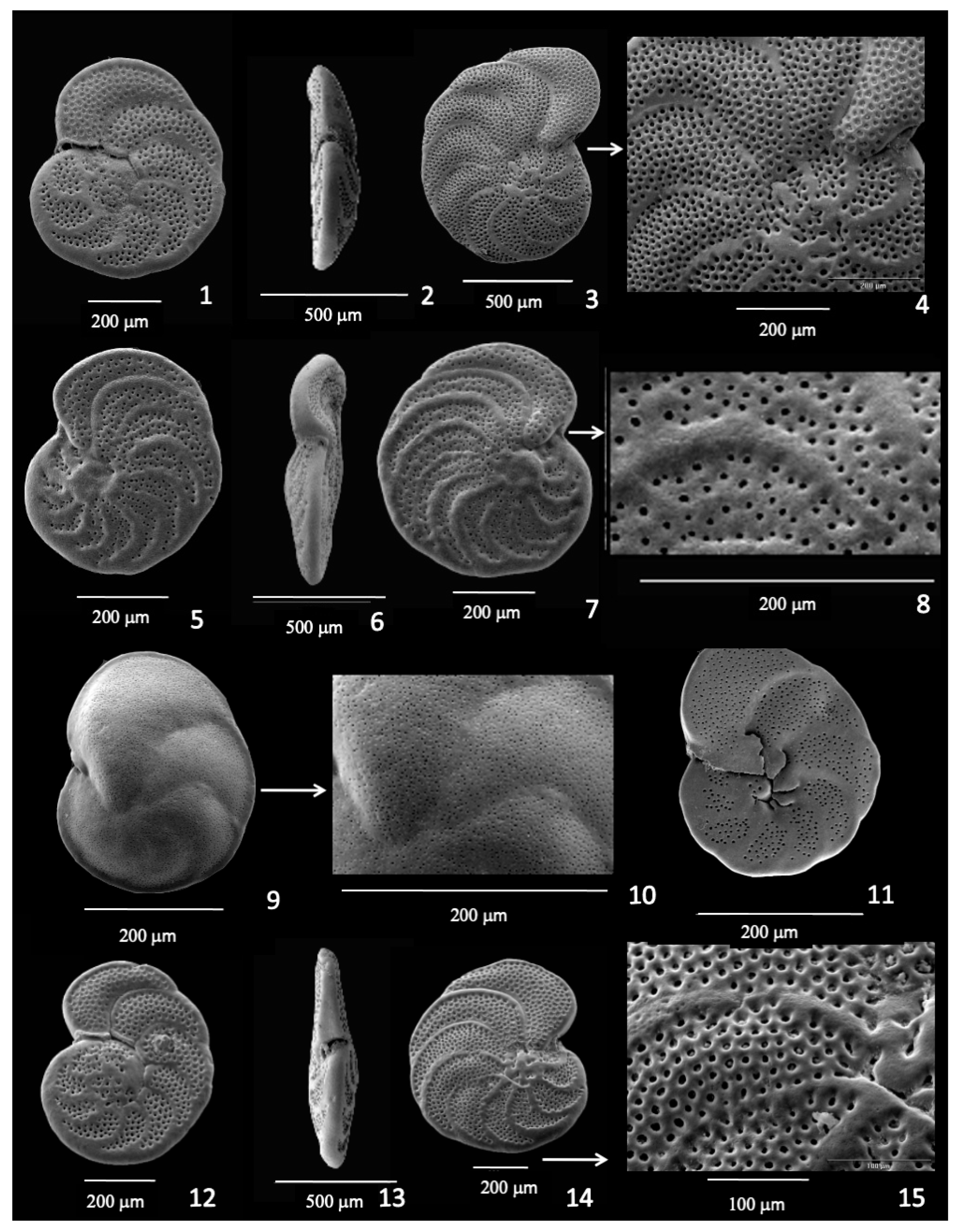
Cibicidoides wuellerstorfi (see [21,49,50] for discussion) is one of the dominant and cosmopolitan species found in the deep oceans and has been widely used for stable isotopic studies because the isotopic signature of its test closely reflects bottom water isotopic composition due its elevated, epibenthic microhabitat [51]. Individuals are attached to spines, spicules, pebbles and other materials on the seafloor and generally live above the sediment–water interface [22,34,52]. The test has a plano-convex shape and attaches to objects on the spiral side (Figure 1(1,2)), exposing the umbilical side of the test to bottom water (Figure 1(3)). The plano-convex shape can be highly variable and, as a result, the species exhibits considerable phenotypic variability. Nova Scotian margin specimens, taken at locations with dissolved oxygen contents of 260–290 µM, are typical of individuals from well-oxygenated regions of the deep sea and have relatively large pores on the spiral side (Figure 1(1,5)), but generally lack pores on the umbilical side (Figure 1(3,6)). In the dysoxic bottom waters of the Sulu Sea (55.8–78.1 µM), a different pattern is found with specimens having abundant pores on the umbilical side exposed to bottom water (Figure 1(4,7–12)). However, the abundance of pores varies with Sulu Sea specimens, with some tests lacking pores on the umbilical side.

3.2. Planulina spp.

Forty-four specimens of Planulina sp. were examined from the California Bight and Monterey Bay sites (Figure 2(1–8); Figure 2(12–15)) and both sites were overlain by low dissolved oxygen content (~38 to 44.5 µM). These epifaunal specimens show porosity on both sides of the test, with pores covering most of the test area.

3.3. Discorbinella bertheloti

This species has a plano-convex test, with pores covering the entire test except for the chamber sutures on the spiral side (Figure 2(9–11)) from the Sulu Sea.

3.4. Cibicidoides mundulus

This species (Figure 3(1,5)) is characterized by a biconvex test (Figure 3(4,5)) and is common in the deep sea. In samples from the Nova Scotian margin, pores are present on the spiral side (Figure 3(1)) but are absent on the umbilical side (Figure 3(2,3)). In contrast, Sulu Sea specimens have abundant pores on both sides of the test (Figure 3(5)).

3.5. Cibicidoides bradyi

This species is not common and occurs in low abundances when present. Pores are found on the umbilical side but are lacking on the spiral side from K104/10 samples (Figure 3(6–8)). In contrast, Sulu Sea specimens have pores distributed over the entire test and are more trochospiral than those found in more well-oxygenated environments, expanding the surface area of the test (Figure 3(9)).

3.6. Oridorsalis umbonatus

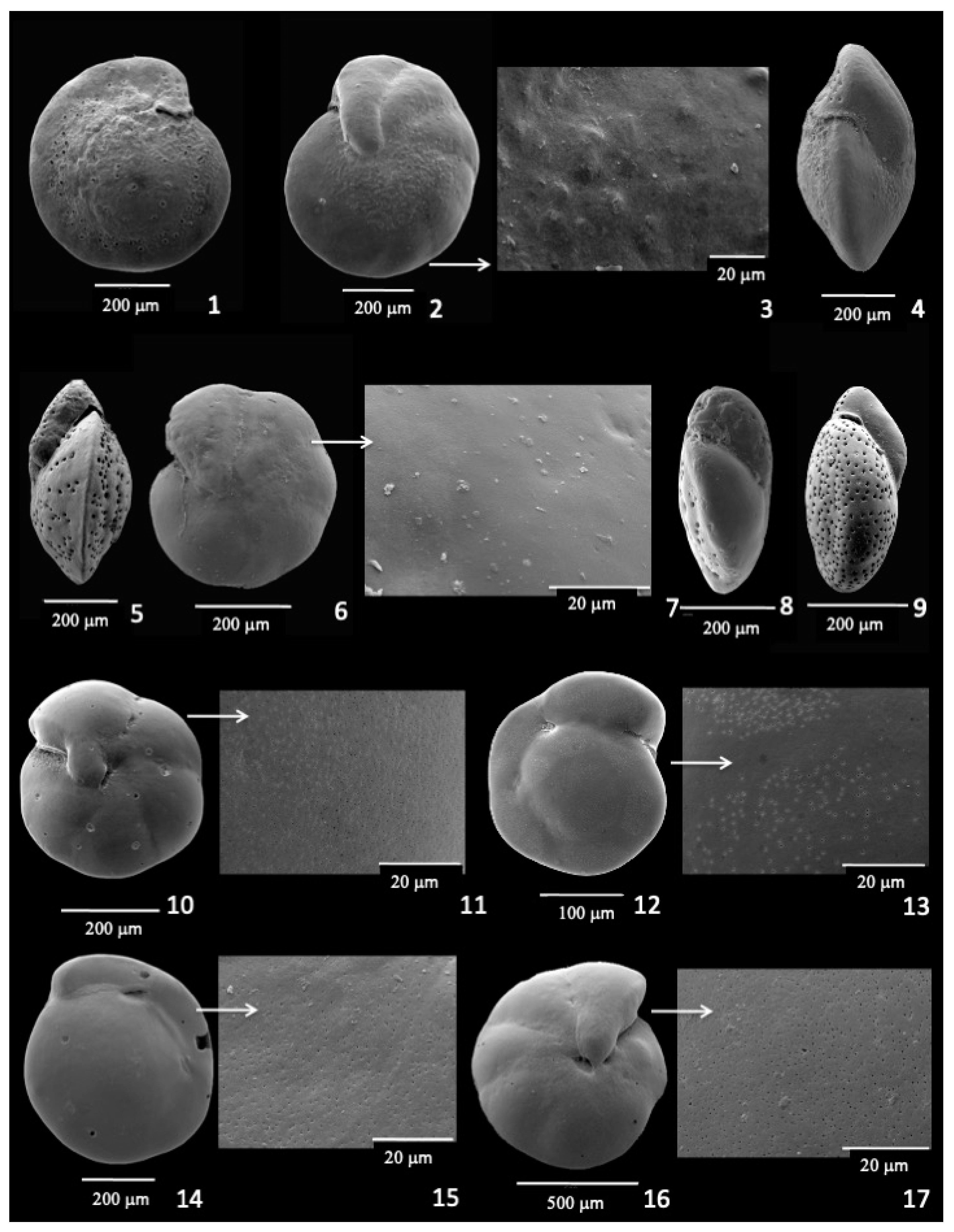
This taxon is a common deep-sea species with a broad distribution. The species was initially thought to have an epifaunal microhabitat [31], but was later suggested to have a microhabitat transitional between epifaunal and infaunal microhabitats, as it was found over the 0–4 cm interval in the Sulu Sea [53]. Oridorsalis umbonatus appears to lack pores on both sides of the test, but a closer examination at higher magnification (~5000×) of 50 specimens reveals small pores on both the umbilical and spiral sides of all of the tests examined. Pores are found on tests from the dysoxic environment of the Sulu Sea (Figure 3(10–13)), as well as the well-oxygenated Nova Scotian margin specimens (Figure 3(14–17)).

3.7. Hoeglundina elegans

This species is a common species with a biconvex test composed of aragonite and was considered as an epifaunal species [31,38,53], but has been characterized more recently as a very shallow infaunal species [54]. The test appears to be smooth on both sides and lacks pores when viewed under a light microscope. However, an examination of 50 specimens shows that pores are present and visible when viewed under higher magnification (~10,000×) and are widespread on individual chambers, but not on the chamber sutures on all specimens examined. Pores are visible on Sulu Sea specimens (Figure 4(1–5)), as well as specimens from the Nova Scotian margin (Figure 4(6–10)). The presence of fine pores on H. elegans in both high- and low-oxygen regions is similar to the pore pattern found with Oridorsalis umbonatus.

3.8. Epistominella umbonifera

This is a common deep-sea species characterized by a convex test (Figure 4(12)) and found in abyssal waters of the oceans above the calcium carbonate compensation depth [55,56]. An examination of specimens from the western North Atlantic Ocean reveals small pores on both the umbilical (Figure 4(11,14)) and spiral sides (Figure 4(13,15)).

3.9. Infaunal Species

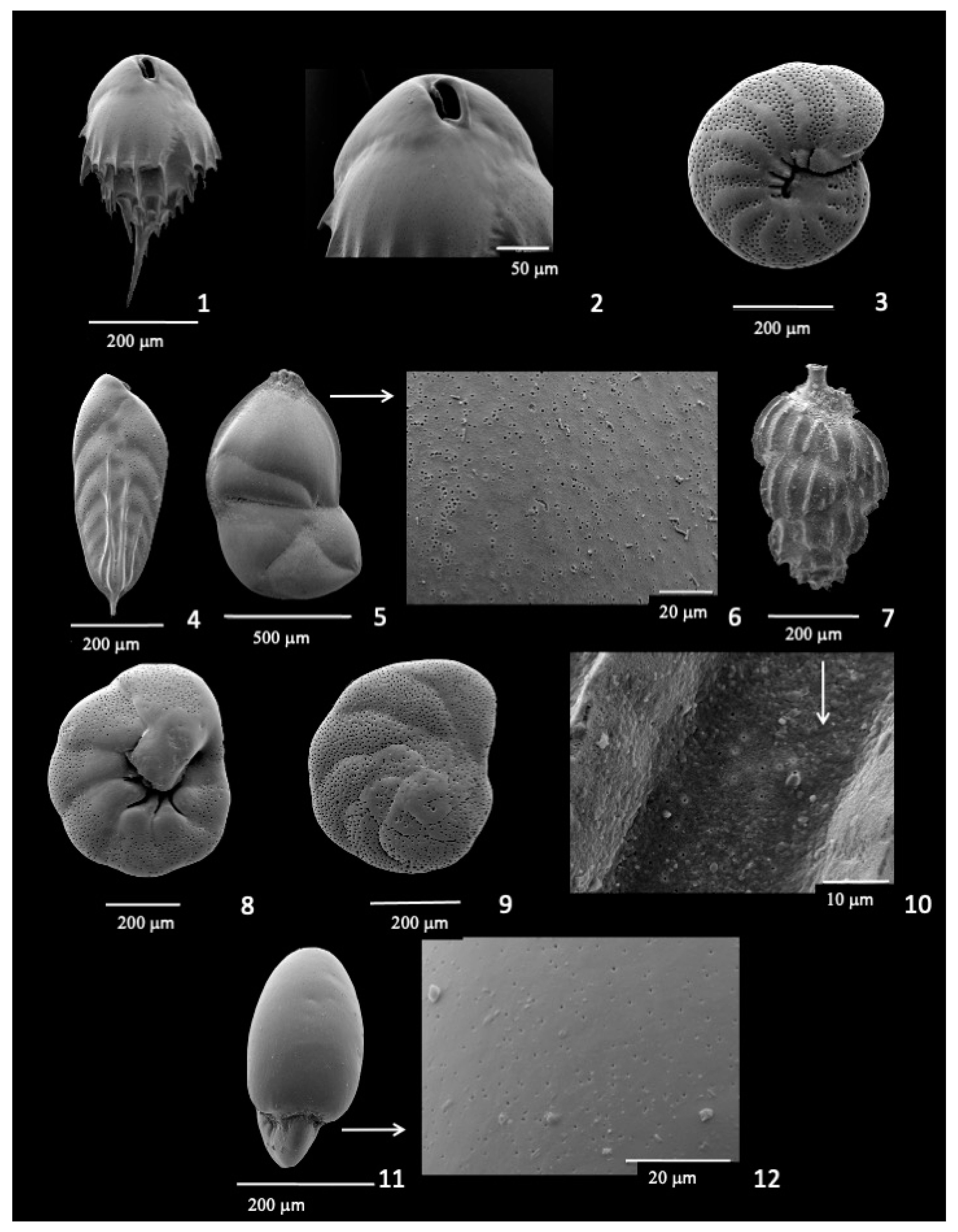
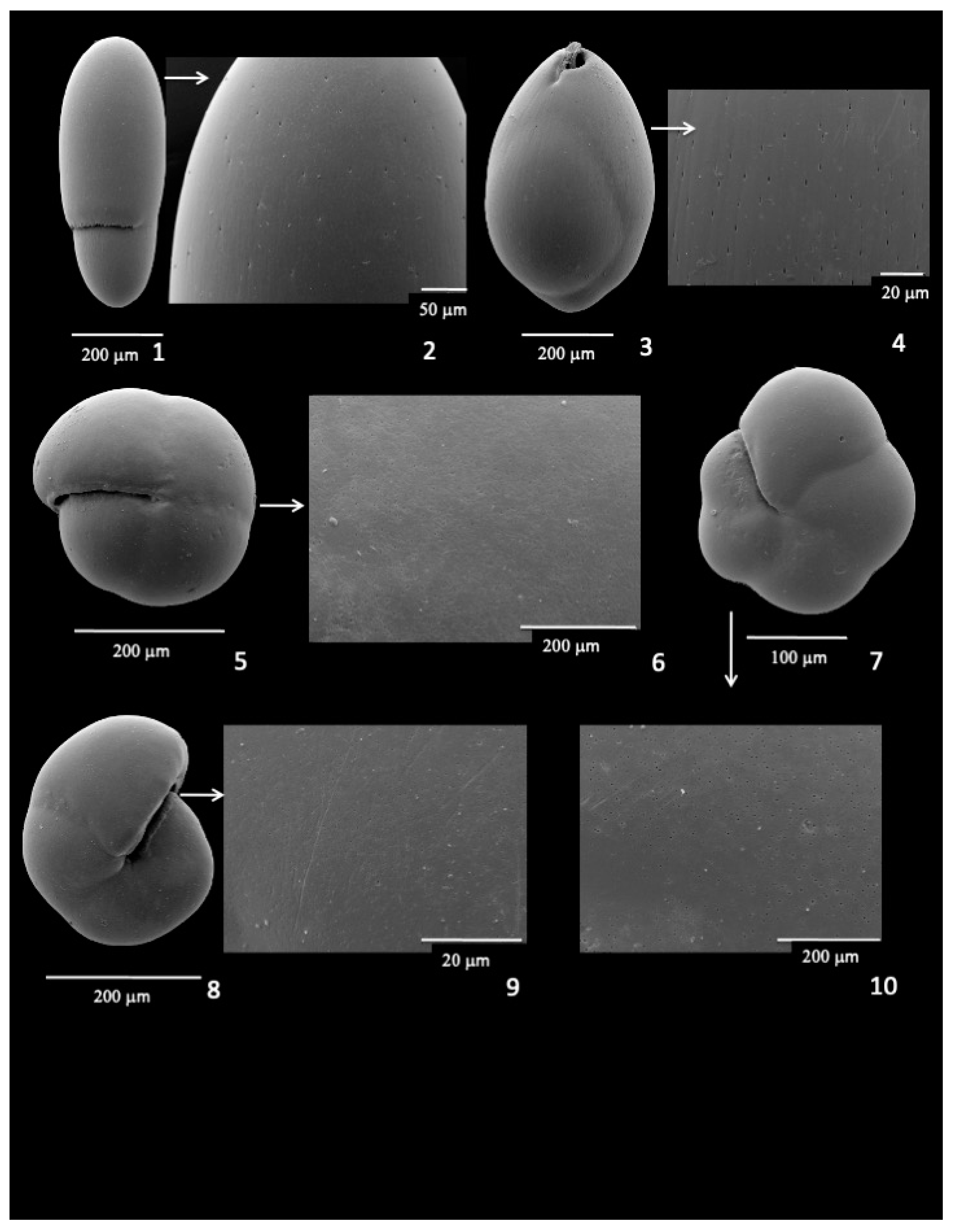
A number of infaunal species are presented to document pore characteristics and patterns. Bulimina mexicana is a shallow infaunal species and has fine pores over the entire test (Figure 5(1,2)). Large pores are found on Melonis affinis (Figure 5(3)) and evenly cover the entire test, except for the chamber sutures. Fine pores cover the entire test of Bolivina sp. (Figure 5(4)) and Lenticulina sp. (Figure 5(5,6)). Uvigerina peregrina is a common taxon on continental margins and had a widespread occurrence in the North Atlantic during the last glacial period. This species (Figure 5(7)) has a shallow infaunal microhabitat (~0–2 cm) and has very small pores on the surface between the ribs (Figure 5(10)), although the pore density is less than other infaunal taxa. Valvulineria mexicana has pores over a large portion of the umbilical side but absent in the central region surrounding the umbilicus (Figure 5(8)), and pores over the entire spiral side (Figure 5(9)). Fine pores over the entire test are also found with specimens of N. grateloupi (Figure 5(11,12)), C. oolina (Figure 6(1,2)), G. affinis (Figure 6(3,4)), P. bulloides (Figure 6(5,6)), P. osloensis (Figure 6(7,10)), and P. simplex (Figure 6(8,9)) from the Nova Scotian margin.

4. Discussion

The assumption is made in this review that specimens of individual species can be identified and grouped based on their test morphology. If cryptic species exist, this grouping might not be accurate. Recent molecular studies of eukaryotic organisms show that higher diversity exists with planktonic foraminifera [57] and shallow water benthic foraminifera [58] than expected from morphological considerations. However, molecular studies of deep-sea benthic foraminiferal taxa [59,60] suggest that a high degree of genetic similarity exists for three widespread species, Epistominella exigua, Cibicidoides wuellerstorfi, and Oridorsalis umbonatus and, therefore, their morphospecies designation accurately reflects their genetic relationships.
Some researchers suggest that the axial profile of cibicidids results more from ecology and mobility than taxonomy [49,61]. Pore characteristics and test shapes of Cibicidoides species can clearly be influenced by environmental conditions [22,23,62]. In addition, recent genetic work on ubiquitous Cibidicoides species shows considerable ecophenotypic plasticity within a single deep-sea species [21,34]. This indicates that at least for some deep-sea taxa, an appreciable degree of morphological variability is possible as a result of environmental conditions rather than taxonomic diversity.
The presence of tiny pores over the tests of both thinly-walled infaunal taxa (Globobulimina and Chilostomella) and more robust species (E. umbonifera, H. elegans, and O. umbonatus) noted in this review may reflect a balance between the need for gas exchange and structural integrity and mobility. Globobulimina and Chilostomella are considered deep infaunal taxa that inhabit fine-grained, oxygen-poor, organic-rich sediments. The thin walls of these taxa have been suggested as an adaptation for gas exchange [63,64] and pores may help facilitate that exchange. The infaunal taxa considered here have a variety of shapes and coiling types, but all exhibit pores over all or most of the test. The size and density of the pores vary between species. Specimens that represent shallow infaunal species, such as Uvigerina, Bolivina, and Pullenia spp., have fine pores; larger pores are found with Melonis affinis, and the deepest dwelling species, C. oolina and G. affinis, have fine pores. Pore size patterns may vary with microhabitat, as some Bolivina specimens found in the upper 1–2 cm have large pores. It is noteworthy that some specimens of a given infaunal species have symbionts while others do not [39], so the metabolic needs reflected in pore patterns may differ between taxa or within a given species. It is well known that symbionts influence the metabolic needs of organisms, including foraminifera [65,66] and the test geochemistry of foraminifera [51]. Nitrate respiration influences metabolic processes of single-celled organisms [67] and probably necessitates metabolic adjustments in foraminifera. Those species that store and respire nitrate or have symbionts are likely to have different requirements than those that do not employ these mechanisms.
Unlike many infaunal taxa, epifaunal Cibicidoides species remain near the sediment–water interface and there is no evidence that they use nitrate for respiration [23]. Based on experiments, it was determined that while most infaunal taxa respired nitrate, Cibicidoides pachyderma does not [17]. Oxygen availability influences pore surface area percentages in epifaunal benthic foraminifera [23], and other factors such as shell thickness [28] and test shape characteristics such as major axis attributes and roundness [64] may also affect pore patterns.
Three biconvex species illustrated here, E. umbonifera, H. elegans, and O. umbonatus, have similar pore characteristics, with pores found over most of the surface of the test. Pores of E. umbonifera can be viewed under low magnification, but pores of H. elegans and O. umbonatus can be seen only under high magnifications (~5000–10,000×). These species are generally found within the top centimeter of surficial sediments. However, results from the Sulu Sea showed that O. umbonatus is found over a 0–4 cm interval, suggesting a transitional microhabitat between epifaunal and deep infaunal habitats. The presence of pores on O. umbonatus from the Sulu Sea is consistent with this suggestion, as oxygen-poor conditions would be found within the sediments as well as in the overlying bottom water. However, the presence of pores in well-oxygenated Nova Scotia samples, with O. umbonatus largely confined to the 0–1 cm interval, suggests that it may live part of the time or full time infaunally, submerged within the top centimeter, rather than living at the sediment–water interface. The biconvex shape of the test also supports this suggestion, as this shape would be advantageous for the organism to move through the sediments. A biconvex test and the presence of pores on both sides of the test characterize E. umbonifera and H. elegans as well. These characteristics suggest that these species live infaunally within this interval for at least the latter part of their ontogeny.
The suggestion that the biconvex species discussed are infaunal for at least part of their life cycle is consistent with observations on the motility of benthic foraminifera. Experiments have shown that with changing conditions, the deep-sea Cibicidoides species C. mundulus and C. pachyderma can change from an epifaunal (exposed to bottom water) to a shallow infaunal (upper 1–2 cm; exposed to pore water) microhabitat [68]. Cibicidoides pachyderma, characterized by a convex test, pores over the entire test, and obtained from 220 m water depth had rates of movement from 1.0 to 23 mm/day and was observed in the laboratory to often cover itself with sediment [69]. Based on these observations, it seems quite reasonable that biconvex taxa are able to move within the upper 1–2 cm of sediment. However, it is noteworthy that C. mundulus, which has a biconvex test, does not have fine pores on the umbilical side in the Nova Scotian samples, in contrast to O. umbonatus and H. elegans.
The presence of fine pores in the deep infaunal taxa, Globobulimina and Chilostomella, is somewhat surprising and contrary to what might be expected, given that these taxa are found in very low-oxygen conditions. An increase in pore size would be advantageous for gas exchange (for O2 or NO3), but the presence of small pores may be related to the movement of the organisms through the sediments and the structural integrity of the test. Large pores would increase surface roughness and resistance (drag) and decrease mobility through the sediments, particularly if the sediments are viscous. Large pores would also weaken the test [28]. The overall capsule shape of these taxa and smooth test surfaces punctuated by very small pores probably enhances the ability of these organisms to move through sediments. Thus, the size of the test pores in deep infaunals may be strongly influenced by the need to have a streamlined test for movement through the sediments. However, the shape of species within these genera can vary considerably and approach sub-spherical in some taxa, which may be due to the physical property of the sediments. It is noteworthy that distributions of some infaunal taxa seem to be influenced by sediment grain size [35]. Furthermore, some infaunal taxa may not have morphologies adapted for movement through the sediment but instead have morphologies that help maintain the organism at or near the sediment–water interface. For example, Uvigerina and Bulimina species have ornamentation over all or part of the test that would increase resistance to movement through the sediment, but the increased surface area would inhibit sinking into the sediments [32]. These taxa are generally found in the upper 1–2 cm of sediments and do not have a broad microhabitat range.
One question that arises from the studies discussed here is whether the pore density and pore size of all species are related to dissolved oxygen availability. As might be expected given the range of oxygen conditions within the sediments, the apparent plasticity of pore characteristics, nitrate respiration, potential symbionts, and anaerobic adaptations, a simple relationship does not exist with infaunal species. A range of pore sizes is found with taxa in the upper ~2 cm and at intermediate depths. The deepest infaunal taxa, G. affinis and C. oolina, are associated with low-oxygen conditions and appear to track redox boundaries within the sediments [70]. It is noteworthy that species of Globobulimina and a growing number of other infaunal taxa have been reported to have the ability to use nitrate for respiration [16,17,18]. This ability may influence the pore characteristics of the test. Based on analyses of Bolivina spissa, Glock et al. [15] suggested that test pore densities of this mobile, shallow infaunal taxon are related to both oxygen and nitrate concentrations in bottom waters. The relationship of pore density and nitrate appeared more sensitive than that between pore density and bottom water oxygen [7,15,71]. It has been suggested that the pores and length of infaunal taxa can be used to assess paleo-oxygen levels [72]. Although the pore characteristics of infaunal taxa offer intriguing insights into the ecology and morphology of benthic foraminiferal taxa, much more research is needed to distinguish the influences of nitrate respiration from oxygen requirements in the sediment.

5. Paleoceanographic Implications

Fossil benthic foraminifera remain one of the best means to evaluate paleoceanographic conditions [33,51] and a variety of paleontological and biogeochemical methods using benthic foraminifera have been employed to assess dissolved oxygen concentrations in ancient bottom waters [30,33]. The mobility of infaunal foraminifera, their ability to respire nitrate, and their exposure to pore-water geochemistry confounds attempts to use infaunal taxa to evaluate paleoceanographic bottom water conditions [28]. As a result of the close, inverse relationship between organic carbon content (productivity) and dissolved oxygen concentrations, it is very difficult to separate the influences of these variables on foraminiferal populations [33,53]. Paleoceanographic proxy methods that use abundances/geochemistry of epifaunal taxa relative to infauna to assess ancient oxygen conditions [73] run the risk of confusing the influence of changes in organic productivity with changes in bottom water mass circulation/oxygenation [23,54,74].
The relationship between pores in the tests of epifaunal foraminifera and the concentration of dissolved oxygen in their environment offers a unique, quantitative means to evaluate dissolved bottom water oxygenation of ancient oceans. Rathburn et al. [23] found a strong correlation between the surface area percentages of pores covering the most recent chambers on the umbilical side (exposed to bottom water) of epifaunal specimens collected alive/recently alive with the dissolved oxygen concentrations of the bottom waters they were living in (Figure 7). This global data set provides modern analog results that can be used to evaluate ancient oxygen levels from the pore characteristics of fossil epifaunal foraminifera [30]. The relationship between the pores of epifauna and the need for oxygen is apparently independent of organic carbon content [23]. Although more work is needed to refine the calibration curve, the pore surface area of epifaunal species, such as C. wuellerstorfi, makes it possible to assess dissolved oxygen concentrations of ancient bottom waters.

6. Conclusions

  • Pores are prominent features of many deep-sea benthic foraminifera and have been suggested to be conduits for gas exchange and, in particular, dissolved oxygen.
  • Scanning electron micrographs are presented that illustrate different pore patterns in twenty species of deep-sea taxa to highlight pore patterns discussed in this review.
  • Although pores were initially thought to be lacking in epifaunal species, recent studies show that both epifaunal and infaunal taxa have pores present in the tests in response to low-oxygen conditions.
  • The pore areas of epifaunal species show a significant inverse relationship with dissolved oxygen and can be used to reconstruct dissolved oxygen conditions during the Cenozoic.
  • Infaunal species have pores present in response to low-oxygen conditions, but pore patterns vary between species and cannot always be simply related to oxygen conditions, since a number of infaunal taxa have been shown to respire nitrate.
  • Three biconvex species illustrated here, E. umbonifera, H. elegans, and O. umbonatus, have pores found over most of the surface of the test. This pore pattern suggests that these species live infaunally for at least the part of their ontogeny.
  • Pore patterns of deep-sea taxa provide important information about the ecology of different species and have considerable potential for reconstructing dissolved oxygen conditions in past oceans.

Author Contributions

Conceptualization: B.H.C.; Scanning Electron Micrographs: B.H.C.; Writing and review: B.H.C. and A.E.R.; Sample acquisition: B.H.C. and A.E.R.; Funding: B.H.C. and A.E.R. All authors have read and agreed to the published version of the manuscript.

Funding

This research was supported by National Science Foundation OCE 9012581 to BHC and National Science Foundation OCE 0550401, National Science Foundation OCE 10-60992, Scripps Institution of Oceanography MLRG minigrant, and CalCOFI Program ship time to AER.

Acknowledgments

We thank Leslie Eibest for assistance in taking the scanning electron micrographs and the Department of Biology, Duke University and College of Engineering, University of Rhode Island, for use of the Scanning Electron Microscope Facility, and M. Elena Pérez and Ashley Burkett for helpful comments and suggestions.

Conflicts of Interest

The authors declare no conflicts of interest.

References

  1. Boltovskoy, E.; Wright, R. Recent Foraminifera; Dr. W. Junk Publishers: The Hague, The Netherlands, 1976; p. 15. ISBN 9061930308. xvii + 515 pp., 133 figs, 17 tables. [Google Scholar]
  2. Sheehan, R.; Banner, F.T. The pseudopodia of Elphidium incertum (Will.). Rev. Esp. Micropaleontol. 1972, 4, 31–63. [Google Scholar]
  3. Dubicka, Z.; Złotnik, M.; Borszcz, T. Test morphology as a function of behavioral strategies-Inferences from benthic foraminifera. Mar. Micropaleontol. 2015, 116, 38–49. [Google Scholar] [CrossRef]
  4. Berthold, W.-U. Ultrastructure and function of wall perforations in Patellina corrugata Williamson, Foraminiferida. J. Foraminifer. Res. 1976, 6, 22–29. [Google Scholar] [CrossRef]
  5. Leutenegger, S.; Hansen, H.J. Ultrastructural and radiotracer studies of pore function in foraminifera. Mar. Biol. 1979, 54, 11–16. [Google Scholar] [CrossRef]
  6. Bernhard, J.M.; Martin, J.B.; Rathburn, A.E. Combined carbonate carbon isotopic and cellular ultrastructural studies of individual benthic foraminifera: 2. Toward an understanding of apparent disequilibrium in hydrocarbon seeps. Paleoceanography 2010, 25, PA4206. [Google Scholar] [CrossRef]
  7. Glock, N.; Schönfeld, J.; Mallon, J. The Functionality of Pores in Benthic Foraminifera in View of Bottom Water Oxygenation: A Review. In Cellular Origin, Life in Extreme Habitats and Astrobiology; Springer: Dordrecht, Netherlands, 2012; pp. 537–552. [Google Scholar]
  8. Bernhard, J.M.; Alve, E. Survival, ATP pool, and ultrastructural characterization of benthic foraminifera from Drammensfjord (Norway): Response to anoxia. Mar. Micropaleontol. 1996, 28, 5–17. [Google Scholar] [CrossRef]
  9. Bernhard, J.M.; Sen Gupta, B.K. Foraminifera of oxygen-depleted environments. In Modern Foraminifera; Springer: Dordrecht, The Netherlands, 1999; pp. 201–216. [Google Scholar]
  10. Bernhard, J.M.; Goldstein, S.T.; Bowser, S.S. An ectobiont-bearing foraminiferan, Bolivina pacifica, that inhabits microxic pore waters: Cell-biological and paleoceanographic insights. Environ. Microbiol. 2010, 12, 2107–2119. [Google Scholar] [CrossRef]
  11. Jauffrais, T.; LeKieffre, C.; Koho, K.A.; Tsuchiya, M.; Schweizer, M.; Bernhard, J.M.; Meibom, A.; Geslin, E. Ultrastructure and distribution of kleptoplasts in benthic foraminifera from shallow-water (photic) habitats. Mar. Micropaleontol. 2017, 138, 46–62. [Google Scholar] [CrossRef]
  12. Gomaa, F.; Utter, D.R.; Powers, C.; Beaudoin, D.J.; Edgcomb, V.P.; Filipsson, H.L.; Hansel, C.M.; Wankel, S.D.; Zhang, Y.; Bernhard, J.M. Multiple integrated metabolic strategies allow foraminiferan protists to thrive in anoxic marine sediments. Sci. Adv. 2021, 7, eabf1586. [Google Scholar] [CrossRef]
  13. Ross, B.J.; Hallock, P. Dormancy in the Foraminifera: A Review. J. Foraminifer. Res. 2016, 46, 358–368. [Google Scholar] [CrossRef]
  14. LeKieffre, C.; Spangenberg, J.E.; Mabilleau, G.; Escrig, S.; Meibom, A.; Geslin, E. Surviving anoxia in marine sediments: The metabolic response of ubiquitous benthic foraminifera (Ammonia tepida). PLoS ONE 2017, 12, e0177604. [Google Scholar] [CrossRef] [PubMed]
  15. Glock, N.; Eisenhauer, A.; Milker, Y.; Liebetrau, V.; Schönfeld, J.; Mallon, J.; Sommer, S.; Hensen, C. Environmental Influences on the Pore Density of Bolivina spissa (Cushman). J. Foraminifer. Res. 2011, 41, 22–32. [Google Scholar] [CrossRef]
  16. Risgaard-Petersen, N.; Langezaal, A.M.; Ingvardsen, S.; Schmid, M.C.; Jetten, M.S.M.; Camp, H.J.M.O.D.; Derksen, J.W.M.; Piña-Ochoa, E.; Eriksson, S.P.; Nielsen, L.P.; et al. Evidence for complete denitrification in a benthic foraminifer. Nature 2006, 443, 93–96. [Google Scholar] [CrossRef]
  17. Piña-Ochoa, E.; Høgslund, S.; Geslin, E.; Cedhagen, T.; Revsbech, N.P.; Nielsen, L.P.; Schweizer, M.; Jorissen, F.; Rysgaard, S.; Risgaard-Petersen, N. Widespread occurrence of nitrate storage and denitrification among Foraminifera and Gromiida. Proc. Natl. Acad. Sci. USA 2010, 107, 1148–1153. [Google Scholar] [CrossRef] [PubMed]
  18. Koho, K.A.; Piña-Ochoa, E.; Geslin, E.; Risgaard-Petersen, N. Vertical migration, nitrate uptake and denitrification: Survival mechanisms of foraminifers (Globobulimina turgida) under low oxygen conditions. FEMS Microbiol. Ecol. 2011, 75, 273–283. [Google Scholar] [CrossRef]
  19. Orsi, W.D.; Morard, R.; Vuillemin, A.; Eitel, M.; Wörheide, G.; Milucka, J.; Kucera, M. Anaerobic metabolism of Foraminifera thriving below the seafloor. ISME J. 2020, 14, 2580–2594. [Google Scholar] [CrossRef]
  20. Menon, A.G.; Davis, C.V.; Nürnberg, D.; Nomaki, H.; Salonen, I.; Schmiedl, G.; Glock, N. A deep-learning automated image recognition method for measuring pore patterns in closely related bolivinids and calibration for quantitative nitrate paleo-reconstructions. Sci. Rep. 2023, 13, 19628. [Google Scholar]
  21. Burkett, A.M.; Rathburn, A.E.; Pérez, M.E.; Levin, L.A.; Cha, H.; Rouse, G.W. Phylogenetic placement of Cibicidoides wuellerstorfi (Schwager, 1866) from methane seeps and non-seep habitats on the Pacific margin. Geobiology 2015, 13, 44–52. [Google Scholar] [CrossRef]
  22. Burkett, A.; Rathburn, A.; Pratt, R.B.; Holzmann, M. Insights into the ecology of epibenthic calcareous foraminifera from a colonization study at 4000 m (Station M) in the NE Pacific Ocean. Deep. Sea Res. Part II Top. Stud. Oceanogr. 2020, 173, 104709. [Google Scholar] [CrossRef]
  23. Rathburn, A.E.; Willingham, J.; Ziebis, W.; Burkett, A.M.; Corliss, B.H. A New biological proxy for deep-sea paleo-oxygen: Pores of epifaunal benthic foraminifera. Sci. Rep. 2018, 8, 9456. [Google Scholar] [CrossRef]
  24. Moodley, L.; HESS, C. Tolerance of Infaunal Benthic Foraminifera for Low and High Oxygen Concentrations. Biol. Bull. 1992, 183, 94–98. [Google Scholar] [CrossRef] [PubMed]
  25. Perez-Cruz, L.L.; Machain-Castillo, M.L. Benthic foraminifera of the oxygen minimum zone, continental shelf of the Gulf of Tehuantepec, Mexico. J. Foraminifer. Res. 1990, 20, 312–325. [Google Scholar] [CrossRef]
  26. Kuhnt, T.; Friedrich, O.; Schmiedl, G.; Milker, Y.; Mackensen, A.; Lückge, A. Relationship between pore density in benthic foraminifera and bottom-water oxygen content. Deep. Sea Res. Part I Oceanogr. Res. Pap. 2013, 76, 85–95. [Google Scholar] [CrossRef]
  27. Kuhnt, T.; Schiebel, R.; Schmiedl, G.; Milker, Y.; Mackensen, A.; Friedrich, O. Automated and Manual Analyses of the Pore Density-to-Oxygen Relationship in Globobulimina turgida (Bailey). J. Foraminifer. Res. 2014, 44, 5–16. [Google Scholar] [CrossRef]
  28. Richirt, J.; Champmartin, S.; Schweizer, M.; Mouret, A.; Petersen, J.; Ambari, A.; Jorissen, F.J. Scaling laws explain foraminiferal pore patterns. Sci. Rep. 2019, 9, 9149. [Google Scholar] [CrossRef] [PubMed]
  29. Petersen, J.; Riedel, B.; Barras, C.; Pays, O.; Guihéneuf, A.; Mabilleau, G.; Schweizer, M.; Meysman, J.R.; Jorissen, F.J. Improved methodology for measuring pore patterns in the benthic foraminiferal genus Ammonia. Mar. Micropaleontol. 2016, 128, 1–13. [Google Scholar] [CrossRef]
  30. Lu, W.; Barbosa, C.F.; Rathburn, A.E.; Xavier, P.d.M.; Cruz, A.P.; Thomas, E.; Rickaby, R.E.; Zhang, Y.G.; Lu, Z. Proxies for paleo-oxygenation: A downcore comparison between benthic foraminiferal surface porosity and I./Ca. Palaeogeogr. Palaeoclim. Palaeoecol. 2021, 579, 110588. [Google Scholar] [CrossRef]
  31. Corliss, B.H. Microhabitats of benthic foraminifera within deep-sea sediments. Nature 1985, 314, 435–438. [Google Scholar] [CrossRef]
  32. Corliss, B.H. Morphology and microhabitat preferences of benthic foraminifera from the northwest Atlantic Ocean. Mar. Micropaleontol. 1991, 17, 195–236. [Google Scholar] [CrossRef]
  33. Jorissen, F.J.; Fontanier, C.; Thomas, E. Chapter Seven Paleoceanographical Proxies Based on Deep-Sea Benthic Foraminiferal Assemblage Characteristics. Develpments Mar. Geol. 2007, 1, 263–325. [Google Scholar]
  34. Burkett, A.M.; Rathburn, A.E.; Pérez, M.E.; Levin, L.A.; Martin, J.B. Colonization of over a thousand Cibicidoides wuellerstorfi (foraminifera: Schwager, 1866) on artificial substrates in seep and adjacent off-seep locations in dysoxic, deep-sea environments. Deep. Sea Res. Part I Oceanogr. Res. Pap. 2016, 117, 39–50. [Google Scholar] [CrossRef]
  35. Venturelli, R.A.; Rathburn, A.E.; Burkett, A.M.; Ziebis, W. Epifaunal Foraminifera in an Infaunal World: Insights Into the Influence of Heterogeneity on the Benthic Ecology of Oxygen-Poor, Deep-Sea Habitats. Front. Mar. Sci. 2018, 5, 344. [Google Scholar] [CrossRef]
  36. Tyson, R.V.; Pearson, T.H. Modern and ancient continental shelf anoxia: An overview. Geol. Soc. Lond. Spec. Publ. 1991, 58, 1–24. [Google Scholar] [CrossRef]
  37. Bernhard, J.M.; Buck, K.R.; Barry, J.P. Monterey Bay cold-seep biota: Assemblages, abundance, and ultrastructure of living foraminifera. Deep. Sea Res. Part I Oceanogr. Res. Pap. 2001, 48, 2233–2249. [Google Scholar] [CrossRef]
  38. Greene, H.D.; Maher, N.M.; Paull, C.K. Physiography of the Monterey Bay National Marine Sanctuary and implications about continental margin development. Mar. Geol. 2002, 181, 55–82. [Google Scholar] [CrossRef]
  39. Rathburn, A.E.; Pérez, M.E.; Martin, J.B.; Day, S.A.; Mahn, C.; Gieskes, J.; Ziebis, W.; Williams, D.; Bahls, A. Relationships between the distribution and stable isotopic composition of living benthic foraminifera and cold methane seep biogeochemistry in Monterey Bay, California. Geochem. Geophys. Geosystems 2003, 4. [Google Scholar] [CrossRef]
  40. Waggoner, J.; Rathburn, A.E.; Martin, J.B.; Bernhard, J.M.; Gieskes, J.; Ziebis, W. Foraminiferal Ecology and Stable Isotope Geochemistry of Methane Seeps in Monterey Bay, California. In Proceedings of the American Geophysical Union, Fall Meeting 2007, San Francisco, CA, USA, 10–14 December 2007. [Google Scholar]
  41. Gupta, B.K.S.; Smith, L.E.; Lobegeier, M.K. Attachment of Foraminifera to vestimentiferan tubeworms at cold seeps: Refuge from seafloor hypoxia and sulfide toxicity. Mar. Micropaleontol. 2007, 62, 1–6. [Google Scholar] [CrossRef]
  42. Jackson, G.A. Physical oceanography of the southern California Bight, Chapter 2. In Lecture Notes on Coastal and Estuarine Studies; Springer: New York, NY, USA, 1986; pp. 13–83. [Google Scholar]
  43. Bernhard, J.M.; Reimers, C.E. Benthic foraminiferal population fluctuations related to anoxia: Santa Barbara Basin. Biogeochemistry 1991, 15, 127–149. [Google Scholar] [CrossRef]
  44. Silva, K.A.; Corliss, B.H.; Rathburn, A.E.; Thunell, R.C. Seasonality of living benthic Foraminifera from the San Pedro Basin, California Borderland. J. Foraminifer. Res. 1996, 26, 71–93. [Google Scholar] [CrossRef]
  45. Rathburn, A.E.; Perez, M.E.; Lange, C.B. Benthic-pelagic coupling in the Southern California Bight: Relationships between sinking organic material, diatoms and benthic foraminifera. Mar. Micropaleontol. 2001, 43, 261–271. [Google Scholar] [CrossRef]
  46. Shepherd, A.S.; Rathburn, A.E.; Pérez, M.E. Living foraminiferal assemblages from the Southern California margin: A comparison of the >150, 63–150, and >63 μm fractions. Mar. Micropaleontol. 2007, 65, 54–77. [Google Scholar] [CrossRef]
  47. Corliss, B.H.; Emerson, S. Distribution of rose bengal stained deep-sea benthic foraminifera from the Nova Scotian continental margin and Gulf of Maine. Deep. Sea Res. Part A Oceanogr. Res. Pap. 1990, 37, 381–400. [Google Scholar] [CrossRef]
  48. Schweizer, M.; Pawlowski, J.; Kouwenhoven, T.J.; Guiard, J.; van der Zwaan, B. Molecular phylogeny of Rotaliida (Foraminifera) based on complete small subunit rDNA sequences. Mar. Micropaleontol. 2008, 66, 233–246. [Google Scholar] [CrossRef]
  49. Schweizer, M.; Pawlowski, J.; Kouwenhoven, T.; van der Zwaan, B. Molecular phylogeny of common Cibicidids and related Rotaliida (Foraminifera) based on small subunit rDNA sequences. J. Foraminifer. Res. 2009, 39, 300–315. [Google Scholar] [CrossRef]
  50. Hoogakker, B.; Ishimura, T.; de Nooijer, L.; Rathburn, A.; Schmiedl, G. A review of benthic foraminiferal oxygen and carbon isotopes. Quat. Sci. Rev. 2024, 342, 108896. [Google Scholar] [CrossRef]
  51. Lutze, G.F.; Thiel, H. Epibenthic foraminifera from elevated microhabitats; Cibicidoides wuellerstorfi and Planulina ariminensis. J. Foraminifer. Res. 1989, 19, 153–158. [Google Scholar] [CrossRef]
  52. Rathburn, A.E.; Corliss, B.H. The ecology of living (stained) deep-sea benthic foraminifera from the Sulu Sea. Paleoceanography 1994, 9, 87–150. [Google Scholar] [CrossRef]
  53. Rathburn, A.E.; Corliss, B.H.; Tappa, K.D.; Lohmann, K.C. Comparisons of the ecology and stable isotopic compositions of living (stained) benthic foraminifera from the Sulu and South China Seas. Deep. Sea Res. Part I Oceanogr. Res. Pap. 1996, 43, 1617–1646. [Google Scholar] [CrossRef]
  54. Fontanier, C.; Jorissen, F.; Anschutz, P.; Gwena, A.; Chaillou, L. Seasonal variability of benthic foraminiferal faunas at 1000 M depth in the Bay of Biscay. J. Foraminifer. Res. 2006, 36, 61–76. [Google Scholar] [CrossRef]
  55. Bremer, M.L.; Lohmann, G.P. Evidence for primary control of the distribution of certain Atlantic Ocean benthonic foraminifera by degree of carbonate saturation. Deep. Sea Res. Part A Oceanogr. Res. Pap. 1982, 29, 987–998. [Google Scholar] [CrossRef]
  56. Corliss, B.H. Distribution of Holocene deep-sea benthonic foraminifera in the southwest Indian Ocean. Deep. Sea Res. Part A Oceanogr. Res. Pap. 1983, 30, 95–117. [Google Scholar] [CrossRef]
  57. Darling, K.F.; Wade, C.M.; Kroon, D.; Brown, A.J.L.; Bijma, J. The Diversity and Distribution of Modern Planktic Foraminiferal Small Subunit Ribosomal RNA Genotypes and their Potential as Tracers of Present and Past Ocean Circulations. Paleoceanography 1999, 14, 3–12. [Google Scholar] [CrossRef]
  58. Holzmann, M.; Pawlowski, J. Taxonomic relationships in the genus Ammonia (Foraminifera) based on ribosomal DNA sequences. J. Micropalaeontol. 2000, 19, 85–95. [Google Scholar] [CrossRef]
  59. Pawlowski, J.; Fahrni, J.; Lecroq, B.; Longet, D.; Cornelius, N.; Excoffier, L.; Cedhagen, T.; Gooday, A.J. Bipolar gene flow in deep-sea benthic foraminifera. Mol. Ecol. 2007, 16, 4089–4096. [Google Scholar] [CrossRef] [PubMed]
  60. Brandt, A.; Gooday, A.J.; Brandão, S.N.; Brix, S.; Brökeland, W.; Cedhagen, T.; Choudhury, M.; Cornelius, N.; Danis, B.; De Mesel, I.; et al. First insights into the biodiversity and biogeography of the Southern Ocean deep sea. Nature 2007, 447, 307–311. [Google Scholar] [CrossRef]
  61. Schweizer, M. Evolution and molecular phylogeny of Cibicides and Uvigerina (Rotaliida, Foraminifera). Geol. Ultraiectina 2006, 261, 1–168. [Google Scholar]
  62. Burkett, A.; Rathburn, A.; Acevedo, A.G.; Acevedo, C.G.; Ezpeleta, J. Foraminiferal population dynamics on elevated plastic substrates and in sediments at 4000 m in the Eastern Pacific. Mar. Ecol. Prog. Ser. 2023, 723, 1–18. [Google Scholar] [CrossRef]
  63. Bernhard, J.M. Characteristic assemblages and morphologies of benthic foraminifera from anoxic, organic-rich deposits; Jurassic through Holocene. J. Foraminifer. Res. 1986, 16, 207–215. [Google Scholar] [CrossRef]
  64. Tetard, M.; Licari, L.; Tachikawa, K.; Ovsepyan, E.; Beaufort, L. Toward a global calibration for quantifying past oxygenation in oxygen minimum zones using benthic Foraminifera. Biogeosciences 2021, 18, 2827–2841. [Google Scholar] [CrossRef]
  65. Cai-Li, R.-Y.; Ren, H.; Fang, W.-N.; Yang, E.-W.; Chen, W.-H.; LeKieffre, C.; Branson, O.; Fehrenbacher, J.; Vetter, L.; Jeng, M.-S.; et al. Symbiont regulation of nitrogen metabolism and excretion in tropical planktonic foraminifera. Geochim. et Cosmochim. Acta 2025, 396, 135–145. [Google Scholar] [CrossRef]
  66. Fujita, K.; Okai, T.; Hosono, T. Oxygen metabolic responses of three species of large benthic foraminifers with algal symbionts to temperature stress. PLoS ONE 2014, 9, e90304. [Google Scholar] [CrossRef] [PubMed]
  67. Yuan, H.; Dang, Z.; Li, C.; Zhou, Y.; Yang, B.; Huang, S. Simultaneous oxygen and nitrate respiration for nitrogen removal driven by aeration: Carbon/nitrogen metabolism and metagenome-based microbial ecology. J. Water Process. Eng. 2022, 50, 103196. [Google Scholar] [CrossRef]
  68. Wollenburg, J.E.; Zittier, Z.M.; Bijma, J. Insight into deep-sea life-Cibicidoides pachyderma substrate and pH-dependent behaviour following disturbance. Deep. Sea Res. Part I Oceanogr. Res. Pap. 2018, 138, 34–45. [Google Scholar] [CrossRef]
  69. Bornmalm, L.; Corliss, B.H.; Tedesco, K. Laboratory observations of rates and patterns of movement of continental margin benthic foraminifera. Mar. Micropaleontol. 1997, 29, 175–184. [Google Scholar] [CrossRef]
  70. Jorissen, F.; Gupta, B. Benthic foraminiferal microhabitats below the sediment-water interface. In Modern Foraminifera; Springer: Dordrecht, The Netherlands, 2003; pp. 161–180. [Google Scholar]
  71. Glock, N.; Erdem, Z.; Wallmann, K.; Somes, C.J.; Liebetrau, V.; Schönfeld, J.; Gorb, S.; Eisenhauer, A. Coupling of oceanic carbon and nitrogen facilitates spatially resolved quantitative reconstruction of nitrate inventories. Nat Commun. 2018, 9, 1217. [Google Scholar] [CrossRef] [PubMed]
  72. Wang, F.; Yang, S.; Zhai, B.; Gong, S.; Wang, J.; Fu, X.; Yi, J.; Ning, Z. Pore density of the benthic foraminiferal test responded to the hypoxia off the Changjiang estuary in the East China Sea. Front. Mar. Sci. 2023, 10, 1159614. [Google Scholar] [CrossRef]
  73. Kaiho, K. Benthic foraminiferal dissolved-oxygen index and dissolved-oxygen levels in the modern ocean. Geology 1994, 22, 719–722. [Google Scholar] [CrossRef]
  74. Harzhauser, M.; Beer, C.; Auer, G.; Piller, W.E.; Kranner, M. Calculating dissolved marine oxygen values based on an enhanced Benthic Foraminifera Oxygen Index. Sci. Rep. 2022, 12, 1376. [Google Scholar] [CrossRef]
Figure 1. (1) Cibicidoides wuellerstorfi, spiral side, K104/10: 1-3: 0–0.5 cm (Northwest Atlantic); (2) Cibicidoides wuellerstorfi, edge view, K104/10: 1-3: 0.5–1 cm (Northwest Atlantic); (3) Cibicidoides wuellerstorfi, umbilical side, K104/10: 1-3: 0–0.5 cm (Northwest Atlantic); (4) Cibicidoides wuellerstorfi, umbilical side, MW 12-1: 0–1 cm (Sulu Sea); (5) Cibicidoides wuellerstorfi, spiral side, K104/10: 1-3: 0–0.5 cm; close-up of #1 (Northwest Atlantic); (6) Cibicidoides wuellerstorfi, umbilical side, K104/10: 1-3: 0–0.5 cm, close-up of #3 (Northwest Atlantic); (7) Cibicidoides wuellerstorfi, umbilical side, MW 9-1: 0–1 cm (Sulu Sea); (8) Cibicidoides wuellerstorfi, umbilical side, MW 16-2: 0–1 cm (Sulu Sea); (9) Cibicidoides wuellerstorfi, edge view, MW 10-1: 0–1 cm (Sulu Sea); (10) Cibicidoides wuellerstorfi, umbilical side, MW 9-1: 0–1 cm (Sulu Sea); (11) Cibicidoides wuellerstorfi, umbilical side, MW 10-1: 0–1 cm, close-up view. (Sulu Sea); (12) Cibicidoides wuellerstorfi, umbilical side, MW 12-1: 0–1 cm, close-up view. (Sulu Sea).
Diversity 17 00343 g001
Figure 2. (1) Cibicidoides wuellerstorfi, spiral side, Core 37: 0–1 cm (Monterey Bay); (2) Cibicidoides wuellerstorfi, edge view, Core 37: 0–1 cm (Monterey Bay); (3) Cibicidoides wuellerstorfi, umbilical side, Core 37: 0–1 cm (Monterey Bay); (4) Cibicidoides wuellerstorfi, umbilical side, Core 37: 0–1 cm, close-up of #3. (Monterey Bay); (5) Planulina sp., spiral side, Calcofi, 7-1B, (Southern California Bight); (6) Planulina sp., edge view, Calcoci, 7-1B, (Southern California Bight); (7) Planulina sp., umbilical side, Calcofi, 7-1B, (Southern California Bight); (8) Planulina sp., umbilical side, Calcofi, 7-1B, close-up of #7 (Southern California Bight); (9) Discorbinella bertheloti, umbilical side, MW 12-2: 0–1 cm (Sulu Sea); (10) Discorbinella bertheloti, umbilical side, MW 12-2: 0–1 cm, close-up of #9 (Sulu Sea); (11) Discorbinella bertheloti, spiral side, MW 12-2: 0–1 cm (Sulu Sea); (12) Planulina sp., spiral side, LTC 49: 1–1.5 cm (Monterey Bay); (13) Planulina sp., edge view, LTC 49: 1–1.5 cm (Monterey Bay); (14) Planulina sp., umbilical side, LTC 49: 1–1.5 cm (Monterey Bay); (15) Planulina sp., umbilical side, LTC 49: 1–1.5 cm, close-up of #14 (Monterey Bay).
Diversity 17 00343 g002
Figure 3. (1) Cibicidoides kullenbergi, spiral side, K104/10: 1-3: 0–0.5 cm (Northwest Atlantic); (2) Cibicidoides kullenbergi, umbilical side, K104/10: 1-3: 0–0.5 cm (Northwest Atlantic); (3) Cibicidoides kullenbergi, umbilical side, K104/10: 1-3: 0–0.5 cm, close-up of #2 (Northwest Atlantic); (4) Cibicidoides kullenbergi, edge view, K104/10: 1-3: 0–0.5 cm. (Northwest Atlantic); (5) Cibicidoides kullenbergi, edge view, MW 12-1: 0–1 cm (Sulu Sea); (6) Cibicides bradyi, umbilical side, K104/10: 1-2: 1.5–2 cm (Northwest Atlantic); (7) Cibicides bradyi, umbilical side, K104/10: 1-2: 1.5–2 cm, close-up of #6 (Northwest Atlantic); (8) Cibicides bradyi, edge view, K104/10: 1-2: 1.5–2 cm (Northwest Atlantic); (9) Cibicides bradyi, edge view, MW 12-2: 0–1 cm (Sulu Sea); (10) Oridorsalis umbonatus, umbilical side, MW 10-1: 0–1 cm (Sulu Sea); (11) Oridorsalis umbonatus, umbilical side, MW 10-1: 0–1 cm, close-up of #10 (Sulu Sea); (12) Oridorsalis umbonatus, spiral side, MW 10-1: 0–1 cm (Sulu Sea); (13) Oridorsalis umbonatus, spiral side, MW 10-1: 0–1 cm, close-up of #12 (Sulu Sea); (14) Oridorsalis umbonatus, spiral side, K104/10: 1-1: 0–0.5 cm (Northwest Atlantic); (15) Oridorsalis umbonatus, spiral side, K104/10: 1-1: 0–0.5 cm, close-up of #14 (Northwest Atlantic); (16) Oridorsalis umbonatus, umbilical side, K104/10: 1-1: 0–0.5 cm (Northwest Atlantic); (17) Oridorsalis umbonatus, umbilical side, K104/10: 1-1: 0–0.5 cm, close-up of #16 (Northwest Atlantic).
Diversity 17 00343 g003
Figure 4. (1) Hoeglundina elegans, edge view, MW 12-1: 0–1 cm (Sulu Sea); (2) Hoeglundina elegans, spiral side, MW 12-1: 0–1 cm (Sulu Sea); (3) Hoeglundina elegans, spiral side, MW 12-1: 0–1 cm, close-up of #2 (Sulu Sea); (4) Hoeglundina elegans, umbilical side, MW 12-1: 0–1 cm (Sulu Sea); (5) Hoeglundina elegans, umbilical side, MW 12-1: 0–1 cm, close-up of #4 (Sulu Sea); (6) Hoeglundina elegans, edge view, K104/10: 1-3: 0–0.5 cm (Northwest Atlantic); (7) Hoeglundina elegans, umbilical side, K104/10: 1-3: 0–0.5 cm (Northwest Atlantic); (8) Hoeglundana elegans, spiral side, K104/10: 1-3: 0–0.5 cm, close-up of #9 (Northwest Atlantic); (9) Hoeglundina elegans, spiral side, K104/10: 1-3: 0–0.5 cm (Northwest Atlantic); (10) Hoeglundina elegans, umbilical side, K104/10: 1-3: 0–0.5 cm, close-up of #7 (Northwest Atlantic); (11) Epistominella umbonifera, umbilical side, Oc86/2: 5-3: 0–1 cm (Northwest Atlantic); (12) Epistominella umbonifera, edge view, Oc86/2: 5-3: 0–1 cm (Northwest Atlantic); (13) Epistominella umbonifera, spiral side, Oc86/2: 5-3: 0–1 cm (Northwest Atlantic); (14) Epistominella umbonifera, umbilical side, Oc86/2: 5-3: 0–1 cm, close-up of #11 (Northwest Atlantic); (15) Epistominella umbonifera, spiral side, Oc86/2: 5-3: 0–1 cm, close-up of #13 (Northwest Atlantic).
Diversity 17 00343 g004
Figure 5. (1) Bulimina mexicana, MW 12-2: 0–1 cm (Sulu Sea); (2) Bulimina mexicana, MW 12-2:0–1 cm (Sulu Sea); (3) Melonis affinis, MW 12-2: 0–1 cm (Sulu Sea); (4) Bolivina sp., K104/10: 8-1: 0.5–1 cm (Northwest Atlantic); (5) Lenticulina sp., K104/10: 4-2: 0–0.5 cm (Northwest Atlantic); (6) Lenticulina sp., K104/10: 4-2: 0–0.5 cm, close-up of #5 (Northwest Atlantic); (7) Uvigerina peregrina, K104/10: 2-1: 0–0.5 cm (Northwest Atlantic); (8) Valvulineria mexicana, MW 18-2: 7–8 cm (Sulu Sea); (9) Valvulineria mexicana, MW 18-2: 8–9 cm (Sulu Sea); (10) Uvigerina peregrina, K104/10: 2-1: 0–0.5 cm, close-up of #7 (Northwest Atlantic); (11) Nonion grateloupi, K104/10: 4-2: 0–0.5 cm (Northwest Atlantic); (12) Nonion grateloupi, K104/10: 4-2: 0–0.5 cm, close-up of #12 (Northwest Atlantic).
Diversity 17 00343 g005
Figure 6. (1) Chilostomella oolina, K104/10: 2-1: 5–6 cm (Nova Scotia); (2) Chilostomella oolina, K104/10: 2-1: 5–6 cm, close-up of #1 (Northwest Atlantic); (3) Globobulimina affinis, K104/10: 8-1: 2–2.5 cm (Northwest Atlantic); (4) Globobulimina affinis, K104/10: 8-1: 2–2.5 cm, close-up of #3 (Northwest Atlantic); (5) Pullenia bulloides, K104/10: 1-1: 0.5–1 cm (Northwest Atlantic); (6) Pullenia bulloides, K104/10: 1-1: 0.5–1 cm, close-up of #5 (Northwest Atlantic); (7) Pullenia osloensis, K104/10: 1-1: 1.5–2 cm (Nova Scotia); (8) Pullenia simplex, K104/10: 1-3: 1–1.5 cm (Northwest Atlantic); (9) Pullenia simplex, K104/10: 1-3: 1–1.5 cm, close-up of #8 (Northwest Atlantic); (10) Pullenia osloensis, K104/10: 1-1: 1.5–2 cm, close-up of #7 (Northwest Atlantic).
Diversity 17 00343 g006
Figure 7. Comparison of pore surface area on the penultimate and antepenultimate chambers of living/recently living epifaunal foraminifera and dissolved oxygen concentration of the bottom waters [23]. Large red dots represent average values of specimens; smaller dots represent individual values of specimen chambers. Lines with bars represent standard deviations.
Diversity 17 00343 g007
Table 1. Summary of core locations, water depths, temperature, oxygen, and salinity for samples used for SEM illustrations.
Site CoreLatitude Longitude Depth (m) Temp. C Oxygen (μM) Salinity
NW Atlantic K104
141°40.65′ N 64°11.43′ W 29753–4°>260–29034.90–34.95
241°59.56′ N64°39.07′ W 22253–4°>260–29034.90–34.95
442°14.99′ N65°02.65′ W10753–4°>260–29034.90–34.95
842°34.38′ N69°52.61′ W2024–7°>260–29034.25–34.75
Bermuda RiseOC86/2:5-335°22.8′ N65°20′ W 4800 2.3° >260–290 34.9
Sulu Sea
108°20.6 N118°57.6 E198010.15°57.234.46
1212 8°02.9 N118°22.4 E 510 11.06° 78.5 34.46
Monterey Bay
Core 3736°44.757 N 122°16.690 W 100644.534.5
STC5236°44.73 N 122°16.673 W 100244.5 34.5
LTC4936°44.249 N 122°16.678 W 1006 44.5 34.5
California Bight
Core 7-1B32°33.89 N 118°37.27 W 1050 3.13–3.23°38.4–43.3 33.44
Disclaimer/Publisher’s Note: The statements, opinions and data contained in all publications are solely those of the individual author(s) and contributor(s) and not of MDPI and/or the editor(s). MDPI and/or the editor(s) disclaim responsibility for any injury to people or property resulting from any ideas, methods, instructions or products referred to in the content.