Article

Massilia paldalensis sp. nov., Isolated from Stream Bank Soil

Department of Life Science, College of Natural Sciences, Kyonggi University, Suwon 16227, Gyeonggi-do, Republic of Korea
*
Author to whom correspondence should be addressed.
Diversity 2025, 17(5), 327; https://doi.org/10.3390/d17050327
Submission received: 24 December 2024 / Revised: 12 April 2025 / Accepted: 28 April 2025 / Published: 1 May 2025
(This article belongs to the Special Issue Microbial Diversity and Culture Collections Hotspots in 2024)

Abstract

:
A novel rod-shaped, Gram-negative, motile, aerobic and heavy metal-resistant bacterial strain, designated TN1-12T, was isolated from stream bank soil in Paldal district, Suwon City, Republic of Korea. Growth occurred at 10–40 °C (opt 30 °C), NaCl concentrations up to 2% (w/v) and pH 5.0–8.0 (opt pH 7.0). Based on the 16S rRNA gene sequence, the closest relatives of strain, TN1-12T, are Massilia putida 6NM-7T (98.21% similarity), Massilia forsythiae GN2-R2T (98.00%), Massilia rhizosphaerae NEAU-GH312T (97.79%), Massilia aurea AP13T (97.78%) and Massilia niabensis 5420S-26T (97.71%). The predominant ubiquinone is Q-8. The G+C content of the DNA is 66.7 mol%. The major polar lipids are diphosphatidylglycerol, phosphatidylglycerol, phosphatidylethanolamine. The major fatty acids are C16:0, summed feature 3 (C16:1ω7c and/or iso-C15:0 2-OH), 17:0 cyclo and summed feature 8 (C18:1 ω7c and/or C18:1 ω6c). DNA–DNA hybridization and Average Nucleotide Identity data showed values below 26% and 85%, respectively, confirming that TN1-12T represents a novel species. Based on the genotypic, chemotaxonomic and physiological data presented in this study, we propose that strain TN1-12T represents a novel species within the genus Massilia with the name Massilia paldalensis sp. nov. (=KACC 23946T = CGMCC 1.65296T).

1. Introduction

The genus Massilia was originally described by La Scola et al. with the type species, Massilia timonae, isolated from human blood samples of cerebellar lesions [1]. Members of this genus are characterized by Gram-negative, aerobic, motile rods and high G+C contents (62.4–69.4 mol%). The predominant quinone is Q-8. Summed feature 3 and C16:0 are the major fatty acids. Phosphatidylglycerol (PG), phosphatidylethanolamine (PE) and diphosphatidylglycerol (DPG) are the main polar lipids [2]. At the time of writing, 66 species names have been validly reported (https://lpsn.dsmz.de/genus/massilia, accessed on 23 November 2024). The species of the genus Massilia can be found in various environmental sources, such as soil, air, humans, drinking water and flowers, etc. [1,3,4,5,6]. Genus Massilia have members with a variety of functions, such as plant growth-promoting properties, against pathogenic bacteria Ralstonia solanacearum, tolerance to heavy metals, and secondary metabolites that have antimicrobial activity, etc. [3,7,8,9]. Studies have shown that the genus Massilia plays an important role in ecosystems, indicating the potential significance of these strains for agricultural applications.
Riverbank soils are host to diverse bacterial communities, often exposed to multiple environmental influences. Bacterial populations in these ecosystems often exhibit adaptive traits that allow them to thrive under fluctuating conditions [10]. Thus, studying the bacterial diversity in streambank soils in Paldal County may lead to the discovery of new bacterial species with critical functions and unique physiological characteristics.
This study aims to confirm its classification as a new species, contributing to the understanding of soil microbial diversity and the taxonomic expansion of the genus Massilia. Furthermore, a comparative analysis of its morphological and physiological traits with closely related strains revealed that TN1-12T exhibits resistance to certain heavy metals. Given that heavy metals persist in soils indefinitely and pose significant environmental and agricultural concerns, investigating the biosynthetic potential of TN1-12T may uncover natural solutions for promoting environmental sustainability.

2. Materials and Methods

2.1. Isolation and Cultivation

During the investigation of soil bacteria, strain TN1-12T was chosen for study due to its undefined taxonomic position. Strain TN1-12T was isolated from stream bank soil in Paldal district, Suwon city, Republic of Korea (37.28136° N, 127.01818° E). One gram of soil was suspended in sterile 0.85% NaCl (w/v; saline solution). 100 µL of six-fold serial dilutions were spread on R2A agar (Reasoner’s 2A agar, MB Cell) and cultured at 30 °C for 1 week. Single colonies were selected and purified by repeated streaking on R2A agar plates. Strain TN1-12T was picked up based on colony morphology and then stored in R2A broth supplemented with 25% (v/v) glycerol at −80 °C for further study.
Strain TN1-12 T was deposited in KACC 23946 and CGMCC 1.65296. Based on the 16S rRNA gene sequencing result, the closest reference strains Massilia putida KACC 21418T, Massilia forsythia KACC 21261T, M. rhizosphaerae DSM 109722T, Massilia aurea KACC 11884T and Massilia niabensis KACC 12632T were chosen for the comparative study.

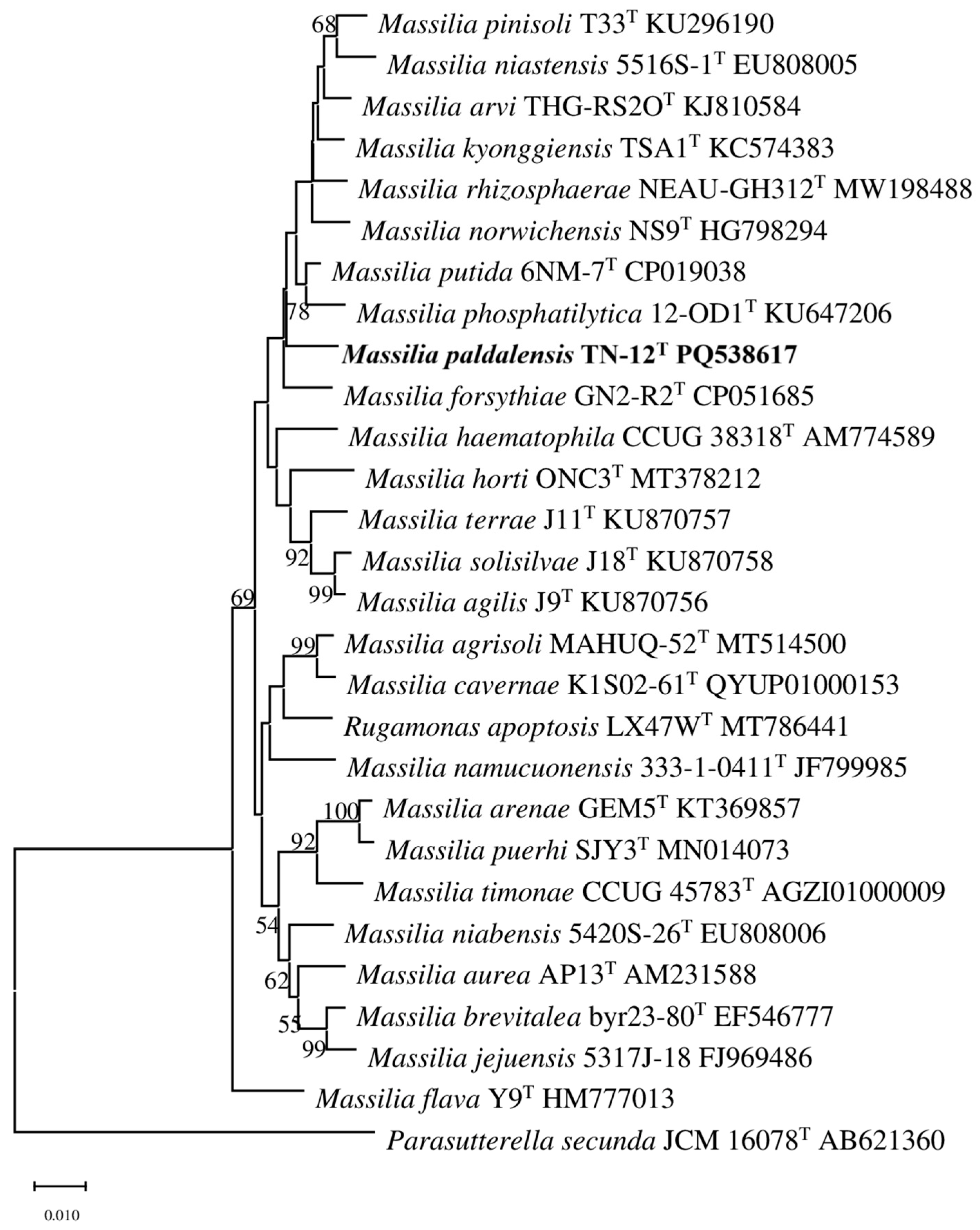
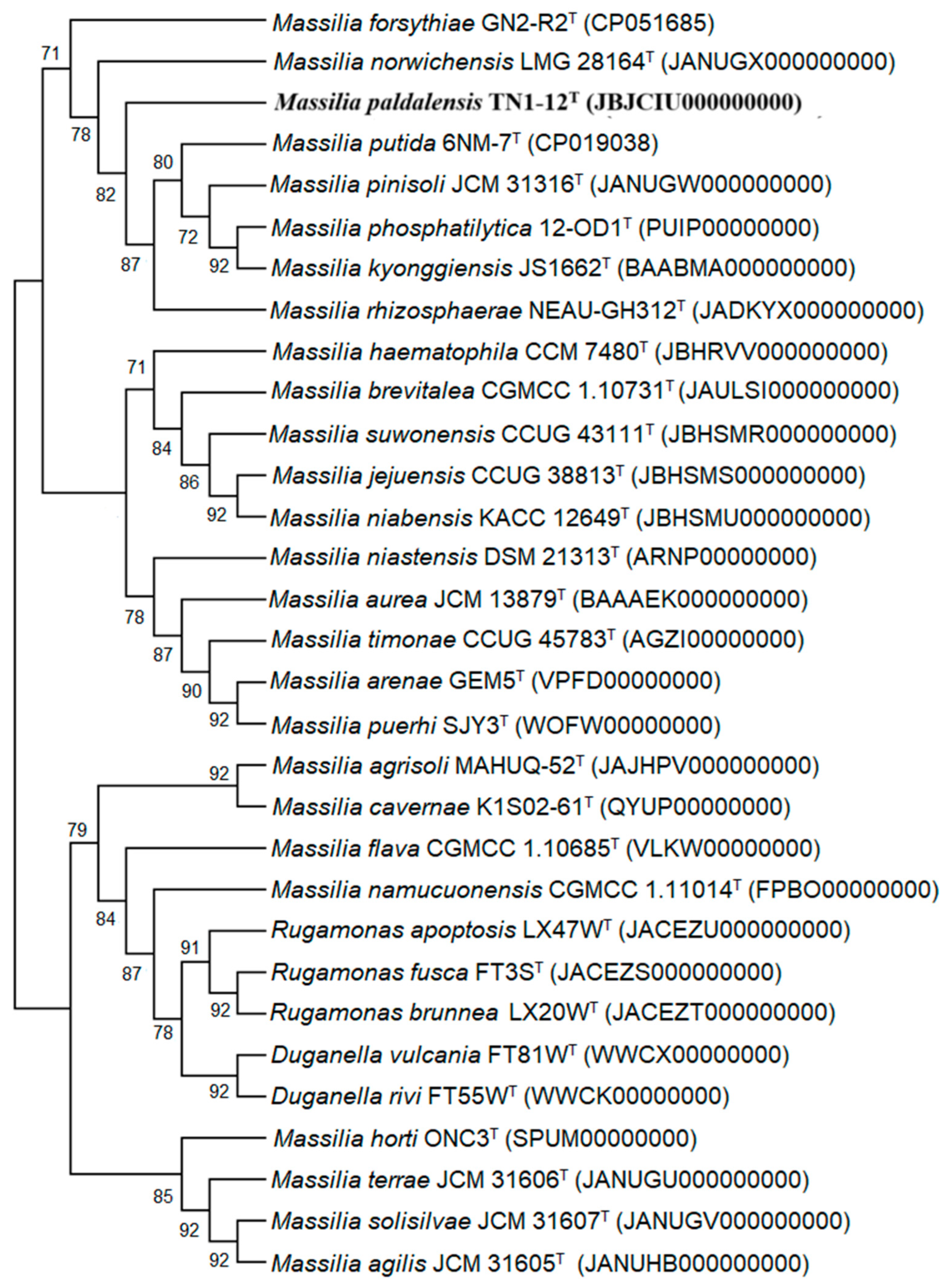
2.2. 16S rRNA Gene, Genome and Phylogenetic Analyses

Genomic DNA was extracted and purified by using the InstaGene Matrix Kit (Bio-Rad, Hercules, CA, USA) according to the kit protocol [11]. The 16S rRNA gene was PCR-amplified with the commercial bacterial primer (forward primer 27F and reverse primer 1492R) [12] and sequenced by Macrogen (Seoul, Republic of Korea). The closest phylogenetic neighbors of strain TN1-12T were identified by performing a search of validly published bacterial species using the EzBioCloud database (www.ezbiocloud.net, accessed on 23 November 2024). The retrieved closest related sequences along with TN1-12T were aligned by the SILVA alignment program (https://www.arb-silva.de/aligner/, accessed on 23 November 2024) [13]. Phylogenetic trees were reconstructed using the maximum-likelihood (ML), neighbor-joining (NJ) [14] and maximum-parsimony (MP) methods in the software MEGA X version 10.0.5 [15] with 1000 bootstrap replicates [16] and the Kimura two-parameter model [17]. Parasutterella secuda JCM 16078T was used as an outgroup for the phylogenetic tree.
Whole-genome shotgun sequencing of strain TN1-12T was accomplished by Macrogen (Republic of Korea) using the Illumina HiSeq 2500 platform (San Diego, CA, USA). Raw reads were quality checked using FastQC version 0.11.7 [18], filtered using Trimmomatic version 0.38 [19]. The genome was then assembled using SPAdes de novo version 3.15.0 [20] and the completeness of the genome assembly was assessed using BUSCO version 5.1.3 [21]. The UBCG (updated bacterial core genes) method was used to reconstruct a phylogenetic tree by concatenated alignments of 92 core genes [22]. The DNA G+C content and genome annotation were determined using the NCBI Prokaryotic Genome Annotation Pipeline (PGAP) version 6.8 [23] and the Rapid Annotation utilizing Subsystem Technology (RAST) server (https://rast.nmpdr.org/, accessed on 26 November 2024) [24]. To estimate pairwise relatedness between TN1-12T and close reference strains, Average Nucleotide Identity (ANI) was calculated utilizing the OrthoANIu (www.ezbiocloud.net/tools/ani, accessed on 23 December 2024) algorithm [25]. DNA–DNA relatedness was clarified using the GGDC web server (http://ggdc.dsmz.de/ggdc.php, accessed on 23 December 2024) [26]. Secondary metabolite biosynthesis gene clusters were analyzed by antiSMASH version 7 [27].

2.3. Physiology and Chemotaxonomy

After 24 h incubation at 30 °C, the cell of TN1-12T was examined by a zoom stereo microscope (SZ61, Olympus, Tokyo, Japan) and transmission electron microscopy (Bio-TEM H-7650, Hitachi, Tokyo, Japan). Growth of TN1-12T was observed on different media, including Reasoner’s 2A agar (R2A, MB Cell, Seoul, Korea), low salt Luria Bertani agar (LB; MB Cell), MacConkey agar (MB Cell), Nutrient agar (NA; MB Cell), and Tryptone Soya agar (TSA; MB Cell), at 30 °C for a week. Motility was assessed in sulfide indole motility (SIM, CM0435; Oxoid, Basingstoke, UK) medium. Growth under anaerobic conditions was checked in a BBL GasPak anaerobic jar (BD, Franklin Lakes, NJ, USA) at 30 °C for 7 days on R2A agar. The sodium chloride tolerance was evaluated by cultivating the strain on R2A broth at various NaCl concentrations (0–4%, in 0.5% increments) for a week at 30 °C. A growth test at different temperatures (4, 10, 15, 20, 25, 28, 30, 35. 37 and 40 °C) and a pH range of 4.0–10 (at intervals of 0.5 pH unit) was investigated in R2A broth for 7 days. For pH tests, the R2A broth was buffered with phosphate citrate buffer (pH 4.0–6.0), Sorensen’s phosphate buffer (pH 6.5–8.0), Tris buffer (pH 8.5–9.0), carbonate buffer (pH 9.5–10.0) and adjusted with NaOH or HCl. A series of phenotypic experiments, such as catalase and oxidase activities, gram staining, hydrolysis of casein, starch, gelatin, DNA, and Tween 80, were evaluated by using the methods of Smibert and Krieg [28]. Additional enzyme activities and carbon source utilization of TN1-12T and the reference strains were analyzed using API ZYM and API 20NE test kits (bioMérieux, Marcy-l’Étoile, France), following the manufacturers protocol. When comparing the physiological characteristics with related strains, the heavy metal resistance was tested by the agar plate method, strains inoculated on R2A agar supplemented with CdCl2.5H2O, CuSO4.5H2O and ZnSO4.7H2O at different concentrations (0, 0.2, 0.5, 1.0, 1.5, 2.0 mM) [7]. Growth was observed after 7 days at 30 °C.
For the determination of cellular fatty acids, strain TN1-12T and reference strains were harvested under the same conditions (at 30 °C for 2 days). The fatty acid compositions were analyzed with a GC system (Agilent 6890N, Santa Clara, CA, USA), according to the Sherlock Microbial Identification System (MIDI) protocol, with the TSBA6 database used for standard identification and quantification [29]. The lyophilized biomass of TN1-12T was used for the analysis of respiratory quinones and polar lipids. Quinones were extracted and analyzed according to the methods described by Minnikin et al. [30] and Hiraishi et al. [31] using a reversed-phase HPLC system. Polar lipids were extracted as previously described by Minnikin et al. [30]. The lipids were separated on thin-layer chromatography (TLC) and identified by spraying with detective reagents (ethanolic molybdatophosphoric acid (Sigma-Aldrich, St. Louis, MO, USA) for total lipids, molybdenum blue spray reagent (Sigma-Aldrich) for phospholipids, and ninhydrin (Sigma Life Science, Burlington, MA, USA) for aminolipids).

3. Results

3.1. 16S rRNA Gene, Genome and Phylogenetic Analyses

The 16S rRNA gene sequence had 1477 bp and has been deposited at GenBank database under the accession PQ538617. Strain TN1-12T was closest to M. putida 6NM-7T (98.21% sequence similarity), followed by M. forsythiae GN2-R2T (98.00%), M. rhizosphaerae NEAU-GH312T (97.79%), M. aurea AP13T (97.78%) and M. niabensis 5420S-26T (97.71%). All three phylogenetic trees clearly showed that strain TN1-12 is a member of the genus Massilia (Figure 1, Figures S1 and S2). The NB phylogenetic tree based on 16S rRNA gene sequences showed that strain TN1-12 is closely related to M. forsythiae GN2-R2T, M. phosphatilytica 12-OD1T, and M. putida 6NM-7T (Figure 1). This phylogenetic relationship was confirmed by the UBCG phylogenetic tree based on the whole genome. Strain TN1-12T was clustered with species in the genus Massilia and forms a clear clade (Figure 2).
The de novo assembled genome sequence of strain TN1-12T was found to be 6,225,423 bp long and composed of 37 contigs with an N50 of 1,047,983 bp, a G+C content of 66.7 mol%. High sequencing depth of 130.77 ensures genome assembly accuracy and reliability. Additionally, BUSCO analysis identified 98.39% of the expected single-copy orthologs, further confirming good assembly with minimal contamination. According to the PGAP annotation, the complete genome of strain TN1-12T was annotated to have 5578 protein-coding sequences (CDSs), 88 RNA genes (9 rRNA, 75 tRNA and 4 ncRNA genes) and 30 pseudo genes. Detailed genomic features are presented in the Table S1. The RAST analysis results showed that 23% (1307) of the genes were involved in essential biological processes, as detailed in Figure S3. The analysis revealed 55 genes indicating virulence, disease and defense, of which 41 genes were involved in antibiotic and toxic compound resistance (explaining the heavy metal resistance capacity of strain TN 1-12T). In addition, 5 genes related to secondary metabolisms consisting of one alkaloid biosynthesis from L-lysine, four auxin biosynthesis, and several genes responsible for flagella motility were identified. Based on the antiSMARH software, five BGCs were identified as follows: terpene, NAPAA, RiPP-like, acyl amino-acids and redox-cofactor (Figure S4). DNA–DNA hybridization results between strain TN1-12T and the close relatives M. putida KACC 21418T, M. forsythiae KACC 21261T, M. rhizosphaerae DSM 109722T, M. aurea KACC 11884T and M. niabensis KACC 12632T were 25.4, 25.5, 25.5, 21.4, and 21.9%, respectively. Strain TN1-12T revealed OrthoANI values of 77.21–82.22% compared with 15 related strains (Table S2). Both DNA–DNA relatedness and ANI values were all below the criteria for species delineation (threshold value of 70% for dDDH and 95–96% for ANI) [32]. These results supported strain TN1-12T being considered to be a new species of the genus Massilia.

3.2. Physiology and Chemotaxonomy

Strain TN1-12T was rod-shaped (0.4–0.8 × 2.0–3.8 μm) and motile by means of a single polar flagellum (Figure S5). Colonies were yellowish white, round, smooth with distinct margins and 0.2–1.0 mm in diameter when cultured for 1 day on R2A agar and turned yellow, irregular and rough after 2–3 days at 30 °C. This difference was also observed in the species M. tieshanensis KACC 14940T; these colonies were difficult to scrape after 2–3 days incubation [3]. Strain TN1-12T is characterized by Gram-negative staining, aerobic, catalase, motile, capable of hydrolyzing urea and arginine, similar to the closely related species. Besides some common features with its closest relatives, strain TN1-12T revealed some morphological, physiological and biochemical differences from related type strains. Growth of strain TN1-12T occurs at pH between 5.0 and 8.0 (optimum pH 7.0), temperature between 10 and 40 °C (optimum 30 °C) with 0–2% (w/v) NaCl (optimum 0%). Detailed differential characteristics from the closest type strain in the genus Massilia related to physiological, biochemical and morphological traits are provided in Table 1. The main distinctions are oxidase positive and hydrolytic activity of the different substrates (starch, casein, gelatin, esculin and Tween 80), as well as assimilation and enzyme activity properties. In addition, among the strains tested, strain TN1-12T had the highest resistance to 1.5 mM Cd2+, 1.5 mM Cu2+ and 2.0 mM Zn2+. The related strain M. putida KACC 21418T was resistant to 1.0 mM Cd2+, 0.5 mM Cu2+ and 0.5 mM Zn2+. M. forsythia KACC 21261T and M. niabensis KACC 12632T were hardly found to be resistant to heavy metals (Table S3). The metal resistance level of strain TN1-12T was found to be quite high when compared with other strains in previous reports. The strain had the highest Cd2+ tolerance (1.5 mM) in the genus Massilia, followed by M. timonae AY512824 (1 mM) [33]. Cu2+ tolerance was only lower than M. tieshanensis KACC 14940T [3] and Zn2+ tolerance was lower than M. timonae AY512824 [33]. These differences support the strain TN1-12T as a potential novel species within the genus Massilia.
The fatty acid result of strain TN1-12T and two reference strains are shown in Table 2. The major fatty acids (>10%) were partially similar to the characteristics of genus Massilia as follows: summed feature 3 (C15:0 iso 2-OH and/or C16:1 ω7c, 54.2%), C16:0 (31.5%), 17:0 cyclo (18.82%) and summed feature 8 (C18:1 ω7c and/or C18:1 ω6c, 10.41%). The proportions of anteiso-branched chain in M. forsythia KACC 21261T and some iso-branched chain in M. putida KACC 21418T, M. rhizosphaerae DSM 109722T, M. aurea KACC 11884T, and M. niabensis KACC 12632T are less than 0.5%. The difference in fatty acid proportions (Table 2) and the absence of anteiso- and iso-branched chain fatty acids distinguished strain TN1-12T from its reference strains. The major respiratory quinone of strain TN1-12T was ubiquinone 8 (Q-8), the same as a common feature among members of the genus. The main polar lipids consisted of diphosphatidylglycerol (DPG), phosphatidylglycerol (PG), phosphatidylethanolamine (PE), followed by phospholipid (PL), two aminophospho lipid (APL) and three lipids (L) (Figure S6).

4. Discussion

Strain TN1-12T exhibited major fatty acids, polar lipids, G+C content, quinones, and several characteristics (such as being Gram-negative, aerobic, and motile) similar to its closest relatives in the genus Massilia. However, its morphological, physiological, and biochemical features clearly differentiate it from reference strains. Notable differences include phenotypic changes after 2–3 days of cultivation and the absence of anteiso- and iso-branched chain fatty acids. Moreover, average nucleotide identity (ANI) and digital DNA–DNA hybridization (dDDH) values, based on species delineation thresholds, strongly support that strain TN1-12ᵀ represents a novel species within the genus Massilia, for which the name Massilia paldalensis sp. nov. is proposed. The discovery of this new soil species and its detailed taxonomic and genetic characterization contribute to a deeper understanding of microbial diversity in soil ecosystems.
Additionally, strain TN1-12T demonstrated remarkably high resistance to heavy metals compared to previously reported Massilia species (Table S3). Studying this resistance may offer insights into the strain’s ecological role in soil environments and its potential in bioremediation applications. The presence of genes related to antibiotic and toxic compound resistance indicates the strain’s adaptability to polluted environments and its possible capability to metabolize pollutants. Further research is required to experimentally confirm the mechanisms underlying its metal resistance and to investigate microbial interactions with soil and plants.
Moreover, the detection of secondary metabolism genes, including those involved in alkaloid biosynthesis from L-lysine and auxin biosynthesis, suggests that M. paldalensis may influence plant growth and provide various beneficial biological functions to host organisms. These characteristics indicate that Massilia paldalensis may play an essential role in maintaining ecosystem balance through pollutant detoxification and nutrient cycling. The identification of this novel bacterial species is a significant step toward expanding our knowledge of microbial ecology and discovering organisms with valuable applications in agriculture and environmental biotechnology.

Description of Massilia paldalensis sp. nov.

Massilia paldalensis (pal.dal.en’sis. N.L. fem. adj. paldalensis of or belonging to Paldal, referring to the Paldal district where the soil sample was collected).
Its characteristics include being an aerobic, rod-shaped (0.4–0.8 × 2.0–3.8 μm), Gram-stain-negative, oxidase-positive, catalase-positive, motile with a single polar flagellum, non-spore-forming bacterium. The colonies are yellowish white, circular, and smooth with entire edges on nutrient agar plates, and 0.2–1.0 mm in diameter after 24 h of incubation at 30 °C. Some differences are observed after 2–3 days of incubation, on which colonies are yellow, irregular and rough. Growth is found to occur at 10–40 °C (opt 30 °C), and at pH 5.0–8.0 (opt pH 7.0) in the presence of 0–2% (w/v) NaCl. Cells can grow on R2A agar, TSA agar, NA agar, LB agar but not on MacConkey agar. Urease reaction is negative. Hydrolyses of substrates, such as starch, casein, Tween 80, esculin and gelatin, can be conducted. β-galactosidase and nitrate reduction is positive but indole production, glucose fermentation, and arginine dihydrolase are negative. In API 20NE strips, the assimilation of d-glucose, l-arabinose, d-mannose, maltose, potassium gluconate, capric acid, malic acid and trisodium citrate is positive. The assimilation is negative for d-mannitol, N-acetyl-glucosamine, adipic acid, and phenylacetic acid. In API ZYM test strip, α-glucosidase, β-glucosidase, alkaline phosphatase, esterase (C4), lipase (C8), leucine arylamidase, valine arylamidase, acid phosphatase and naphthol-AS-BI-phosphohydrolase are positive, but α-galactosidase, β-galactosidase, lipase (C14), cystine arylamidase, trypsin, α-chymotrypsin, β-glucuronidase, N-acetyl-β-glucosaminidase, α-mannosidase and α-fucosidase are negative. The predominant ubiquinone is Q-8. The G+C content of the DNA is 66.7 mol%. The main polar lipids are diphosphatidylglycerol, phosphatidylglycerol, phospholipid, phosphatidylethanolamine, two aminophospho lipid and three lipids. The major fatty acids are C16:0, summed feature 3 (C16:1ω7c and/or iso-C15:0 2-OH), 17:0 cyclo and summed feature 8 (C18:1 ω7c and/or C18:1 ω6c).
The type strain TN1-12T (=KACC 23946T = CGMCC 1.65296T) was isolated from stream bank soil in Suwon City, Korea. The GenBank accession numbers are PQ538617 (16S rRNA) and JBJCIU000000000 (genome).

Supplementary Materials

The following supporting information can be downloaded at: https://www.mdpi.com/article/10.3390/d17050327/s1, Figure S1: Maximum-likelihood phylogenetic tree based on the 16S rRNA gene sequence showing the positions of strain TN1-12T and other reference strains. Parasutterella secuda JCM 16078T was used as an outgroup for the phylogenetic tree. Bar, 0.02 substitutions per nucleotide position; Figure S2: Maximum-parsimony tree showing the phylogenetic position of strain TN1-12T and its related taxa. Only bootstrap values over 50% (percentages of 1000 replications) are shown at branch points; Figure S3: Functional classes of protein-coding genes of Massilia paldalensis TN1-12T; Figure S4: The Prediction Informatics for Secondary Metabolomes of Massilia paldalensis TN1-12T; Figure S5: Transmission electron microscopy of Massilia paldalensis TN1-12T grown on R2A broth for 24 h at 28 °C; Figure S6: Polar lipids of strain TM1-12T after spraying 5% ethanolic phosphomolybdic acid (1); ninhydrin (2); molybdenum blue spray reagent (3); Table S1: General genome features of Massilia paldalensis TN1-12T with closely related strains; Table S2: The average nucleotide identity and digital DNA–DNA hybridization values between the genome sequences of Massilia paldalensis TN1-12T and closely related strains; Table S3: Minimum inhibitory concentrations (MICs) of three metal ions tested on bacterial strains TN1-12T and reference strain.

Author Contributions

N.T.A.N. conceived, designed, and conducted all of the experiments. N.T.A.N. and J.K. interpreted all of the data and read, discussed, edited, and approved the final draft of the manuscript. J.K. coordinated and supervised the study. All authors have read and agreed to the published version of the manuscript.

Funding

This work was supported by Kyonggi University’s Graduate Research Assistantship 2023.

Institutional Review Board Statement

Not applicable.

Data Availability Statement

The whole-genome sequence of Massilia paldalensis TN1-12T has been deposited in GenBank under the accession number JBJCIU000000000. The 16S rRNA gene sequence of strain TN1-12T is available in GenBank under the accession number PQ538617. All other relevant data supporting the findings of this study are available from the corresponding author upon reasonable request.

Conflicts of Interest

The authors declare no conflicts of interest.

References

  1. La Scola, B.; Birtles, R.J.; Mallet, M.N.; Raoult, D. Massilia timonae gen. nov., sp. nov., isolated from blood of an immunocompromised patient with cerebellar lesions. J. Clin. Microbiol. 1998, 36, 2847–2852. [Google Scholar] [CrossRef] [PubMed]
  2. Kämpfer, P.; Lodders, N.; Martin, K.; Falsen, E. Revision of the genus Massilia La Scola et al. 2000, with an emended description of the genus and inclusion of all species of the genus Naxibacter as new combinations, and proposal of Massilia consociata sp. nov. Int. J. Syst. Evol. Microbiol. 2011, 61, 1528–1533. [Google Scholar] [CrossRef] [PubMed]
  3. Du, Y.; Yu, X.; Wang, G. Massilia tieshanensis sp. nov., isolated from mining soil. Int. J. Syst. Evol. Microbiol. 2012, 62 Pt 10, 2356–2362. [Google Scholar] [CrossRef]
  4. Weon, H.-Y.; Kim, B.-Y.; Hong, S.-B.; Jeon, Y.-A.; Koo, B.-S.; Kwon, S.-W.; Stackebrandt, E. Massilia niabensis sp. nov. and Massilia niastensis sp. nov., isolated from air samples. Int. J. Syst. Evol. Microbiol. 2009, 59 Pt 7, 1656–1660. [Google Scholar] [CrossRef]
  5. Gallego, V.; Sánchez-Porro, C.; García, M.T.; Ventosa, A. Massilia aurea sp. nov., isolated from drinking water. Int. J. Syst. Evol. Microbiol. 2006, 56 Pt 10, 2449–2453. [Google Scholar] [CrossRef]
  6. Heo, J.; Won, M.; Lee, D.; Han, B.-H.; Hong, S.-B.; Kwon, S.-W. Duganella dendranthematis sp. nov. and Massilia forsythiae sp. nov., isolated from flowers. Int. J. Syst. Evol. Microbiol. 2022, 72, 005487. [Google Scholar] [CrossRef]
  7. Feng, G.-D.; Yang, S.-Z.; Li, H.-P.; Zhu, H.-H. Massilia putida sp. nov., a dimethyl disulfide-producing bacterium isolated from wolfram mine tailing. Int. J. Syst. Evol. Microbiol. 2016, 66, 50–55. [Google Scholar] [CrossRef] [PubMed]
  8. Li, C.; Cao, P.; Du, C.; Zhang, X.; Bing, H.; Li, L.; Sun, P.; Xiang, W.; Zhao, J.; Wang, X. Massilia rhizosphaerae sp. nov., a rice-associated rhizobacterium with antibacterial activity against Ralstonia solanacearum. Int. J. Syst. Evol. Microbiol. 2021, 71, 005009. [Google Scholar] [CrossRef]
  9. Dahal, R.H.; Chaudhary, D.K.; Kim, J. Genome insight and description of antibiotic producing Massilia antibiotica sp. nov., isolated from oil-contaminated soil. Sci. Rep. 2021, 11, 12864. [Google Scholar]
  10. Kour, D.; Kaur, T.; Devi, R.; Yadav, A.; Singh, M.; Joshi, D. Beneficial microbiomes for bioremediation of diverse contaminated environments for environmental sustainability: Present status and future challenges. Environ. Sci. Pollut. Res. 2021, 28, 24917–24939. [Google Scholar] [CrossRef]
  11. Weisburg, W.G.; Barns, S.M.; Pelletier, D.A.; Lane, D.J. 16S ribosomal DNA amplification for phylogenetic study. J. Bacteriol. 1991, 173, 697–703. [Google Scholar] [CrossRef] [PubMed]
  12. Frank, J.A.; Reich, C.I.; Sharma, S.; Weisbaum, J.S.; Wilson, B.A.; Olsen, G.J. Critical evaluation of two primers commonly used for amplification of bacterial 16S rRNA genes. Appl. Environ. Microbiol. 2008, 74, 2461–2470. [Google Scholar] [CrossRef]
  13. Pruesse, E.; Peplies, J.; Glöckner, F.O. SINA: Accurate high-throughput multiple sequence alignment of ribosomal RNA genes. Bioinformatics 2012, 28, 1823–1829. [Google Scholar] [CrossRef]
  14. Saitou, N.; Nei, M. The neighbor-joining method: A new method for reconstructing phylogenetic trees. Mol. Biol. Evol. 1987, 4, 406–425. [Google Scholar] [PubMed]
  15. Kumar, S.; Stecher, G.; Li, M.; Knyaz, C.; Tamura, K. MEGA X: Molecular Evolutionary Genetics Analysis across Computing Platforms. Mol. Biol. Evol. 2018, 35, 1547–1549. [Google Scholar] [CrossRef] [PubMed]
  16. Felsenstein, J. Confidence limits on phylogenies: An approach using the bootstrap. Evolution 1985, 39, 783–791. [Google Scholar] [CrossRef]
  17. Kimura, M. A simple method for estimating evolutionary rates of base substitutions through comparative studies of nucleotide sequences. J. Mol. Evol. 1980, 16, 111–120. [Google Scholar] [CrossRef]
  18. Andrews, S. FastQC: A quality control tool for high throughput sequence data. In Babraham Bioinformatics; Babraham Institute: Babraham, UK, 2010. [Google Scholar]
  19. Bolger, A.M.; Lohse, M.; Usadel, B. Trimmomatic: A flexible trimmer for Illumina sequence data. Bioinformatics 2014, 30, 2114–2120. [Google Scholar] [CrossRef]
  20. Bankevich, A.; Nurk, S.; Antipov, D.; Gurevich, A.A.; Dvorkin, M.; Kulikov, A.S.; Lesin, V.M.; Nikolenko, S.I.; Pham, S.; Prjibelski, A.D.; et al. SPAdes: A new genome assembly algorithm and its applications to single-cell sequencing. J. Comput. Biol. 2012, 19, 455–477. [Google Scholar] [CrossRef]
  21. Simão, F.A.; Waterhouse, R.M.; Ioannidis, P.; Kriventseva, E.V.; Zdobnov, E.M. BUSCO: Assessing genome assembly and annotation completeness with single-copy orthologs. Bioinformatics 2015, 31, 3210–3212. [Google Scholar] [CrossRef]
  22. Na, S.I.; Kim, Y.O.; Yoon, S.H.; Ha, S.M.; Baek, I.; Chun, J. UBCG: Up-to-date bacterial core gene set and pipeline for phylogenomic tree reconstruction. J. Microbiol. 2018, 56, 280–285. [Google Scholar] [CrossRef]
  23. Tatusova, T.; DiCuccio, M.; Badretdin, A.; Chetvernin, V.; Nawrocki, E.P.; Zaslavsky, L.; Lomsadze, A.; Pruitt, K.D.; Borodovsky, M.; Ostell, J. NCBI prokaryotic genome annotation pipeline. Nucleic Acids Res. 2016, 44, 6614–6624. [Google Scholar] [CrossRef] [PubMed]
  24. Aziz, R.K.; Bartels, D.; Best, A.A.; DeJongh, M.; Disz, T.; Edwards, R.A.; Formsma, K.; Gerdes, S.; Glass, E.M.; Kubal, M.; et al. The RAST Server: Rapid annotations using subsystems technology. BMC Genom. 2008, 9, 75. [Google Scholar] [CrossRef]
  25. Yoon, S.-H.; Ha, S.-M.; Lim, J.; Kwon, S.; Chun, J. A large-scale evaluation of algorithms to calculate average nucleotide identity. Antonie Van Leeuwenhoek 2017, 110, 1281–1286. [Google Scholar] [CrossRef]
  26. Meier-Kolthoff, J.P.; Auch, A.F.; Klenk, H.-P.; Göker, M. Genome sequence-based species delimitation with confidence intervals and improved distance functions. BMC Bioinform. 2013, 14, 60. [Google Scholar] [CrossRef] [PubMed]
  27. Blin, K.; Shaw, S.; Augustijn, H.E.; Reitz, Z.L.; Biermann, F.; Alanjary, M.; Fetter, A.; Terlouw, B.R.; Metcalf, W.W.; Helfrich, E.J.; et al. antiSMASH 7.0: New and improved predictions for detection, regulation, chemical structures and visualisation. Nucleic Acids Res. 2023, 51, W46–W50. [Google Scholar] [CrossRef] [PubMed]
  28. Smibert, R.M.; Krieg, N.R. Phenotypic characterization. In Methods for General and Molecular Bacteriology; Gerhardt, P., Murray, R.G.E., Wood, W.A., Krieg, N.R., Eds.; American Society for Microbiology: Washington, DC, USA, 1994; pp. 607–654. [Google Scholar]
  29. Sasser, M. Identification of bacteria by gas chromatography of cellular fatty acids. Tech. Note 1990, 101, 1–6. [Google Scholar]
  30. Minnikin, D.E.; O’donnell, A.G.; Goodfellow, M.; Alderson, G.; Athalye, M.; Schaal, A.; Parlett, J.H. An integrated procedure for the extraction of bacterial isoprenoid quinones and polar lipids. J. Microbiol. Methods 1984, 2, 233–241. [Google Scholar] [CrossRef]
  31. Hiraishi, A.; Ueda, Y.; Ishihara, J.; Mori, T. Comparative lipoquinone analysis of influent sewage and activated sludge by high-performance liquid chromatography and photodiode array detection. J. Gen. Appl. Microbiol. 1996, 42, 457–469. [Google Scholar] [CrossRef]
  32. Wayne, L.G. International Committee on Systematic Bacteriology: Announcement of the report of the ad hoc Committee on Reconciliation of Approaches to Bacterial Systematics. Zentralblatt Bakteriol. Mikrobiol. Hygiene A 1988, 268, 433–434. [Google Scholar]
  33. Abou-Shanab, R.A.; van Berkum, P.; Angle, J.S. Heavy metal resistance and genotypic analysis of metal resistance genes in gram-positive and gram-negative bacteria present in Ni-rich serpentine soil and in the rhizosphere of Alyssum murale. Chemosphere 2007, 68, 360–367. [Google Scholar] [CrossRef] [PubMed]
Figure 1. Neighbor-joining tree created using mega X based on 16S rRNA gene sequences showing relationships between strain TN1-12T (bold; a novel type strain as a new species) and other related species. Only bootstrap values above 50% (expressed as percentages of 1000 replications) are shown. Bar, 0.01 nucleotide exchanges per site.
Diversity 17 00327 g001
Figure 2. Genome-based phylogenetic tree is reconstructed using the UBCG showing the phylogenetic placement of the strain TN1-12T (bold; a novel type strain as a new species) with closely related species of the genus Massilia. GenBank accession numbers are given in parentheses. Bar, 0.1 substitutions per site.
Diversity 17 00327 g002
Table 1. Differential characteristics of strain TN1-12T and closely related species: 1, strain TN1-12T; 2, M. putida KACC 21418T; 3, M. forsythiae KACC 21261T; 4, M. rhizosphaerae DSM 109722T; 5, M. aurea KACC 11884T; 6, M. niabensis KACC 12632T. Data in columns 1–3, 5–6 are from this study except where marked. Data for 4, M. rhizosphaerae DSM 109722T are from Chenxu Li et al. (2021) [8]. +, Positive; −, negative; ND, not determined.
Characterictics123456
Isolation sourceSoilWolfram mine
tailing [7]
Flowers [6]Rhizosphere soil of riceDrinking water [5]Air [4]
MediaR2A, TSA, NA, LBR2A, PYE [7]R2A [6]R2A, NA, ISP2PCA, R2A, TSB, NA [5]R2A, NA [4]
Oxidase++++
Catalase+++ND++
Urea
NaCl tolerance (%, w/v)0–20–10–10–50–20–1
Temperature for growth10–4020–3710–3010–4010–3010–35
pH (optimum)5–8 (7)6–8 (7)4.5–8.5 (7)4–8 (7)4.5–9 (7.5)6.5–9 (7)
Hydrolysis of:
Casein+ND+
Starch+++++
Gelatin+++
Esculin+++
Tween 80++
Assimilation of:
d-Glucose+++++
l-Arabinose+++
d-Mannose++++
d-Mannitol
N-acetyl-glucosamine++
Maltose+++++
Potassium gluconate+++
Capric acid++
Adipic acid++
Malic acid++++
Trisodium citrate++
Phenylacetic acid+
Enzyme activities:
Lipase (C14)+
Cystine arylamidase+
Trypsin++
α-chymotrypsin+++
α-galactosidase++
β-galactosidase+++
β-glucuronidase+
β-glucosidase+++++
N-acetyl- β-glucosaminidase+++
α-mannosidase+
DNA G+C content (mol%)66.766.8 [7]66.5 [6]66.366.0 [5]67.8 [4]
Table 2. Fatty acid compositions of strain TN1-12T and two closely related species. Strain: 1, TN1-12T; 2, M. putida KACC 21418T; 3, M. forsythiae KACC 21261T; 4, M. rhizosphaerae DSM 109722T; 5, M. aurea KACC 11884T; 6, M. niabensis KACC 12632T. Data in columns 1–3, 5–6 are from this study. Data for 4, M. rhizosphaerae DSM 109722T are from Chenxu Li et al. (2021) [8]. −, <0.5% or not detected.
Fatty Acid123456
10:00.620.56
10:0 3OH6.093.694.835.73
12:03.731.452.278.32
12:0 3OH0.880.5
14:01.793.522.681.33
14:0 2OH3.272.312.93
14:0 iso0.7
15:0 iso0.89
15:0 anteiso14.83
16:031.5026.2130.8239.77.2622.55
16:0 iso1.416.46
17:0 anteiso17.22
17:0 iso2.49
17:0 cyclo18.826.69
17:1 w8c0.59
18:00.56
18:1 w9c7.86
18:1 w7c 11-methyl1.31
Summed Feature 3 *23.8526.3746.5047.93.1651.10
Summed Feature 8 **10.4126.069.379.825.0810.83
* Summed feature 3 consists of C16:1 w7c/C16:1 w6c; ** Summed feature 8 consists of C18:1 w7c/C18:1 w6c.
Disclaimer/Publisher’s Note: The statements, opinions and data contained in all publications are solely those of the individual author(s) and contributor(s) and not of MDPI and/or the editor(s). MDPI and/or the editor(s) disclaim responsibility for any injury to people or property resulting from any ideas, methods, instructions or products referred to in the content.

