Article

Ecological Role of the Clam Jukesena foveolata (Bivalve, Cyamiidae) Inferred by Digenean Parasites in Subantarctic Waters

by
Cecilia Trani
1,2,
Cintia Debora Medina
3,
Florencia Cremonte
2 and
Carmen Gilardoni
2,4,*
1
Laboratory of Genomic and Bioinformatic of Pathogens, Institute of Biosciences, Biotechnology and Translational Biology, Department of Physiology, Molecular and Cellular Biology, Faculty of Exact and Naturals Sciences, University of Buenos Aires, Intendente Güiraldes 2160, Buenos Aires 1428, Argentina
2
Laboratory of Parasitology (LAPA), Institute of Biology of Marine Organisms (IBIOMAR), Centro Nacional Patagónico (CCT CONICET-CENPAT), Boulevard Brown 2915, Puerto Madryn 9120, Argentina
3
Institute of Austral Diversity and Evolution, Centro Nacional Patagónico (CCT CONICET-CENPAT), Boulevard Brown 2915, Puerto Madryn 9120, Argentina
4
Department of Zoology, Faculty of Science, University of South Bohemia, Branišovská 1760, 370 05 České Budějovice, Czech Republic
*
Author to whom correspondence should be addressed.
Diversity 2025, 17(4), 233; https://doi.org/10.3390/d17040233
Submission received: 22 February 2025 / Revised: 20 March 2025 / Accepted: 20 March 2025 / Published: 25 March 2025
(This article belongs to the Section Marine Diversity)

Abstract

:
Bivalves serve as the first intermediate hosts in the life cycles of some digeneans, but little is known about their larval stages along the southwestern Atlantic coast. These larvae may provide valuable insights into the ecological role of mollusks in parasite life cycles and marine food webs. This study offers a morphological, histological, and molecular (ITS2, 28S rDNA) description of the larval stages of three digenean species parasitizing the bivalve Jukesena foveolata in the Beagle Channel, Tierra del Fuego Province, Argentina. Sporocysts containing cercariae of a monorchiid species were found infecting the gonads and digestive glands (p = 2%), while metacercariae were encysted in the foot (p = 7%). Additionally, metacercariae of Renicola sp. (Renicolidae) were located between the tubules of the digestive gland (p = 2%), and metacercariae of Bartolius sp. (Gymnophallidae) were found in the extrapallial space (between the mantle and the valve, p = 13.5%), where they elicited a histopathological reaction with the secretion of a calcareous, igloo-shaped coating. The monorchiid larvae are described here for the first time in this bivalve, with fish as their definitive hosts. Metacercariae of Renicola sp. and Bartolius sp., both of which use coastal birds as definitive hosts, have been previously recorded in other mollusks from the Argentine coast. This study contributes to our understanding of the life cycles of digeneans from the southwestern Atlantic coast and the role of bivalve hosts in marine food webs.

1. Introduction

Mollusks, particularly gastropods, have been identified as primary intermediate hosts for digeneans. However, some marine families use bivalves as the first intermediate hosts [1,2]. To date, the digenean families known to use marine bivalves as the first intermediate hosts include Bucephalidae Poche, 1907, Aporocotylidae Odhner, 1912, Monorchiidae Odhner, 1911, and Fellodistomidae Nicoll, 1909, whose definitive hosts are fish [3], as well as Gymnophallidae, whose definitive hosts are coastal birds [4]. While most digenean species have complex life cycles, in many cases, only part of the cycle is known due to the difficulty in distinguishing diagnostic morphological characters in the larvae [3]. However, molecular tools now allow us to determine whether different developmental stages in different hosts belong to the same species [5]. Ribosomal DNA (rDNA) is highly repetitive and contains variable regions (ITS) that are useful for species identification, alternating with conserved regions (18S, 28S) that are suitable for phylogenetic studies [6]. In recent years, these markers have been widely used to study digeneans, e.g., [7,8].
The clam Jukesena foveolata (Cooper & Preston, 1910) is a small bivalve (2–5 mm) first described in the Malvinas (Falkland) Islands and more recently recorded along the coasts of Tierra del Fuego Province, Argentina, and New Zealand [9]. However, knowledge of its biology, ecology, and geographical distribution is still limited. The aim of this study is to describe the parasites found in J. foveolata from the Beagle Channel, Tierra del Fuego Province, located on the Southwestern Atlantic coast, using both morphological and molecular analyses. It also aims to determine the infection sites and identify any lesions through histological examination. This research contributes to our understanding of the life cycles of digeneans and their role in the biology and ecology of hosts along the South Atlantic coast.

2. Materials and Methods

2.1. Study Site and Sample Collection

During the summer of 2018, two sediment samples were collected from a sandy intertidal area at Los Yámanas Beach, Beagle Channel, Tierra del Fuego Province (54°50′17.06″ S, 68°21′40.89″ W) using a fine sieve with a mesh size of approximately 500 µm. The samples were transported to the laboratory, where 700 live clams and 200 hemi-valves of Jukesena foveolata were separated. The average length of the clams was 3 mm, ranging from 2 to 5 mm. A total of 200 live clams were conditioned in aquariums in a temperature-controlled chamber (4 °C) until examination. Another 200 clams were preserved in 96% ethanol for molecular studies, while the remaining 300 clams were immediately fixed in a 10% seawater formalin solution for histological and morphological analysis

2.2. Parasites’ Morphological Study

The live clams were dissected using a Gillette blade, and their valves and visceral mass were examined under a stereomicroscope and a light microscope. The parasites found were isolated, stained with neutral red, and then fixed. Their morphology was studied while alive and photographed using a light microscope equipped with a camera. The developmental stages (sporocysts, cercariae, and metacercariae) were fixed in a 10% formalin solution, stained with acetic carmine, dehydrated through an ascending ethanol series, cleared with methylsalicylate, and mounted on glass slides with Canada balsam. Drawings were made with the aid of a drawing attachment, and photographs were taken with a Leica DFC280 camera (Leica Geosystems AG, Heerbrugg, Switzerland) connected to the microscope via software. Measurements are provided in micrometers (μm), with the mean followed by the range in parentheses. Specimens of each species were deposited in the Parasitological Collection at the Instituto de Biología de Organismos Marinos (CNP-Par), CCT CONICET-CENPAT, Puerto Madryn, Argentina. For each parasite species, infection indices, including prevalence, mean infection intensity, and mean abundance, were calculated following the method of Bush et al. [10].

2.3. DNA Extraction, PCR Amplification, and Sequencing

Specimens of each parasite species, stored in 96% ethanol, were selected for molecular studies. DNA extraction was performed on two specimens of each digenean species (sporocyst and/or metacercaria) using the GenElute Mammalian Genomic DNA Miniprep Kit (Sigma, St. Louis, MO, USA), following the manufacturer’s instructions. The ITS1 and 28S regions of the ribosomal DNA (rDNA) were amplified by PCR. The PCR reactions were carried out in a total volume of 50 µL, containing 10× buffer (200 mM Tris–HCl pH 8.4, 500 mM KCl), 0.2 mM of each dNTP, 1.5 mM MgCl2, 0.4 μM of each primer, and 1 U of platinum Taq polymerase. A total of 2 μL of genomic DNA was used as the template. The ITS1 region was amplified using the forward primer 18S-ITS1: 5′-CCGTCGCTACTACCGATTGAA-3′ and the reverse primer 5.8S-ITS1: 5′-CGCAATGTGCGTTCAAGATGTC-3′. The 28S region was amplified using the forward primer 28S-28S: 5′-GTGAATACCCGCTGAACTTAAGC-3′ and the reverse primer 28S-28S: 5′-TCTCCTTGGTCCGTGTTTCAA-3′ [4]. The cycling conditions included an initial denaturation at 94 °C for 5 min, followed by 40 cycles of 30 s at 94 °C, 30 s at 54 °C (annealing) for ITS1 and 52 °C for 28S, and 2 min at 72 °C, with a final extension step of 10 min at 72 °C. Amplified PCR products were electrophoretically separated in a 1% agarose gel stained with Green Gel. Negative controls were always included in the PCR to monitor for contamination. Amplicons from positive bands were sent to Macrogen Korea Inc. (Seoul, Republic of Korea) for purification and sequencing using the Sanger method. Sequences were cleaned and aligned using FinchTV 1.4.0 and MultiAlin [11]. The consensus sequences of the ITS1 and 28S regions have been deposited in GenBank (see accession numbers in the Taxonomic Summary section).

2.4. Phylogenetic Analysis

The partial ITS1 and 28S sequences obtained were analyzed along with other sequences from the same family available in GenBank (Table S1). Concatenated alignments were performed using MAFFT Version 7 software [12] and MEGA X [13]. Phylogenetic and molecular evolutionary analyses were conducted on the aligned nucleotide sequences of 28S using both the maximum likelihood (ML) method with the online W-IQTREE tool and Bayesian inference (BI) with BEAST 1.8.0 software [14]. Additionally, these analyses were performed for the ITS1 sequences of the Monorchiidae family, as this sequence failed to amplify for parasites from the Renicolidae and Gymnophallidae families. For the ML analyses, the selected substitution models were: HKY + I + G for the 28S and ITS1 trees of the Monorchiidae family and GTR + F + G4 for the 28S trees of the Renicolidae and Gymnophallidae families [15]. Node support was assessed using the ultrafast bootstrap test with 1000 replications [16].
For the BI analyses, the most appropriate evolutionary models were determined using the corrected Akaike information criterion in the jModelTest 2.1.1 software [17]. The selected models for each analysis were: GTR + G for the 28S trees of Renicolidae and Gymnophallidae, TVM + G for the ITS1 tree of Monorchiidae, and GTR + I + G for the 28S tree of Monorchiidae. The BI analysis was run for 10,000,000 generations (ngen = 10,000,000) with two runs, each containing four simultaneous Markov Chain Monte Carlo (MCMC) chains (nchains = 4), discarding 10% of the initial chains as burn-in (burnin = 1000). A tree was constructed with a consensus greater than 50%. Model parameter estimates were evaluated using Tracer v1.5 software. Node support is presented as posterior probability. For each analysis, the tree was rooted using species from closely related families as outgroups (Table S1). Both the ML and BI trees were visualized using FigTree v1.4.4 software [18].

2.5. Histological Studies

The 300 clams fixed in 10% formalin were subjected to decalcification in formic acid for 7 days, then repeatedly washed in tap water. They were dehydrated through an ascending ethanol series and processed for routine histological studies [19]. Sections 6 µm thick were stained with hematoxylin and eosin (H&E) and examined under a light microscope.

3. Results

Larval stages of three digenean species were found in the 200 live clams analyzed. Sporocysts containing cercariae of Monorchiidae gen. et sp. were recorded in the gonad and digestive gland, while metacercariae of the same species were found in the foot. Two other digeneans in the metacercaria stage were also identified: Renicola sp. (Renicolidae) in the digestive gland, and Bartolius sp. (Gymnophallidae) in the extrapallial space (i.e., between the mantle and valve).

3.1. Family Monorchiidae Odhner, 1911

Monorchiidae gen. et sp.

3.1.1. Description

Sporocyst (measurements based on four fixed specimens) (Figure 1A): Colorless, thin-walled, ovoid to elongated, 345 (367–399) µm long and 113 (110–126) µm wide. Mother sporocysts filled with germinal balls and daughter sporocysts containing germinal balls 10 (5–12) and cercariae 5 (4–8) in different developmental stages.
Cercaria (measurements based on 10 fixed specimens) (Figure 1B,C): Body elongated, 184 (118–224) µm long by 49 (40–59) µm wide, uniformly covered with spines. Seven pairs of penetration glands with cytons located close to the caecal bifurcation. Oral sucker opening subterminally, (25–43) µm long and 28 (20–34) µm wide. Ventral sucker, 30 (21–39) µm long by 30 (26–37) µm wide. Pre-pharynx very short 13 (8–17) µm long. Pharynx well developed, 14 (10–16) µm long and 13 (11–17) µm wide. Caeca reaching close to half of the hindbody. Prominent cystogenous cells scattered through body parenchyma. Genital primordia present, located between the ventral sucker and the proximal part of the excretory vesicle. Testes ovoid, located anteriorly to the excretory vesicle. Ovary pretesticular. Excretory bladder oval shaped, with thick wall and narrow lumen. Tiny and spiny knob tail, 20 (16–31) µm long by 14 (10–23) µm wide.
Metacercaria (measurements based on 10 fixed specimens) (Figure 1D): Cyst rounded, translucent and membranous, 90 (67–131) µm in diameter; thin wall 4 (3–5) µm thick. Oral sucker 26 (18–45) µm long by 23 (18–32) µm wide. Ventral sucker, 30 (27–36) µm long by 29 (25–33) µm wide.

3.1.2. Taxonomic Summary

Prevalences: 2% (4/200, sporocyst), 7% (14/200, metacercaria)
Mean intensity: 1.38 (1–2) (metacercaria)
Infection sites: gonad and digestive gland (sporocysts with cercariae) and foot (metacercaria)
Collection numbers: CNP-Par 207 (metacercaria in histological section), CNP-Par
208 (whole mounted cercariae)
GenBank accession numbers: OQ297754.1 (sporocyst), OQ297756.1 (metacercaria)

3.1.3. Molecular Analysis

The sequence encoding the ITS1 region yielded products of 643 bp (cercaria) and 530 bp (metacercaria), while the sequence encoding the 28S region provided a product of 838 bp (metacercaria). For the 28S region, both Maximum Likelihood (ML) and Bayesian Inference (BI) analyses resulted in well-supported trees with identical topologies. In both trees, species within the Monorchiidae family formed a monophyletic group, distinct from the outgroup Bulbocirrus aulostomi Yamaguti, 1965. The species described here clustered in a well-supported clade alongside Proctotrema sp. from Gaimardia trapesina (Lamarck) and Postmonorcheides maclovinus from Lasaea adansoni (Gmelin) in Argentinean Patagonia. Two other species of Proctotrema, P. adansoni and P. prominens, formed a separate clade distant from the studied species, also recorded along the Patagonian coast (Figure 2A). For the ITS1 region, both ML and BI trees exhibited similar topologies with strong support (Figure 2B). The larval stage sequences (cercaria and metacercaria) were identical and grouped in a well-supported clade with Proctotrema sp. from G. trapesina, Proctotrema bartolii from the clam Darina solenoides, and Postmonorcheides maclovinus from the clam Lasaea adansoni, all from the Patagonian coast.

3.1.4. Histopathology

The sporocysts invaded the digestive gland and gonad, leading to complete castration. They were typically found in the connective tissue between the digestive gland tubules and surrounding the gonadal acini. These tubules and/or acini were compressed, deformed, and ruptured, with the tissue replaced by sporocysts (Figure 3A). Metacercariae were located within the foot musculature, and a hemocytic infiltration was observed around the cyst (Figure 3C).

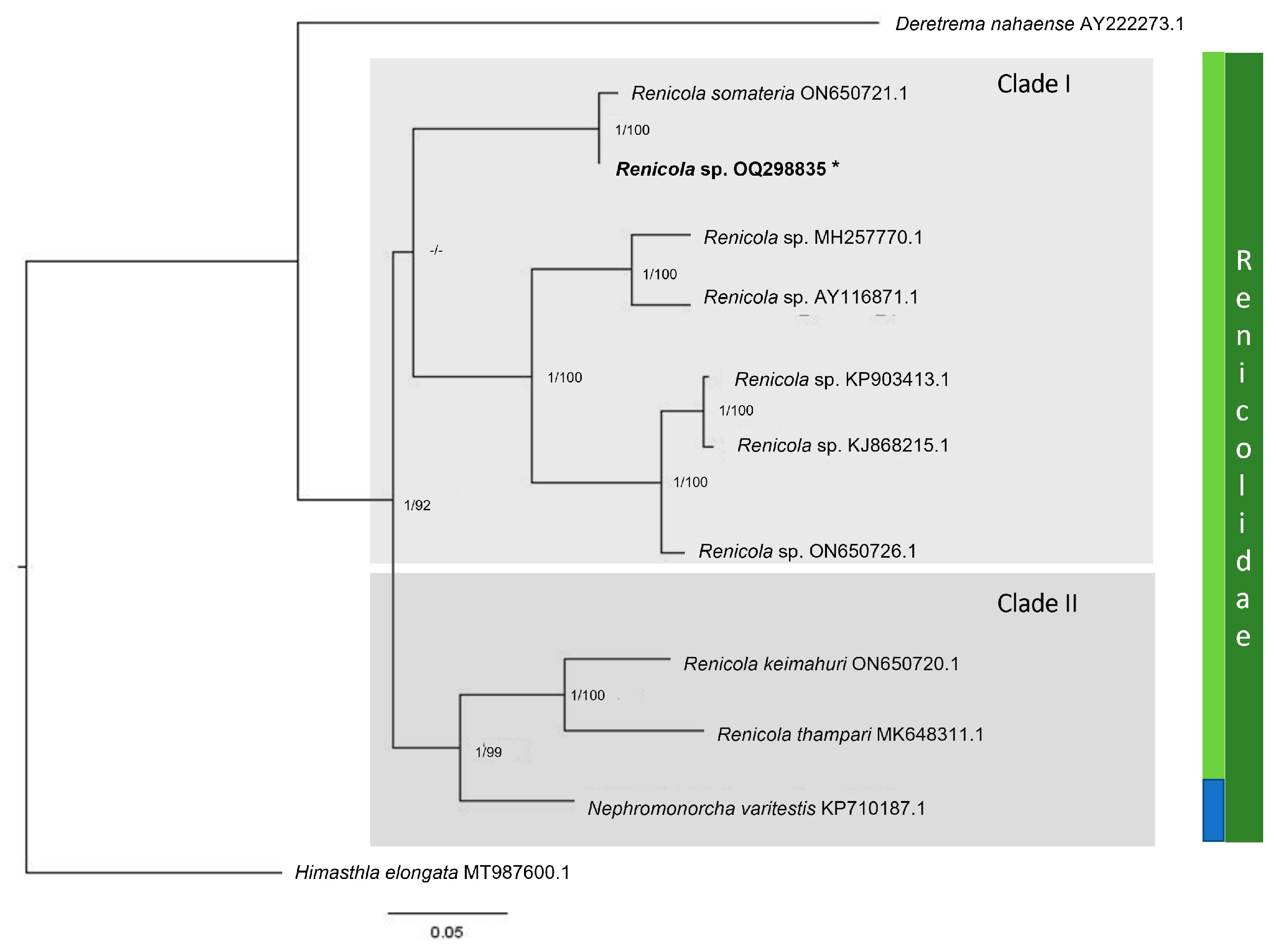
3.2. Renicolidae Dollfus, 1939

Renicola Cohn, 1904
Renicola sp.

3.2.1. Description

Metacercaria (measurements based on four fixed specimens) (Figure 1E): Cyst rounded and refringent, 132 (96–190) µm in diameter, thick and strong wall, 111 (99–120) µm in thickness. An oral sucker, ventral sucker, and a large excretory vesicle with dark excretory granules are observed.

3.2.2. Taxonomic Summary

Prevalence: 2% (4/200)
Mean intensity: 1
Infection site: Digestive gland
Collection number: CNP-Par 206 (metacercaria in histological section)
GenBank accession number: OQ298835.1

3.2.3. Molecular Analysis

The 28S rDNA yielded a single product of 852 bp. Both Maximum Likelihood (ML) and Bayesian Inference (BI) analyses produced trees with identical topologies (Figure 4). The Renicolidae family formed a monophyletic clade distinct from the two outgroup species, Deletrema naharnce Yamaguti, 1942, and Himasthla elongata (Mehlis, 1831). Both analyses revealed two distinct clades: Clade I includes seven Renicola species, while Clade II consists of two Renicola species and one species from the genus Nephromonorcha Leonov, 1958. The species described here belongs to Clade I and is most closely related to Renicola somateria Belopol’skaja, 1952 (Figure 4).

3.2.4. Histopathology

The metacercaria were found encysted among the digestive gland tubules without encapsulation or any host reaction (Figure 3D).

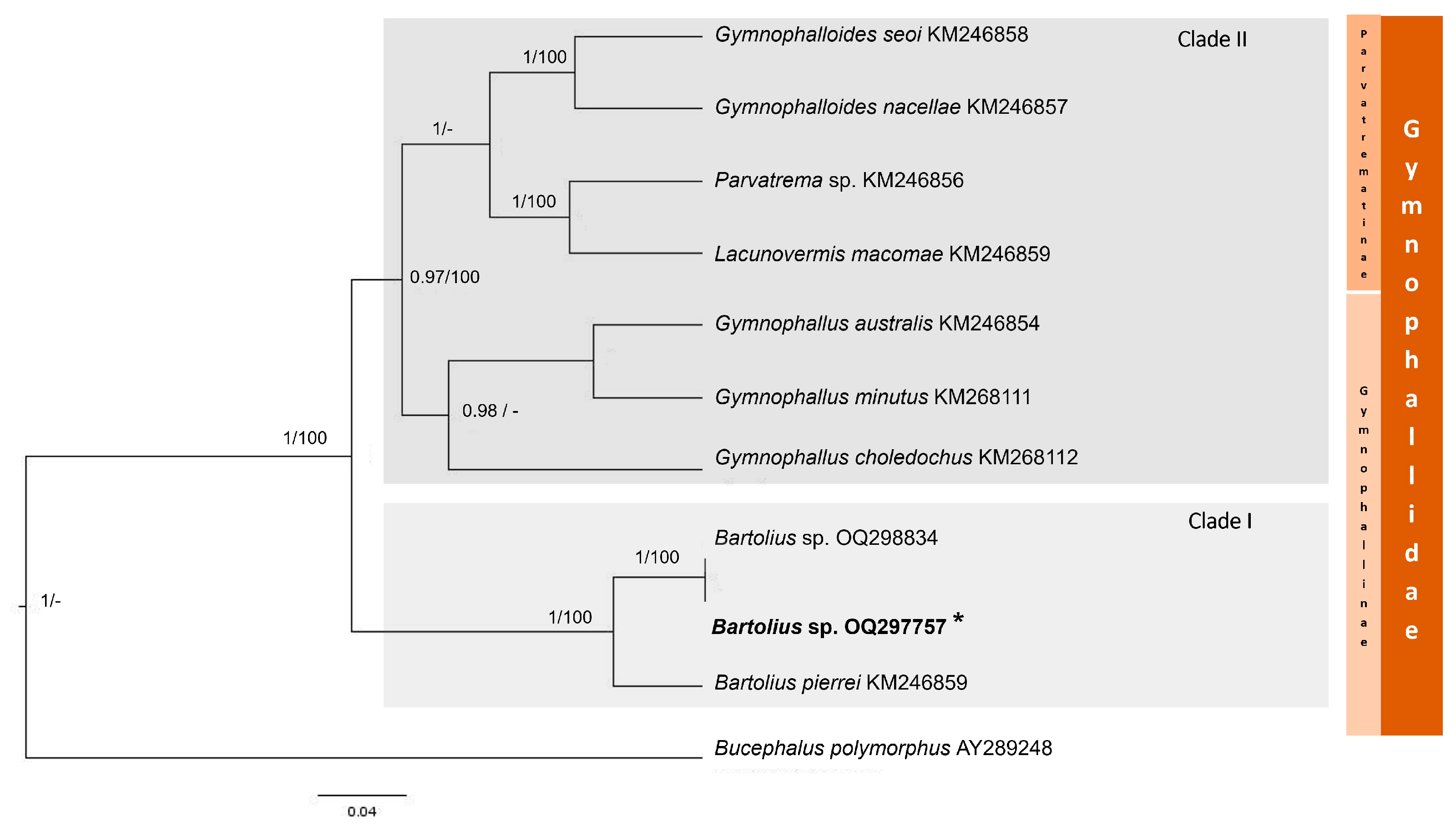
3.3. Family Gymnophallidae Odhner, 1905

Genus Bartolius Cremonte, 2001
Bartolius sp.

3.3.1. Description

Metacercaria (measurements based on 10 fixed specimens) (Figure 1F,G): Body small, elongated, and spinose, 250 (206–290) µm long by 109 (85–142) µm wide. Oral sucker subterminal, large, 84 (64–107) µm long by 69 (54–79) µm wide. Pre-pharynx absent; developed pharynx, 30 (21–33) µm long by 25 (21–28) µm wide. Caeca short, 97 (80–140) µm long by 35 (25–48) µm wide, without dorsal diverticula. Ventral pit absent. Ventral sucker 37 (29–45) µm long by 37 (30–42) µm wide, located in the middle third of the body. Seminal vesicle and pars prostatica absent. Testes opposite or slightly diagonal, located at the level of, or slightly posterior to, the ventral sucker. Left testis, 19 (12–25) µm long by 21 (15–25) µm wide, and right testis, 17 (12–24) µm long by 17 (12–22) µm wide. Ovary post-testicular, 5 (10–21) µm long by 18 (9–28) µm wide.

3.3.2. Taxonomic Summary

Prevalence: 13.5% (27/200)
Mean intensity: 1
Infection site: extrapallial space (between the shell and the mantle)
Collection number: CNP-Par 209 (whole mounted metacercariae)
GenBank accession number: OQ297755.1

3.3.3. Molecular Analysis

The 28S rDNA yielded a product of 724 bp. Maximum Likelihood (ML) and Bayesian Inference (BI) analyses produced well-supported trees with identical topologies (Figure 5). In both trees, species from the Gymnophallidae family form a monophyletic group, distinct from the outgroup Bucephalus polymorphus von Baer, 1827. Additionally, both analyses revealed two clades (posterior probability BI: 1.0; bootstrap ML: 100%). Clade I includes two species of the genus Bartolius, both recorded along the southwestern Atlantic coast. Clade II contains two subclades: one with species of Gymnophallus and the other with species of Gymnophalloides, Parvatrema, and Lacunovermis, all of which belong to the subfamily Parvatrematinae. The genera Gymnophallus and Bartolius are part of the subfamily Gymnophallinae. The species described here shows 100% similarity with the 28S sequence of Bartolius sp. recorded in the clam Gaimardia trapesina along the Patagonian coast.

3.3.4. Histopathology

The presence of Bartolius sp. metacercariae in the extrapallial space induced alterations (hyperplasia and metaplasia) in the outer mantle epithelium, where a calcareous, igloo-shaped covering was secreted, surrounding each larva (Figure 3E,F). The calcareous covering consistently displayed a lateral, moderately flattened, circular opening through which the anterior end of the larva was exposed.

4. Discussion

This study provides the first record of three species of larval digeneans infecting the little-known clam Jukesena foveolata, including sporocysts with cercariae and metacercariae of Monorchiidae gen. et sp., metacercariae of Renicola sp. (Renicolidae), and Bartolius sp. (Gymnophallidae). These findings suggest that the clam serves as both the first and second intermediate host in the life cycle of the monorchiid species, and as a second intermediate host for the renicolid and gymnophallid species. A comprehensive understanding of larval digeneans is crucial for elucidating parasite life cycles and provides insights into the role of hosts in food webs. The monorchiid species uses fish as definitive hosts [20], while renicolids and gymnophallids utilize birds [21,22]. As a result, J. foveolata emerges as a key prey species for coastal fishes and birds that forage in the sandy intertidal zone of the Beagle Channel. These findings contribute to the growing body of knowledge on digenean parasites along the southwestern Atlantic coast, particularly in the southernmost tip of Patagonia, where research has been limited, e.g., refs. [5,23].
The first species described, the monorchiid cercaria, was identified based on the characteristics outlined by Cable [24] and Schell [25], which include: a distome body shape, pharyngeate structure, spinose tegument, the presence or absence of eyespots, absence of a stylet, a non-thin-walled excretory vesicle, and a long, slender tail that is either shorter, collar-like, or features a tiny knob or brevifurcate structure. Cremonte et al. [26] classified monorchiid cercariae into four morphological groups. The first group (1) includes cercariae with a well-developed tail, usually ocellate; the second group (2) consists of cercariae with shorter or collar-like tails; the third group (3) comprises cercariae with a short furca and no tail stem; and the fourth group (4) includes cercariae with a tiny knob tail and no ocelli. The cercaria described here belongs to the fourth group, which also includes two species found on the Patagonian coast: a monorchiid cercaria in the clam Eucallista purpurata [26] and Proctotrema bartolii Carballo, Laurenti & Cremonte, 2011 [27] in D. solenoides. Additionally, two other monorchiid cercariae have been recorded in this area: P. maclovini infecting L. adansoni, and Proctotrema sp. in G. trapesina. Cercariae of P. maclovini and P. bartolii show a high genetic similarity (93%) with the species described here. However, the cercaria of P. maclovini belongs to the first group [28]. Although the measurements, shapes, and sizes of diagnostic structures (body length, oral and ventral suckers, and pharynx) are similar to those of the species described here, they differ in the number (7 vs. 12) and location of the penetration glands (near the caeca bifurcation vs. at the level of the ventral sucker). The cercaria recorded here shows a high morphological similarity to P. bartolii in the shape and location of some diagnostic structures (elongated body, seven pairs of penetration glands, tiny tail). However, this cercaria has a smaller body length (184 µm vs. 478 µm) and a smaller ventral sucker (28 µm vs. 50 µm). Furthermore, the cytons of P. bartolii are located at the level of the pharynx, whereas in the cercaria described here, they are positioned posterior to the caeca bifurcation. Another difference is the extent of the caeca; in P. bartolii, the caeca reach the posterior end of the body, while in the species described here, they only reach the proximal end of the excretory vesicle. The cercaria of Proctotrema sp. recorded in G. trapesina shows a higher genetic similarity (97%) but differs in the size of diagnostic structures (body length, oral and ventral suckers, pharynx), being smaller than the cercaria described here, and in the number (7 vs. 5) and length of the penetration glands (close to the caeca bifurcation vs. extending to the level of the pharynx) (Gilardoni, unpublished data). The cercaria of the family Monorchiidae recorded in E. purpurata is larger than the cercaria described here and has only two pairs of penetration glands, with cytons extending to the anterior end. Species in the genera Proctotrema and Postmonorcheides primarily differ in the number of testes: Proctotrema species have a single testis, while Postmonorcheides species have two testes [29]. The monorchiid species studied here is placed in a strong clade with P. bartolii and P. maclovini from the Patagonian coast.
The second species described here as Renicola sp. presents a thick-walled cyst, an excretory vesicle filled with dark-colored granules, and metacercariae encysting in the digestive gland [28].
The diagnostic features (absence of a ventral pit, post-testicular ovary) place the third species studied here in the genus Bartolius [30]. The specimens of Bartolius sp. found in J. foveolata belong to the same species previously identified in the clam G. trapesina from the Beagle Channel [23], a finding that was corroborated both morphologically and molecularly. Morphologically, the metacercaria described here has a smaller body, as well as smaller suckers and pharynx compared to those found in G. trapesina. These differences could be attributed to the developmental stage of the metacercariae or to differential phenotypic plasticity resulting from size variations between the host clams. The valves of G. trapesina can reach up to 23 mm [23], whereas those of J. foveolata reach up to 5 mm (present study).
Furthermore, this study provides valuable insights into a bivalve species that has been under-documented in the past. This small clam was first recorded on the Malvinas Islands in 1910 and was later observed in Tierra del Fuego province. Historically classified within Veneroidea, it was assigned to the Cyamiidae family [9]. In recent decades, the number of digenean records infecting marine mollusks, particularly along the Patagonian coast, has increased. This marks the fourth documented instance of a monorchiid cercaria along the southwestern Atlantic coast [26,27,31]. Additionally, two renicolid species (sporocysts with cercariae) have been recorded in the gastropods Nacella magellanica (Gmelin) and Trophon geversianus (Pallas) [32,33]. Metacercariae have also been observed in the mytilid species Mytilus platensis d’Orbigny, Perumytilus purpuratus (Lamarck), Aulacomya atra (Molina), and Lasaea adansoni (Gmelin) [28]. Moreover, metacercariae were identified in adults of the kelp gull, Larus dominicanus Lichtenstein [34]. Molecular analysis, based on the ITS1 region, reveals the presence of two distinct species. The first species uses T. geversianus (the first intermediate host), mytilids (the second intermediate hosts), and L. dominicanus (the definitive host) in its life cycle, while the second species uses N. magellanica (the first intermediate host) [28]. Further molecular studies of this rDNA region will aid in determining the species found in J. foveolata and its relationship with one of the aforementioned species. Within the Gymnophallidae family, metacercariae have been documented in the Argentine Sea, including three unidentified species in Gaimardia trapesina (Lamarck), Neolepton cobbi (Cooper & Preston), and L. adansoni [28].
Two life cycles have been comprehensively elucidated. Bartolius pierrei Cremonte, 2001, utilizes the clam Darina solenoides (P. P. King, 1832) as both the first and second intermediate hosts, with L. dominicanus and Calidris canutus rufa (Wilson) serving as the definitive hosts [30]. Gymnophalloides nacellae Cremonte, Pina, Gilardoni, Rodrigues, Chai & Ituarte uses G. trapesina as the first intermediate host, N. magellanica as the second intermediate host, and the black oystercatcher Haematopus ater Vieillot & Oudart as the definitive host [5]. Another life cycle, for Gymnophallus australis Szidat, has been partially elucidated, revealing its use of mytilids as second intermediate hosts and L. dominicanus as the definitive host [35,36]. The metacercaria discussed here was previously recorded in G. trapesina in the same region (Beagle Channel, Tierra del Fuego province) [23], and this study further expands our understanding of the trematode diversity infecting marine mollusks in Argentina.
While there has been a notable increase in the prevalence of adult parasites in our country, research into the life cycles of these parasites remains limited due to the lack of parasitological descriptions and molecular data on larval stages in invertebrates. Research in this area not only aids in understanding life cycles but also opens opportunities to use parasites in ecological and ecosystem studies.

Supplementary Materials

The following supporting information can be downloaded at: www.mdpi.com/article/10.3390/d17040233/s1, Table S1: Digenean species included in phylogenetic trees for Monorchiidae, Renicolidae, and Gymnophallidae families. Sequences provided by this study are highlighted in bold letter [37,38,39,40,41,42,43,44,45,46,47,48,49,50].

Author Contributions

Conceptualization, C.G.; field sampling, C.G.; sample dissection, C.G. and C.T.; DNA extraction, C.D.M. and C.G.; molecular analyses, C.G. and C.D.M.; histology, C.T.; taxonomic identification, C.G. and C.T.; funding acquisition, C.G. and F.C.; writing—preparing the original draft, C.T. and C.G.; writing—revising and editing, C.G. and F.C. All authors have read and agreed to the published version of the manuscript.

Funding

This research was funded by Conservation, Food and Health Foundation and Agencia Nacional de Promoción Científica y Tecnológica (C.G., PICT 2019-0837 FC 2020-2120).

Institutional Review Board Statement

Fieldworks and animal manipulation were conducted with permits provided by the Wildlife Secretary of Tierra del Fuego (Resolution Number Nº 0292/2016).

Data Availability Statement

Sequences provided in this study are available in GenBank https://www.ncbi.nlm.nih.gov/genbank/ (accessed on 20 January 2023).

Acknowledgments

Authors thank Maria Emilia Diez and Antonella Frizzera for their help in the field, to CADIC (Ushuaia) for the use of laboratory facilities, to Diego Zelaya for his help with information about the clam, and Tomas Chalde and Natalia Dellabianca for helping us with the information about fish and birds in the study area.

Conflicts of Interest

The authors declare no conflicts of interest. The authors CM, CG, and FC are members of the Consejo Nacional de Investigaciones Científicas y Técnicas (CONICET) and CT is a member of the Universidad de Buenos Aires (UBA).

Abbreviations

The following abbreviations are used in this manuscript:
MLMaximum Likelihood
BIBayesian Inference
H&EHematoxylin and Eosin
MCMCMarkov Chain Monte Carlo

References

  1. Kinne, O. Diseases of Marine Animals: General aspects, Protozoa to Gastropoda. In Diseases of Marine Animals; Kinne, O., Ed.; John and Wiley and Sons: Chichester, UK, 1980; Volume 1, p. 466. [Google Scholar]
  2. Cremonte, F. Enfermedades de moluscos bivalvos de interés comercial causadas por metazoos. In Enfermedades de Moluscos Bivalvos de Interés en Acuicultura; Figueras, A., Novoa, B., Eds.; Fundación Observatorio Español de Acuicultura: Madrid, Spain, 2011; ISBN 84-00-09288-0. [Google Scholar]
  3. Lauckner, G. Introduction, Bivalvia to Scaphopoda. In Diseases of Marine Animals; Kinne, O., Ed.; Biologische Anstalt Helgoland: Hamburg, Germany, 1983; Volume 2, pp. 477–977. [Google Scholar]
  4. Cremonte, F.; Gilardoni, C.; Pina, S.; Rodrigues, P.; Ituarte, C. Revision of the Family Gymnophallidae Odhner, 1905 (Digenea) Based on Morphological and Molecular Data. Parasitol. Int. 2015, 64, 202–210. [Google Scholar]
  5. Gilardoni, C.; Di Giorgio, G.; Bagnato, E.; Pina, S.; Rodrigues, P.; Cremonte, F. A Potential Zoonotic Parasite, the Digenean Gymnophalloides nacellae, on the Magellanic Coast in the Southwestern Atlantic Ocean: Its Life Cycle and Geographical Distribution. Pol. Biol. 2020, 43, 725–734. [Google Scholar] [CrossRef]
  6. Adlard, R.D.; Barker, S.C.; Blair, D.; Cribb, T.H. Comparison of the Second Internal Transcribed Spacer (Ribosomal DNA) from Populations and Species of Fasciolidae (Digenea). Int. J. Parasitol. 1993, 23, 423–425. [Google Scholar] [CrossRef]
  7. Olson, P.D.; Cribb, T.H.; Tkach, V.V.; Bray, R.A.; Littlewood, D.T.J. Phylogeny and Classification of the Digenea (Platyhelminthes: Trematoda). Int. J. Parasitol. 2003, 33, 733–755. [Google Scholar] [CrossRef] [PubMed]
  8. Gilardoni, C.; Etchegoin, J.; Cribb, T.; Pina, S.; Rodrigues, P.; Diez, M.E.; Cremonte, F. Cryptic Speciation of the Zoogonid Digenean Diphterostomum flavum n. sp. Demonstrated by Morphological and Molecular Data. Parasite 2020, 27, 44. [Google Scholar]
  9. Zelaya, D.G.; Güller, M.; Ituarte, C. Filling a Blank in Bivalve Taxonomy: An Integrative Analysis of Cyamioidea (Mollusca: Bivalvia). Zool. J. Linn. Soc. 2019, 190, 558–591. [Google Scholar]
  10. Bush, A.O.; Lafferty, K.D.; Lotz, J.M.; Shostak, A.W. Parasitology Meets Ecology on Its Own Terms: Margolis et al. Revisited. J. Parasitol. 1997, 83, 575–583. [Google Scholar]
  11. Corpet, F. Multiple Sequence Alignment with Hierarchical Clustering. Nucleic Acids Res. 1988, 16, 10881–10890. [Google Scholar]
  12. Katoh, K.; Standley, D.M. MAFFT Multiple Sequence Alignment Software Version 7: Improvements in Performance and Usability. Mol. Biol. Evol. 2013, 30, 772–780. [Google Scholar]
  13. Kumar, S.; Stecher, G.; Li, M.; Knyaz, C.; Tamura, K. MEGA X: Molecular Evolutionary Genetics Analysis across Computing Platforms. Mol. Biol. Evol. 2018, 35, 1547. [Google Scholar]
  14. Drummond, A.J.; Suchard, M.A.; Xie, D.; Rambaut, A. Bayesian Phylogenetics with BEAUti and the BEAST 1.7. Mol. Biol. Evol. 2012, 29, 1969–1973. [Google Scholar] [CrossRef] [PubMed]
  15. Nguyen, L.-T.; Schmidt, H.A.; Von Haeseler, A.; Minh, B.Q. IQ-TREE: A Fast and Effective Stochastic Algorithm for Estimating Maximum-Likelihood Phylogenies. Mol. Biol. Evol. 2015, 32, 268–274. [Google Scholar] [CrossRef] [PubMed]
  16. Hoang, D.T.; Chernomor, O.; Von Haeseler, A.; Minh, B.Q.; Vinh, L.S. UFBoot2: Improving the Ultrafast Bootstrap Approximation. Mol. Biol. Evol. 2018, 35, 518–522. [Google Scholar] [CrossRef] [PubMed]
  17. Darriba, D.; Taboada, G.L.; Doallo, R.; Posada, D. jModelTest 2: More Models, New Heuristics and Parallel Computing. Nat. Methods 2012, 9, 772. [Google Scholar] [CrossRef]
  18. Molecular Evolution, Phylogenetics and Epidemiology. Available online: https://tree.bio.ed.ac.uk/software/figtree/ (accessed on 20 March 2023).
  19. Cremonte, F.; Figueras, A.; Burreson, E.M. A Histopathological Survey of Some Commercially Exploited Bivalve Molluscs in Northern Patagonia, Argentina. Aquaculture 2005, 249, 23–33. [Google Scholar] [CrossRef]
  20. Wee, N.Q.-X.; Cribb, T.H.; Cutmore, S.C. Four New Monorchiids from Marine Teleost Fishes of Moreton Bay and the Great Barrier Reef, Australia, Including the Proposal of a New Genus. Parasitol. Int. 2022, 89, 102566. [Google Scholar] [CrossRef]
  21. Galaktionov, K.V.; Solovyeva, A.I.; Blakeslee, A.M.; Skírnisson, K. Overview of renicolid digeneans (Digenea, Renicolidae) from marine gulls of northern Holarctic with remarks on their species statuses, Phylogeny and Phylogeography. Parasitology 2023, 150, 55–77. [Google Scholar] [CrossRef]
  22. Bowers, E.A.; James, B.L. Studies on the Morphology, Ecology and life-cycle of Meiogytnnophallus minutus (Cobbold, 1859) comb. nov. (Trematoda: Gymnophallidae). Parasitology 1967, 57, 281–300. [Google Scholar] [CrossRef]
  23. Ituarte, C.F.; Cremonte, F.; Deferrari, G. Mantle-Shell Complex Reactions Elicited by Digenean Metacercariae in Gaimardia trapesina (Bivalvia: Gaimardiidae) from the Southwestern Atlantic Ocean and Magellan Strait. Dis. Aquat. Org. 2001, 48, 47–56. [Google Scholar] [CrossRef]
  24. Cable, R.M. Marine cercariae of Puerto Rico. Sci. Surv. Porto Rico Virgin Islands 1956, 16, 491–577. [Google Scholar]
  25. Schell, S.C. Handbook of Trematodes of North America North of Mexico; University Press of Idaho: Montreal, QC, Canada, 1985; 263p. [Google Scholar]
  26. Cremonte, F.; Kroeck, M.A.; Martorelli, S.R. A New Monorchiid Cercaria (Digenea) Parasitising the Purple Clam Amiantis purpurata (Bivalvia: Veneridae) in the Southwest Atlantic Ocean, with Notes on Its Gonadal Effect. Folia Parasitol. 2001, 48, 217–223. [Google Scholar] [CrossRef]
  27. Gilardoni, C.; Carballo, M.C.; Cremonte, F. The Life Cycle and Geographical Distribution of the Monorchiid Proctotrema bartolii (Digenea) in the Clam Darina solenoides from the Patagonian Coast, Argentina. J. Helminthol. 2013, 87, 392–399. [Google Scholar] [PubMed]
  28. Bagnato, E.; Gilardoni, C.; Di Giorgio, G.; Cremonte, F. A Checklist of Marine Larval Trematodes (Digenea) in Molluscs from Argentina, Southwestern Atlantic Coast. Check List 2015, 11, 1706. [Google Scholar] [CrossRef]
  29. Madhavi, R. Family Monorchiidae Odhner, 1911. In Keys to the Trematoda; CABI: Wallingford, UK, 2008; Volume 3, pp. 145–175. [Google Scholar]
  30. Cremonte, F. Bartolius pierrei ng, n. sp. (Digenea: Gymnophallidae) from the Península Valdés, Argentina. Syst. Parasitol. 2001, 49, 139–147. [Google Scholar] [CrossRef] [PubMed]
  31. Bagnato, E.; Gilardoni, C.; Pina, S.; Rodrigues, P.; Cremonte, F. Redescription and Life Cycle of the Monorchiid Postmonorcheides maclovini Szidat, 1950 (Digenea) from the Southwestern Atlantic Ocean: Morphological and Molecular Data. Parasitol. Int. 2015, 65, 44–49. [Google Scholar] [CrossRef]
  32. Gilardoni, C.; Etchegoin, J.; Diaz, J.; Ituarte, C.; Cremonte, F. A Survey of Larval Digeneans in the Commonest Intertidal Snails from Northern Patagonian Coast, Argentina. Acta Parasitol. 2011, 56, 163–179. [Google Scholar] [CrossRef]
  33. Gilardoni, C.; Di Giorgio, G.; Bagnato, E.; Cremonte, F. Survey of trematodes in intertidal snails from Patagonia, Argentina: New larval forms and diversity assessment. J. Helminthol. 2019, 93, 342–351. [Google Scholar] [CrossRef]
  34. Gilardoni, C.; Lorenti, E.; Diaz, J.I.; Leonardi, S.; Cremonte, F. Parasitological Survey of Coastal Birds from the Magellanic Coast, Southwestern Atlantic Ocean. An. Acad. Bras. Ciências 2023, 95, e20201392. [Google Scholar] [CrossRef]
  35. Cremonte, F.; Vázquez, N.; Ituarte, C. The Development of Gymnophallus australis Szidat, 1962 (Digenea: Gymnophallidae) from the Patagonian Coast (Argentina) from Metacercaria to Adult, with an Amended Diagnosis of Gymnophallus Odhner, 1905. Syst. Parasitol. 2008, 69, 23–31. [Google Scholar]
  36. Diaz, J.I.; Cremonte, F.; Navone, G.T. Helminths of the kelp gull, Larus dominicanus, from the northern Patagonian coast. Parasitol. Res. 2011, 109, 1555–1562. [Google Scholar] [CrossRef]
  37. Atopkin, D.M.; Besprozvannykh, V.V.; Ngo, H.D.; Van Ha, N.; Van Tang, N.; Ermolenko, A.V.; Beloded, A.Y. Morphometric and molecular data of the two digenean species Lasiotocus lizae Liu, 2002 (Monorchiidae) and Paucivitellosus vietnamensis sp. n. (Bivesiculidae) from Mullet Fish in Tonkin Bay, Vietnam. J. Helminthol. 2017, 91, 346–355. [Google Scholar] [CrossRef] [PubMed]
  38. Jousson, O.; Bartoli, P.; Pawlowski, J. Cryptic Speciation among Intestinal Parasites (Trematoda: Digenea) Infecting Sympatric Host Fishes (Sparidae). J. Evol. Biol. 2000, 13, 778–785. [Google Scholar] [CrossRef]
  39. Bartoli, P.; Jousson, O.; Russell-Pinto, F. The Life Cycle of Monorchis parvus (Digenea: Monorchiidae) Demonstrated by Developmental and Molecular Data. J. Parasitol. 2000, 86, 479–489. [Google Scholar] [CrossRef] [PubMed]
  40. Carella, F.; Culurgioni, J.; Aceto, S.; Fichi, G.; Pretto, T.; Luise, D.; Gustinelli, A.; De Vico, G. Postmonorchis sp. inq. (Digenea: Monorchiidae) metacercariae Infecting Natural Beds of Wedge Clam Donax trunculus in Italy. Dis. Aquat. Org. 2013, 106, 163–172. [Google Scholar] [CrossRef]
  41. Searle, E.L.; Cutmore, S.C.; Cribb, T.H. Monorchiid trematodes of the painted sweetlips, Diagramma labiosum (Perciformes: Haemulidae), from the Southern Great Barrier Reef, Including a New Genus and Three New Species. Syst. Parasitol. 2014, 88, 195–211. [Google Scholar] [CrossRef] [PubMed]
  42. Cribb, T.H.; Wee, N.-X.; Bray, R.A.; Cutmore, S.C. Monorchis lewisi n.sp. (Trematoda: Monorchiidae) from the Surf Bream, Acanthopagrus australis (Sparidae), in Moreton Bay, Australia. J. Helminthol. 2018, 92, 100–108. [Google Scholar] [CrossRef]
  43. Tkach, V.V.; Pawlowski, J.; Mariaux, J.; Swiderski, Z.; Littlewood, D.T.J.; Bray, R.A. Molecular Phylogeny of the Suborder Plagiorchiata and Its Position in the System of Digenea. In Interrelationships of the Platyhelminthes; Littlewood, D.T.J., Bray, R.A., Eds.; CRC Press: London, UK, 2001; Chapter 17; pp. 186–193. [Google Scholar]
  44. Patitucci, K.F.; Kudlai, O.; Tkach, V.V. Nephromonorcha varitestis n. sp.(Digenea: Renicolidae) from the American White Pelican, Pelecanus Erythrorhynchos in North Dakota, USA. Comp. Parasitol. 2015, 82, 254–261. [Google Scholar]
  45. De León, G.P.-P.; Hernández-Mena, D. Testing the higher-level phylogenetic classification of digenea (Platyhelminthes, Trematoda) based on nuclear rDNA sequences before entering the Age of the ‘next-generation’ Tree of Life. J. Helminthol. 2019, 93, 260–276. [Google Scholar] [CrossRef]
  46. O’Dwyer, K.; Faltýnková, A.; Georgieva, S.; Kostadinova, A. An integrative taxonomic investigation of the diversity of digenean parasites infecting the intertidal snail Austrolittorina unifasciata Gray, 1826 (Gastropoda: Littorinidae) in Australia. Parasitol. Res. 2015, 114, 2381–2397. [Google Scholar] [CrossRef]
  47. Huston, D.C.; Cutmore, S.C.; Cribb, T.H. Molecular systematics of the digenean community parasitising the cerithiid gastropod Clypeomorus batillariaeformis Habe & Kusage on the Great Barrier Reef. Parasitol. Int. 2018, 67, 722–735. [Google Scholar]
  48. O’Dwyer, K.; Blasco-Costa, I.; Poulin, R.; Faltýnková, A. Four Marine Digenean Parasites of Austrolittorina spp. (Gastropoda: Littorinidae) in New Zealand: Morphological and Molecular Data. Syst. Parasitol. 2014, 89, 133–152. [Google Scholar] [PubMed]
  49. Cremonte, F.; Pina, S.; Gilardoni, C.; Rodrigues, P.; Chai, J.-Y.; Ituarte, C. A new species of Gymnophallid (Digenea) and an amended diagnosis of the Genus Gymnophalloides Fujita, 1925. J. Parasitol. 2013, 99, 85–92. [Google Scholar] [PubMed]
  50. Lee, S.-H.; Chai, J.-Y.; Seo, M.; Choi, M.-H.; Kim, D.-C.; Lee, S.-K. Two Cases of Gymnophalloides seoi Infection Accompanied by Diabetes Mellitus. Korean J. Parasitol. 1995, 33, 61–64. [Google Scholar] [CrossRef] [PubMed]
Figure 1. (AD) Monorchiidae gen. et sp. (A) sporocyst with cercariae, in vivo (B) cercaria, line drawing (C) cercaria in vivo stained with neutral red, (D) metacercaria inside foot, in vivo, (E) Renicola sp., unstained metacercaria in vivo, (F,G) Bartolius sp. (F) metacercaria, line drawing, (G) fixed metacercaria stained with acetic carmine. Abbreviations: c, caeca; e, esophagus; ev, excretory vesicle; os, oral sucker; ov, ovary; pgc, penetration glands cytons; ph, pharynx; t, testis; vs, ventral sucker. White arrows are references for e, pgc and ph to do not overlap the indicated stuctures. Scale bars: (A,E) 100 μm, (BD,F,G) 50 μm.
Diversity 17 00233 g001
Figure 2. Phylogenetic trees for Monorchiidae gen. et sp. obtained by the Bayesian Inference (BI) and Maximum Likelihood (ML) methods. Posterior probability values (BI) and bootstrap values (ML) associated with the branches are shown as BI/ML; support values lower than 0.90 (BI) and 90 (ML) are showed with a dash. The species here described is indicated in bold and with an asterisk. (A) 28S rDNA. The sequence Bulbocirrus aulostomi (FJ788470.1) was included as an outgroup. The species within the grey box are those recorded in the Argentinean Sea. (B) ITS1 rDNA. The sequence Homalometron manteri (KC603478) was included as an outgroup. The species inside the purple box are the species found in the Argentinean Sea.
Diversity 17 00233 g002
Figure 3. Histological sections (H&E) of the clam Jukesena foveolata; (A) general view of gonad and digestive gland replaced by sporocysts of Monorchiidae gen. et sp., foot and demibranch without parasites; (B) detail of a cercaria Monorchiidae gen. et sp. inside the sporocyst; (C) metacercaria Monorchiidae gen. et sp. (arrowhead) evidencing a hemocytic reaction in foot (arrow); (D) metacercaria Renicola sp. in digestive gland without a hemocytic reaction (arrowhead) and thick cyst wall and eosinophilic (arrow); (E) metacercaria Bartolius sp. (head arrow) in the general extrapallial space covered by the altered mantle epithelium (the space between the shell matrix and mantle epithelium is an artifact due to decalcification procedure) with metaplasia and hyperplasia of the outer mantle epithelium (arrow); (F) detail of the metacercaria with its calcareous covering. Abbreviations: c: cercaria, cc: calcareous covering, dg: digestive gland, d: demibranch, es: extrapallial space, f: foot, m: metacercaria, me: mantle epithelium, os: oral sucker, pam: posterior adductor muscle, s: sporocyst, sh: shell, vs: ventral sucker. Scale bars: (A) 200 μm, (BF) 100 μm.
Diversity 17 00233 g003
Figure 4. Phylogenetic tree (28S rDNA) for Renicola sp. obtained by the Bayesian Inference (BI) and Maximum Likelihood (ML) methods. Posterior probability values (BI) and bootstrap values (ML) associated with the branches are shown as BI/ML; support values lower than 0.90 (BI) and 90 (ML) are showed with a dash. The sequences Himasthla elongata (MT987600.1) and Deretrema nahaense (AY222273.1) were included as outgroups. The species described in the clam Jukesena foveolata is indicated in bold and with an asterisk. The species observed within the green bar belong to the genus Renicola. The species inside the blue bar belongs to the genus Nephromonorcha.
Diversity 17 00233 g004
Figure 5. Phylogenetic tree (28S rDNA) for Bartolius sp. obtained by the Bayesian Inference (BI) and Maximum Likelihood (ML) methods. Posterior probability values (BI) and bootstrap values (ML) associated with the branches are shown as BI/ML; support values lower than 0.90 (BI) and 90 (ML) are showed with a dash. The sequence Bucephalus polymorphus (AY289248) was included as an outgroup. The species demarcated with the orange bar belong to the family Gymnophallidae, which in turn is composed of the subfamilies Parvatrematinae (dark orange) and Gymnophallinae (light orange). The species described in the clam Jukesena foveolata is indicated in bold and with an asterisk.
Diversity 17 00233 g005
Disclaimer/Publisher’s Note: The statements, opinions and data contained in all publications are solely those of the individual author(s) and contributor(s) and not of MDPI and/or the editor(s). MDPI and/or the editor(s) disclaim responsibility for any injury to people or property resulting from any ideas, methods, instructions or products referred to in the content.

