Article

Integrated Taxonomic Analysis of Biomphalaria (Hygrophila: Planorbidae) from the Brazilian Amazon

by
Larissa de Souza Barros
1,*,
Anderson Costa Silva
2,
Jéssica Aires dos Santos
3,
Ayla Monique Santos da Silva
2,
Andressa Teixeira Ramos
2,
Bruno Braulino Batista
4,
Lincoln Lima Corrêa
4 and
Sheyla Regina Marques Couceiro
2
1
Programa de Pós-Graduação em Sociedade Ambiente e Qualidade de Vida, Universidade Federal do Oeste do Pará, Santarém 68040-255, PA, Brazil
2
Laboratório de Ecologia e Taxonomia de Invertebrados Aquáticos, Bacharelado em Ciências Biológicas, Universidade Federal do Oeste do Pará, Santarém 68040-255, PA, Brazil
3
Programa de Pós-Graduação em Fisiologia Vegetal, Universidade Federal de Lavras, Lavras 37200-900, MG, Brazil
4
Instituto de Ciência e Tecnologia das Águas, Universidade Federal do Oeste do Pará, Santarém 68040-255, PA, Brazil
*
Author to whom correspondence should be addressed.
Diversity 2025, 17(4), 227; https://doi.org/10.3390/d17040227
Submission received: 8 January 2025 / Revised: 5 March 2025 / Accepted: 5 March 2025 / Published: 25 March 2025
(This article belongs to the Special Issue Tropical Aquatic Biodiversity)

Abstract

:
Identification of individuals of Biomphalaria is a challenging task, since morphological aspects alone are not sufficient to distinguish between species, which share many similar characteristics. However, the accurate identification of species of Biomphalaria is crucial for monitoring of schistosomiasis, since these species are intermediate hosts of the parasite Schistosoma mansoni, which causes the disease, which is prevalent in the north region of Brazil. In this context, the objective of this study was to identify specimens of Biomphalaria that occur in Mapiri Lake, in the lower Amazon region, in Santarém, Pará, Brazil. An integrated approach was used for identification of specimens of Biomphalaria, which included embryological and morphological analyses (comparison of diagnostic characteristics between species of the genus), as well as molecular assays using the Sanger sequencing method with dideoxy chain termination, as a method to reinforce the precision of species identification. The results establish the first record of B. amazonica in the state of Pará. This species has a development cycle consistent with that observed for other species of the genus Biomphalaria but possesses morphological characteristics that make accurate identification at the species level difficult, which reinforces the need for the molecular analyses. The first record of B. amazonica in the state of Pará in this study enlarges the distribution area of this species in Brazil, which demonstrates the importance of research focused on the identification of species of Amazonian mollusks as an auxiliary tool that can be used to combat schistosomiasis.

1. Introduction

Biomphalaria (Mollusca) are widely known to be intermediate hosts of Schistosoma mansoni Sambon, 1907, a parasite that causes schistosomiasis [1]. The most recent global estimate of the human population infected by S. mansoni is about 83 million people in 54 countries [2], and is the schistosome that most infects humans [3].
However, not all species of Biomphalaria are susceptible to the parasite. Among the 37 species of Biomphalaria, at least nine are susceptible to the parasite, and another nine species are considered potential intermediate hosts [4]. In Brazil, there are at least three species of Biomphalaria known to be susceptible to the biological cycle of schistosomes, and these species are B. glabrata Say, 1818; B. straminea Dunker, 1848; and B. tenagophila d’Orbigny, 1835 [5]. These species have been recorded in Brazil, principally for the South region, according to the database SpeciesLink (https://specieslink.net/, accessed on 20 June 2023); however, there are studies that have registered these species in the Northeast and the Amazon regions [3].
Despite studies that have documented B. glabrata, B. straminea, and B. tenagophila in the Amazon region, the majority of the specimens collected in the region have been identified only to the genus level, a fact that was identified using the SpeciesLink database. Identification only at the genus level is the result of the difficulty of identifying taxonomically at the species level, principally when identification is based only on morphological characteristics [6].
Morphological characterization used for identification of species of Biomphalaria uses, as principal anatomical identifiers, the reproductive anatomy and the renal tube [7,8]. According to Ohlweiler et al. [9], the difficulty resides in the plasticity of this genus. There are several species that have very similar characteristics, and it is difficult to identify them only through morphology. As a result, complex taxonomies have been created that are based on morphological characteristics, such as the Tenagophila and Straminea complexes.
The mtDNA markers (COI, Cytb, ND4) are, in most cases, uniparental inherited and are widely used in population studies, as they present broad intraspecific polymorphism and evolve faster than nDNA [10]. Among the mitochondrial markers currently known, two stand out in genetic studies with animals: the cytochrome c oxidase I gene (COI) and the mtDNA control region (also known as D-loop). The high evolutionary rate, generally presented by these two markers, allows them to be used in several types of studies, including phylogeography, genetic barcoding (in the case of COI), reconstruction of demographic history, and adaptive diversification. The genetic information of these markers is obtained through the sequencing of DNA fragments. Currently, DNA sequencing is the method that offers the highest level of resolution for genetically characterizing individuals and populations, but it is considered the most expensive of all [11].
During recent decades, new tools have been incorporated into the process of morphological identification of Biomphalaria, which have become part of a “new systematics”, which is based on integrative taxonomy that incorporates morphological, molecular, histological, and even paleontological characteristics [12]. Molecular techniques involving cytochrome c oxidase I (COI) have aided in the identification of neotropical gastropods [9] including Biomphalaria. However, authors such as Kawano et al. [12] and Ohlweiler et al. [9] suggest that the isolated use of molecular techniques should not substitute morphological identification due to the practicality of its use.
In this context, the principal objective of this study was to identify species of Biomphalaria that occur in Mapiri Lake, in the city of Santarém in the lower Amazon region, using an approach that is integrated across three methods: embryology, morphology, and molecular biology.

2. Materials and Methods

2.1. Approval from the Ethics Committee

This study was registered, in accordance with Brazilian legislation, in the National System for the Management of Genetic Heritage and Associated Traditional Knowledge of the Brazilian government, under registration number A0A0058 (Lincoln Lima Corrêa). All captures of mollusks and initial research procedures were conducted according to the ethical principles established by the Brazilian Code for Animal Experimentation (COBEA), and by the Research Ethics Committee of the Federal University of Western Pará (CEUA license nº 0920220220), according to federal norms as stipulated in the law 11.794, of 8 October 2008.

2.2. Sample Collection Area

Two sample collections were conducted between July and August 2022 in Mapiri Lake (2°25′48,2″ S 54°44′48,8″ W), located in the city of Santarém. Mapiri Lake discharges into the Tapajós River, one of the principal rivers of the lower Amazon region (Figure 1). Approximately one hundred and fifty specimens of Biomphalaria were collected from macrophytes using an aquatic net and tweezers. After capture, the specimens were stored in plastic pots containing water and then taken for processing to Aquatic Invertebrate Ecology and Taxonomy Laboratory (LETIA) of the Federal University of Western Pará (UFOPA., Santarém, Pará, Brazil).

2.3. Embryonic Cycle

In order to understand the embryonic cycle, 100 specimens of Biomphalaria were kept in two aquariums oxygenated using an air pump, with 50 individuals in each, and monitored daily for four weeks. The specimens were fed using fresh lettuce leaves and observed daily. Styrofoam sheets were used as a substrate for oviposition and for close monitoring of the egg laying process.
When the egg masses were observed in the styrofoam, they were moved to small aquariums that were oxygenated and monitored daily using a stereomicroscope (Leica S8AP0). The time from the embryonic stage to hatching was quantified, with the following sequence of observed aspects: blastula, characterized as a mass of cells still without clear differentiation; gastrula, marked by the beginning of cellular organization; trochophore, the stage in which the shell begins to form; veliger, when the shell begins to twist to the right, the body is progressively covered, and the initial development of the eyes, tentacles, and foot occurs; and, finally, hippo, in which the tentacles and eyes are well developed and the shell covers almost the entire body. Water temperature and turbidity were monitored at 2-day intervals with a multiparameter meter model Akso AK88. The average values for temperature in the aquariums were 25 °C (±0.70), pH was 6.71 (±0.49), and dissolved oxygen was 16.2 mg/L (±10.67).

2.4. Morphological Identification

For the dissection procedure, 30 specimens of Biomphalaria were anesthetized with 0.05% nembutal following the method in Barbosa [13]. Soft tissue was separated from the shell for visualization using a stereomicroscope (Leica S8AP0). The parameters for morphological analysis included shell measurements (diameter, width, and number of spirals) and soft tissue aspects (mantle, presence or absence of a renal crest, penile pouch, dorsal vaginal wall, with or without wrinkles, foreskin, deferential canal, muscle, and spermatheca). The data were processed and organized, and then submitted to cluster analysis with cophenetic correlation, using the software R, version 4.2.1.
Data for the shell and soft tissue were compared with those available from taxonomic keys such as Thorp and Covich’s Freshwater Invertebrates [14,15,16].
Illustrations of the morphological aspects of these specimens were made to improve visualization of anatomical characteristics. The soft tissues and reproductive apparatus of the organisms were deposited in the Aquatic Invertebrate Ecology and Taxonomy Laboratory (LETIA) of the Federal University of Western Pará (UFOPA).

2.5. Molecular Analysis

Samples from specimens of the genus Biomphalaria were preserved in Formol PA for analysis in the Molecular Biology Research and Development Laboratory (OMIKKA), using the Sanger sequencing method with dideoxy chain termination. The primers used were LCO1490 (5′-GGTCAACAAATCATAAAGATATTGG-3′) and HCO2198 (5′-TAAACTTCAGGGTGACCAAAAAATCA-3′), as described in Moszczynska et al. [17] for amplification of the subunit I of cytochrome c oxidase (gene COI).
The resulting DNA sequences were aligned and analyzed using the ClustalW algorithm [18] in version 7.1.3.0 of the BioEdit software [19]. Subsequently, the nucleotide sequences of the specimens were submitted for analysis in the BLASTn database in GenBank [20], available at (www.ncbi.nlm.nih.gov/, accessed on 20 June 2023).
Evaluation of the phylogenetic position of the studied mollusks was conducted by aligning in Bioedit the sequences obtained in this study along with the nine most similar sequences identified using BLASTn, with up to 90% similarity, and then analyzing the maximum likelihood estimation using PhyML 3.0 [21], available at (www.atgc-montpellier.fr/phyml/, accessed on 20 June 2023).
A phylogenetic tree was generated by the maximum likelihood estimation method through application of neighbor-joining algorithms and BioNJ to a pairwise distance matrix estimated using the maximum composite likelihood (MCL) method, and then selecting the topology with the highest value of the log of the highest likelihood. The tree was designed at scale, with branch lengths measured at the number of substitutions at each point. The analysis involved 28 haplotypes of the genus Biomphalaria and a terrestrial gastropod haplotype as an outgroup. All positions having gaps and missing data were eliminated.
The genetic or divergence dissimilarity matrix was generated using the p-distance function of the program MEGA 12.0 [22]. The GenBank sequence (MG514686.1) for Helisoma_anceps was adopted as an external analysis group. The nine GenBank sequences used for molecular analysis were DQ084824, MZ778983, and MZ778984 from B. glabrata; MZ778899, MZ778901, and MZ778902 from B. straminea; MZ778925 and MZ778865 from B. kuhniana, and M Z778865 from B. amazonica. For the Asian continent haplotypes, we used the haplotypes MF179836, MF17983, MF179834, and MF179833 of the B. straminea species from GenBank. For the haplotypes of the African continent, we used the haplotypes according to DeJong et al. (2001) [23] and Jørgensen et al. (2007) [24], where we performed the phylogenetic analysis of the taxa of these works for COI. Representative DNA sequences for this study are found in GenBank accession number PV252083.
The phylogenetic analysis was performed as suggested by a manuscript reviewer using the online platform of the IQ TREE program, which evaluated the alignment uploaded by the user, calculating the optimal model based on the BIC criteria, and reconstructing a tree with two supports (SH-aLRT and ML based on 1000 generations).

3. Results

3.1. Aspects of the Embryonic Cycle

A total of 32 egg masses were monitored in the laboratory. The egg masses were found on the part of the styrofoam cooler in contact with the water and were contained in a gelatinous capsule that was light yellow in color with a slightly opaque content, which made it possible to count the eggs inside (Figure 2). Each mass had on average five eggs (±2.78 eggs) with each egg having an average size of 50 µm (±6.45 µm), with the range of eggs per mass being one to fourteen eggs. Among the 32 egg masses monitored, just 16 of them hatched correctly. In general, hatching occurred 6 days after the eggs were laid (Figure 3). The masses that did not hatch correctly were those that had problems in their development.
The development cycle of the eggs was compared to that described in Biomphalaria glabrata for Camey & Verdonk [25]. The observed development phases were also consistent with those quantified by Kawano et al. [12].

3.2. Morphological Aspects

Recently hatched specimens of Biomphalaria had a translucent shell, which enabled visualization of the renal tube without a crest. The shell opening was large and oval shaped, and the animal body had dark pigments on the head (Figure 4).
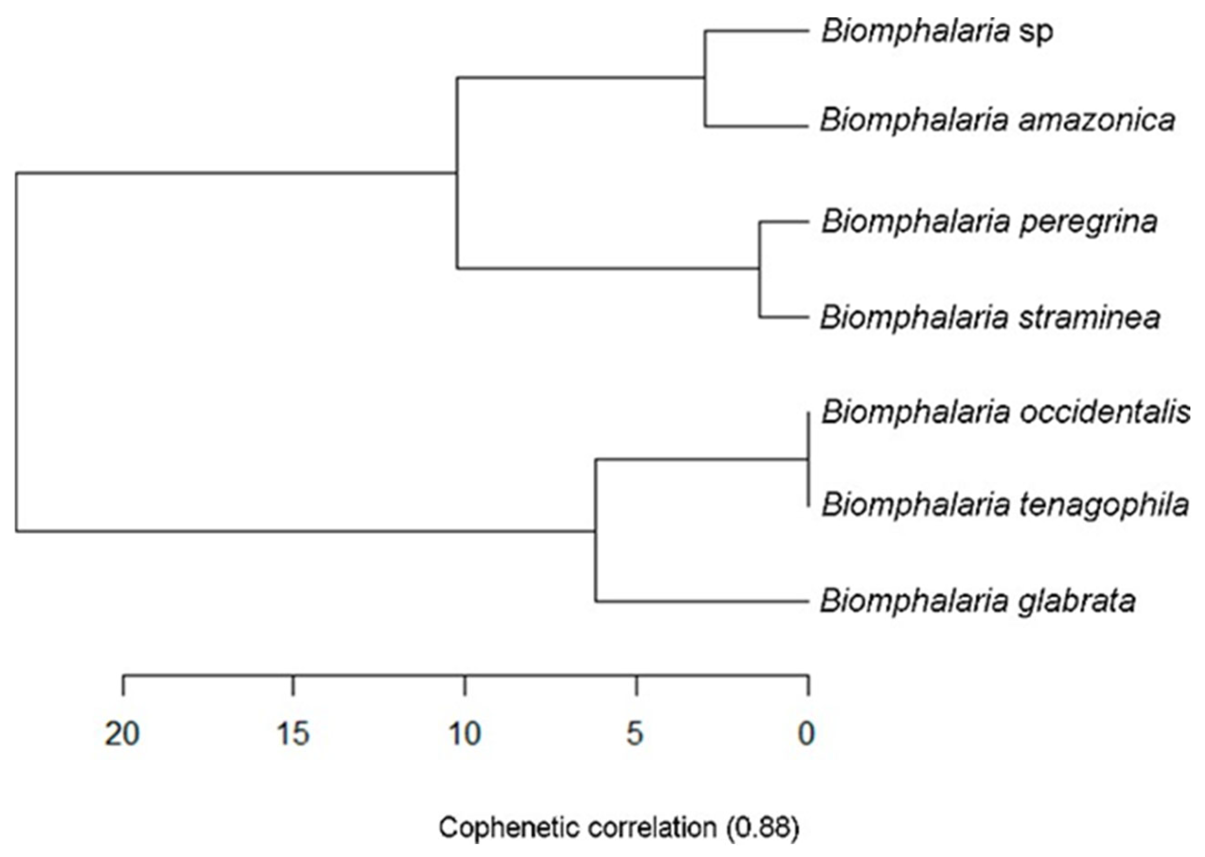
The laboratory-hatched specimens that were closer to adult age, which were used for morphological identification, had a small brown shell about 5 mm (±0.60 mm) in diameter and 2.5 mm in width (±0.30 mm), with 5 spirals that rapidly grew in diameter (the central spiral to the left) and a rounded edge. Compared to other species of Biomphalaria, shell measurements of diameter, width, and number of spirals were similar to B. amazonica [7], which was corroborated by the cluster analysis, with a cophenetic correlation of 0.88 (Figure 5).
The specimens of Biomphalaria collected in Mapiri Lake have a central spiral that is deeper on the right side of the shell, with a funneling cavity. The external spiral declines to the left, with a mantle that has pigments that begin to blend together near the edge. These specimens did not have a renal crest. With respect to the reproductive organs, it was difficult to precisely differentiate them compared to other species of the genus. However, there was a wide foreskin with a sheath that was shorter than the foreskin, a narrow and delicate deferential canal, spermatheca form varying between claviform and oval, vaginal walls without wrinkles, and the appearance of just one muscle between the junction of the foreskin and the penile sheath (Figure 6).
The observed shell and reproductive system characteristics are in agreement with those described by Paraense [7,26], and indicate that these specimens sampled from Mapiri Lake belong to the species B. amazonica.

3.3. Molecular Biology

The results from the molecular biology analysis agreed with those from the morphological analysis, confirming that the specimens collected at Mapiri Lake belong to the species B. amazonica (Figure 7, Table A1). Representative DNA sequences for this study are found in GenBank accession number PV252083.

4. Discussion

The species Biomphalaria amazonica is widely distributed throughout the neotropical zone, especially in the North and Central–West regions of Brazil, and northern Bolivia [5]. In Brazil, the states that have the greatest number of records of B. amazonica are Amazonas, Acre, and Rondônia [16]. The adult shell of B. amazonica can reach 8 mm in diameter and 2.5 mm in width, which makes these shells smaller than most other species of Biomphalaria [26]. The specimens sampled in Mapiri Lake were even smaller, but large quantities of them were found adhered to macrophytes along the edges of the lake, suggesting that juvenile populations are thriving in this lake, a common sign that a body of water is a breeding area [27], which is most likely the case for Mapiri Lake. This is the first record of B. amazonica in the state of Pará, specifically for the lower Amazon region, which enlarges the distribution area of this species in Brazil [28,29].
The period of the development cycle quantified in this study for B. amazonica was not different from those observed for other species such as B. straminea, B. tenagophila, and B. glabrata, thus demonstrating a uniform pattern of development for this group. The development phases were consistent with those quantified by Kawano et al. [12], namely:
  • Blastula: 10 to 23 h after fertilization;
  • Gastrula: 24 h after fertilization of the ovule, the beginning of growth, differentiation, and cell movement occurs;
  • Trochophore: 40 to 65 h after fertilization of the ovule, the embryo becomes elongated, kidney-shaped, with slow movements initially, subsequently becoming more rapid.
  • Veliger: About 120 h after fertilization of the ovule; the shell becomes dislocated and twisted to the right side of the animal and the eyes become visible, as well as the foot and mouth;
  • Hippo: The veliger is more developed, and the tentacles, eyes, and foot become clearly visible and defined, along with the curving form of the main axis and nearly complete covering of the body by the shell. The mollusk then hatches at about 144 h (between 6 and 9 days) after fertilization of the ovule
The hatching of B. amazonica on the sixth day of the cycle could also be influenced by the water temperature maintained in the laboratory (25 °C), which agrees with Kawano et al. [10], who related a similar result of hatching after 6 days in water at the same temperature. The percentage of hatching by B. amazônica in the laboratory was lower than that for other studies of the species B. tenagophila and B. glabrata, 90.5% and 94.8% of hatched snails, respectively [30]. This observation suggests the need for adaptation to the challenges of maintenance and breeding in a laboratory [31]. In the current study, the specimens were fed lettuce, and this might have affected egg production. For example, Valente [31] tested different diets and showed that lettuce resulted in a low number of eggs per egg mass and hatched eggs. Therefore, studies that relate data on fertility and birth rates of Biomphalaria are of great importance to the understanding of the biology of these organisms in the natural environment where they are found.
With respect to species identification, the similarities in the morphological characteristics with other species of Biomphalaria complicate precise identification at the species level [32], especially for juvenile specimens [27]. The shell structure of the species in the current study follows a pattern similar to that of B. straminea and B. peregrina for the number of spirals, peripheral form, and shell opening, and is different only with respect to rapid diameter growth, which is slower for B. peregrine [26]. In summary, this study used a molecular marker (COI) as a model in different genetically distinct Biomphalaria species. The results of this study provided important evidence on the adaptive diversification and speciation of B. amazonica in the state of Pará, with implications even for the taxonomy of this group, when compared with species of the same genus between continents [33].
The absence of a renal crest, which is an important diagnostic parameter for B. glabrata, is also observed in species such as B. amazônica, B. peregrina, B. tenagophila, and B. occidentalis [26]. Other aspects are also important diagnostic characteristics, such as vaginal wrinkling, which is most frequently seen in B. sraminea, as well as the presence of a vaginal pouch, which is absent in B. occidentalis [26]. There are many similarities for other morphological characteristics, which can easily lead to errors in identification. In this context, molecular analysis becomes an essential tool for precise identification of organisms belonging to B. amazonica, and its use should be consistently applied as an indispensable part of species identification.
The species B. amazonica was not observed to be naturally parasitized by S. mansoni; however, studies have indicated it susceptibility to this parasite, demonstrating that B. amazonica could be a potential intermediate host [4]. Furthermore, Carvalho et al. [34], in a study using molecular analysis, suggested that the capacity to be an intermediate host of S. mansoni could be acquired by other species that could be infected by this parasite.
Considering that schistosomiasis in Brazil has been expanding, including into areas where this disease has historically been absent [16], the possible presence of B. amazonica parasitized by S. mansoni in Mapiri Lake could represent a serious health risk for the human communities in the region that depend on this lake for fishing and cultural and recreational activities. This alert becomes even more serious when the environmental conditions of Mapiri Lake are considered, as the lake has received residual solids and sewage from the urban area of the city of Santarém [35,36], even though this area is legally a permanent protection area (APP). These conditions favor the occurrence and growth in population of the mollusks Biomphalaria [32].
There is an urgent need for more in-depth studies on the occurrence and dynamics of populations of Biomphalaria, as well as on the parasite–host relationship with S. mansoni in Mapiri Lake. This research should be established as a continuous series of actions, beginning with the implementation of sanitary measures by the public authorities related to the environmental health of Amazonian water bodies with the goal of combating schistosomiasis, and special attention should be given to species of Biomphalaria that are present in the region. Furthermore, the current study and future ones will aid in the identification of high-risk areas, thus promoting the efficient use of resources to prevent the proliferation of schistosomiasis parasites.

5. Conclusions

This study relates the presence of B. amazonica in Mapiri Lake, in the city of Santarém, thus establishing the first record of this species in the state of Pará. The findings of this study increase knowledge about the regions where this species occurs in Brazil, including the lower Amazon region in its area of distribution. The results of this study have the potential to guide monitoring and identification activities around Mapiri Lake as well as across the Amazon region, and provide valuable information about the life cycle of this species. Studies such as this will contribute to the identification of high-risk areas for the proliferation of schistosomiasis parasites and will aid in the efficient use of resources to promote the health of Amazonian lakes and their surrounding communities.

Author Contributions

Conceptualization, L.d.S.B., A.C.S. and J.A.d.S.; methodology, L.d.S.B., A.C.S., J.A.d.S., A.M.S.d.S. and A.T.R.; software, L.d.S.B., L.L.C. and S.R.M.C.; validation, L.d.S.B.; B.B.B., L.L.C. and S.R.M.C.; formal analysis, L.d.S.B. and S.R.M.C.; investigation, L.d.S.B., A.C.S., J.A.d.S., A.M.S.d.S., A.T.R., B.B.B., L.L.C. and S.R.M.C.; resources, L.d.S.B.; B.B.B., L.L.C. and S.R.M.C.; data curation, L.d.S.B., A.C.S., J.A.d.S., A.M.S.d.S. and A.T.R.; writing—original draft preparation, L.d.S.B., A.C.S. and J.A.d.S.; writing—review and editing, L.d.S.B., J.A.d.S., B.B.B., L.L.C. and S.R.M.C.; visualization, L.d.S.B., B.B.B., L.L.C. and S.R.M.C.; supervision, B.B.B., L.L.C. and S.R.M.C.; project administration, L.d.S.B. and S.R.M.C.; funding acquisition, L.d.S.B. and S.R.M.C. All authors have read and agreed to the published version of the manuscript.

Funding

Coordenação de Aperfeiçoamento de Pessoal de Nível Superior—Brasil (Capes)-Project: 88881.918081/2023-01. "Biodiversidade e promoção da saúde na qualidade de vida e no desenvolvimento socioeconômico na Amazônia".

Institutional Review Board Statement

Not applicable.

Data Availability Statement

Data are contained within the article and Appendix A.

Acknowledgments

We thank the Universidade Federal do Oeste do Pará (Ufopa) and Coordenação de Aperfeiçoa-mento de Pessoal de Nível Superior—Brasil (Capes) for granting a postgraduate scholarship and research funding.

Conflicts of Interest

The authors declare no conflicts of interest. The funders had no role in the design of the study; in the collection, analyses, or interpretation of data; in the writing of the manuscript; or in the decision to publish the results.

Appendix A

Table A1. Dissimilarity matrix, based on the estimate of evolutionary divergences between the sequences of the cytochrome c oxidase subunit I (COI) gene of species of the genus Biomphalaria, considering the number of bases that vary between the species highlighted.
Biomphalaria amazonica PV252083Biomphalaria amazonica MZ778865Biomphalaria kuhniana MZ778865Biomphalaria kuhniana MZ778925Biomphalaria straminea MZ778902Biomphalaria straminea MZ778901Biomphalaria straminea MZ778899Biomphalaria glabrata MZ778984Biomphalaria glabrata MZ778983Biomphalaria glabrata DQ084824Biomphalaria tenagophila MF380482Biomphalaria tenagophila MF380481Biomphalaria tenagophila MF380479Biomphalaria tenagophila MF380477Biomphalaria choanomphala DQ084828Biomphalaria choanomphala HM768906Biomphalaria choanomphala HM768905Biomphalaria sudanica DQ084844Biomphalaria camerunensis DQ084827Biomphalaria angulosa DQ084826Biomphalaria pfeifferi MG780212Biomphalaria pfeifferi MG780211Biomphalaria pfeifferi MG780210Biomphalaria pfeifferi MG780209Biomphalaria straminea MF179836Biomphalaria straminea MF179835Biomphalaria straminea MF179834Biomphalaria straminea MF179833
B. amazonica PV252083
B. amazonica MZ7788650.000
B. kuhniana MZ7788650.9210.921
B kuhniana MZ7789250.0050.0050.921
B straminea MZ7789020.0090.0090.9210.005
B straminea MZ7789010.0050.0050.9210.0090.005
B straminea MZ7788990.0000.0000.9210.0050.0090.005
B glabrata MZ7789840.0090.0090.9240.0140.0190.0140.009
B glabrata MZ7789830.0090.0090.9210.0140.0180.0140.0090.000
B glabrata DQ0848240.8850.8850.9110.8850.8850.8850.8850.8770.880
B tenagophila MF3804820.9120.9120.9250.9120.9120.9120.9120.9100.9120.926
B tenagophila MF3804810.9110.9110.9250.9110.9110.9110.9110.9100.9110.9300.000
B tenagophila MF3804790.9110.9110.9250.9110.9110.9110.9110.9100.9110.9300.0000.000
B tenagophila MF3804770.9120.9120.9250.9120.9120.9120.9120.9100.9120.9260.0000.0000.000
B choanomphala DQ0848280.8880.8880.9350.8880.8840.8840.8880.8820.8840.8980.9160.9160.9160.916
B choanomphala HM7689060.8900.8900.9110.8900.8900.8900.8900.8820.8850.0180.9260.9300.9300.9260.898
B choanomphala HM7689050.8900.8900.9110.8900.8900.8900.8900.8820.8850.0140.9220.9250.9250.9220.8980.005
B sudanica DQ0848440.0140.0140.9250.0190.0230.0190.0140.0140.0140.8840.9160.9160.9160.9160.8880.8880.888
B camerunensis DQ0848270.0050.0050.9210.0090.0140.0090.0050.0050.0050.8840.9120.9110.9110.9120.8880.8890.8890.009
B angulosa DQ0848260.8900.8900.9350.8900.8850.8850.8900.8820.8850.8940.9170.9160.9160.9170.0000.8940.8940.8880.889
B pfeifferi MG7802120.9050.9050.8800.9050.9100.9100.9050.9050.9050.9150.9400.9400.9400.9400.9300.9150.9150.9050.9050.930
B pfeifferi MG7802110.8950.8950.8850.9000.9050.9000.8950.8950.8950.9100.9350.9350.9350.9350.9200.9100.9100.8950.8950.9200.090
B pfeifferi MG7802100.9050.9050.8800.9050.9100.9100.9050.9050.9050.9150.9400.9400.9400.9400.9300.9150.9150.9050.9050.9300.0000.090
B pfeifferi MG7802090.9100.9100.8750.9100.9150.9150.9100.9100.9100.9200.9500.9500.9500.9500.9400.9200.9200.9100.9100.9400.0700.1200.070
B straminea MF1798360.9080.9080.8830.9080.9130.9130.9080.9060.9080.9170.9420.9410.9410.9420.9270.9170.9170.9080.9080.9270.0050.0910.0050.076
B straminea MF1798350.9080.9080.8830.9080.9130.9130.9080.9060.9080.9170.9420.9410.9410.9420.9270.9170.9170.9080.9080.9270.0050.0910.0050.0760.000
B straminea MF1798340.9080.9080.8830.9080.9130.9130.9080.9060.9080.9170.9420.9410.9410.9420.9270.9170.9170.9080.9080.9270.0050.0910.0050.0760.0000.000
B straminea MF1798330.9080.9080.8830.9080.9130.9130.9080.9060.9080.9170.9420.9410.9410.9420.9270.9170.9170.9080.9080.9270.0050.0910.0050.0760.0000.0000.000

References

  1. Pepe, M.S.; Caldeira, R.L.; Carvalho, O.d.S.; Muller, G.; Jannotti-Passos, L.K.; Rodrigues, A.P.; Amaral, H.L.; Berne, M.E.A. Biomphalaria molluscs (Gastropoda: Planorbidae) in Rio Grande do Sul, Brazil. Mem. Do Inst. Oswaldo Cruz 2009, 104, 783–786. [Google Scholar] [CrossRef] [PubMed]
  2. Chitsulo, L.; Engels, D.; Montresor, A.; Savioli, L. The global status of schistosomiasis and its control. Acta Trop. 2000, 77, 41–51. [Google Scholar] [CrossRef] [PubMed]
  3. Morgan, J.A.T.; Dejong, R.J.; Snyder, S.D.; Mkoji, G.M.; Loker, E.S. Schistosoma mansoni and Biomphalaria: Past history and future trends. Parasitology 2001, 123, 211–228. [Google Scholar] [CrossRef] [PubMed]
  4. Teodoro, T.M.; Jannotti-Passos, L.K.; Carvalho, O.d.S.; Grijalva, M.J.; Baús, E.G.; Caldeira, R.L. Hybridism between Biomphalaria cousini and Biomphalaria amazonica and its susceptibility to Schistosoma mansoni. Mem. Inst. Oswaldo Cruz 2011, 106, 851–855. [Google Scholar] [CrossRef]
  5. Simões, L.F.; Bastos, L.D.B.; Camargo, E.A.F.; Neves, M.F.; Linhares, A.X.; Magalhães, L.A.; Zanotti-Magalhães, E.M. Host-parasite relationship between Biomphalaria amazonica (Paraense, 1966) and Schistosoma mansoni (Sambon, 1907). Braz. J. Biol. 2016, 77, 340–346. [Google Scholar] [CrossRef]
  6. Velásquez, L.E.; Caldeira, R.L.; Estrada, V.; Carvalho, O.S. Morphological and polymerase chain reaction-restriction fragment lenght polymorphism characterization of Biomphalaria kuhniana and Biomphalaria amazonica from Colombia. Mem. Inst. Oswaldo Cruz 2002, 97, 997–1004. [Google Scholar] [CrossRef]
  7. Paraense, W.L. Biomphalaria Amazonica and B. cousini, two new species of neotropical planorbid molluscs. Rev. Bras. Biol. 1966, 26, 115–126. [Google Scholar] [PubMed]
  8. Paraense, W.L. Estado atual da sistemática dos planorbídeos brasileiros. Arch. Mus. Nac. 1975, 55, 105–128. [Google Scholar]
  9. Ohlweiler, F.P.; Rossignoli, T.d.J.; Palasio, R.G.S.; Tuan, R. Taxonomic diversity of Biomphalaria (Planorbidae) in São Paulo state, Brazil. Biota Neotropica 2020, 20, e20200975. [Google Scholar] [CrossRef]
  10. Avise, J.C. Phylogeography: The History and Formation of Species; Harvard University Press: Cambridge, MA, USA, 2000. [Google Scholar] [CrossRef]
  11. Bueno-Silva, M. Genética molecular e sistemática animal: Um breve histórico, contribuições e desafios. Estud. de Biol. 2012, 34, 157. [Google Scholar] [CrossRef]
  12. Kawano, T.; Nakano, E.; Watanabe, L.C. Estudo do desenvolvimento embrionário de Biomphalaria glabrata (Mollusca, Planorbidae) e suas aplicações. In Schistosoma Mansoni & Esquistossomose: Uma Visão Multidisciplinary; Fiocruz Editora: Rio de Janeiro, Brazil, 2008. [Google Scholar]
  13. Barbosa, F.S. Tópicos em Malacologia Médica; Fiocruz Editora: Rio de Janeiro, Brazil, 1995; Available online: https://books.scielo.org/id/npy7z (accessed on 20 June 2023).
  14. Rogers, D.C.; Damborenea, C.; Thorp, J. Thorp and Covich’s Freshwater Invertebrates, 4th ed.; Academic Press: Cambridge, MA, USA, 2020. [Google Scholar]
  15. Ohlweiler, F.P. Manual de Gastrópodes Límnicos e Terrestres do Estado de São Paulo Associados às Helmintoses; Redes Editora: São Paulo, Brazil, 2010. [Google Scholar]
  16. dos Santos Carvalho, O.; Coelho, P.M.Z.; Lenzi, H.L. Schistosoma Mansoni & Esquistossomose: Uma Visão Multidisciplinar; Fiocruz Editora: Rio de Janeiro, Brazil, 2008; Available online: https://books.scielo.org/id/37vvw (accessed on 20 June 2023).
  17. Moszczynska, A.; Locke, S.A.; McLaughlin, J.D.; Marcogliese, D.J.; Crease, T.J. Development of primers for the mitochondrial cytochrome c oxidase I gene in digenetic trematodes (Platyhelminthes) illustrates the challenge of barcoding parasitic helminths. Mol. Ecol. Resour. 2009, 9, 75–82. [Google Scholar] [CrossRef] [PubMed]
  18. Kearse, M.; Moir, R.; Wilson, A.; Stones-Havas, S.; Cheung, M.; Sturrock, S.; Buxton, S.; Cooper, A.; Markowitz, S.; Duran, C.; et al. Geneious Basic: An integrated and extendable desktop software platform for the organization and analysis of sequence data. Bioinformatics 2012, 28, 1647–1649. [Google Scholar] [CrossRef] [PubMed]
  19. Hall, T.A. BioEdit: A User-Friendly Biological Sequence Alignment Editor and Analysis Program for Windows 95/98/NT. Nucleic Acids Symp. Ser. 1999, 41, 95–98. [Google Scholar]
  20. Benson, D.A.; Cavanaugh, M.; Clark, K.; Karsch-Mizrachi, I.; Lipman, D.J.; Ostell, J.; Sayers, E.W. GenBank. Nucleic Acids Res. 2013, 41, D36–D42. [Google Scholar] [CrossRef]
  21. Guindon, S.; Dufayard, J.-F.; Lefort, V.; Anisimova, M.; Hordijk, W.; Gascuel, O. New Algorithms and Methods to Estimate Maximum-Likelihood Phylogenies: Assessing the Performance of PhyML 3.0. Syst. Biol. 2010, 59, 307–321. [Google Scholar] [CrossRef]
  22. Tamura, K.; Stecher, G.; Kumar, S. MEGA11: Molecular Evolutionary Genetics Analysis Version 11. Mol. Biol. Evol. 2021, 38, 3022–3027. [Google Scholar] [CrossRef]
  23. DeJong, R.J.; Morgan, J.A.T.; Paraense, W.L.; Pointier, J.-P.; Amarista, M.; Ayeh-Kumi, P.F.K.; Babiker, A.; Barbosa, C.S.; Brémond, P.; Canese, A.P.; et al. Evolutionary Relationships and Biogeography of Biomphalaria (Gastropoda: Planorbidae) with Implications Regarding Its Role as Host of the Human Bloodfluke, Schistosoma mansoni. Mol. Biol. Evol. 2001, 18, 2225–2239. [Google Scholar] [CrossRef]
  24. Jørgensen, A.; Kristensen, T.K.; Stothard, J.R. Phylogeny and biogeography of African Biomphalaria (Gastropoda: Planorbidae), with emphasis on endemic species of the great East African lakes. Zool. J. Linn. Soc. 2007, 151, 337–349. [Google Scholar] [CrossRef]
  25. Camey, T.; Verdonk, N.H. The Early Development of the Snail Biomphalaria glabrata (Say) and the Origin of the Head Organs. Neth. J. Zool. 1969, 20, 93–121. [Google Scholar] [CrossRef]
  26. Paraense, W.L.; Carvalho, O.S.; Coelho, P.M.Z.; Lenzi, H.L. Histórico do gênero Biomphalaria, morfologia e sistemática morfológica. In Schistosoma Mansoni: Uma Visão Multidisciplinar; Carvalho, O.S., Coelho, P.M.Z., Lenzi, H.L., Eds.; Editora Fiocruz: Rio de Janeiro, Brazil, 2008; pp. 285–308. [Google Scholar]
  27. David, N.F.; Cantanhede, S.P.D.; Monroe, N.B.; Pereira, L.P.L.A.; Silva-Souza, N.; Abreu-Silva, A.L.; Tchaicka, L. Spatial distribution and seasonality of Biomphalaria spp. in São Luís (Maranhão, Brazil). Parasitol. Res. 2018, 117, 1495–1502. [Google Scholar] [CrossRef]
  28. Paraense, W.L. Biomphalaria kuhniana (Clessin, 1883), planorbid mollusc from South America. Mem. Inst. Oswaldo Cruz 1988, 83, 1–12. [Google Scholar] [CrossRef]
  29. Species Link Network. Available online: https://specieslink.net/search/ (accessed on 10 January 2024).
  30. Kawazoe, U. Alguns aspectos da biologia de Biomphalaria glabrata (Say, 1818) e Biomphalaria tenagophila (D’Orbigny, 1835) (Pulmonata, planorbidae): II-Fecundidade e fertilidade. Rev. De Saúde Pública 1977, 11, 47–64. [Google Scholar] [CrossRef] [PubMed]
  31. Valente, W.C.G.; Garcia, J.S.; Neto, J.d.M.R.; Schirato, G.V.; Faro, M.J. Avaliação da biologia reprodutiva de moluscos da espécie Biomphalaria glabrata (Say, 1818) expostos a diferentes tipos de alimentação e substratos. Biotemas 2022, 35, 1–14. [Google Scholar] [CrossRef]
  32. Bezerra, F.S.d.M.; Pinheiro, M.C.C.; Filho, J.D.d.S.; de Castro, I.M.N.; Caldeira, R.L.; Sousa, M.S.; Cavalcante, A.B.; Júnior, A.N.R. Identification of Biomphalaria sp and other freshwater snails in the large-scale water transposition project in the Northeast of Brazil. Rev. Inst. Med. Trop. Sao Paulo 2018, 60, e41. [Google Scholar] [CrossRef]
  33. GenBank Overview. Available online: https://www.ncbi.nlm.nih.gov/genbank/ (accessed on 10 January 2024).
  34. Carvalho, O.S.; Caldeira, R.L.; Simpson, A.J.G.; Vidigal, T.H.D.A. Genetic variability and molecular identification of Brazilian Biomphalaria species (Mollusca: Planorbidae). Parasitology 2001, 123, 197–209. [Google Scholar] [CrossRef]
  35. Miranda, G.S.; Rodrigues, J.G.M.; Lira, M.G.S.; Nogueira, R.A.; Gomes, G.C.C.; Miranda, B.S.; De Araújo, A.; Silva-Souza, N. Moluscos límnicos como hospedeiros de trematódeos digenéticos de uma região metropolitana da ilha do Maranhão, Brasil. Sci. Plena 2016, 12. [Google Scholar] [CrossRef]
  36. Carmo, A.M.; dos Amaral Sousa, K.; dos Santos Rodrigues, R.T.; Social, S. A cidade como extensão das águas: O processo de antropização e os impactos socioambientais no lago Mapiri em Santarém–PA. O direito à moradia nas cidades da Amazônia, 18. In Anais do III Seminário de Direito à Cidade em Santarém–PA; Universidade Federal do Oeste do Pará: Santarém, Brazil, 2022; Available online: https://www.ufopa.edu.br (accessed on 20 June 2023).
Figure 1. Mapiri Lake—location in Santarém Hydrography, Geography. Produced in Qgis software 3.28.
Diversity 17 00227 g001
Figure 2. Appearance of the egg mass under a stereomicroscope (A) 200 µm. Schematic illustration of the egg masses (B), namely: capsule (Cp), egg (Ov), embryo (Em d).
Diversity 17 00227 g002
Figure 3. Schematic representation of the cycle presented for specimens of Biomphalaria found in Mapiri Lake in Santarém, PA, and the main stages of development observed in the laboratory. Aquatic environment and macrophyte locations (A); adult specimen of Biomphalaria (B); egg mass in blastula (C) on the first day of the cycle; gastrula (D) after 24 h; trochophore (E) on the fourth day of the cycle; veliger (F) on the fifth day of the cycle; hippo (G) on the sixth day of the cycle; adult organism of post-hatch Biomphalaria molluscs (H).
Diversity 17 00227 g003
Figure 4. Aspects of the newborn mollusk. Head (A); shell (B); renal tube (C); tentacles (D); pigments (E); eyes (F); shell opening (G); foot (H).
Diversity 17 00227 g004
Figure 5. Result of cluster analysis using morphological characters of Biomphalaria. Graphic result showing similarity between Biomphalaria specimens collected in Mapiri Lake (Biomphalaria sp) and Biomphalaria amazonica shells. Cophenetic correlation of 0.88.
Diversity 17 00227 g005
Figure 6. Aspects of the specimens collected in Mapiri Lake, morphologically identified as Biomphalaria amazonica. Right side of the shell (A); shell opening (B); left side (C); aspects of the mantle (D); aspects from the penile complex (E): vas deferens (cn), penile sac (bp), retractor muscle (mr), foreskin (pp); complexed vaginal aspects (F): spermatheca (sp), vaginal pouch (bv), uterus (ur).
Diversity 17 00227 g006
Figure 7. Maximum likelihood tree of species of the genus Biomphalaria based on COX1, available in GenBank. The numbers above the branches indicate the confidence level: 1. Correspondence to haplotypes analyzed from the African continent. 2. Haplotypes analyzed from the American continent. 3. Haplotypes analyzed from the Asian continent. Representative DNA sequences for this study are found in GenBank accession number PV252083.
Diversity 17 00227 g007
Disclaimer/Publisher’s Note: The statements, opinions and data contained in all publications are solely those of the individual author(s) and contributor(s) and not of MDPI and/or the editor(s). MDPI and/or the editor(s) disclaim responsibility for any injury to people or property resulting from any ideas, methods, instructions or products referred to in the content.