Article

Fossil Samaras of Acer in the Lower Miocene of Central Inner Mongolia, China, and Their Phytogeographical Implications

1
The Third Institute of Geology and Minerals Exploration, Gansu Provincial Bureau of Geology and Minerals Exploration and Development, Lanzhou 730050, China
2
Gold Mine Resource Exploration and Utilization Technology Innovation Center of Gansu Province, Lanzhou 730050, China
3
School of Earth Science and Resources, Chang’an University, Xi’an 710054, China
*
Author to whom correspondence should be addressed.
Diversity 2025, 17(3), 218; https://doi.org/10.3390/d17030218
Submission received: 20 December 2024 / Revised: 24 February 2025 / Accepted: 10 March 2025 / Published: 19 March 2025

Abstract

:
Samara fossils of Acer were unearthed from the Early Miocene Hannuoba Formation in central Inner Mongolia, China. Based on macro- and micro-characteristics, they were identified as Acer pretataricum sp. nov. of section Ginnala, and cf. Acer mono of section Platanoidea. We reconstructed the dispersal routes of these two sections according to their fossil records. During the Early Eocene, section Ginnala was confined to North America. In the Late Eocene, this section expanded westward to East Asia. It was distributed widely in East Asia. In the Late Tertiary, it potentially recolonized the European and American continents. Meanwhile, section Platanoidea was distributed disjunctively in East Asia and North America during the Eocene. Members of this section likely expanded westward from East Asia into Europe in the Oligocene. By the Miocene, it had achieved extensive distribution in the Northern Hemisphere. However, by the Pliocene, it was confined to East Asia and Western Europe. It may have spread eastward from East Asia to North America during the Holocene, finally forming its current existence in North America and the Eurasian continent. This investigation reveals distinct differences in the dispersal pathways of two sections, suggesting that the reconstruction of dispersal routes for Acer taxa should be conducted separately at the section level.

1. Introduction

The genus Acer, belonging to the family Sapindaceae, is a diverse group with around 200 living species globally. These species are predominantly found in the temperate zones of Asia, Europe, and North America [1]. There are approximately 140 species of Acer in China, classified into 14 different sections, which makes it a pivotal center for analyzing both the geographical and species diversity of the genus Acer [2,3,4,5]. The earliest Acer fossils found in North America and the western coastal area of the Bering Strait date to the Late Cretaceous [6]. However, more reliable fruit fossils for taxonomy were found in southern Alaska, USA, from the Upper Paleocene, identified as A. alaskense Wolfe et Tanai [7,8]. During the Eocene, Acer witnessed a notable expansion across the world (Figure 1). Towards the end of the Eocene, Acer spread to the Kamchatka Peninsula on the Siberian side of the Bering Land Bridge [7]. Budantsev et al. [9] proposed that the primary taxa within this genus diversified in western North America during the Middle to Late Eocene, and then potentially spread to the Asian continent via the Bering Land Bridge and the North Atlantic Land Bridge. The distribution range of Acer became smaller in area during the Oligocene compared to the Eocene, likely due to cool down from the Late Eocene to the Oligocene [10]. In the Miocene, the distribution range of Acer reached its peak, possibly due to the favorable warm climate during the Miocene for plant survival [11]. After the Late Miocene, the distribution range of Acer significantly decreased again, with a reduction in species diversity, maybe because of further temperature declines and alternating cycles of glacial and interglacial periods during the Quaternary [12]. Following climatic warming in the Holocene, Acer plants continued to spread to form their current distribution pattern. The earliest known Acer fossils were from the Upper Cretaceous, found in western North America, suggesting that the genus seems to originate from North America. However, Xu [13] argued that East Asia has been the center for the modern distribution of Acer, providing conditions conducive to the emergence of primitive Acer groups. It is suggested by Pax [14] and Pojarkova [15] that Acer originated in the mountainous regions of East Asia. Studies on molecular phylogenetics have also indicated that the ancestors of the Acer genus originated from East Asia [16,17]. However, the hypothesis of East Asian origin lacks the most direct supporting evidence from Acer fossil records.
There has been ongoing debate regarding the dispersal pathways of the Acer genus [7,13,18,19,20], with three main viewpoints currently prevailing. Wolfe and Tanai [7] and Huang [18] argue that the Acer genus originated in North America and subsequently dispersed westward and eastward to East Asia and Europe, respectively. Xu [13] posits that after originating in East Asia, the Acer genus spread westward through Europe and northern Africa to North America, eastward to North America, and southward through the Malay Peninsula to Indonesia. Kvacek [19] and Boulter et al. [20] suggest that some Acer plants dispersed from North America to Europe before the opening of the North Atlantic in the Early Eocene. Additionally, some Acer plants migrated westward from East Asia to Europe during the Oligocene. These differing viewpoints may be related to the selection of different groups of Acer plants used to reconstruct the dispersal pathways of the entire genus. There are 23 groups of Acer globally, and the varying climatic adaptations of these groups have led to different distribution ranges throughout geological history [7,20,21,22,23,24,25,26,27,28,29] (Figure 1), which may also result in differing dispersal pathways. Therefore, studying the dispersal of Acer plants according to different groups can provide more accurate results for reconstructing the dispersal pathways of the entire genus.
We collected 12 fossil samaras of Acer from the Early Miocene Hannuoba Formation in Zhuozi County, Ulanqab City, Inner Mongolia. This study conducted detailed observations and analyses of the fossil characteristics from both macro and micro perspectives, comparing them with the morphological features of both extant and fossil Acer that have been reported, accurately identifying their taxonomic positions, and ultimately classifying these fossils into two groups of the Acer genus. Additionally, this paper summarizes the global fossil records of these two groups, conducts a paleogeographical study, and infers the ancient geographical dispersal pathways of the two groups.

2. Materials and Methods

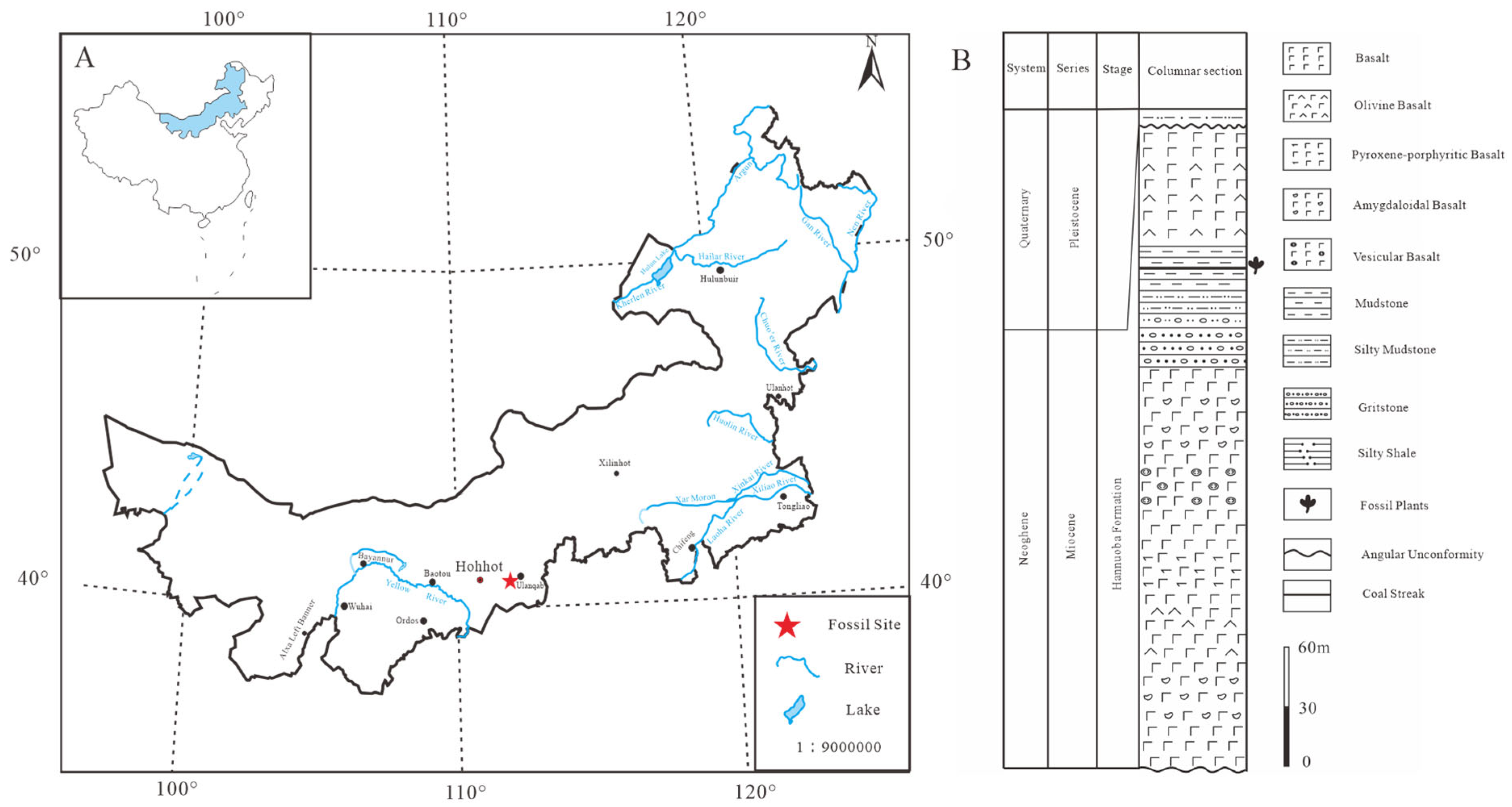
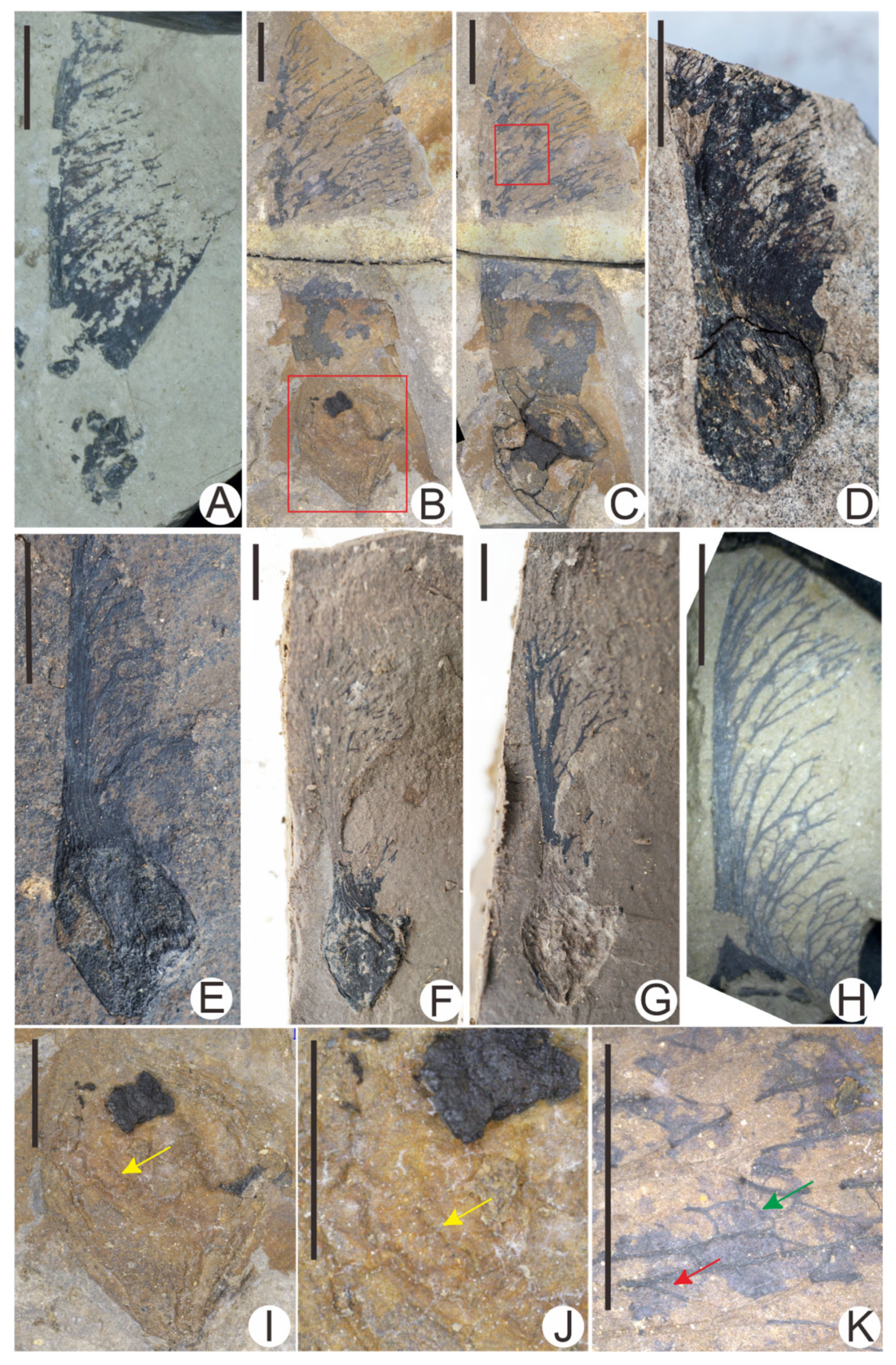
2.1. Research Materials

In this study, 12 Acer fossils were collected from the Hannuoba Formation in Zhuozi County, Ulanqab City, Inner Mongolia (40°45′01″ N, 112°48′46″ E). The Hannuoba Formation is extensively distributed across Hebei Province, Inner Mongolia, and northern Shanxi, primarily consisting of a sequence of gray, black, and purplish-gray olivine basalt, interbedded with sedimentary rocks, including gray to gray-black mudstone, gray-white marl, black oil shale, and local low-quality coal seams. Abundant well-preserved plant fossils and a limited number of gastropod and bivalve fossils were found in the interbedded sediments (Figure 2) [30,31,32]. The aforementioned lithological and paleontologic evidence together points to a fluvial or lacustrine depositional environment, further implying a near-autochthonous burial of these fossils. The abundant plant and animal fossils previously reported within the mudstone interlayers of the Hannuoba basalt in Hebei Province infer the geological age of this stratigraphic unit as the Miocene [33,34]. Additionally, Wang et al. [30] performed K-Ar dating on the Hannuoba basalt in Zhangjiakou, determining its geological age to be 18–24 Ma, correlating with the Late Oligocene to Early Miocene. Li et al. [35] also conducted K-Ar dating on the Hannuoba basalt in Zuoyun County, Shanxi Province, and obtained a geological age of 19.35–22.84 Ma, which corresponds to the Early Miocene. In addition, Zhang et al. [36] selected four Hannuoba basalt specimens from the nearby Jining District in Inner Mongolia for K-Ar isotopic dating. Their findings indicate that the primary eruption period of the basalt in the Jining area occurred from the Late Oligocene to the Miocene, which can be further categorized into three eruption cycles: approximately 33 Ma, 22.8–22.1 Ma, and 12.2–9.4 Ma, respectively. The basalt assemblage from the Hannuoba Formation in Erlongshan, Zhuozi County, examined in this study, closely resembles the rock characteristics of the Hanqing Ba profile in the Jining area. This similarity suggests that the basalt in this study may correspond to the second eruption cycle, leading to the inference of a geological age of 22.8–22.1 Ma belonging to the Early Miocene. Consequently, we deduce that the geological age of fossiliferous sedimentary interlayers in the Hannuoba Formation of Zhuozi County is likely determined to be the Early Miocene.
Living specimens of Acer samaras were collected from Xi’an, and both living and fossil specimens are preserved in the Geological Museum of Chang’an University.

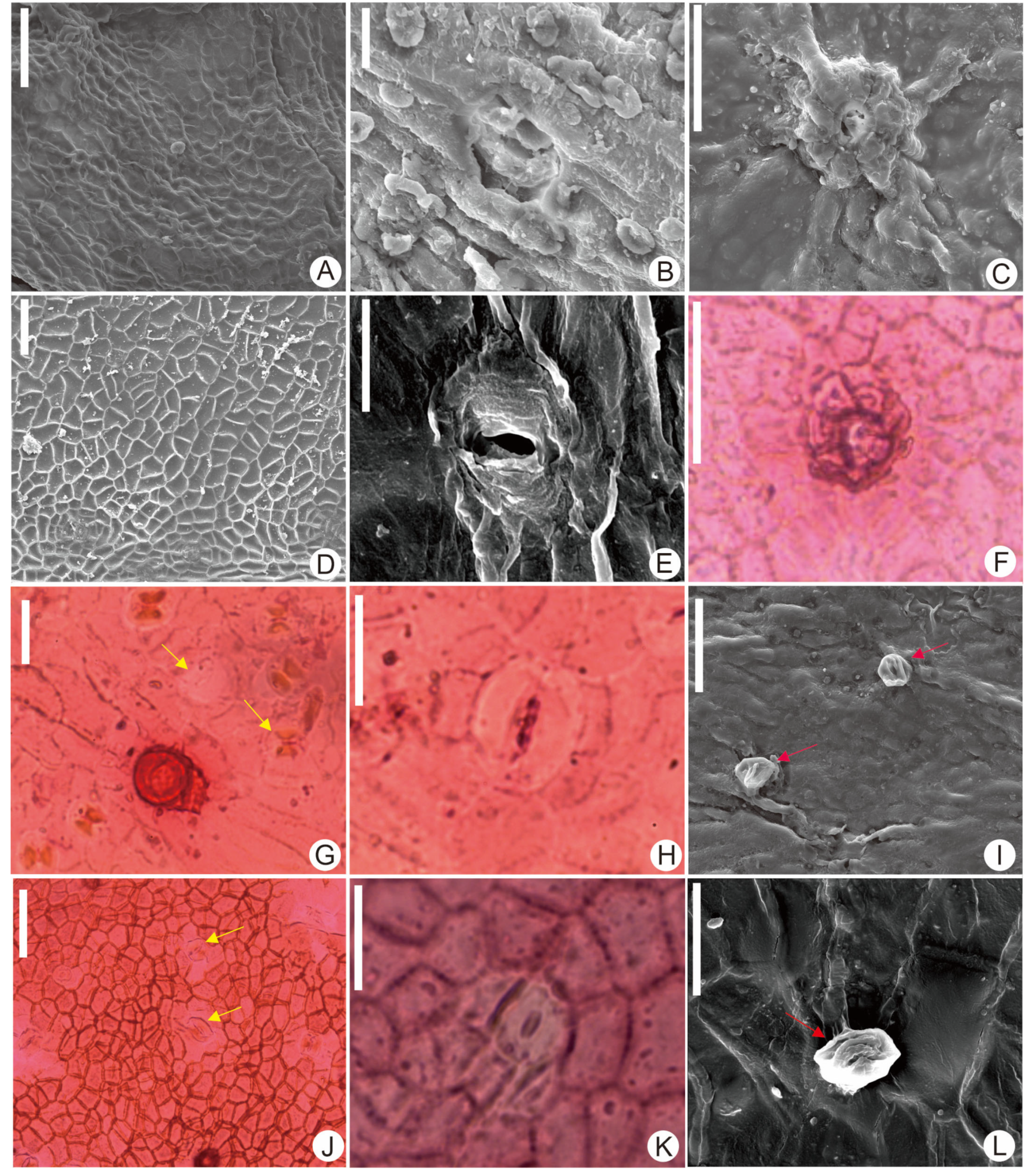
2.2. Processing of Fossil Specimens

Initially, samara fossils of Acer were observed and photographed using a VHX-1000 ultra-depth three-dimensional microscope (Keyence, Osaka, Japan) to capture both macro and micro characteristics. Following this, experiments were conducted on the cuticle of the winged fruit, employing the improved transparent leaf method, as described by Liang et al. [37]. The fossil cuticle was soaked in a 20% HCl solution for 10 h, then thoroughly washed with distilled water until neutral. Subsequently, a 40% HF solution was applied, and the specimen was soaked for approximately 12 h before being rinsed again with distilled water until neutral. Next, a 70% HNO3 solution was added, and the specimen was soaked for 6 h. Once the specimen turned yellow-brown, it was washed with distilled water until neutral. After this, two drops of 10% ammonia (NH4OH) were introduced; if yellow-brown misty or filamentous substances were released, the specimen was immediately washed with distilled water until a neutral state was reached, and then observed under a microscope. If the cuticle surface displayed no adsorbed impurities and the cell structure was clear, it was promptly stained with safranine solution. If impurities remained present, the ammonia treatment was repeated until the cell structure was clarified. Finally, the specimens were sealed with glycerin and nail polish, assigned identification numbers, and photographed under a DM1000 light microscope (Leica, Wetzlar, Germany). Additionally, some specimens underwent gold sputtering treatment and were subsequently observed and photographed using an FEI Quanta 650 FEG scanning electron microscope (FEI, Hillsboro, OR, USA).

2.3. Processing of Living Specimens

For the current samaras, the specimens were first washed with distilled water and then placed in a beaker containing a mixed solution of CH3COOH and H2O2 (1:1). The beaker was then placed in a water bath at 70 °C and heated for 5 to 7 h. Once the specimens became completely transparent, they were washed with distilled water until a neutral state was reached. After separating the upper and lower epidermis, 1% safranine solution for staining was dropped in. After 5 to 10 s, the specimens were rinsed thoroughly with distilled water; then, a slide was prepared with glycerin and sealed with nail polish. Finally, we observed and photographed the specimens under a DM1000 light microscope. Additionally, some specimens were selected for gold sputtering treatment and photographed using a Quanta 650 scanning electron microscope.

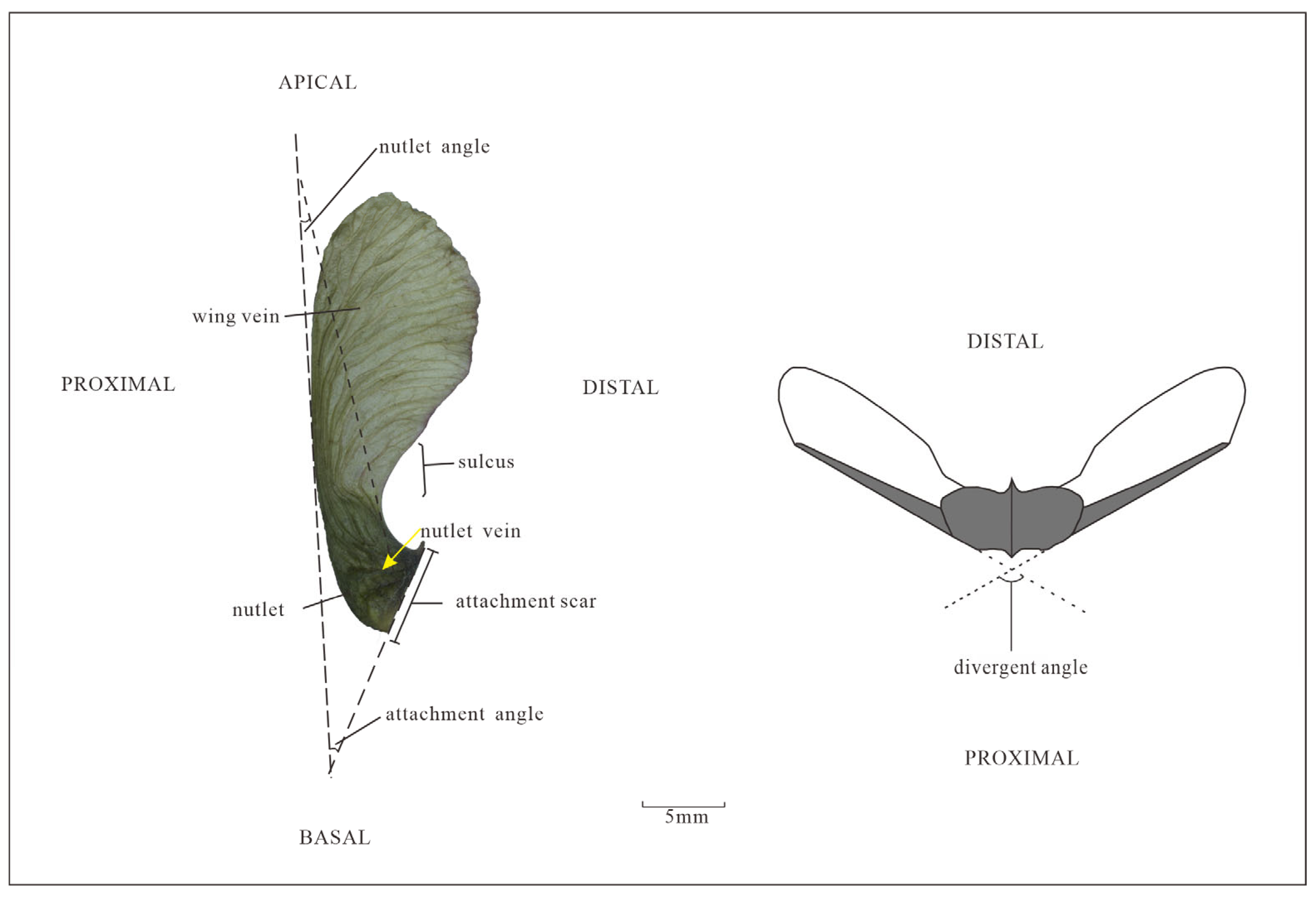
2.4. Terms Describing the Morphological Characteristics of Acer Samaras
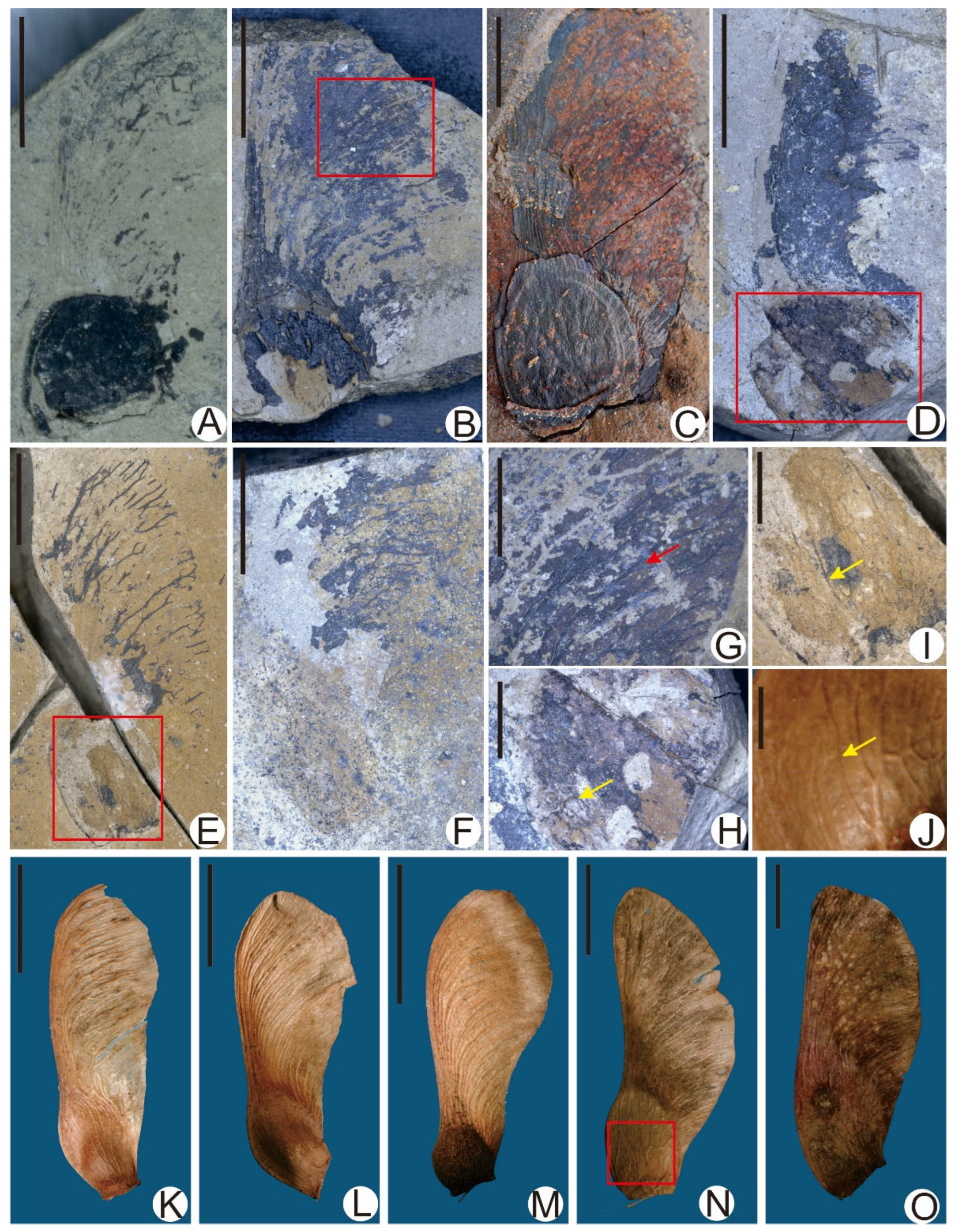
The morphological characteristics of the samaras of the Acer specimens in this article are described using the terminology of Wolfe and Tanai [7] and Xu [38], as detailed in Figure 3.

3. Results

Family—Aceraceae.
Genus—Acer Linn., 1753.
Section—Ginnala Nakai., 1966.
Species—Acer pretataricum Xiao and Wang sp. nov.
Etymology: The species name “pretataricum” indicates that this fossil represents an ancient species of Acer tataricum, which is morphologically similar to the extant Acer tataricum.
Holotype: ELS-19-347 (Figure 4A).
Paratypes: ELS-21-234 (Figure 4B,C), ELS-19-228 (Figure 4D), ELS-18-97 (Figure 4E), ELS-19-374 (Figure 4F,G), ELS-19-146 (Figure 4H).
Stratigraphy horizon: Hannuoba Formation, Lower Miocene.
Diagnosis: Fruits are asymmetric, samaroid. The nutlet exhibits a slightly inflated and oval shape with convex veins on its surface. The divergent angle is acute or nearly erect, while the groove forms an obtuse angle. Notably, the wing veins are well defined, featuring 10 to 15 primary veins that run nearly parallel. The curved primary veins are accompanied by one to three bifurcations, resulting in a network-like appearance.
Description: The samaras are of 2.1–4.2 cm in length and 0.42–1.2 cm in width, with a length-to-width ratio of 3.1–4. At the base, there are nutlets, measuring 0.55–0.9 cm in length and 0.43–0.8 cm in width, with a length-to-width ratio of 1.1–2.2, slightly inflated; nutlets are curved and oval-shaped, with convex veins on the surface; the tip is narrow oval to triangular, with relatively clear surface veins and a reticulate pattern; nutlet angels are 7–15°. Attachment scars appear on the inner side of nutlets, measuring 0.4–0.75 cm in length, with a more pronounced distal ridge and an inconspicuous proximal ridge; the attachment angles are about 36–45°, and the divergent angles are acute or nearly erect. The length of the wings is 1–3 cm, shaped from acute triangular to elongated oval; the distal ridge of the wing extends from near the top of the nutlets, protruding and forming an obtuse groove with nutlets; the ridge is straight or slightly convex, with clear and developed wing veins and 10–15 main veins extending from the top of the nutlets, converging along the proximal ridge of the wing, bending and bifurcating towards the distal ridge, with bending angles of the main vein of about 100–150°. The primary veins are nearly parallel with significant curvature, convex, bifurcating 1–3 times, many forming a network.
The cuticle of the fruit wing is thin, characterized by irregularly shaped cells that vary from quadrilateral to hexagonal forms. These cells are 13–18 μm in length and 7–9 μm in width. The anticlinal walls are shallowly wavy, and multicellular hair bases can be observed. The stomatal apparatus is anomocytic, with sunken guard cells, and the stomatal openings are lower than the subsidiary cells. The stomas are 24–26 μm in length and 17–18 μm in width (Figure 5).
Comparison and Discussion: The fossils are preserved as half of a samara with a single wing. They are asymmetrical. The base of the nutlet shows an attachment scar, located on the distal ridge of the samara, with no sign of a subsidiary wing. At least 39 genera from 11 families of plants exhibit such asymmetrical samaras [39]. Samaras with asymmetrical wing shapes or sometimes with double or triple wings include Helietta Tul., Diatenopteryx Radlk., Thouinidium Radlk., Thouinia Poit., Acridocarpus Guill. & Perr., Gaudichaudia Kunth, Sphedamnocarpus Planch. ex Benth. & Hook. f., Banisteriopsis C.B. Rob., Ectopopterys W.R. Anderson, and Acer Linn. However, only the samaras of Acer have attachment scars concentrated on the distal ridge of the samaras and lack a subsidiary wing [1,29,39,40,41,42]. Therefore, the present fossils should obviously be divided into Acer according to their morphological characteristics.
The nutlet exhibits a slightly inflated shape with convex veins on its surface. The base is inclined, one side is wider while the other is narrower, and the divergent angle is acute or nearly erect. These macro morphological characteristics are quite similar to those of section Ginnala within Acer, while being significantly different from the samaras of other sections within Acer [1]. The current samara fossils were compared with previously reported Acer fossil species (Table 1), revealing that the current specimen resembles the following 13 species in overall morphology: Acer subginnala Guo [43], A. pseudoginnala Tanai et Onoe [44], A. ashwilli Wolfe et Tanai, A. glabroides Wolfe et Tanai, A. hillsi Wolfe et Tanai, A. knolli Wolfe et Tanai, A. browni Wolfe et Tanai, A. busamarum Wolfe et Tanai, A. tigilense Chelebaeva, A. whitebirdense Wolfe et Tanai, A. schorni Wolfe et Tanai, A. eomedianum Wolfe et Tanai, and A. niklasi Wolfe et Tanai [7]. Among these 13 species, A. subginnala, A. pseudoginnala, A. ashwilli, A. browni, A. schorni, A. tigilense, and A. glabroides have attachment angles all less than 35°, with acute divergent angles, which differs from the current fossil’s connecting angle of 36–45° and acute to nearly upright divergent angle. In comparison with the remaining six species, the current fossil has oval, curved nutlets, thus excluding A. hillsi (hemicycle nutlet), A. knolli (elongated oval nutlet), and A. glabroides (triangular nutlet). The samara of A. busamarum (length 4.7–10 cm, width 1.1–2.8 cm) is significantly larger than the current fossil samara (length 2–4 cm, width 0.42–1.2 cm). Nutlets of Acer whitebirdense are larger (length 1–2.7 cm, width 0.5–1.6 cm), accounting for 1/3–1/2 of the total length of the samara, while the current fossil nutlet is relatively smaller, measuring 0.55–0.9 cm in length and 0.43–0.8 cm in width. Nutlets of Acer niklasi have nearly parallel veins on the surface, which can be distinguished from the reticulate veins of the current fossil. Overall, the current fossil exhibits significant morphological differences from previously reported fossil samaras, and thus cannot be classified under any previously established fossil species.
A morphological comparison was made between the current fossils and the samaras of extant Acer species. First, based on characteristics such as the overall external morphology of the samara, the presence of sulci, and the shape of the wings—ranging from acute triangular to elongated oval—seven extant Acer species similar to the current fossil were selected, including Acer tataricum L. (Figure 6G,H), Acer buergerianum Miq (Figure 6A), Acer caudatum Wall (Figure 6B), Acer davidii Frach. (Figure 6C), Acer tetramerum Pax (Figure 6F), Acer monspessulanum L. (Figure 6D), and Acer pseudoplatanus L. (Figure 6E). According to the divergent angle of the samara, A. davidii and A. monspessulanum can be excluded, as the divergent angle of the current species is acute or nearly upright (70–90°), while A. davidii has an obtuse or nearly horizontal divergent angle and A. monspessulanum has a very small divergent angle, generally not exceeding 60°. Based on the shape and venation characteristics of the small nutlet, A. buergerianum and A. pseudoplatanus can be excluded, as the small nutlet of this fossil has a curved oval, slightly inflated shape, and has a reticulate venation on its surface, whereas A. buergerianum has a distinctly inflated small nutlet with longitudinal veins on its surface, and A. pseudoplatanus has a spherical small nutlet with longitudinal veins. Comparing the remaining three species, A. caudatum (with a length-to-width ratio of 2.5–3, broad and short type) can be distinguished from this fossil (with a length-to-width ratio of 3.1–4, slender type) based on the length-to-width ratio of the samara. Then, comparing the morphological characteristics of the wing veins, it was found that A. tetramerum has unconnected wing veins that do not form a reticulate pattern, which is significantly different from the interconnected and short branches of the wing veins in this fossil. Finally, it was found that only A. tataricum has a morphology similar to the current fossil samara, including key features such as samara 2–4 cm in length, the presence of sulci, a divergent angle that is acute or nearly upright, bifurcated wing veins, small veins forming a grid-like pattern, and small nutlets that are flat or slightly inflated with a reticulate venation on the surface. The only difference is the width of the samara, with A. tataricum being about 0.5 cm, while the current fossils are about 0.5–1 cm. In addition, a further comparison of the cuticular microstructural features of the current fossil with the samara of extant A. tataricum revealed that both share the same main structural characteristics, including the following: the epidermal cells of the samara are irregular in shape, varying from quadrilateral to hexagonal, with multicellular hair bases visible; the stomatal apparatus is arranged in a ring; guard cells are depressed and lower than subsidiary cells; and stomatal openings measuring 20–30 μm in length and 15–20 μm in width.
In summary, the current samara fossils are most similar in morphological characteristics to extant A. tataricum, but the width of the samara differs slightly from that of extant A. tataricum. Therefore, these fossils are designated as a new species, Acer pretataricum Xiao and Wang sp. nov.
Family—Aceraceae.
Genus—Acer Linn., 1753.
Section—Platanoidea Pax., 1885.
Species—cf. Acer mono Maxim,. 1856.
Materials examined: ELS-18-70 (Figure 7A), ELS-19-84 (Figure 7B), ELS-18-188 (Figure 7C), ELS-19-99 (Figure 7E), ELS-21-232 (Figure 7D), ELS-18-258 (Figure 7F), and ELS-18-258 (Figure 7F).
Stratigraphy horizon: Hannuoba Formation, Lower Miocene.
Location: Erlongshan, Zhuozi County, Ulanqab City, Inner Mongolia (112°85′ N, 40°77′ E).
Description: The samaras are of 2–2.5 cm in length and 0.75–1.2 cm in width, with a length-to-width ratio of 2.8–3.2. At the base, the nutlets are 0.65–0.9 cm in length and 0.5–0.7 cm in width, with a length-to-width ratio of 1.1–1.5, flattened; the nutlets are oval-shaped, with 6–8 veins on the surface extending from the attachment scars to the top of the nutlets, nearly parallel to the long axis of the nutlets, and the reticulate veins are relatively clear; the nutlet angles are 7–12°. The attachment scars are 0.35–0.55 cm in length, located on the distal ridge of the nutlets; the connection angles are approximately 40–80°, and the divergent angles are either acute or obtuse. The wings are 1–2 cm in length, ranging from elongated oval to elongated elliptical; the ventral edge of the wing extends from near the attachment scar of the nutlet, completely covering the nutlet, with no groove developed; the ridge is slightly convex or concave, with clear and well-developed wing veins, consisting of 10–15 main veins that extend from the top of the nutlet, bending and branching towards the ventral edge without healing, with divergent angles of approximately 120–170°. The wing veins are nearly parallel, with noticeable curvature, branching 1–3 times, forming a network.
Comparison and Discussion: These fossils do not have sulci, and the nutlets are flat, with the divergent angles being either acute or obtuse. Among Acer sections, only sections Hyptiocarpa and Platanoidea lack sulci and have flat nutlets. However, section Hyptiocarpa has an acute divergent angle, and thus the fossil is classified under section Platanoidea [1]. Based on the overall shape of the samara, the presence or absence of sulci, and the shape of the wings, the current six fossil specimens were compared with fossil species of the genus Acer, revealing morphological similarities with the following fossil species: Acer hueberi Wolfe et Tanai, Acer oligomedianum Wolfe et Tanai, Acer medianum Knowlton [7], Acer protomiyabei Endo, and Acer palaeoplatanoides Endo [45], and Acer subpictum Saporta [23]. However, among these six fossil species, the samara lengths of Acer hueberi and Acer subpictum are 2.6–3.1 cm and 2.7–5 cm, respectively, which are significantly longer than the current fossil samara (2–2.5 cm); the connection angle of Acer oligomedianum is 25–40°, and the surface of the nutlet has nearly parallel veins. However, they are not parallel to the long axis of the nutlet. The connection angle of the current fossil is 40–80°, and the veins on the surface of the nutlet extend from the attachment scar to the top of the nutlet, being nearly parallel to the long axis of the nutlet, showing significant differences. Acer medianum is distinguished by its larger samara (4–5 cm long, 0.9–1.6 cm wide) and longer attachment scar (0.7–1 cm) compared to the current fossil’s smaller samara (2–2.5 cm long, 0.75–1.2 cm wide) and shorter attachment scar (0.35–0.55 cm); Acer protomiyabei has a larger connection angle (90–135°), while Acer palaeoplatanoides has a longer attachment scar (0.5–0.9 cm), both showing significant differences from the current fossil (Table 1). After the detailed comparison above, it was found that the morphology of the aforementioned Acer fossils differs from the current fossil; thus, these samara fossils cannot be directly classified as any named fossil species.
Subsequently, a morphological comparison analysis was conducted between the fossils and extant species of the genus Acer (Figure 7). Based on morphological characteristics, such as the absence of sulci, wing shape being elongated oval to elongated elliptical, nutlet angles of 7–12°, and connection angles of 40–80°, four extant Acer species similar to the current fossils were selected: Acer grosseri Pax (Figure 7K), Acer pensylvanicum L. (Figure 7L), Acer rufinerve Sieb. et Zucc (Figure 7M), and Acer mono Maxim., 1856 (Figure 7N,O) from section Macrantha. However, further comparison revealed that the three extant species from sect. Macrantha still exhibit differences in some key characteristics compared to the current fossils. First, based on the size of the divergent angles of the samaras, A. pensylvanicum can be excluded, as this extant species has an acute divergent angle, while the current fossil has a divergent angle that is either acute or obtuse. Then, based on the shape of the nutlet, A. rufinerve can be excluded; the nutlet of the current fossil is oval, flattened, or slightly swollen, while A. rufinerve has a spherical and significantly swollen nutlet. Additionally, the surface of the current fossil’s nutlet has nearly parallel veins, which distinguishes it from A. grosseri, whose nutlet surface has longitudinal veins. Finally, when comparing this fossil with Acer mono from section Platanoidea, it was found that their main characteristics are quite similar, including wing lengths of 2–2.5 cm, the absence of sulci, a divergent angle that is acute or nearly obtuse, and small nuts that are 0.5–0.8 cm wide and have a flattened or slightly swollen shape. However, the small nuts of these fossils are slightly longer than those of extant Acer mono (Table 2). At the same time, due to the poor preservation state of this fossil, the characteristics of the epidermal cells of the small nuts could not be obtained. Therefore, these fossils are temporarily named as a similar species to Acer mono from section Platanoidea, cf. Acer mono Maxim.
Table 1. Comparison of the present fossil’s samaras with similar fossil species.
SpeciesGL *
(cm)
GW *
(cm)
NL *
(cm)
NW *
(cm)
NOS *AA *NA *NI *WR *SulusASL * (cm)AgeLocalityReference
Acer subginnala31.21.30.55Elliptical11–15°8–12°-Slightly straightAuxetic0.6–0.7MioceneQinghai, China[43]
Acer pseudoginnala4.21.20.90.6Elliptical15°--StraightAuxetic0.7MioceneFukushima and Hokkaido, Japan[46]
Acer ashwilli2.3–3.40.6–1.00.5–0.90.3–0.6Elliptical20–30°30–45°Moderately inflatedConvexAuxetic-Early EoceneOregon, U.S.A.[7]
Acer hillsi>2.510.50.5Suborbicular40°20°-StraightAuxetic-Middle EoceneWashington, U.S.A.[7]
Acer browni3.3–41–1.20.8–1.00.7–0.8Elliptical to orbiculate25–30°40°Flattened to strongly inflated ConvexAuxetic0.6–0.7MioceneOregon, Washington, U.S.A. and Queen Charlotte Island, Canada[7]
Acer busamarum4.7–101.1–2.80.7–20.6–1.5Ovate20–40°45–60°Strongly inflatedStraightAuxetic0.9–2.2MioceneColombia, U.S.A.[47]
Acer schorni1.6–2.80.5–10.6–0.80.4–0.7Suborbicular10–30°25–80°Strongly inflated Straight to convexAuxetic0.2–0.5MioceneOregon, Idaho, Nevada, U.S.A.[7]
Acer tigilense1.7–2.70.5–0.70.5–0.70.3–0.5Elliptical20–30°20–30°Slightly inflatedConvexAuxetic0.3–0.5MioceneColombia, U.S.A.[7]
Acer whitebirdense2.5–6.70.6–1.51–2.70.5–1.6Oblate20–55°10–40°Moderately inflatedConvexAuxetic0.2–0.7MioceneColombia, U.S.A.[7]
Acer knolli2.8–4.20.6–1.21–1.70.3–0.7Lanceolate40–45°10–15°Moderately inflatedStraightAuxetic0.4–0.8Middle MioceneWashington, U.S.A.[7]
Acer eomedianum3.20.8–0.90.70.5–0.6Elliptical20–35°15–20°Moderately inflatedStraightAuxetic0.6EoceneNevada, Montana, U.S.A.[7]
Acer niklasi3–3.61.0–1.30.7–1.00.5–0.7Elliptical35–40°10°Moderately inflatedConvexAuxetic0.3–0.4MioceneIdaho, Washington, U.S.A.[7]
Acer glabroides3.410.70.6Triangular40°20°Moderately inflatedStraightAuxetic0.6Early EoceneOregon, U.S.A.[7]
Acer hueberi2.6–3.10.8–1.20.6–0.70.6–0.7Hemicycle35–70°35–50°Flattened Slightly convexObsolete0.4–0.5EoceneMontana, U.S.A.[7]
Acer oligomedianum2.1–3.70.5–0.90.7–10.5–0.7Elliptical25–40°10–30°-Straight to
convex
Obsolete0.5–0.7OligoceneOregon, U.S.A.[7]
Acer medianum4–50.9–1.61.2–1.50.7–1.2Elliptical25–30°10–35°Moderately inflatedStraight to convexObsolete0.7–1Late MioceneColumbia, Nevada and Idaho, U.S.A.[7]
Acer protomiyabei1.8–3.50.7–1.10.7–1.20.7–1.2Orbiculate90–135°-Strongly inflatedSinuousObsolete0.6–1MioceneNorth Korea[45]
Acer palaeoplatanoides2.4–2.70.7–1.40.6–10.5–0.9Suborbicular60–90°-Flattened Straight to sinuousObsolete0.5–0.9MioceneNorth Korea[45]
Acer subpictum2.7–50.7–1.20.4–10.3–0.5Ovate to oblong30–45°20–35°-Straight to sinuousObsolete0.2–0.4MioceneYunnan, China[23]
Acer pretataricum sp. nov. 2–40.42–1.20.55–0.90.43–0.8Ovate36–45°7–15°Slightly inflatedStraight to slightly convexAuxetic0.4–0.75Early MioceneInner Mongolia, ChinaThis paper
cf. Acer mono2–2.50.75–1.20.65–0.90.5–0.7Elliptical40–80°7–12°Flattened to slightly inflatedSlightly convex or sinuousObsolete0.35–0.55Early MioceneInner Mongolia, ChinaThis paper
* GL = general length; GW = general width; NL = nutlet length; NW = nutlet width; NOS = nutlet outline shape; AA = attachment angle; NA = nutlet angle; NI = nutlet inflation; WR = wing ridge; ASL = attachment scar length.

4. Discussion

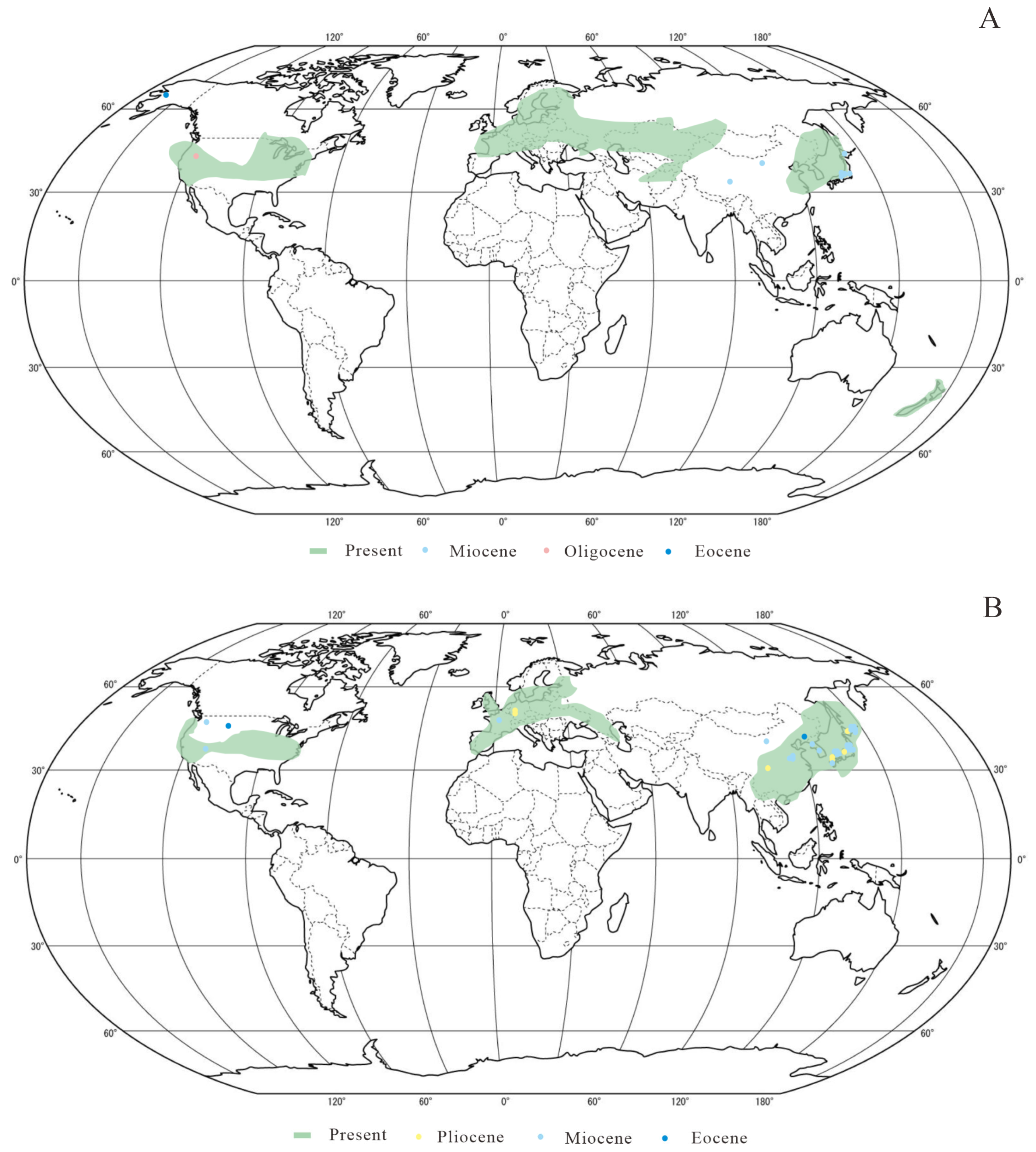
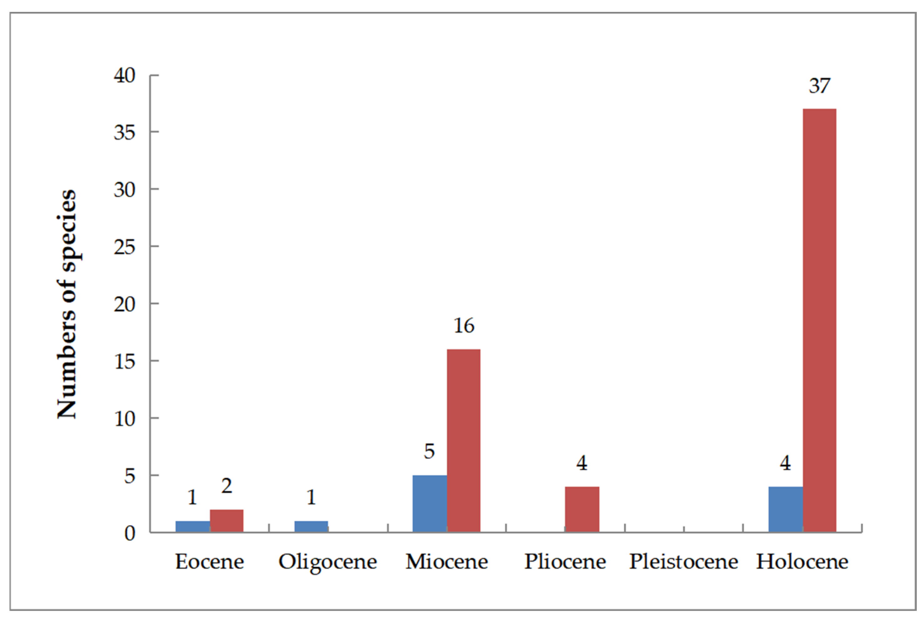
4.1. Paleogeographic Analysis of Sections Ginnala and Platanoidea

The earliest fossil record of section Ginnala is represented by the samara fossil of Acer sp. from the Eocene in Alaska [7]. Molecular phylogenetic analyses suggest that this section diverged approximately 48 million years ago [17], indicating a potential origin during the Eocene. During this epoch, Acer plants were likely able to thrive in high-latitude areas, attributable to the generally warm global climate, which is consistent with findings that large temperate plant communities flourished in these areas during the Eocene [48]. In the Oligocene, the plants of this section were restricted to the western regions of North America [7]. However, with the onset of the Miocene, section Ginnala became more widespread (Figure 8A), predominantly in the mid-latitude areas of East Asia [43,44,49] (Table 3). During the Miocene, the climate in eastern Asia was relatively warm [10,50,51], providing favorable conditions for the growth of Acer species. Based on the change in species numbers of section Ginnala [7,43,44,46,49] (Figure 9), we hypothesize that after the early Oligocene, the section may have dispersed from North America to East Asia via the Bering Land Bridge. This land bridge served as a crucial biological exchange route between the Americas and the Eurasian continent prior to the Pliocene, facilitating significant exchanges of temperate plant species [52]. There is a notable absence of sect. Ginnala fossil records from the Pliocene and Pleistocene. At present, section Ginnala is distributed across the Eurasian continent, North America, and New Zealand of the Southern Hemisphere, which indicates that the current climate is relatively suitable for the harboring of this section. Distinct to the distribution of sect. Ginnala fossils, the habitats of this section expand to the Southern Hemisphere, which may be attributed to anthropogenic activities, including its introduction and cultivation [53].
The earliest fossil records of section Platanoidea are traced back to the Eocene, and include Acer subpictum Saporta from East Asia [54] and Acer hueberi Wolfe et Tanai from North America [7]. Additionally, molecular phylogenetic research has indicated that this section diverged approximately 43 million years ago [17]. The integration of the aforementioned evidence suggests that this section originated prior to the late Eocene. According to two groups of section Platanoidea fossils in North America and East Asia, it is hypothesized that this section may have spread between East Asia and North America via the Bering Land Bridge during the Eocene. However, it is a pity that fossils of section Platanoidea from the Oligocene are not reported as yet (Figure 8B). Similar to section Ginnala, the Miocene species of section Platanoidea are abundant (Figure 9), displaying a wide distribution across the Americas, Europe, and Asia, with a concentration in East Asia (Figure 8B). However, during the Pliocene, the species number of sect. Platanoidea decreased from 16 to 4 (Figure 9), and the distribution range of this section became smaller in area. The majority of them were located in the mid-latitude areas of East Asia, and some were in Western Europe, but not in North America (Figure 8B). This phytogeographical change may be due to Late Tertiary extinctions having greater impacts in Europe and America than in East Asia [55]. Today, section Platanoidea is again widely distributed across the temperate regions of North America and Eurasia, which likewise seems to suggest that the present climate conditions are favorable for the survival of section Platanoidea.
Table 3. Acer section Ginnala and Platanoidea fossil records.
SectionSpeciesOrganAgeLocalityReference
Section
Ginnala
Acer tataricum L.LeafEarly MioceneInner Mongolia, China[56]
Acer pretataricum XiaoSamaraEarly MioceneInner Mongolia, ChinaThis paper
Acer subginnala GuoSamaraMioceneQinghai, China[43]
Acer pseudoginnala Tanai et OnoeSamaraMioceneHokkaido, Japan[46]
SamaraMioceneFukushima, Japan[44]
Acer prototataricum Tanai et Suzuki SamaraMioceneHokkaido, Japan[46]
LeafMioceneHokkaido and Honshu, Japan[49]
Acer ashwilli Wolfe et TanaiLeaf and samaraEarly OligoceneOregon, U.S.A.[7]
Acer sp.SamaraEoceneAlaska, U.S.A.[7]
Section
Platanoidea
Acer cf. cappadocicum GleditschLeaf and samaraPlioceneFrankfurt, Germany[57]
Acer cf. platanoides L.Leaf and samaraPlioceneFrankfurt, Germany[57]
LeafMioceneFrance[57]
Acer pictum ThunbgLeafPlioceneNagasaki, Japan[58]
Acer subpictum SaportaLeafPlioceneSichuan, China[51]
Leaf and samaraEarly Miocene to Late PlioceneJapan[49]
Leaf and samaraMioceneShandong, China[54]
Leaf and samaraMioceneHokkaido, Japan[46]
Leaf and samaraEoceneLiaoning, China[54]
Acer scolliae MacGinitieLeaf and samaraEarly MioceneNevada, U.S.A.[7]
Late MioceneColombia, U.S.A.[7]
Acer cf. mono MaximSamaraEarly MioceneInner Mongolia, ChinaThis paper
Acer chiharae Huzioka et NishidaLeafMioceneNiigata, Japan[59]
Acer huziokae Tanai LeafMioceneTottori, Japan[27]
Acer rotundatum HuziokaLeaf and samaraMioceneSouth Korea[27]
Acer shanwangense Tanai LeafMioceneShandong, China[27]
Acer palaeoplatanoides EndoSamaraMioceneFukushima, Japan[27]
MioceneHokkaido, Japan[46]
MioceneJapan[49]
SamaraMioceneNorth Korea[45]
Acer Matsuii Tanai et OnoeSamaraMioceneFukushima, Japan[44]
Acer sub Mayrii Tanai et OnoeSamaraMioceneKyushu, Japan[46]
Acer protodistylum EndoSamaraMioceneHokkaido, Japan[46]
Acer watarianum Takahashi et M. SuzukiTreeMioceneHokkaido, Japan[60]
Acer protojaponicum Tanai et OnoeLeaf and samaraMioceneFukushima, Japan[44]
Acer florinii Hu et ChaneyLeafMioceneTottori, Japan[61]
LeafMioceneShandong, China[54]
Acer integerrimum (Viviani) MassalongoLeafMioceneTottori, Japan[61]
Acer hueberi Wolfe et TanaiSamaraLate EoceneMontana, U.S.A.[7]

4.2. Dispersal Pathway Comparison Between Acer Section Ginnala and Section Platanoidea

The center of origin and dispersal pathways of the genus Acer have been the subject of considerable debate [7,13,18,19,20]. There are three main hypotheses on the dispersal pathways of the entire genus, but different sections of Acer may follow distinct dispersal routes, leading to varied interpretations of their migration patterns. Consequently, this paper aimed to separately examine and compare the dispersal pathways of section Ginnala and section Platanoidea. The earliest fossil records of sections Ginnala and Platanoidea are found in the Eocene strata of North America and East Asia, respectively [17,51]. Molecular phylogenetic studies further indicate that both groups diverged during the Eocene [17]. Based on fossil evidence, this paper hypothesizes that section Ginnala may have originated in North America during the Eocene and subsequently spread to East Asia via the Bering Land Bridge during the Oligocene, establishing a widespread presence in East Asia by the Miocene. In contrast, section Platanoidea had already dispersed to both East Asia and North America during the Eocene, likely migrating between these regions via the Bering Land Bridge. During the Oligocene, section Platanoidea expanded from East Asia to Western Europe via the Turgai Strait, and by the Miocene, it exhibited a distribution across North America, East Asia, and Western Europe, demonstrating a broader range compared to section Ginnala during the same periods. This comparison reveals significant differences in the dispersal pathways and distribution ranges of sections Ginnala and Platanoidea, which may be attributed to their ecological adaptability. Notably, section Platanoidea appears to exhibit a greater capacity for climate adaptation.
In summary, the dispersal pathways of different sections within Acer are distinct. When employing various sections as specimens to reconstruct the overall dispersal history of the genus, differing conclusions may emerge. This variability contributes to the ongoing debates surrounding the evolutionary processes of Acer. Therefore, it is essential to study the dispersal pathways of Acer by categorizing them into specific sections.

Author Contributions

Conceptualization, H.D. and L.X.; field sampling, data processing, and measurement, Y.W., X.W., D.J., M.W. and J.L.; writing—original draft preparation, H.D., M.W. and L.X.; writing—review and editing, X.W. and L.X. All authors contributed suggestions and revised the article. All authors have read and agreed to the published version of the manuscript.

Funding

This research was funded by the Second Batch of Mineral Resources and Paleontological Fossil Protection Program in Gansu Province of China in 2022 (NO. 2022-109); the Program from the third Geological and Mineral Exploration Institute, Gansu Bureau of Geology and Mineral Resources (NO. HT2023270439); the Fundamental Research Funds for the Central Universities, CHD (Nos. 300102272206); and the Natural Science Basic Research Program in Shaanxi Province of China (Grant No. 2023-JC-YB-223).

Institutional Review Board Statement

Not applicable.

Data Availability Statement

The original contributions presented in this study are included in the article Further inquiries can be directed to the corresponding author.

Acknowledgments

We thank Xing Wang, Hongyu Wang, Jian Wang, and Man Yuan for collecting the fossils.

Conflicts of Interest

The authors declare no conflicts of interest.

References

  1. Xu, T.Z.; Chen, Y.S.; de Jong, P.C.; Oterdoom, H.J.; Chang, C.S. Aceraceae. In Flora of China; Wu, Z.Y., Raven, P.H., Eds.; Science: Beijing, China, 2008; Volume 11, pp. 515–553. [Google Scholar]
  2. Li, L.; Lin, L.-J.; Zhu, Z.-Y.; Ding, Y.-L.; Kuai, B.-K. Studies on the Taxonomy and Molecular Phylogeny of Acer in China. Acta Hortic. Sin. 2017, 44, 1535–1547. [Google Scholar]
  3. Huang, Y.J.; Zhu, H.; Chen, W.Y.; Zhou, Z.K. Intraspecific variation in samara morphology of Acer and its implication in fossil identification. Plant Divers. Resour. 2013, 35, 295–302. [Google Scholar] [CrossRef]
  4. Xu, T.Z. Geographical distribution of Aceraceae. Yunnan Bot. Res. 1996, 18, 43–50. [Google Scholar]
  5. Dong, P.-B.; Wang, R.-N.; Afzal, N.; Liu, M.-L.; Yue, M.; Liu, J.-N.; Tan, J.-L.; Li, Z.-H. Phylogenetic relationships and molecular evolution of woody forest tree family Aceraceae based on plastid phylogenomics and nuclear gene variations. Genomics. 2021, 113, 2365–2376. [Google Scholar] [CrossRef]
  6. Newberry, J.S. The Later Extinct Floras of North America; Geological Survey Monographs; U.S. Government Printing Office: Washington, DC, USA, 1898; Volume 35, pp. 1–295. [Google Scholar]
  7. Wolfe, J.A.; Tanai, T. Systematics, phylogeny, and distribution of Acer (maples) in the Cenozoic of western North America. J. Fac. Sci. Geol. Mineral. 1987, 22, 1–246. [Google Scholar]
  8. Crane, P.R.; Manchester, S.R.; Dilcher, D.L. A preliminary survey of fossil leaves and well-preserved reproductive structures from the Sentinel Butte Formation (Paleocene) near Almont, North Dakota. Field Geol. 1990, 20, 1–63. [Google Scholar]
  9. Budantsev, L.Y. The Fossil Flora of the Paleogene Climatic Optimum in North Eastern Asia. In Cenozoic Plants and Climates of the Arctic; Boulter, M.C., Fisher, H.C., Eds.; Springer: Berlin/Heidelberg, Germany, 1994; pp. 297–313. [Google Scholar]
  10. Zachos, J.; Pagani, M.; Sloan, L.; Thomas, E.; Billups, K. Trends, rhythms, and aberrations in global climate 65 Ma to present. Science 2001, 292, 686–693. [Google Scholar] [CrossRef]
  11. LePage, B.A. A new species of Tsuga (Pinaceae) from the Middle Eocene of Axel Heiberg Island, Canada, and an assessment of the evolution and biogeographical history of the genus. Bot. J. Linn. Soc. 2003, 141, 257–296. [Google Scholar] [CrossRef]
  12. Qiu, Y.-X.; Fu, C.-X.; Comes, H.P. Plant molecular phylogeography in China and adjacent regions: Tracing the genetic imprints of Quaternary climate and environmental change in the world’s most diverse temperate flora. Mol. Phylogenetics Evol. 2011, 59, 225–244. [Google Scholar] [CrossRef]
  13. Xu, T.-Z. The systematic evolution and distribution of the genus Acer. Acta Bot. Yunnanica 1998, 20, 383–393. (In Chinese) [Google Scholar]
  14. Pax, F. Aceraceae. In Das Pflanzenreich, 4th ed.; Engler, H.G.A., Ed.; W. Engelmann: Leipig, Germany, 1902. [Google Scholar]
  15. Pojarkova, A.Z. Botanico-geographical survey of the maples in USSR, in cconnection with the history of the whole genus Acer L. Acta Inst. Bot. Acad. Sci. USSR 1933, 1, 224–374. [Google Scholar]
  16. Gao, J.; Liao, P.-C.; Huang, B.H.; Yu, T.; Zhang, Y.Y.; Li, J.Q. Historical biogeography of Acer L. (Sapindaceae): Genetic evidence for Out-of-Asia hypothesis with multiple dispersals to North America and Europe. Sci. Rep. 2020, 10, 21178. [Google Scholar] [CrossRef]
  17. Areces-Berazain, F.; Hinsinger, D.D.; Strijk, J.S. Genome-wide supermatrix analyses of maples (Acer, Sapindaceae) reveal recurring inter-continental migration, mass extinction, and rapid lineage divergence. Genomics 2021, 113, 681–692. [Google Scholar] [CrossRef] [PubMed]
  18. Huang, X.-F. Phylogeny and Historical Biogeography of Acer. Ph.D. Thesis, University of Missouri-St. Louis, St. Louis, MO, USA, 2002. [Google Scholar]
  19. Kvacek, Z. Connecting Links Between the Arctic Paleocene and European Tertiary Floras. In Cenozoic Plants and Climates of the Arctic; Boulter, M.C., Fisher, H.C., Eds.; Springer: Berlin/Heidelberg, Germany, 1994; pp. 251–266. [Google Scholar]
  20. Boulter, M.C.; Benfield, J.N.; Fisher, H.C.; Gee, D.A.; Lhotak, M. The evolution and global migration of the Aceraceae. Philosophical Trans. R. Soc. London. Ser. B Biol. Sci. 1996, 351, 589–603. [Google Scholar]
  21. Wheeler, E.A.; Manchester, S.R. A diverse assemblage of Late Eocene woods from Oregon, western USA. Foss. Impr. 2022, 77, 299–329. [Google Scholar] [CrossRef]
  22. Buechler, W.K.; Dunn, M.T.; Rember, W.C. Late Miocene Pickett Creek Flora of Owyhee County, Idaho. Contrib. Mus. Paleontol. 2007, 31, 305–362. [Google Scholar]
  23. Wang, Y.-F.; Shao, Y.; Li, B.-K.; Liu, K.-N.; Xie, S.-P. Acer leaves and samaras from the late Miocene of Lincang, Yunnan Province. Geol. J. China Univ. 2015, 21, 105–116. [Google Scholar]
  24. Chen, Y.-F.; Wong, W.O.; Hu, Q.; Liufu, Y.-Q.; Xie, Z.-M. A new fossil-species of Acer (Sapindaceae) from the Ningming Basin in Guangxi, South China. Phytotaxa 2017, 298, 158–164. [Google Scholar] [CrossRef]
  25. Jeong, E.K.; Kim, K.; Suzuki, M.; Kim, J.W. Fossil woods from the Lower Coal-bearing Formation of the Janggi Group (Early Miocene) in the Pohang Basin, Korea. Rev. Palaeobot. Palynol. 2009, 153, 124–138. [Google Scholar] [CrossRef]
  26. Jin, J.H. Two Eocene fossil fruits from the Changchang basin of Hainan Island, China. Rev. Palaeobot. Palynol. 2009, 153, 150–152. [Google Scholar] [CrossRef]
  27. Tanai, T. Revisions of Tertiary Acer from East Asia. J. Fac. Sci. Geol. Mineral. 1983, 20, 291–390. [Google Scholar]
  28. Mai, D.H. Die Endokarpien bei der Gattung Acer L. (Aceraceae)-Eine biosystematische Studie. Gleditschia 1984, 11, 17–46. [Google Scholar]
  29. Wang, Z.-E.; Cao, R.; Ding, H.; Huang, Y.-T.; Song, Z.-H.; Ding, S.-T.; Wu, J.-Y. Fossil samaras of Acer L. (Sapindaceae) from the Upper Pliocene of western Yunnan, southwestern China. Palaeobiodiversity Palaeoenvironments 2023, 103, 695–710. [Google Scholar] [CrossRef]
  30. Wang, H.-F.; Dai, T.-M.; Fan, S.-K.; Yang, X.-C. K-Ar daeing of Hannuoba basalts at Zhangjiakou. Geochimica 1985, 3, 206–215. (In Chinese) [Google Scholar]
  31. Zhang, Y.-S. Origin discussion of Cenozoic Hannuoba basalts in Shanxi Province. Geol. Surv. China 2018, 6, 58–67. [Google Scholar] [CrossRef]
  32. Inner Mongolia Autonomous Region Bureau of Geology and Mineral Resources. Regional Geology of Inner Mongolia Autonomous Region; Geology Press: Beijing, China, 1991; pp. 1–725. (In Chinese) [Google Scholar]
  33. Li, C.-K. A Tertiary Beaver from Changpei, Hopei Province. Vertebr. Palasiat. 1962, 1, 72–79. [Google Scholar]
  34. Guo, C.-Q.; Li, Y.; Wu, P.-C.; Yao, J.-X. The phytogeographic significance of Early Miocene mosses from Weichang area, Hebei Province. Geol. Bull. China 2016, 35, 1976–1984. [Google Scholar]
  35. Li, D.-m.; Li, Q. Serial K-Ar Dating along the Volcanic Section in Zuoyun, Shanxi Province. Acta Geosci. Sin. 2003, 24, 559–562. [Google Scholar] [CrossRef]
  36. Zhang, W.-H.; Han, B.-F. K-Ar Chronology and Geochemistry of Jining Cenozoic basaits, Inner Mongolia, and geodynamic Implications. Acta Petrol. Sin. 2006, 22, 1597–1607. (In Chinese) [Google Scholar]
  37. Liang, J.-Q.; Leng, Q.; Höfig, D.F.; Niu, G.; Wang, L.; Royer, D.L.; Burke, K.; Xiao, L.; Zhang, Y.-G.; Yang, H. Constraining conifer physiological parameters in leaf gas-exchange models for ancient CO2 reconstruction. Glob. Planet. Change 2022, 209, 103737. [Google Scholar] [CrossRef]
  38. Xu, T.Z. Samara shape of Aceraceae and its implications in systematics and evolution. Guangxi Zhiwu 1996, 16, 109–122. (In Chinese) [Google Scholar]
  39. Mirle, C.; Burnham, R.J. Identification of asymmetrically winged samaras from the Western Hemisphere. Brittonia 1999, 51, 1–14. [Google Scholar] [CrossRef]
  40. Anderson, W.R.; Gates, B. Barnebya, a New Genus of Malpighiaceae from Brazil. Brittonia 1981, 33, 275. [Google Scholar] [CrossRef]
  41. Gates, B. Banisteriopsis, Diplopterys (Malpighiaceae); New York Botanical Garden Press: New York, NY, USA, 1982. [Google Scholar]
  42. Tan, K.; Dong, S.-p.; Lu, T.; Zhang, Y.-J.; Xu, S.-T.; Ren, M.-X. Diversity and evolution of samara in angiosperm. Chin. J. Plant Ecol. 2018, 42, 806–817. [Google Scholar] [CrossRef]
  43. Guo, S.-x. Miocene flora of Zeku, Qinghai. Acta Palaeontol. Sin. 1980, 5, 406–411+441. (In Chinese) [Google Scholar]
  44. Tanai, T.; Onoe, T. A Miocene flora from the northern part of the Joban Coal Field, Japan. Bull. Geol. Surv. Jpn. 1959, 10, 261–286. [Google Scholar]
  45. Endo, S. On the fossil Acer from Japan, Korea and south Manchuria. Short Pap. Inst. Geol. Paleontol. 1950, 1, 11–17. [Google Scholar]
  46. Tanai, T.; Suzuki, N. Miocene Maples from Southwestern Hokkaido, Japan. Fac. Sci. Geol. Mineral. 1960, 10, 551–570. [Google Scholar]
  47. Knowlton, F.H. Fossil Flora of the John Day Basin, Oregon. U.S. Ceol. Sury. Bull. 1902, 204, 153. [Google Scholar]
  48. Jiang, Y.; Gao, M.; Meng, Y.; Wen, J.; Ge, X.-J.; Nie, Z.-L. The importance of the North Atlantic land bridges and eastern Asia in the post-Boreotropical biogeography of the Northern Hemisphere as revealed from the poison ivy genus (Toxicodendron, Anacardiaceae). Mol. Phylogenetics Evol. 2019, 139, 106561. [Google Scholar] [CrossRef]
  49. Tanai, T. Neogene Floral Change in Japan. Fac. Sci. Geol. Mineral. 1961, 11, 119–398. [Google Scholar]
  50. Mosbrugger, V.; Utescher, T.; Dilcher, D.L. Cenozoic continental climatic evolution of Central Europe. Proc. Natl. Acad. Sci. USA 2005, 102, 14964–14969. [Google Scholar] [CrossRef]
  51. Tao, J.-R.; Zhou, Z.-K.; Liu, Y.-S. Development and Evolution of China’s Flora from the Late Cretaceous to the Cenozoic; Science Press: Beijing, China, 2000; pp. 49–290. [Google Scholar]
  52. Sanmartin, I.; Enghoff, H.; Ronquist, F. Patterns of animal dispersal, vicariance and diversification in the Holarctic. Biol. J. Linn. Soc. 2001, 73, 345–390. [Google Scholar] [CrossRef]
  53. Ranatunga, D.; Hoare, B. Auckland Museum Botany Collection; Version 1.96; Auckland War Memorial Museum: Auckland, New Zealand, 2024; Occurrence Dataset; Available online: https://www.gbif.org/occurrence/2883314201 (accessed on 19 October 2024).
  54. Writing Group of Cenozoic Plants of China (WGCPC) (Ed.) Cenozoic Plants from China. In Fossil Plants of China; Science Press: Beijing, China, 1978; pp. 1–232. (In Chinese) [Google Scholar]
  55. Loidi, J.; Marcenò, C. The Temperate Deciduous Forests of the Northern Hemisphere. A review. Mediterr. Bot. 2022, 43, e75527. [Google Scholar] [CrossRef]
  56. Liang, J.-Q.; Xiao, L.; Li, X.-C.; Wang, X.; Wang, Q. The first discovery of the fossils of Acer from the Miocene epoch in Inner Mongolia. Geol. Rev. 2019, 65, 69–70. [Google Scholar]
  57. Ivan, G.; Johanna, K.E. The genus Acer from the lower/middle Pleistocene Sisian Formation, Syunik region, South Armenia. Rev. Palaeobot. Palynol. 2011, 165, 111–134. [Google Scholar]
  58. Nathorst, A.G. Contributions a la flore fossile du Japon. KgI. Svensk. Vel. Akad. Handt. 1883, 20, 3–92. [Google Scholar]
  59. Huzioka, K.; Nishida, S. The Seki flora of the island Sado. Sodo Mus. Publ. 1960, 3, 1–26. [Google Scholar]
  60. Takahashi, A.; Suzuki, M. Two new fossil woods of Acer and a new combination of Prunus from the Tertiary of Japan. Bot. Mag. Tokyo. 1988, 101, 473–481. [Google Scholar] [CrossRef]
  61. Tanai, T.; Ozaki, K. The genus Acer from the Upper Miocene in Tottori Prefecture, western Japan. J. Fac. Sci. Hokkaido Univ. 1977, 17, 575–606. [Google Scholar]
Figure 1. Distribution of extant and fossil Acer.
Diversity 17 00218 g001
Figure 2. Geographic position of fossil sites and stratigraphic column of the Hannuoba Formation in Inner Mongolia. (A) Geographic position of fossil site. (B) Stratigraphic column of the Hannuoba Formation in Inner Mongolia.
Diversity 17 00218 g002
Figure 3. Sketch of leaf and samara morphology of Acer fossils (modified from [7,38]).
Diversity 17 00218 g003
Figure 4. Characteristics of Acer pretataricum sp. nov. samaras. (A) ELS-19-347. (B) ELS-21-234A; red square representing the position of (I) and (J). (C) ELS-21-234B; red square representing the position of (K). (D) ELS-19-228. (E) ELS-19-374A. (F) ELS-19-374B. (G) ELS-19-146. (H) ELS-18-97. (I,J) Close-up of nutlet with yellow arrow pointing to vein-branching nutlet veins. (K) Close-up of samara wing; red arrow pointing to vein-branching wing veins and green arrow pointing to reticulodromous. (AH) Scale bar = 0.5 cm. (IK) Scale bar = 0.4 cm.
Diversity 17 00218 g004
Figure 5. Epidermal characteristics of fossil samaras of Acer pretataricum sp. nov. (AC,GI) and living A. tataricum (D,E,JL), represented by scanning electron microscope images and light microscope pictures. (A) Outer epidermal cell of fossil A. pretataricum sp. nov. (B,H) Stomata apparatus of fossil A. pretataricum sp. nov. (C) Fossil A. pretataricum sp. nov. trichome base. (D) Inner epidermis of the extant A. tataricum. (E,K) Stomata apparatus of the extant A. tataricum. (F) Extant A. tataricum trichome base. (G) Inner epidermal cell of fossil A. pretataricum sp. nov. with yellow arrow pointing to stomata apparatus. (I) Glandular trichomes of fossil A. pretataricum sp. nov. with red arrow pointing to glandular trichomes. (J) Inner epidermal cell of extant A. tataricum. Yellow arrow pointing to stomata apparatus. (L) Glandular trichomes of extant A. tataricum with red arrow pointing to glandular trichomes. (A,C,D,F,I,J) Scale bar = 50 μm. (B,E) Scale bar = 10 μm. (G,H,K,L) Scale bar = 20 μm.
Diversity 17 00218 g005
Figure 6. Seven extant samaras whose features are similar to those of Acer pretataricum sp. nov. The pictures are from https://www.cvh.ac.cn/index.php (accessed on 25 October 2023). (A) Acer buergerianum Mi. (B) Acer caudatum Wall. (C) Acer davidii Frach. (D) Acer monspessulanum L. (E) Acer pseudoplatanus L. (F) Acer tetramerum Pax. (G,H) Acer tataricum L. (I) Magnified nutlet of Acer tataricum with yellow arrow pointing to nutlet veins. (J) Magnified samara wings of (H); red arrow pointing to vein-branching wing veins and green arrow pointing to reticulodromous. (AJ) Scale bar = 0.8 cm.
Diversity 17 00218 g006
Figure 7. Characteristics of cf. Acer mono samaras and five extant samaras whose features are similar to those of cf. Acer mono. The pictures of (KO) are from https://www.cvh.ac.cn/index.php (accessed on 25 October 2023). (A) ELS-18-70. (B) ELS-19-84; red square representing the position of (G). (C) ELS-18-188. (D) ELS-21-232; red square representing the position of (H). (E) ELS-18-99A; red square representing the position of (I). (F) ELS-19-258. (G) Close-up of samara wings of cf. Acer mono with red arrow pointing to vein-branching wing veins. (H) Close-up of samara wings of cf. Acer mono with yellow arrow pointing to vein-branching nutlet veins. (I) Close-up of samara wings of cf. Acer mono with yellow arrow pointing to vein-branching nutlet veins. (J) Close-up of samara wings of Acer mono with yellow arrow pointing to vein-branching nutlet veins. (K) Acer grosseri Pax. (L) Acer pensylvanicum L. (M) Acer rufinerve Sieb. et Zucc. (N,O) Acer mono Maxim; red square representing the position of (J). (AF) Scale bar = 0.5 cm. (GJ) Scale bar = 0.2 cm. (KO) Scale bar = 0.8 cm.
Diversity 17 00218 g007
Figure 8. Global distribution ranges and fossil records of sections Ginnala and Platanoidea. (A) Section Ginnala; (B) section Platanoidea.
Diversity 17 00218 g008
Figure 9. The species number of sections Ginnala and Platanoidea in each era (statistics of species are from Table 3). Blue columns represent species numbers of section Ginnala and red represent species numbers of section Platanoidea.
Diversity 17 00218 g009
Table 2. Comparison of the present fossil’s samaras with similar extant species of Acer.
SectionSpeciesGL * (cm)GW * (cm)DA *NL * (cm)NW * (cm)NI *NS *SulusNV *
AcerAcer pseudoplatanus L.4–100.5–1.2Variously1.50.5Moderately inflatedSphericalAuxeticLongitudinal
ArgutaAcer tetramerum Pax2–3.51–1.2Acute or nearly erect0.80.6Strongly inflatedLanceolateAuxeticRidged
GinnalaAcer ginnala Maxim2–40.4–1Acute or nearly erect0.5–10.5Flattened to slightly inflatedEllipticalAuxeticReticular
GoniocarpaAcer monspessulanum L.2–2.50.7–1.1Acute0.6–0.90.6–0.8Strongly inflatedOrbiculateAuxeticReticular
IntegrifoliaAcer buergerianum Miq2–2.50.8–1.2Acute or nearly erect3.6–63.6–6Strongly inflatedEllipticalAuxeticLongitudinal
MacranthaAcer davidii Frach.2.5–31–1.5Obtuse or nearly horizontal0.7–1.50.7–1.5Flattened to slightly inflatedEllipticalAuxeticSub-parallel
MacranthaAcer grosseri Pax2.5–2.90.5Acute or nearly
erect
0.70.4Flattened EllipticalObsoleteSub-parallel
MacranthaAcer pensylvanicum L.1.8–2.70.8–1.2Acute0.5–1.10.35–0.6Flattened EllipticalObsoleteLongitudinal
MacranthaAcer rufinerve Sieb. et Zucc.2–3-Obtuse0.40.4Strongly inflatedSphericalObsoleteReticular
PlatanoideaAcer mono Maxim., 18562–2.50.5–1Acute or nearly
horizontal
0.5–1.30.5–0.8Flattened to slightly inflatedEllipticalObsoleteSub-parallel
SpicataAcer caudatum Wall2.5–2.80.7–0.9Acute or nearly erect0.80.6Moderately to strongly inflatedEllipticalAuxeticReticular or sub-parallel
Acer pretataricum sp. nov.2–40.42–1.2Acute or nearly erect0.55–0.90.5–1Slightly inflatedOvateAuxeticReticular
cf. Acer mono Maxim2–2.50.75–1.2Acute or nearly obtuse0.65–0.90.5–0.7Flattened to slightly inflatedEllipticalObsoleteSub-parallel
* GL = general length; GW = general width; DA = divergent angle; NL = nutlet length; NW = nutlet width; NI = nutlet inflation; NS = nutlet shape; NV = nutlet vein.
Disclaimer/Publisher’s Note: The statements, opinions and data contained in all publications are solely those of the individual author(s) and contributor(s) and not of MDPI and/or the editor(s). MDPI and/or the editor(s) disclaim responsibility for any injury to people or property resulting from any ideas, methods, instructions or products referred to in the content.

