Article

Insights into the Taxonomy of the Genus Chrysastrella (Chrysophyceae), with Establishment of Chrysastrellaceae fam. nov.

1
K.A. Timiryazev Institute of Plant Physiology, Russian Academy of Sciences, Botanicheskaya Street 35, 127276 Moscow, Russia
2
Severtsov Institute of Ecology and Evolution, Russian Academy of Sciences, Leninsky Prospect 33, 119071 Moscow, Russia
3
Institute of Biology, Komi Scientific Center, Ural Branch, Russian Academy of Sciences, Kommunisticheskaya Street, 28, 167982 Syktyvkar, Russia
4
Department of Ecology and Technosphere Safety, Murmansk Arctic University, Kapitana Egorova Street 15, 183038 Murmansk, Russia
5
All-Russian Collection of Microorganisms (VKM), Scryabin Institute of Biochemistry and Physiology of Microorganisms, Russian Academy of Sciences, 142290 Pushchino, Russia
*
Author to whom correspondence should be addressed.
Diversity 2025, 17(12), 824; https://doi.org/10.3390/d17120824
Submission received: 26 October 2025 / Revised: 22 November 2025 / Accepted: 25 November 2025 / Published: 28 November 2025
(This article belongs to the Section Freshwater Biodiversity)

Abstract

Chrysastrella is a small genus of the Ochromonas-like chrysophytes, taxonomy and phylogenetic placement of which remained unclear. For a long time Chrysastrella was considered a cyst genus, i.e., a morphogenus based on the structure of stomatocysts, the resting stages of chrysophytes. We isolated several new strains of C. paradoxa from the peat bogs in Murmansk Region and the Republic of Komi (Russia) and studied them using light and scanning electron microscopy as well as molecular techniques. We showed that morphological differences between C. paradoxa, C. minor and C. breviappendiculata are within the range of variability of stomatocysts during development. So, we synonymized C. minor and C. breviappendiculata with C. paradoxa. Molecular phylogenetic analysis based on SSU rDNA and rbcL sequences revealed that Chrysastrella belongs to the order Chrysosaccales. A new monotypic family, Chrysastrellaceae fam. nov., was formally described to accommodate this genus.

1. Introduction

Ochromonas-like chrysophytes comprise unicellular mixotrophic flagellates with two unequal flagella [1]. They are often the most dominant bacterivorous flagellates in freshwater ecosystems, and they present an important link between bacterial production and higher trophic levels [2,3]. Ochromonas is a large genus with nearly 130 species and varieties, many of which are invalid [4]. Molecular studies revealed the polyphyly of the genus Ochromonas [1,2], but its taxonomic revision is ongoing.
The genus Chrysastrella Chodat is a recently redefined genus of the Ochromonas-like chrysophytes. Initially, it was established by Chodat [5] for three species, namely, C. paradoxa, C. minor and C. breviappendiculata, which had spherical cells with a short collar and projections of different lengths on the cell surface. Chodat did not observe the flagellate stage and believed that the non-motile stage predominates in the life cycle of Chrysastrella. However, Pascher [6,7] pointed out that this genus represents typical chrysophycean resting cysts (stomatocysts). Conrad [8] described a genus nearly identical to Chrysastrella, Echinochrysis, with a single species, E. chodatii, and observed the Ochromonas-like zoospores. Later, a number of cyst taxa were added to Chrysastrella [9,10,11,12,13,14,15].
In the 1970s Hibberd discovered a new species of Ochromonas, O. tuberculata (‘tuberculatus’), and studied the ultrastructure of its cells and stomatocysts [16,17]. Molecular phylogeny of the genus Ochromonas was performed by Andersen et al. [1] showing that O. tuberculata is unrelated to the type of Ochromonas, O. triangulata Vysotskij. Since stomatocysts of O. tuberculata were extremely similar to those of Chrysastrella paradoxa, Andersen et al. [1] re-identified the authentic strain of the former species (CCAP 933/27) as C. paradoxa. The other strain (CCMP 1861) was identified as C. breviappendiculata [1] despite the non-significant differences in DNA sequences and morphology of stomatocysts. Both strains formed a separate lineage within the clade of uncertain taxonomic placement (incertae sedis) [1].
This article aims to study morphology and phylogeny of three newly isolated strains of Chrysastrella to clarify the taxonomy and phylogenetic placement of the genus.

2. Materials and Methods

Material for this study was collected from a peat bog on the bank of the Paz River (N 69°23.489′, E 29°45.388′), Murmansk Region, Russia, on 19 June 2019 [18] and an unnamed peat bog in the Vychegda River basin (N 61°40′08″, E 51°02′52″), env. of Syktyvkar, Republic of Komi, Russia, on 18 September 2019. The strains DK1 and DK2 originate from the Murmansk Region. The strain SYKOA Chr-002-19 originates from the Republic of Komi. It was deposited in the Culture Collection of Algae at the Institute of Biology, Komi Scientific Center of the Ural Branch of the Russian Academy of Sciences (SYKOA). The cells were isolated by micropipetting and cultivated in WC medium [19] at 12 °C, under a light intensity of 20–40 μmol photons/m2·s−1, with a light/dark cycle of 12:12 h.
Light microscopical (LM) observations were performed with a Zeiss Axio Scope A1 microscope (Carl Zeiss Microscopy GmbH, Gottingen, Germany) equipped with an oil immersion objective (×100, n.a. 1.4, differential interference contrast) and Axiocam Erc 5s camera (Carl Zeiss Microscopy GmbH, Gottingen, Germany).
For electron microscopical studies of stomatocysts, an aliquot of each monoclonal culture was initially washed by repeated centrifugation in deionized water to remove the culture medium. Drops of the washed cultures were placed on aluminum stubs, air-dried, sputter-coated with gold, and observed with a TESCAN Vega III (TESCAN, Brno, Czech Republic) (Borissiak Paleontological Institute RAS, Moscow, Russia) scanning electron microscope, operated at 10 kV and 8 mm distance.
The genomic DNA of monoclonal strains was extracted using the InstaGene™ Matrix kit (Bio-Rad, Hercules, CA, USA) following the manufacturer’s protocol. Amplification of partial SSU rRNA gene fragments (1624 bp) was carried out using primer pairs 18S-F [20] and 18L [21]. The rbcL cpDNA fragment (656 bp) was amplified using primers rbcL_2F [22] and Synura_rbcLR [23]. Amplification, purification and sequencing of studied fragments were performed as described in the previous studies [24,25].
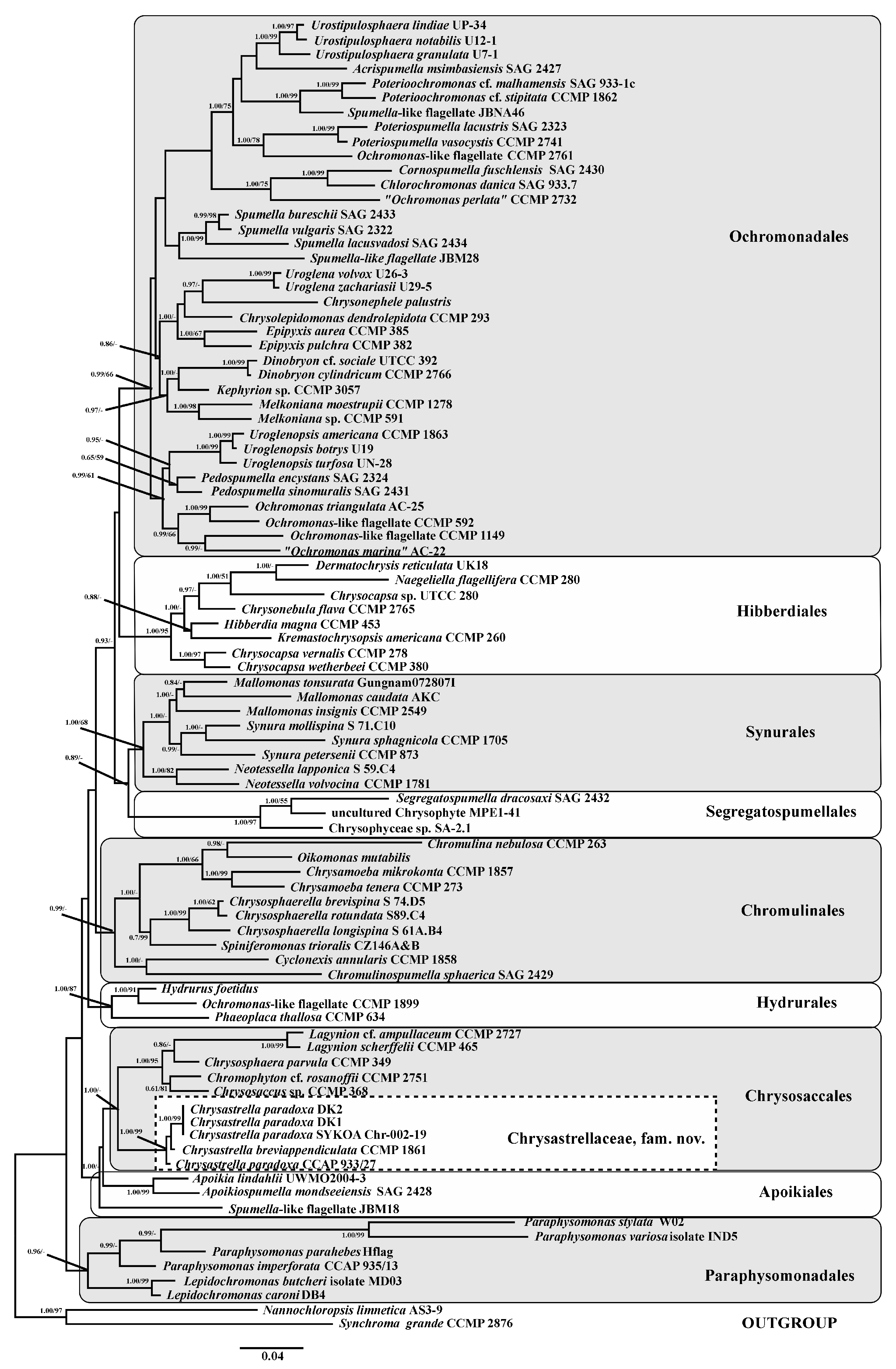
Received sequences were checked manually and assembled after using MEGA X [26]. Newly determined sequences and GenBank sequences of 85 different Ochromonadales, Hibberdiales, Synurales, Segregatospumellales, Chromulinales, Hydrurales, Chrysosaccales, Apoikiales and Paraphysomonadales strains were included in the alignment. Two strains, Nannochloropsis limnetica Krienitz, Hepperle, H.-B. Stich & W. Weiler (isolate AS3-9) and Synchroma grande R. Schnetter (strain CCMP 2876), were added to the dataset as outgroup. The GenBank accession numbers of sequences used in the analysis are listed in Table S1. The sequences were aligned using either the global SILVA alignment in the SINA v1.2.11 software [27] for SSU rDNA, or MAFFT v7 with auto-strategy [28] for rbcL cpDNA. The resulting SSU rDNA + rbcL cpDNA dataset (2338 bp) was partitioned into different genetic regions, and the most appropriate substitution model for each partition was estimated separately, using the Bayesian information criterion (BIC) in the jModelTest 2.1.10 [29]. The most fit model was selected: GTR + G + I for the SSU rDNA. For each codon position of the protein-coding rbcL cpDNA gene, the best model was also tested. The BIC-based model selection procedure selected the following models: GTR + G + I for the first and third codon positions and GTR + I for the second codon position. Bayesian Inference (BI) analysis was conducted with MrBayes-3.2.5 [30]. Three “hot” and one “cold” Markov chains were run for 15 × 106 cycles in two repetitions with the selection of each 100th generated tree. Phylogenetic tree and posterior branching probabilities were obtained after discarding the first 25% to produce estimated parameter models of nucleotide substitutions and likelihood. The Markov Chain converged, and the average standard deviation of split frequencies (ASDSF) was equal to 0.004797. The maximum likelihood phylogeny (ML) was constructed using MEGA X with the GTR + G+ I model, also chosen by jModelTest 2.1.10, and 1000 replicas were used to calculate bootstrap values. Viewing and editing of trees were carried out in the programs FigTree (ver 1.4.2) and Adobe Photoshop CC (19.0).

3. Results

3.1. Morphological Observations

Cell morphology of all three examined strains was identical. Cells were ovate (18.5–18.7 µm long, 13.5–13.9 µm wide) to round (16–22 µm in diameter), highly metabolic, with an obliquely truncated anterior end and rounded posterior end. The cell outline had a verrucose appearance due to the presence of round (2.0–2.3 µm in diameter) discobolocysts. There was a single large parietal chloroplast with a red eyespot at the anterior part. Two unequal flagella arose subterminally from the oblique anterior end. The longer flagellum was slightly longer than the cell (~24 µm), and the short flagellum was 1.5–2 µm long (Figure 1A,B).
Stomatocysts of all investigated strains were similar. However, in strains DK1 and DK2, only mature stomatocysts have been documented (Figure 1C,D), whereas in strain SYKOA Chr-002-19 stomatocysts have been observed in different maturity stages (Figure 1E,F). Stomatocysts were spherical (12.6–16.1 μm in diameter) with a cylindrical to obconical collar (2.0–3.9 μm wide, 0.6–1.2 μm high) with a folded and turned-down apical margin. The pore was regular (0.6–0.8 μm in diameter) and surrounded by a flat planar annulus. The cyst surface was smooth and ornamented with spines, the length of which varied (0.5–7.7 µm long) depending on the maturity.

3.2. Phylogenetic Analysis

Phylogenetic analysis using maximum likelihood and Bayesian inference from a concatenated dataset of nuclear-encoded small subunit rRNA and plastid-encoded rbcL genes revealed that Chrysastrella is a member of the order Chrysosaccales (Figure 2).
Our strains DK1, DK2 and SYKOA Chr-002-19 were genetically identical. They form a clade together with the authentic strain Ochromonas tuberculata CCAP 933/27 (=Chrysastrella paradoxa) and the strain C. breviappendiculata CCMP 1861 with high Bayesian (1.00) and ML (99) supports. We refer all these strains to a single species, Chrysastrella paradoxa.

4. Discussion

The limited number of diagnostic features made the structure of stomatocyst crucial for species delimitation in Ochromonas sensu lato (e.g., [31,32,33,34]). Even now stomatocyst morphology remains important in taxonomy of some chrysophyte genera, like Uroglena Ehrenberg and Urostipulosphaera Pusztai & Škaloud [35,36], Spumella Cienkowsky [37,38] or Dinobryon Ehrenberg [39].
Mature stomatocysts of Chrysastrella paradoxa have a distinctive morphology, including the numerous long spines, sometimes with branched ends, which makes species identification possible. Our previous study based on an abundant natural material revealed that stomatocysts of this species are highly variable [40]. All three taxa described by Chodat (C. paradoxa, C. minor and C. breviappendiculata) [5] were collected from the same site (Lac de Champex, Switzerland) and might represent a single species. Stomatocysts of C. minor are similar to those of C. paradoxa but differ in smaller size, lacking a collar, lesser number of spines and another type of its branching [5]. However, all these features can significantly vary [40]. C. breviappendiculata is characterized by the presence of extremely short spines [5]. Such spines are typical of immature stomatocysts. Indeed, we observed similar immature stomatocysts from the natural population [40] and in the culture of our strain SYKOA Chr-002-19 (see Figure 1E).
Andersen et al. [1] identified the strain CCMP 1861 as C. breviappendiculata despite the cyst diameter and spine length (Figure S35, [1]) corresponding to stomatocysts of C. paradoxa, not C. breviappendiculata. Since our strains and strain CCMP 1861 do not have significant nucleotide differences, we believe that they belong to the same species, C. paradoxa. Additionally, stomatocyst ultrastructure of our strain SYKOA Chr-002-19 and the strain CCMP 1861 are also extremely similar.
Although stomatocysts of Echinochrysis chodatii look similar to those of C. paradoxa, they are larger (16–26 µm), and the collar consists of several projections [8]. The flagellate stage of E. chodatii does not have a verrucose cell outline, so it is unlikely that it belongs to Chrysastrella.
There are several Ochromonas-like chrysophytes with verrucose cell outline, namely, Ochromonas crenata G.A. Klebs, O. verrucosa Skuja, O. sphaerocystis Matvienko, O. hovassei Bourrelly, etc., which are morphologically similar to Chrysastrella paradoxa. Kalina [41] attempted to revise O. crenata and synonymized all similar taxa with it, ignoring the differences in the structure of stomatocysts. According to Andersen et al. [1], O. sphaerocystis formed a clade X within the Ochromonadales, so it is unrelated to Chrysastrella. Unfortunately, stomatocysts have not been described for O. verrucosa and O. hovassei, which does not allow their morphological comparison with C. paradoxa. Thus, synonymization of other verrucose Ochromonas-like taxa with Chrysastrella seems to be premature until additional study.
The taxonomic placement of Chrysastrella is still confusing. Originally, Chodat [5] placed Chrysastrella within the family Chrysostomataceae (‘Chrysostomatacées’) together with other cyst genera, namely, Chrysostomum Chodat, Phaeocitrus Chodat, Selenophaea Chodat and Clathrostomum Chodat. However, he noted that this classification is provisional [5]. Deflandre [10] amended the family Chrysostomataceae to include the cyst genera Carnegia Pantoscek, Clericia Frenguelli and Outesia Frenguelli.
In the phylogenetic analysis performed by Andersen et al. [1], Chrysastrella formed a clade I of uncertain placement (incertae sedis). According to Bock et al. [42], Chrysastrella belongs to the “Chrysosacca-Apoikiida” clade (Chrysosaccales + Chrysastrella and Apoikiales). Malavasi et al. [43] placed it within the Chrysosaccales. Our phylogenetic analysis also confirmed that Chrysastrella belongs to this order (see Figure 2).
The order Chrysosaccales was described by Bourrelly [44] to include palmelloid chrysophytes (Phaeosphaera W. West et G.S. West, Chrysosaccus Pascher, Chalkopyxis Pascher and Heimiochrysis Bourrelly). However, the current concept of this order based on the available SSU rDNA and rbcL sequences differs drastically, and now the order Chrysosaccales includes also the Chromulina-like (Chromophyton Woronin) and Ochromonas-like (Chrysastrella) flagellates, loricate flagellates (Chrysococcus G.A. Klebs) and even amoeboid loricate chrysophytes (Lagynion Pascher and Stylococcus Chodat) [45,46,47].
The sister clade is represented by the order Apoikiales (“Apoikiida”), which comprises colorless bacterivorous chrysophytes classified into three monotypic genera, Apoikia E. Kim et al., Apoikiospumella Boenigk and Pseudapoikia T. Pietsch, F. Nitsche, and H. Arndt [48,49,50].
Since Chrysastrella forms a distinct lineage within Chrysosaccales, a new family is proposed here:
Chrysastrellaceae Kapustin, fam. nov.
Solitary, mixotrophic, chrysophytes with two visible unequal flagella. Contains a single genus, Chrysastrella. Type: Chrysastrella Chodat.
Chrysastrella Chodat in Bull. Soc. Bot. Gen. 13: 86. 1921 [5] emend. Kapustin.
Type (vide Deflandre in Ann. Protistol. 4: 161. 1934 [10]): Chrysastrella paradoxa Chodat.
Unicellular; cells are ovate to round, highly metabolic, anterior end obliquely truncate, posterior end rounded; cell outline verrucose; chloroplast large parietal with red stigma; two unequal flagella; stomatocysts spherical with cylindrical to obconical collar; cyst surface ornamented with spines of various lengths. Monotypic.
Chrysastrella paradoxa Chodat in Bull. Soc. Bot. Gen. 13: 86. 1921 [5].
Lectotype (designated here): [icon in] Bull. Soc. Bot. Gen. 13: fig. 10: 1–7 [5].
Synonyms:
=Chrysastrella minor Chodat in Bull. Soc. Bot. Gen. 13: 86. 1921 [5].
Lectotype (designated here): [icon in] Bull. Soc. Bot. Gen. 13: fig. 10: 8 [5].
=Chrysastrella breviappendiculata Chodat in Bull. Soc. Bot. Gen. 13: 87. 1921 [5].
Lectotype (designated here): [icon in] Bull. Soc. Bot. Gen. 13: fig. 9: 11, 13 [5].
=Chrysastrella chodatii Zanon in Acta Pont. Acad. Sci. 11: 50. 1947 [12].
Lectotype (designated here): [icon in] Acta Pont. Acad. Sci. 11: Pl. 1: fig. 5 [12].
=Ochromonas tuberculata (‘tuberculatus’) D.J. Hibberd in Br. Phycol. J. 5: 119. 1970 [16].
Taxa to be excluded from the genus Chrysastrella:
The following taxa were described based on stomatocyst structure only. However, their stomatocysts significantly differ from those of Chrysastrella paradoxa. Since Chrysastrella is no longer an artificial morphogenus, these taxa should be excluded from it.
Chrysastrella aculeata (G.I. Dolgoff) Deflandre [10].
Chrysastrella americana (Lemmermann) Deflandre [10].
Chrysastrella andina Frenguelli [13].
Chrysastrella armata Deflandre [9].
Chrysastrella caudata Rampi [11].
Chrysastrella deceptionis J. Frenguelli & H.A. Orlando [15].
Chrysastrella deflandrei Rampi [11].
Chrysastrella furcata (G.I. Dolgoff) Deflandre [10].
Currently, this name is regarded as a synonym of Chrysococcus furcatus K.H. Nicholls [18,51,52].
Chrysastrella furcata var. fibulata Frenguelli [14].
Chrysastrella incerta Rampi [11].
Chrysastrella ovata Frenguelli [13].
Chrysastrella palmeri Deflandre [10].
Chrysastrella rasumowskoensis (V.I. Dolgoff) Deflandre [10].
It is probably identical to stomatocysts of Ochromonas ostreaeformis Swale & J.H. Belcher [53].
Chrysastrella reicheltii (‘reichelti’) (Pantocsek & Greguss) Deflandre [10].

Supplementary Materials

The following supporting information can be downloaded at: https://www.mdpi.com/article/10.3390/d17120824/s1, Table S1: GenBank accession numbers of the strains of Chrysophyceae used in this study.

Author Contributions

Conceptualization, D.K.; methodology, D.K. and N.M.; validation, D.K. and M.K.; formal analysis, D.K., N.M., I.S. and A.I.; investigation, D.K., N.M. and I.S.; resources, D.K., A.I. and I.S.; writing—original draft preparation, D.K., N.M. and I.S.; writing—review and editing, D.K., N.M., I.S., A.I. and M.K.; visualization, D.K., N.M. and I.S.; supervision, M.K.; project administration, D.K.; funding acquisition, D.K. All authors have read and agreed to the published version of the manuscript.

Funding

This study was supported by the Russian Science Foundation (project No. 24-24-00372; https://rscf.ru/project/24-24-00372/ (accessed on 26 October 2025)).

Institutional Review Board Statement

Not applicable.

Data Availability Statement

Data is contained within this article.

Acknowledgments

The authors are grateful to Roman Rakitov (Borissiak Paleontological Institute, RAS) for the technical assistance with the SEM.

Conflicts of Interest

The authors declare no conflicts of interest.

References

  1. Andersen, R.A.; Graf, L.; Malakhov, Y.; Yoon, H.S. Rediscovery of the Ochromonas type species Ochromonas triangulata (Chrysophyceae) from its type locality (Lake Veysove, Donetsk region, Ukraine). Phycologia 2017, 56, 591–604. [Google Scholar] [CrossRef]
  2. Boenigk, J.; Pfandl, K.; Stadler, P.; Chatzinotas, A. High diversity of the ‘Spumella-like’ flagellates: An investigation based on the SSU rRNA gene sequences of isolates from habitats located in six different geographic regions. Environ. Microbiol. 2005, 7, 685–697. [Google Scholar] [CrossRef]
  3. Pietsch, T.; Arndt, H. Comparison of mixotrophic and heterotrophic chrysomonads of similar size regarding bacterivory and growth rate. Eur. J. Protistol. 2024, 95, 126109. [Google Scholar] [CrossRef]
  4. Kapustin, D.A.; Iurmanov, A.A.; Kulikovskiy, M.S. A nomenclator of the genus Ochromonas sensu lato (Chrysophyceae). Phytotaxa 2025. accepted. [Google Scholar]
  5. Chodat, R. Matériaux pour l’histoire des Algues de la Suisse. Bull. Soc. Bot. Genève 1921, 13, 66–114. [Google Scholar]
  6. Pascher, A. Neue oder wenig bekannte Protisten. XII. Arch. Protististenk. 1924, 48, 492–508. [Google Scholar]
  7. Pascher, A. Die braune Algenreihe der Chrysophyceen. Arch. Protistenk. 1925, 52, 489–564. [Google Scholar]
  8. Conrad, W. Recherches sur les flagellates de nos eaux saumâtres. II. Chrysomonadines. Arch. Protistenk. 1926, 56, 167–231. [Google Scholar]
  9. Deflandre, G. Monographie du genre Trachelomonas Ehr. (Suite et fin). Rev. Gén. Bot. 1927, 39, 73–98. [Google Scholar]
  10. Deflandre, P. Sur l’abus de l’emploi en paléontologie du nom de genre Trachelomonas. Ann. Protistol. 1934, 4, 151–165. [Google Scholar]
  11. Rampi, L. Note sur les Chrysostomatacées du dépôt de Crognuolo (Monte Amiata). Bull. Soc. Fr. Microsc. 1939, 8, 15–20. [Google Scholar]
  12. Zanon, V. Saggio systematica dell Crisostomacee deposito Quaternario di Crisostomacee in Roma (Nota preventiva). Acta Pont. Acad. Sci. 1947, 11, 43–62. [Google Scholar]
  13. Frenguelli, J. Nuevas especies argentinas del género Chrysastrella (Crysostomataceae). Not. Mus. La Plata 1945, 10, 99–105. [Google Scholar]
  14. Frenguelli, J. Analisis microscopico de una segunda serie de muestras de la turbera del rio de la Mision, Rio Grande, Tierra del Fuego, extraidos por el Dr. Väinö Auer. Ann. Acad. Sci. Fenn. Ser. A. III Geol.-Geogr. 1953, 34, 1–52. [Google Scholar]
  15. Frenguelli, J.; Orlando, H.A. Diatomeas y silicoflagelados del sector Antártico Sudamericano. Inst. Antárt. Argent. Publ. 1958, 5, 1–191. [Google Scholar]
  16. Hibberd, D.J. Observations on cytology and ultrastructure of Ochromonas tuberculatus sp. nov. (Chrysophyceae), with special reference to the discobolocysts. Br. Phycol. J. 1970, 5, 119–143. [Google Scholar] [CrossRef]
  17. Hibberd, D.J. Ultrastructure of cyst formation in Ochromonas tuberculata (Chrysophyceae). J. Phycol. 1977, 13, 309–320. [Google Scholar] [CrossRef]
  18. Kapustin, D. Diversity and taxonomy of chrysophytes from the Pasvik State Nature Reserve (Russia). Water 2024, 16, 2990. [Google Scholar] [CrossRef]
  19. Guillard, R.R.L.; Lorenzen, C.J. Yellow-green algae with Chlorophyllide C1,2. J. Phycol. 1972, 8, 10–14. [Google Scholar] [CrossRef]
  20. Katana, A.; Kwiatowski, J.; Spalik, K.; Zakryś, B.; Szalacha, E.; Szymańska, H. Phylogenetic position of Koliella (Chlorophyta) as inferred from nuclear and chloroplast small subunit rDNA. J. Phycol. 2001, 37, 443–451. [Google Scholar] [CrossRef]
  21. Hamby, R.K.; Sims, L.; Issel, L.; Zimmer, E. Direct ribosomal RNA sequencing: Optimization of extraction and sequencing methods for work with higher plants. Plant Mol. Biol. Rep. 1988, 6, 175–192. [Google Scholar] [CrossRef]
  22. Daugbjerg, N.; Andersen, R.A. Phylogenetic analysis of the rbcL sequences from haptophytes and heterokont algae suggest their chloroplasts are unrelated. Mol. Biol. Evol. 1997, 14, 1242–1251. [Google Scholar] [CrossRef] [PubMed]
  23. Gusev, E.S.; Čertnerová, D.; Škaloudová, M.; Škaloud, P. Exploring Cryptic Diversity and Distribution Patterns in the Mallomonas kalinae/rasilis Species Complex with a Description of a New Taxon—Mallomonas furtiva sp. nov. J. Eukaryot. Microbiol. 2018, 65, 38–47. [Google Scholar] [CrossRef]
  24. Martynenko, N.; Gusev, E.; Kapustin, D.; Kulikovskiy, M. A New Cryptic Species of the Genus Mychonastes (Chlorophyceae, Sphaeropleales). Plants 2022, 11, 3363. [Google Scholar] [CrossRef]
  25. Martynenko, N.; Kezlya, E.; Gusev, E. Description of a New Species of the Genus Cryptomonas (Cryptophyceae: Cryptomonadales), Isolated from Soils in a Tropical Forest. Diversity 2022, 14, 1001. [Google Scholar] [CrossRef]
  26. Kumar, S.; Stecher, G.; Li, M.; Knyaz, C.; Tamura, K. MEGA X: Molecular Evolutionary Genetics Analysis across Computing Platforms. Mol. Biol. Evol. 2018, 35, 1547–1549. [Google Scholar] [CrossRef]
  27. Pruesse, E.; Peplies, J.; Glöckner, F.O. SINA: Accurate high-throughput multiple sequence alignment of ribosomal RNA genes. Bioinformatics 2012, 28, 1823–1829. [Google Scholar] [CrossRef]
  28. Darriba, D.; Taboada, G.L.; Doallo, R.; Posada, D. jModelTest 2: More models, new heuristics and parallel computing. Nat. Methods 2012, 9, 772. [Google Scholar] [CrossRef] [PubMed]
  29. Katoh, K.; Rozewicki, J.; Yamada, K.D. MAFFT online service: Multiple sequence alignment, interactive sequence choice and visualization. Brief. Bioinform. 2019, 20, 1160–1166. [Google Scholar] [CrossRef] [PubMed]
  30. Ronquist, F.; Huelsenbeck, J.P. MrBayes 3: Bayesian phylogenetic inference under mixed models. Bioinformatics 2003, 19, 1572–1574. [Google Scholar] [CrossRef]
  31. Doflein, F. Untersuchungen über Chrysomonadinen. I. Ochromonas granularis Dofl. II. Über Chrysamoeba radians Klebs. Arch. Protistenk. 1922, 44, 149–213. [Google Scholar]
  32. Doflein, F. Untersuchungen über Chrysomonadinen. III. Arten von Chromulina und Ochromonas aus dem badischen Schwarzwald und ihre Cystenbildung. Arch. Protistenk. 1923, 46, 267–327. [Google Scholar]
  33. Matvienko, A.M. De chrysomonade nova e viciniis Charkov. Bot. Mat. Otd. Spor. Rast. 1949, 6, 21–25. (In Russian) [Google Scholar]
  34. Matvienko, A.M. Chrysomonadineae paludis Charkoviensis Mochovatoje. Bot. Mat. Otd. Spor. Rast. 1951, 7, 10–18. (In Russian) [Google Scholar]
  35. Pusztai, M.; Škaloud, P. Elucidating the evolution and diversity of Uroglena-like colonial flagellates (Chrysophyceae): Polyphyletic origin of the morphotype. Eur. J. Phycol. 2019, 54, 404–416. [Google Scholar] [CrossRef]
  36. Pusztai, M.; Škaloud, P. Species delimitation within the colonial flagellates Uroglena, Uroglenopsis and Urostipulosphaera (Chrysophyceae). Eur. J. Phycol. 2022, 57, 79–95. [Google Scholar] [CrossRef]
  37. Findenig, B.M.; Antonis Chatzinotas, A.; Boenigk, J. Taxonomic and Ecological Characterization of Stomatocysts of Spumella-Like Flagellates (Chrysophyceae). J. Phycol. 2010, 46, 868–881. [Google Scholar] [CrossRef]
  38. Jeong, M.; Kim, J.I.; Nam, S.W.; Shin, W. Molecular Phylogeny and Taxonomy of the Genus Spumella (Chrysophyceae) Based on Morphological and Molecular Evidence. Front. Plant Sci. 2021, 12, 758067. [Google Scholar] [CrossRef] [PubMed]
  39. Jeong, M.; Wang, Y.; Kim, J.I.; Shin, W. Multigene phylogeny reveals a cryptic diversity in the genus Dinobryon (Chrysophyceae) with integrative description of five new species. Front. Plant Sci. 2023, 14, 1150814. [Google Scholar] [CrossRef]
  40. Kapustin, D.; Sterlyagova, I.; Patova, E. Morphology of Chrysastrella paradoxa stomatocysts from the Subpolar Urals (Russia) with comments on related morphotypes. Phytotaxa 2019, 402, 295–300. [Google Scholar] [CrossRef]
  41. Kalina, T. Morphologie und Artbegrenzung von Ochromonas crenata Klebs (Chrysomonadales). Acta Univ. Carolinae–Biol. 1964, 2, 149–153. [Google Scholar]
  42. Bock, C.; Olefeld, J.L.; Vogt, J.C.; Albach, D.C.; Boenigk, J. Phylogenetic and functional diversity of Chrysophyceae in inland waters. Org. Divers. Evol. 2022, 22, 327–341. [Google Scholar] [CrossRef]
  43. Malavasi, V.; Pusztai, M.; Jadrná, I.; Škvorová, Z.; Škaloud, P. Morphological diversity and phylogeny of the palmelloid chrysophyte genera Chrysotilos and Globulochrysis, gen. nov. Eur. J. Phycol. 2024, 59, 279–289. [Google Scholar] [CrossRef]
  44. Bourrelly, P. Recherches sur les Chrysophycées. Morphologie, Phylogénie, Systématique. Rev. Algol. Mém. Hors- Sér. 1957, 1, 1–412. [Google Scholar]
  45. Kristiansen, J.; Škaloud, P. Chrysophyta. In Handbook of the Protists, 2nd ed.; Archibald, J.M., Simpson, A.G.B., Slamovits, C.H., Eds.; Springer International Publishing AG: Cham, Switzerland, 2017; pp. 331–366. [Google Scholar] [CrossRef]
  46. Andersen, R.A.; Graf, L.; Malakhov, Y.; Kim, H.; Yoon, H.S. Stylococcus aureus and S. brevis sp. nov. (Chrysophyceae) and their phylogenetic relationship to Lagynion. Phycologia 2025, 64, 282–296. [Google Scholar] [CrossRef]
  47. Malavasi, V.; Pusztai, M.; Jankowska, K.; Zakryś, B.; Škaloud, P. Revisiting Chrysococcus (Chrysophyceae): New phylogenetic evidence and evolutionary implications. Fottea 2025, 25, 121–127. [Google Scholar] [CrossRef]
  48. Kim, E.; Yubuki, N.; Leander, B.S.; Graham, L.E. Ultrastructure and 18S rDNA Phylogeny of Apoikia lindahlii comb. nov. (Chrysophyceae) and its Epibiontic Protists, Filos agilis gen. et sp. nov. (Bicosoecida) and Nanos amicus gen. et sp. nov. (Bicosoecida). Protist 2010, 161, 177–196. [Google Scholar] [CrossRef]
  49. Grossmann, L.; Bock, C.; Schweikert, M.; Boenigk, J. Small but Manifold—Hidden Diversity in “Spumella-like Flagellates”. J. Eukaryot. Microbiol. 2016, 63, 419–439. [Google Scholar] [CrossRef]
  50. Pietsch, T.; Nitsche, F.; Arndt, H. High molecular diversity in the functional group of small bacterivorous non-scaled chrysomonad flagellates. Eur. J. Protistol. 2022, 86, 125915. [Google Scholar] [CrossRef]
  51. Nicholls, K.H. Chrysococcus furcatus (Dolg.) comb. nov.: A new name for Chrysastrella furcata (Dolg.) Defl. based on the discovery of the vegetative stage. Phycologia 1981, 20, 16–21. [Google Scholar] [CrossRef]
  52. Kapustin, D.A.; Kapustina, N.V. New records of Chrysococcus furcatus (Chrysophyceae) in Russia. Inland Water Biol. 2018, 11, 384–386. [Google Scholar] [CrossRef]
  53. Swale, E.M.F.; Belcher, J.H. Ochromonas ostreaeformis nov. sp., a large compressed chrysomonad. New Phytol. 1966, 65, 267–272. [Google Scholar] [CrossRef]
Figure 1. (AF) Morphology of Chrysastrella paradoxa and its stomatocysts: (A) Vegetative cell, strain DK1, LM. (B) Vegetative cell, strain SYKOA Chr-002-19, LM; note the presence of the short (black arrow) and long (white arrow) flagella, and numerous discobolocysts (white arrowheads). (C) Mature stomatocyst, strain DK1, SEM. (D) Mature stomatocyst, strain DK2, SEM. (E) Immature stomatocyst, strain SYKOA Chr-002-19, SEM. (F) Mature stomatocyst, strain SYKOA Chr-002-19, SEM. Scale bars: (AD) 10 µm. (E,F) 5 µm.
Diversity 17 00824 g001
Figure 2. Phylogeny of the Chrysophyceae obtained by Bayesian inference of the concatenated SSU rDNA and rbcL dataset. The Bayesian posterior probability (≥0.60) and maximum likelihood bootstrap values (≥50%) are shown to the left and right of the fraction line, respectively. For each sequence, taxonomic designations, and, if known, strain information are provided. Scale bar shows the estimated number of substitutions per site.
Diversity 17 00824 g002
Disclaimer/Publisher’s Note: The statements, opinions and data contained in all publications are solely those of the individual author(s) and contributor(s) and not of MDPI and/or the editor(s). MDPI and/or the editor(s) disclaim responsibility for any injury to people or property resulting from any ideas, methods, instructions or products referred to in the content.