Article

Indigenous Bacterial Endophytes as Sustainable Alternatives for Management of Green Mould Disease in Agaricus bisporus

1
Department of Plant Protection, Faculty of Agriculture, Siirt University, 56100 Siirt, Türkiye
2
Mushroom Program, Bergama Vocational Training School, Ege University, 35700 Izmir, Türkiye
3
Department of Plant Protection, Faculty of Agriculture, Ege University, 35100 Izmir, Türkiye
4
Greenhouse Program, Bergama Vocational Training School, Ege University, 35700 Izmir, Türkiye
*
Authors to whom correspondence should be addressed.
Diversity 2025, 17(11), 757; https://doi.org/10.3390/d17110757
Submission received: 18 September 2025 / Revised: 22 October 2025 / Accepted: 24 October 2025 / Published: 28 October 2025
(This article belongs to the Section Microbial Diversity and Culture Collections)

Abstract

Trichoderma aggressivum f. aggressivum is a major pathogen responsible for the green mould disease in Agaricus bisporus, causing significant yield losses. This study evaluated the effects of native bacterial strains as biocontrol agents against T. aggressivum f. aggressivum in the cultivation of Agaricus bisporus. Bacterial strains were collected from mushroom caps and screened for plant growth-promoting traits, including siderophore production, phosphate solubilisation, indole-3-acetic acid synthesis, chitinolytic, and proteolytic activities. In vitro antagonism assays identified Pseudomonas chlororaphis (Pl 4/2), Bacillus wiedmannii (Pl 6/1), and Bacillus cereus (Pl 5/2) as the most promising candidates. In vivo assays under controlled compost conditions revealed that Pl 5/2 significantly enhanced mycelial growth in A. bisporus. Field trials have confirmed its strong biocontrol potential, with disease severity reductions comparable to the fungicide Prochloraz. Furthermore, Pl 5/2 markedly increased the mushroom yield and the improved cap number and weight in A. bisporus. These results demonstrate the dual functionality of B. cereus Pl 5/2 in suppressing green mould and promoting yield, supporting its potential integration into sustainable, chemical-free mushroom production systems.

1. Introduction

Mushrooms, classified as macrofungi within the phylum Basidiomycota, are primarily consumed for their nutritional value [1,2]. Current estimates suggest the existence of over 140,000 species of mushrooms in the natural environment; however, only approximately 2000 of these species are recognised as edible. Among these, approximately 25 species—including Agaricus bisporus, Pleurotus ostreatus, Lentinus edodes, Cordyceps militaris, Hericium erinaceus, Volvariella volvacea, Ganoderma lucidum, and Auricularia spp.—are commercially cultivated and consumed [3,4]. Edible mushrooms are known for their nutritional benefits and are characterised by low caloric content and a wealth of proteins, lipids, minerals, and vitamins [5]. Furthermore, mushrooms possess over 100 therapeutic properties, making them noteworthy for their medicinal applications. These properties stem from a diverse array of bioactive compounds that exhibit antiparasitic, antibacterial, antioxidant, anti-inflammatory, anticancer, antitumor, anticoagulant, cytotoxic, hypolipidemic, antithrombotic, hypocholesterolemic, anti-HIV, antidiabetic, and hepatoprotective activities [6,7]. According to data from the Food and Agriculture Organisation Statistical (FAOSTAT), global mushroom production has increased significantly, reaching 43 million metric tonnes (MT) during the period from 2018 to 2019 [8]. With projections indicating an increase in mushroom consumption, this trend suggests a continued growth trajectory in production. In 2020, the global mushroom market was valued at USD 45.3 billion, and it is anticipated to expand at a compound annual growth rate (CAGR) of 7% from 2022 to 2027 [1,9]. Agaricus bisporus is one of the most extensively cultivated mushroom species globally [10,11]. This mushroom species is predominant in Europe and North America and is commercially available in a diverse array of shapes and sizes [11,12]. A. bisporus is abundant in metabolites and other biologically active compounds including amino acids, simple sugars, indole, phenolic compounds, fatty acids, sterols, statins, vitamins, trace elements, and minerals [13]. On average, the fruiting bodies of this mushroom contain approximately 30% protein, 35% carbohydrates, and less than 5% of other components (such as sterols, saponins, tannins, terpenoids, minerals, and vitamins), in addition to water content [14,15]. A. bisporus is susceptible to various diseases caused by viruses, bacteria, and fungi, which can significantly affect their cultivation and commercial production. The most destructive of these diseases is green mould. Green mould disease, induced by various species of Trichoderma, has emerged as a notable global threat to mushroom cultivation globally, leading to crop loss. This disease affects multiple mushroom species including Ganoderma sichuanense, P. ostreatus, A. bisporus, and Lentinus edodes [16]. The production of affected mushrooms leads to yield reductions ranging from 30% to 100% in A. bisporus [17]. This disease has been documented across Europe, North and South America, Canada, and Australia, with varying levels of production losses noted in white button mushrooms (A. bisporus) [18]. Green mould is characterised by the presence of white mycelia from rapidly growing colonies that change colour to dark green following extensive sporulation on the substrate. Infected crops exhibit a dark green appearance, indicative of T. aggressivum sporulation on the compost surface [17]. This condition, commonly referred to as green mould disease or Trichoderma compost moulds, develops during the later stages of colonisation in the compost and casing layer of the affected sites [19]. This disease inhibits mushroom growth, thereby delaying or preventing the production of fruiting bodies, which may result in poor-quality specimens due to damage or discolouration [20]. T. aggressivum is capable of surviving in fungal compost in both the presence and absence of mushrooms; however, it reproduces more frequently in the presence of mushrooms [20]. T. aggressivum produces metabolites that are toxic to mushrooms [18,21] competes for nutrients, and lyses mushroom cells by secreting hydrolytic enzymes [22]. Benzimidazole-based fungicides have been employed to chemically manage green mould disease [23]. However, these fungicides are no longer used to control mushroom infections in several European countries, and their use has been restricted in North America due to the emergence of resistance [24]. To mitigate the spread of infection, any occurrence of T. aggressivum within a mushroom crop should be promptly steamed and discarded [19]. Therefore, it is imperative to develop alternative management strategies to address green mould in A. bisporus cultivation. In this context, biological control has emerged as a prominent option due to its distinctive characteristics including specificity to target microorganisms, cost-effectiveness, and environmental sustainability [25]. Diverse microbial populations, including bacteria, play a significant roles in the degradation of lignocellulosic materials, which are essential for mushroom growth. They can also help suppress pathogens through competitive exclusion and the production of antimicrobial compounds, reducing the risk of disease in cultivated mushrooms [26]. The aim of this study was to evaluate the activity of a unique bacterial strain obtained from mushroom production areas against the green mould pathogen T. aggressivum under in vitro, in vivo, and field trial conditions. Additionally, this study sought to assess the potential application of successful strains in the mushroom production area.

2. Materials and Methods

2.1. Mushrooms Strains and Composts

Spawn of Agaricus bisporus (cv. Sylvan A15) inoculated compost and peat was purchased from a local mushroom company, PE-MA Mushroom Production and Trad Inc. Foça, İzmir, Turkiye

2.2. Green Mould Strain Used in This Study

T. aggressivum f. aggressivum strain BT1/4 (Accession number: PX069521) was used as the test pathogen for this study. This disease strain was isolated from caps of Agaricus bisporus in the mushroom facility at the Ege University Bergama Vocational School (39.10217° N, 27.14695° E) in 2023. Its morphological identification was carried out by Prof. Dr. Gülay Turhan. The isolate was identified based on the ITS1–5.8S–ITS2 rDNA region sequence analysis using ITS1 and ITS4 primers, following the protocol of White et al. [27].

2.3. Isolation of Bacteria Used in This Study

The bacterial isolates utilised in this study were obtained from various mushroom caps collected from mushroom production areas across various regions of Turkiye (Figure 1).
To isolate bacteria, various mushroom caps were subjected to thorough washing with distilled water. Subsequently, they were disinfected using a 10% sodium hypochlorite solution for a duration of 5 min, followed by treatment with 70% ethanol for an additional 5 min. The mushroom samples were rinsed three times with sterile distilled water to remove any residual disinfectants. To confirm the efficacy of the disinfectants, 50 μL of water from the final wash was streaked onto KingB medium agar as a control. Any samples that exhibited growth from the final wash plating were discarded. The sample tissues were then inoculated onto KingB medium using imprinting techniques [28]. Single colonies were isolated and preserved in pure cultures at −86 °C in a 15% (v/v) glycerol solution.

2.4. Pre-Assessment Tests of Bacteria

The isolated bacteria were evaluated based on morphological characteristics such as colony colour, colony shape, and topology. The bacterial isolates were also assessed in terms of Gram reaction using the 3% KOH test [29]. Hypersensitivity tests on tobacco and pectolytic activity assessments on potato were conducted to evaluate any potential environmental risks. Bacterial isolates that tested positive in any of the assessments were excluded [30].

2.5. Determination of Bacteria as Plant Growth-Promoting Aspects Under In Vitro Conditions

Determination of siderophore production: The CAS agar that was used to determine siderophore production was prepared in accordance with the methodology established by Sun et al. [31]. A growth medium was formulated containing 20 g of glucose, 3.5 g of (NH4)2SO4, 1.5 g of L-asparagine, 0.02 g of L-methionine, 0.010 g of L-histidine, 1 g of KH2PO4, 0.5 g of MgSO4, 0.5 g of NaCl, and Chrome Azurol S (CAS) solution. The preparation was carried out as follows: Solution A was prepared by dissolving 0.07 g of CAS in 50 mL of deionised water, followed by the addition of 10 mL of 1 mmol/L FeCl3. Solution B was prepared by dissolving 0.06 g of hexadecyltrimethylammonium bromide (HDTMA) in 40 mL of deionised water. Subsequently, Solution A was gradually added to Solution B along the wall of the beaker and gently shaken to achieve a homogeneous mixture, resulting in Solution C, the blue CAS solution. All media and solutions were sterilised at 121 °C for 15 min. Bacterial isolates were inoculated onto the CAS agar plate and incubated at 25 °C for 7 days. All experiments were conducted in triplicate. Colonies that exhibited orange zones were further inoculated using the streak plate method on a fresh CAS agar plate, with each single colony isolated by repeating the process several times.
Determination of phosphate solubilisation activity: NBRIP agar was used to assess the phosphate solubilisation activity following the methodology described by Nautiyal [32]. NBRIP agar plates (The National Botanical Research Institute’s Phosphate solubilising medium) were prepared with the following composition: glucose (10 g), (NH4)2SO4 (0.1 g), MgSO4·7H2O (0.25 g), KCl (0.2 g), and agar (15 g). The medium was supplemented with 5 g of rock phosphate sourced from the Khouribga phosphate mine and dissolved in 1000 mL of distilled water, with the pH adjusted to 6.8. To inhibit fungal growth, cycloheximide was added into the NBRIP medium. The NBRIP agar plates were subsequently incubated for 7 days at 25 ± 2 °C. During this incubation period, the formation of clear zones of solubilisation surrounding the colony facilitated the identification of phosphate-solubilising bacteria (PSBs). The diameter of the clear zone was measured in millimetres. [33].
Determination of chitinolytic activity: The method outlined by Rangel et al. [34] was used to assess chitinolytic activity. To isolate bacteria capable of hydrolysing chitin, each isolate was cultivated on a chemically defined medium, hereafter referred to as Chitinase medium (w/v, 0.65% Na2HPO4, 0.3% KH2PO4, 0.05% NaCl, 1% NH4Cl, 0.02% MgSO4, 0.01% yeast extract, 1% colloidal chitin), which contained 1.6% (w/v) agar. Isolates were incubated for up to 14 days at 28 °C and selected based on their chitinolytic capabilities, as indicated by the formation of a hydrolysis halo. The diameter of the halo was measured in millimetres.
Determination of proteolytic activity: Proteolytic activity was performed by the method described by Chung et al. [35]. The proteolytic activities of the isolates were qualitatively assessed using skim milk agar. Skim milk medium was prepared by combining 1% (w/v) skim milk, 0.01% (v/v) Triton X-100, and 1.5% (w/v) Bacto agar. Isolates were inoculated on the skim milk medium and cultured at 28 °C for 7 days. Proteolytic activity was determined based on the presence of clear zones around the colonies. The diameter of the clear zone was measured in millimetres.
Determination of indole-3-acetic acid production: Indole-3-acetic acid (IAA) production and estimation were conducted in accordance with the methodology outlined by Gang et al. [36].The biosynthesis of IAA in the cultures was induced by supplementing the nutrient broth with 0.1% (w/v) L-tryptophan. The cultures were incubated in the dark at a temperature of 25 °C on an orbital shaker operating at 120 rpm. IAA production and secretion were quantified in culture supernatants after 24 h using the Salkowski reagent. In brief, 1 mL of culture supernatant was combined with 1 mL of Salkowski reagent and incubated in the dark for 30 min. The development of a pink colour was measured spectrophotometrically at 535 nm, and IAA was quantified using an IAA standard. OD values obtained using the regression equation y = 0.0393x − 0.1293 were converted to ppm.
Determination of antagonistic activity against T. aggressivum f. aggressivum: One mycelial plug of each isolate of T. aggressivum f. sp aggressivum was placed approximately 2 cm from the edge of a Petri dish (9 cm in diameter) containing PDA, while a loopful of each bacterial isolate was streaked on the opposite side. This procedure was replicated for each interaction and incubated at 25 °C for seven days. In the control treatments, only mycelial plugs of the isolates of T. aggressivum f. aggressivum were positioned. In each experimental condition, the radial growth of T. aggressivum f. aggressivum was measured using a ladder and compared with the control. After five days, the growth inhibition of T. aggressivum f. aggressivum caused by each bacterial isolate was assessed [37].
Selection of bacteria for in vivo bioassay: Weighted relative superiority (WRS) was used for the selection of bacteria. For the calculation of the WRS, a coefficient was given for each character, which was determined by the researchers according to the degree of importance. In our study, the coefficients were determined as 5 for phosphate solubilising ability, 5 for proteolytic activity, 6 for siderophore production, 6 for IAA production, 7 for chitinolytic activity and 10 for antagonistic activity.
WRS =   5 × P h   a c + 5 × P r   a c + 6 × S i d   a c + 6 × I A A   a c + 7 × C h   a c + 10 × A n t   a c 5 × P h   b c + 5 × P r   b c + 6 × S i d   b c + 6 × I A A   b c + 7 × C h   b c + 10 × A n t   b c   × 100
In the formula; Ph is the phosphate solubilising activity, Pr is the proteolytic activity, Sid is the siderophore activity, IAA is the ability to produce indole acetic acid, Ch is the chitinolytic activity, Ant is the antagonistic activity, a is the value of the isolate in that character, b is the highest value of that character among the isolates, and c is the average among the isolates of that character [38].

2.6. Determination of the Effects of Bacteria Against T. aggressivum f. aggressivum via In Vivo Bioassay

The most successful isolates identified through in vitro growth promoting tests were selected for in vivo bioassay studies. The objective of this process was to assess whether bacteria that exhibited efficacy under in vitro conditions demonstrated any ineffectiveness or adverse effects prior to their application in production environments, thus facilitating the elimination of such isolates before their utilisation in producer conditions. The in vivo bioassay was conducted in the growth chamber located in the Plant Protection Department of the Faculty of Agriculture at Ege University. For this purpose, plastic containers measuring 179 × 135 × 58 mm and possessing a volume of 750 cc were utilised, adhering to the producer’s conditions but on a micro scale. For the A. bisporus in vivo bioassay, initially, the T. aggressivum f. aggressivum strain was inoculated onto potato dextrose agar (PDA) medium and allowed to grow for a duration of seven days. Subsequently, a suspension of the developing T. aggressivum f. aggressivum strain was prepared at a concentration of 106 spores/mL using a haemocytometer. The disease agent was inoculated into the casing soil of A. bisporus. Mycelia of the mushroom were incorporated into 3% of the compost and subsequently applied to the disease-inoculated casing soil. Subsequently, bacterial suspensions with an OD600 of 0.1 (approximately 108 cfu/mL), which demonstrated efficacy in vitro, were applied to the cover soil via spraying. The mixture was then maintained in a controlled chamber environment with a temperature of 25 °C and a light:dark cycle of 16:8 h for further development. Four weeks later, the mycelial area of the mushrooms developed on the casing soil surface was assessed by in vivo bioassays using ImageJ v1.53 [39].

2.7. Assessment of the Efficacy of Bacteria Against T. aggressivum f. aggressivum Under Controlled Mushroom Cultivation Conditions

The field trial was conducted from 2023 to 2024 at the mushroom production facilities of Ege University Bergama Vocational School. Isolates that demonstrated efficacy in the in vivo assays were subsequently evaluated under mushroom production conditions against both T. aggressivum f. aggressivum and the mushroom yields were assessed.
For the A. bisporus field trials, containers measuring 47 × 32 × 20 cm were utilised. Consistent with the in vivo experiment, the strain of Trichoderma aggressivum f. aggressivum was cultivated on PDA medium for a duration of seven days. A suspension with a concentration of 106 conidia/mL was then prepared from the resultant fungal culture using a haemocytometer. The pathogenic strain was inoculated into the casing soil for A. bisporus. The beneficial bacterial isolates were inoculated into kingB medium and incubated for 48 h. The resulting isolates were then adjusted to an OD600:0.1 (108 cfu/mL) concentration using a UV–Vis spectrophotometer (PG Instrument T90). As a reference control, a fungicide containing 450 g/L of the active ingredient Prochloraz (MIRAGE 45 EC) was applied at the recommended dosage of 1.25 mL/m2. The applications were conducted one day after the casing time. The trial design included the following treatments presented in Table 1.
During the mycelial growth period of A. bisporus, the temperature of the production rooms was set to 24 ± 2 °C and humidity to 85–90% and no irrigation was performed during this period. After the spawn run was complete, the casing layer was applied to the colonised composts at a depth of 4 cm for each treatment. The compost temperature was maintained at 24 ± 2 °C for 5–6 days. After this period, the temperature of the production was gradually reduced from 24 ± 2 °C to an average of 16 ± 2 °C, and fresh air was introduced into the production room to reduce the CO2 concentration to approximately 1000 ppm to promote pinhead formation. Throughout this process, irrigations were applied to maintain the moisture in the casing layer. During the rest of the cultivation process, the temperature and relative humidity were maintained at 16 ± 2 °C and 85–90%, respectively [40]. After four weeks, the emergence of disease was assessed using the 0–5 scale defined by Roberti et al. [41], where a score of 0 indicates no colonisation, 1 indicates sporadic growth with a few small green areas, 2 indicates growth with less than 20% of the substrate colonised by green mould, 3 indicates 20–50% colonisation, 4 indicates 51–70% colonisation, and 5 indicates more than 70% colonisation by green mould. The obtained scale values were transformed into a percentage of disease severity (DS%) utilising the Townsend–Heurberger formula. [42]. The DS% obtained was calculated using the Abbott formula to determine the percentage of efficacy (E%) [43]. The resultant mushroom caps were also assessed in terms of quantity and weight to evaluate the yield. The yield results were calculated using the Abbott formula to determine the E% [43].

2.8. Molecular Identification of Bacterial Isolates

DNA from each bacterium was extracted and purified using the DNA Purification Kit (Thermo Fisher Scientific Inc Company, Waltham, MA, USA). The 16S rRNA was amplified with universal oligonucleotide primers, specifically 27F (5′-AGA GTT TGA TCC TGG CTC AG-3′) and 1492R (5′-GGT TAC CTT GTT ACG ACT T-3′) [44]. The PCR products were separated using 1.5% agarose gels (Condalab), stained with a safe DNA dye (RedSafe), and visualised under ultraviolet light. Sequence analysis was conducted by Medsantek Company (Istanbul, Turkiye). The sequences obtained were compared through BLAST v2.17.0 searches (http://www.ncbi.nlm.nih.gov) accessed on 23 October 2025 to identify the closest sequence matches and were subsequently deposited in GenBank.

2.9. Statistical Analysis

In vitro and in vivo experiments were conducted within a field trial framework utilising a randomised plot design with three replicates as well as a randomised block with a design featuring four replicates. The data collected were analysed employing the ‘agricolae’ package [45] in the R v.4.2.3 statistical software environment, utilising one-way analysis of variance (one-way ANOVA) and Tukey’s multiple comparison test at a 95% confidence level.

3. Results

3.1. Results of In Vitro Assays

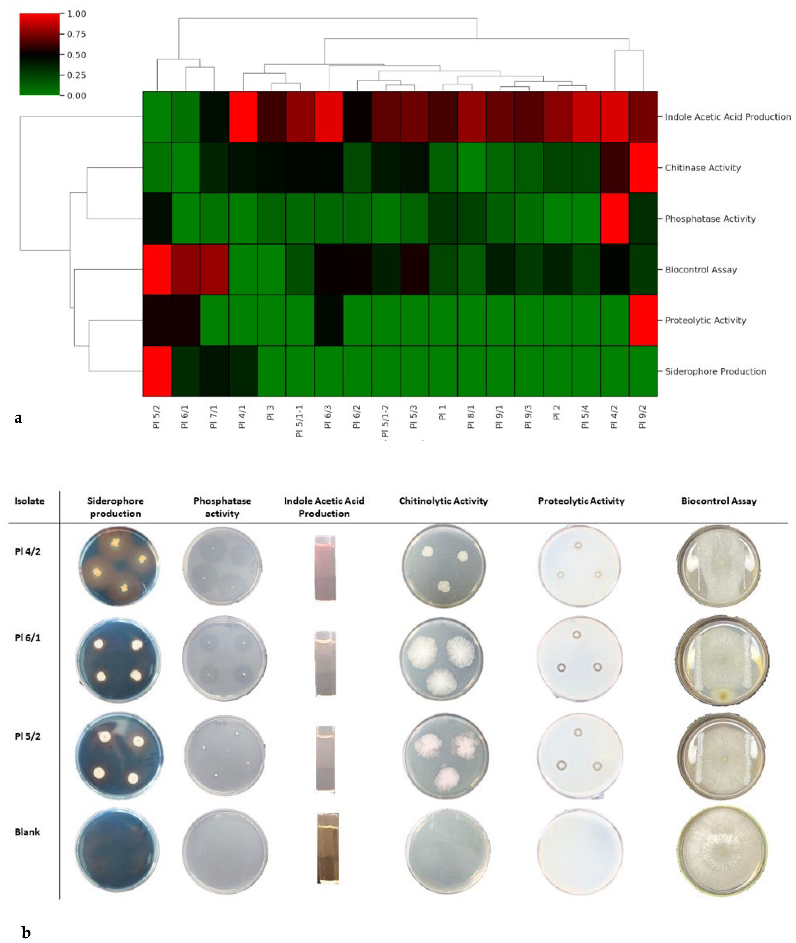
Eighteen strains were identified in the present study. Of these strains, 50% were derived from Izmir, 27.78% from Antalya, and 22.22% from Ankara. Upon evaluation of the Gram reaction, it was determined that 77.78% of the strains were Gram-negative, while 22.22% were Gram-positive. Assessment of the pectolytic activity and hypersensitivity reactions in tobacco revealed that none of the strains exhibited pectolytic activity or hypersensitivity reactions. In terms of siderophore production, the Pl 4/2 demonstrated the highest value, yielding 15.88 mm of siderophore. Regarding phosphorase solubilisation, the Pl 4/1 exhibited the greatest solubilisation capability, with a measurement of 16.13 mm. For indole-3-acetic acid (IAA) production, Pl 9/2 performed the best, producing 361.33 µg/mL. In relation to chitinase activity, four strains demonstrated chitinase activity, with Pl 9/2 showing the highest chitin degradation activity, measured at a zone of 2.63 mm. Additionally, in terms of proteolytic activity, Pl 5/2 exhibited the highest activity, recorded at 2.40 mm. In the context of in vitro biocontrol testing, Pl 6/1 achieved the highest activity, with a measurement of 2.1 mm (Table 2, Figure 2).
Based on the results of in vitro assays, Pl 4/1, Pl 6/1, and Pl 5/2 were selected for in vivo evaluation utilising a weighted relative superiority (Figure 2b).

3.2. Results of In Vivo Assay

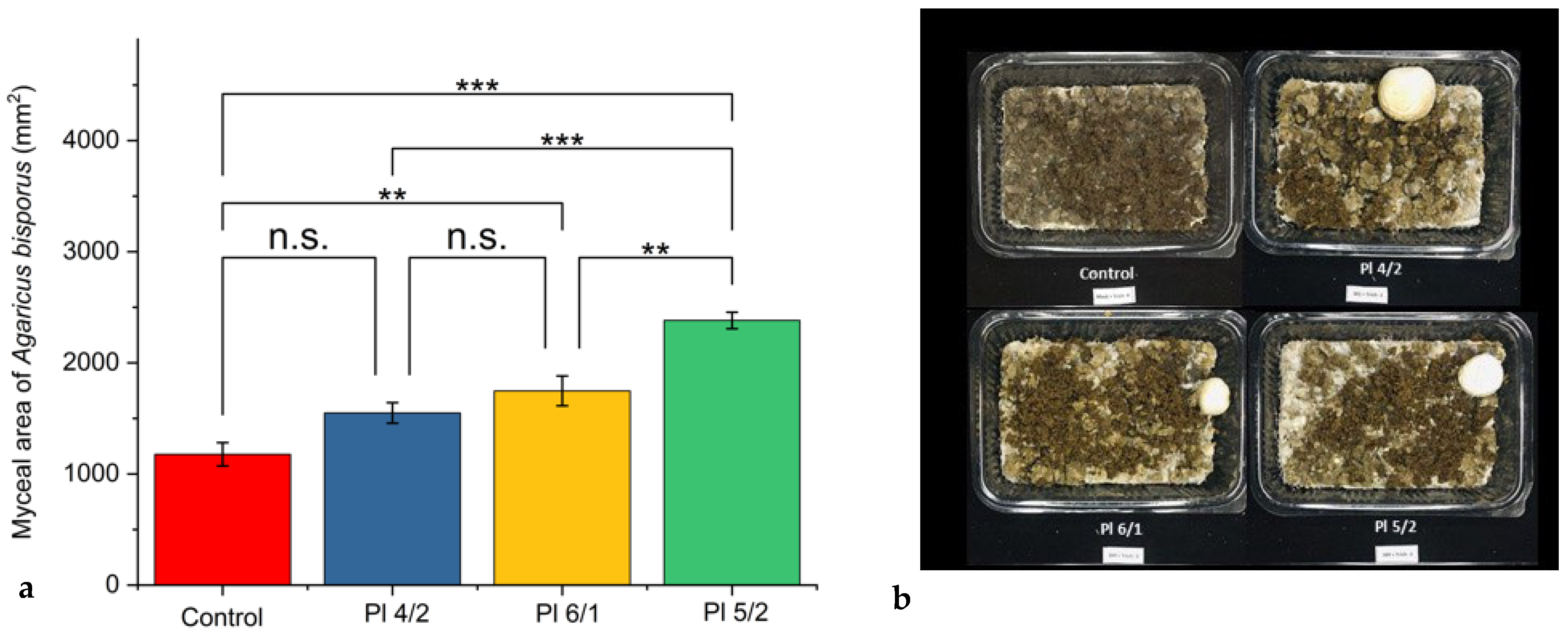
Based on the results obtained from the in vitro tests, the Pl 4/2, Pl 6/1, and Pl 5/2 strains, selected using WRS, were subjected to in vivo assays to evaluate their efficacy prior to the field trials and assess any potential adverse effects on mushroom production. In the in vivo assay, conducted with four replicates, the area covered by mushroom mycelia was quantified using ImageJ software and expressed in mm2. In the in vivo assay conducted on A. bisporus, Pl 5/2 exhibited the highest mycelial growth, measuring 2381.19 ± 74.07 mm2, surpassing all other strains and demonstrating a statistically significant difference in all comparisons (p < 0.001). In contrast, when comparing Pl 6/1 (1747.60 ± 133.83 mm2) with Pl 4/2 (1548.95 ± 92.42 mm2), the observed difference was not statistically significant (p ≥ 0.05). However, when these strains were compared with a control group with a mycelial growth of 1176.93 ± 104.85 mm2, the differences were found to be statistically significant (p < 0.001). Although Pl 4/2 demonstrated an increase in mycelial growth compared with the control group, this difference did not reach statistical significance (p ≥ 0.05) (Figure 3).

3.3. Results of Field Trials on Mushroom Cultivation Conditions

3.3.1. Results Related to the Effects on Disease Severity

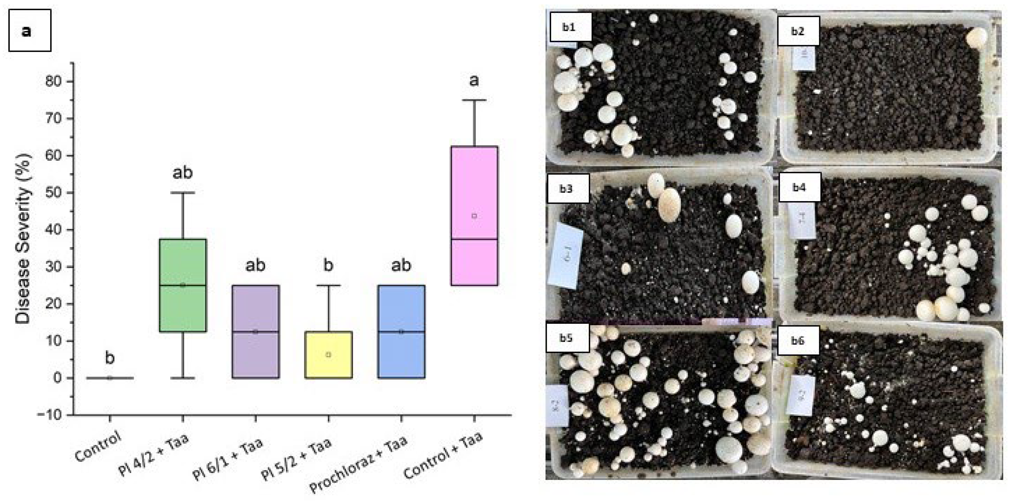
The field trial conducted under controlled mushroom cultivation conditions was executed as described in Section 2.7 of the Materials and Methods. The results of the trial, which included four replicates, were analysed using one-way analysis of variance (ANOVA) and Tukey’s multiple comparison test at a 95% confidence level (Figure 4).
In evaluating the results of the A. bisporus trial, Pl 5/2 (DS%: 6.25 ± 6.25; E%: 85.71) was statistically categorised within the same group as the control treatment, indicating its efficacy in suppressing the disease. Conversely, Pl 6/1 (DS%: 12.50 ± 7.22; E%: 71.43), Pl 4/2 (DS%: 25.00 ± 10.21; E%: 42.86), and Prochloraz (DS%: 12.50 ± 7.22; E%: 71.43) were grouped together, exhibiting statistically insignificant differences among them. However, these treatments demonstrated a statistically significant reduction compared with the control treatment containing T. aggressivum f. aggressivum.

3.3.2. Results Related to Their Impact on Yield

The evaluation conducted on Agaricus bisporus revealed that the application of bacterial strains significantly influenced both the number of mushroom caps and the total weight, as demonstrated in Table 3.
In A. bisporus, no statistically significant difference was observed among treatments regarding cap number (p > 0.05), as all treatments were classified within the same letter category. However, significant differences were identified in terms of the total weight. The highest average was recorded for the Pl 5/2 application (1268.00 ± 560.35 g; E%: 212.89), which was statistically classified in group ‘a’. In contrast, the control group exhibited the lowest average (273.50 ± 76.30 g; E%:−32.51) and was classified in a statistically distinct group ‘b’. These results indicate that Pl 5/2 effectively enhanced the biomass of A. bisporus. Overall, it was determined that applications utilising bacterial strains positively affected A. bisporus. Notably, the Pl 5/2 was distinguished by its substantial biomass production in A. bisporus., whether applied alone or under Taa pressure. This situation underscores the dual potential of biological control agents for disease suppression and yield enhancement.

3.4. Results of Molecular Identification

The strains were chosen for identification after antagonistic activities in the in vitro tests and not in the in vivo. For the molecular diagnosis of beneficial bacteria, the universal primer pairs 27F/1492R, designed to target the 16S rRNA region, was employed, and amplification was conducted in accordance with the established protocol. The resultant PCR products were subjected to sequence analysis (Medsantek Company, Istanbul, Turkiye), and the bacterial strains were identified through BLAST analysis of the sequence results obtained from the NCBI database (Table 4). Based on the sequence results, the strains were classified as Pseudomonas chlororaphis (Pl 4/2), Bacillus wiedmannii (Pl 6/1), and Bacillus cereus (Pl 5/2) (Table 4).

4. Discussion

This study proposes an integrated approach to biocontrol by examining the antagonistic potential of endophytic bacterial strains against T. aggressivum, with a focus on in vitro, in vivo, and field trial efficacy. In vitro assays assessing plant growth-promoting traits, including siderophore production, phosphate solubilisation, indole-3-acetic acid synthesis as well as chitinolytic and proteolytic activities, demonstrated significant variability among the strains. These traits are not only essential for enhancing plant or fungal growth, but also play a critical role in the indirect suppression of pathogens [46]. The siderophore production demonstrated by Pl 4/2 and the chitinolytic activity observed in Pl 5/2 and Pl 6/1 indicate that competitive iron acquisition and enzymatic degradation of the fungal cell walls may serve as significant mechanisms of antagonism. Notably, siderophores have the capacity to sequester iron within the rhizosphere or mycosphere, thereby limiting its availability to pathogenic organisms [47]. Furthermore, chitinases and proteases actively degrade the structural components of fungal cell walls including chitin and proteins [48]. The elevated proteolytic activity and moderate chitinolytic capability of Pl 5/2, alongside its significant in vitro antagonism zone of 1.09 mm, highlight its multifaceted potential for biocontrol [49,50]. Furthermore, the observed production of indole-3-acetic acid may enhance mycelial growth by modulating auxin signalling pathways in fungal systems; however, this interaction necessitates further investigation [51]. The translation of in vitro performance to in vivo efficacy under controlled compost conditions substantiated the bioactivity of the bacterial strains. Pl 5/2 demonstrated a statistically significant enhancement in mycelial expansion in A. bisporus, significantly surpassing that of the untreated control and competing strains. Its capacity to facilitate vigorous colonisation in the presence of Trichoderma aggressivum indicates that this strain not only inhibits the pathogen, but may also promote fungal resilience or growth through direct mechanisms. Interestingly, Pl 4/2, despite demonstrating promising in vitro plant growth promoting traits, particularly in indole-3-acetic acid and siderophore production, did not result in statistically significant enhancements in A. bisporus mycelial growth. This discrepancy may explain the intricate interactions between bacterial metabolites, host species physiology, and substrate conditions. Furthermore, it emphasises the limitations of relying exclusively on plant growth promoting traits to predict in vivo performance, a conclusion corroborated by previous research [52]. Field trial results further validated the antagonistic efficacy of Pl 5/2. In A. bisporus, Pl 5/2 again achieved the lowest disease severity score (6.25%), thereby reinforcing its biocontrol efficacy in A. bisporus. In contrast, Pl 4/2 did not significantly reduce the disease severity in A. bisporus, despite exhibiting relatively high levels of siderophore and indole-3-acetic acid production. This observation indicates that while these traits may facilitate fungal growth promotion, they do not necessarily result in effective disease suppression, particularly in the presence of an aggressive pathogen such as T. aggressivum, which utilises rapid mycelial colonisation and the production of antifungal metabolites [20]. A significant outcome of this study is the demonstrated dual benefit of Pl 5/2 regarding both disease suppression and yield enhancement. The application of Pl 5/2 resulted in the highest total biomass (1268 g), which was nearly five times greater than that of the untreated control. These results are consistent with prior research that underscores the ability of Bacillus spp. to augment mushroom yield through mechanisms such as substrate modification, enhanced nutrient availability, and stress mitigation [25,53]. The dual functionality of Pl 5/2 renders it particularly appealing for commercial applications, as it provides both protective and productivity advantages within a single agent. The molecular characterisation of the strains supports their taxonomic classification within genera recognised for environmental resilience and biocontrol potential.
PI 4/2, identified as Pseudomonas chlororaphis, is a bacterium renowned for its significant role in biological control, primarily due to its capacity to synthesise a diverse array of antimicrobial metabolites. These metabolites include phenazines, pyrrolnitrine, 2-hexyl, 5-propyl resorcinol, siderophores, and volatile organic compounds (VOCs), all of which contribute to their antagonistic activities against plant pathogens, nematodes, and insects [54]. A notable feature of P. chlororaphis is the production of phenazines, such as phenazine-1-carboxylic acid (PCA) and its derivatives, which are critical for its biocontrol efficacy, particularly against fungal pathogens [55]. Furthermore, this bacterium synthesises siderophores, including pyoverdine, which facilitate iron acquisition and confer a competitive advantage in the rhizosphere [54]. The VOCs produced by P. chlororaphis have also demonstrated considerable potential as biological fumigants; for instance, VOCs derived from P. chlororaphis subsp. aureofaciens SPS-41 have been shown to inhibit pathogenic fungi, thus offering a natural alternative to chemical fumigants [56]. The strain Pseudomonas chlororaphis PA23 exhibited significant efficacy in protecting canola (Brassica napus) from the necrotrophic fungus Sclerotinia sclerotiorum, with the application of PA23 to the plant surfaces resulting in a 91.1% reduction in lesions on petals. This strain activates unique defence mechanisms within the plant, thereby enhancing its innate defence capabilities [57]. Additionally, the strain Pseudomonas chlororaphis PCL1606 has been utilised to combat the soil-borne fungal pathogen Rosellinia necatrix, which adversely affects various woody crops including avocados. Through the production of the antifungal compound 2-hexyl, 5-propyl resorcinol (HPR), PCL1606 effectively reduces the presence of the pathogen in the soil, thus safeguarding avocado plants [58,59]. Pseudomonas chlororaphis zm-1, isolated from the rhizosphere of Anemarrhena asphodeloides, has demonstrated potential in managing peanut stem rot caused by Sclerotium rolfsii, as this strain produces phenazine compounds that significantly inhibit the growth of the pathogen, providing an effective biocontrol strategy for this economically significant disease [60]. PI 6/1 was identified as Bacillus wiedmannii; in particular, the strain known as B. wiedmannii biovar thuringiensis has shown promise as a biological control agent. It is a specialised mosquitocidal pathogen, effective against mosquito species such as Aedes aegypti and Culex pipiens, which are vectors for various diseases. This strain possesses multiple genes encoding Cry proteins, which are well-known for their insecticidal properties, making it a promising candidate for controlling mosquito populations [61]. Moreover, B. wiedmannii has been investigated for its capacity to produce polyhydroxybutyrates (PHBs), which are biodegradable plastics. The strain AS-02, identified as B. wiedmannii, demonstrated efficient PHB production from agricultural waste, emphasising its utility in sustainable practices [62]. Bacillus species, in general, are well-regarded in biological control due to their ability to produce bioactive compounds that inhibit pathogens, induce systemic resistance in plants, and compete for nutrients. These characteristics render Bacillus spp. versatile agents in the biocontrol of plant diseases. Key mechanisms include the production of antimicrobial compounds and competition in the rhizosphere, which collectively assist in managing plant diseases sustainably [56,63]. PI 5/2, identified as Bacillus cereus, employs multiple mechanisms as a biological control agent, particularly through the process of competitive exclusion. This strategy encompasses various approaches that enhance its capacity to suppress pathogenic organisms. Key modes of action include competitive exclusion facilitated by rapid growth rates, efficient nutrient uptake, and siderophore production [64]. In addition, B. cereus is capable of biofilm formation, which cultivates a protective environment that enhances its persistence and functionality as a biocontrol agent. B. cereus can form biofilms via pathways analogous to those of Bacillus subtilis or through alternative mechanisms, thereby enabling it to thrive on plant roots and offer protection against pathogens. These biofilms play a crucial role in ability of the bacterium to function as a biological control agent by creating barriers against pathogen colonisation [65]. Moreover, B. cereus exhibits antifungal activity through the production of enzymes such as chitinases. For instance, chitinase has demonstrated inhibitory effects on the germination of fungal conidia, further contributing to its biocontrol potential, particularly against fungal pathogens such as Botrytis elliptica [66]. One strain, Bacillus cereus AR156, has demonstrated effectiveness in controlling a wide spectrum of plant diseases by acting as a microbial elicitor of plant immune responses. This strain modifies plant root exudates, altering their composition in ways that enhance both the growth and biocontrol efficacy of B. cereus while suppressing pathogens such as Ralstonia solanacearum [67]. Furthermore, B. cereus can also be applied in postharvest scenarios. For example, B. cereus AR156 effectively mitigates grey mould disease in postharvest strawberries by enhancing the fruit’s reactive oxygen-scavenging and defence-related enzyme activities. This suppression is primarily attributed to the induction of host responses at the transcriptional level, which increases the expression of defence-related genes [68]. Additionally, B. cereus can contribute to biological control through the production of antifungal chitinases, as demonstrated by B. cereus 28-9, which inhibits the conidial germination of pathogens such as Botrytis elliptica [66].

5. Conclusions

In conclusion, this study underscores the effect of native bacteria, specifically Bacillus cereus Pl 5/2, as biocontrol agents for managing Trichoderma aggressivum f. aggressivum in the cultivation of Agaricus bisporus. The utilisation of these bacteria not only alleviates the incidence of green mould disease, but also enhances yield, thereby providing a sustainable and environmentally friendly alternative to traditional fungicides. The incorporation of such biological solutions into mushroom production systems has the potential to substantially advance the goals of chemical-free agriculture.

Author Contributions

Conceptualisation, U.Ş., M.Ҫ. and H.Ö.; Methodology, U.Ş. and M.Ҫ.; Software, U.Ş.; Validation, H.Ö., F.A. and E.E.; Formal analysis, N.G. and U.Ş.; Investigation, U.Ş. and M.Ҫ.; Resources, E.E. and M.Ҫ.; Data curation, U.Ş.; Writing—original draft preparation, U.Ş.; Writing—review and editing: H.Ö., M.Ҫ. and F.A.; Visualisation, U.Ş.; Supervision, H.Ö.; Project administration, H.Ö.; Funding acquisition, H.Ö. and M.Ҫ. All authors have read and agreed to the published version of the manuscript.

Funding

This research received no external funding.

Data Availability Statement

Dataset available on request from the authors due to ongoing related research that includes the same dataset.

Acknowledgments

We would like to express our gratitude to the Directorate of the Ege University Bergama Vocational School and Bergama Municipality for their support in providing the field area and test materials. We would like to extend our gratitude to Gülay Turhan (retired academic member of the Ege University Faculty of Agriculture Department of Plant Protection) for her support in diagnosing the morphological characteristics of the fungal disease agent.

Conflicts of Interest

The authors declare no conflicts of interest.

Abbreviations

The following abbreviations are used in this manuscript:
ANOVAAnalysis of variance
BLASTBasic Local Alignment Search Tool
CASChrome Azurol S
cfu/mLColony forming unit per millilitre)
FAOSTATFood and Agriculture Organisation Statistical Database
HDTMAHexadecyltrimethylammonium bromide
IAAIndole-3-acetic acid
NBRIPNational Botanical Research Institute’s phosphate solubilising medium
NCBINational Centre for Biotechnology Information
OD600Optic density 600 nm
PCRPolymerase chain reaction
PDAPotato dextrose agar

References

  1. Hamza, A.; Mylarapu, A.; Krishna, K.; Biotechnology, D.K. An Insight into the Nutritional and Medicinal Value of Edible Mushrooms: A Natural Treasury for Human Health. J. Biotechnol. 2024, 381, 86–99. [Google Scholar] [CrossRef] [PubMed]
  2. Mowna Sundari, T.; Alwin Prem Anand, A.; Jenifer, P.; Shenbagarathai, R. Bioprospection of Basidiomycetes and Molecular Phylogenetic Analysis Using Internal Transcribed Spacer (ITS) and 5.8 S RRNA Gene Sequence. Sci. Rep. 2018, 8, 10720. [Google Scholar] [CrossRef] [PubMed]
  3. Heleno, S.A.; Barros, L.; Sousa, M.J.; Martins, A.; Ferreira, I.C.F.R. Study and Characterization of Selected Nutrients in Wild Mushrooms from Portugal by Gas Chromatography and High Performance Liquid Chromatography. Microchem. J. 2009, 93, 195–199. [Google Scholar] [CrossRef]
  4. Rathore, H.; Prasad, S.; Sharma, S. Mushroom Nutraceuticals for Improved Nutrition and Better Human Health: A Review. PharmaNutrition 2017, 5, 35–46. [Google Scholar] [CrossRef]
  5. Huang, K.; El-Seedi, H.R.; Xu, B. Critical Review on Chemical Compositions and Health-Promoting Effects of Mushroom Agaricus blazei Murill. Curr. Res. Food Sci. 2022, 5, 2190–2203. [Google Scholar] [CrossRef]
  6. Ajith, T.A.; Janardhanan, K.K. Indian Medicinal Mushrooms as a Source of Antioxidant and Antitumor Agents. J. Clin. Biochem. Nutr. 2007, 40, 157–162. [Google Scholar] [CrossRef]
  7. Heng, H.H.Q.; Stevens, J.B.; Liu, G.; Bremer, S.W.; Ye, K.J.; Reddy, P.; Wu, G.S.; Wang, Y.A.; Tainsky, M.A.; Ye, C.J. Stochastic Cancer Progression Driven by Non-clonal Chromosome Aberrations. J. Cell. Physiol. 2006, 208, 461–472. [Google Scholar] [CrossRef]
  8. Singh, I.; Thakur, P. Impact of Fungi on the World Economy and Its Sustainability: Current Status and Potentials. In Fungal Resources for Sustainable Economy: Current Status and Future Perspectives; Springer: Singapore, 2023; pp. 3–37. ISBN 9789811991035. [Google Scholar]
  9. Niego, A.G.; Rapior, S.; Thongklang, N.; Raspé, O.; Jaidee, W.; Lumyong, S.; Hyde, K.D. Macrofungi as a Nutraceutical Source: Promising Bioactive Compounds and Market Value. J. Fungi 2021, 7, 397. [Google Scholar] [CrossRef]
  10. Royse, D.J.; Baars, J.; Tan, Q. Current Overview of Mushroom Production in the World. In Edible and Medicinal Mushrooms: Technology and Applications; Wiley-Blackwell: Hoboken, NJ, USA, 2017; pp. 5–13. [Google Scholar]
  11. Baars, J.J.P.; Scholtmeijer, K.; Sonnenberg, A.S.M.; van Peer, A. Critical Factors Involved in Primordia Building in Agaricus bisporus: A Review. Molecules 2020, 25, 2984. [Google Scholar] [CrossRef]
  12. Anonymous Concerning the Marketing and Commercial Quality Control of Cultivated Mushrooms. Available online: https://www.unece.org/fileadmin/DAM/trade/agr/standard/standard/fresh/FFV-Std/English/24_CultivatedMushrooms.pdf (accessed on 1 May 2025).
  13. Muszynska, B.; Kala, K.; Rojowski, J.; Grzywacz, A.; Opoka, W. Composition and Biological Properties of Agaricus bisporus Fruiting Bodies—A Review. Pol. J. Food Nutr. Sci. 2017, 67, 173–181. [Google Scholar] [CrossRef]
  14. Eswari, S.; Bhavan, P.S.; Kalpana, R.; Dharani, C.; Manjula, T.; Sarumathi, K.; Rajkumar, G. Phytochemical Characterization of the Mushroom Agaricus bisporus and Assessment of Its Nutritional Ability in the Place of Fishmeal for Survival and Growth of the Freshwater Prawn Macrobrachium rosenbergii Post-Larvae. Integrative Food. Nutr. Metab. 2019, 6, 1–11. [Google Scholar]
  15. Tirta Ismaya, W.; Tjandrawinata, R.R.; Rachmawati, H. Lectins from the Edible Mushroom Agaricus bisporus and Their Therapeutic Potentials. Molecules 2020, 25, 2368. [Google Scholar] [CrossRef] [PubMed]
  16. An, X.-Y.; Cheng, G.-H.; Gao, H.-X.; Li, X.-F.; Yang, Y.; Li, D.; Li, Y. Phylogenetic Analysis of Trichoderma Species Associated with Green Mold Disease on Mushrooms and Two New Pathogens on Ganoderma sichuanense. J. Fungi 2022, 8, 704. [Google Scholar] [CrossRef] [PubMed]
  17. Samuels, G.J.; Dodd, S.L.; Gams, W.; Castlebury, L.A.; Petrini, O. Trichoderma Species Associated with the Green Mold Epidemic of Commercially Grown Agaricus bisporus. Mycologia 2002, 94, 146–170. [Google Scholar] [CrossRef] [PubMed]
  18. Goltapeh, E.M.; Danesh, Y.R. Studies on Interaction between Trichoderma Species and Agaricus bisporus Mycelium. In Science and Cultivation of Edible Fungi, Proceedings of the 15th International Congress on the Science and Cultivation of Edible Fungi, Maastricht, The Netherlands, 15–19 May 2000; CRC Press: Boca Raton, FL, USA, 2000; pp. 661–666. [Google Scholar]
  19. Fletcher, J.T.; Gaze, R.H. Mushroom Pest and Disease Control: A Color Handbook; Elsevier: Amsterdam, The Netherlands, 2007; ISBN 0123739845. [Google Scholar]
  20. Largeteau, M.L.; Savoie, J.-M. Microbially Induced Diseases of Agaricus bisporus: Biochemical Mechanisms and Impact on Commercial Mushroom Production. Appl. Microbiol. Biotechnol. 2010, 86, 63–73. [Google Scholar] [CrossRef]
  21. Savoie, J.-M.; Iapicco, R.; Largeteau-Mamoun, M.L. Factors Influencing the Competitive Saprophytic Ability of Trichoderma harzianum Th2 in Mushroom (Agaricus bisporus) Compost. Mycol. Res. 2001, 105, 1348–1356. [Google Scholar] [CrossRef]
  22. Guthrie, J.L.; Castle, A.J. Chitinase Production during Interaction of Trichoderma aggressivum and Agaricus bisporus. Can. J. Microbiol. 2006, 52, 961–967. [Google Scholar] [CrossRef]
  23. Rinker, D.L.; Alm, G. Management of Green Mould Disease in Canada. In Science and Cultivation of Edible Fungi: Proceedings of the 15th International Congress on the Science and Cultivation of Edible Fungi, Maastricht, The Netherlands, 15–19 May 2000; A. A. Balkema Publisher: Rotterdam, The Netherlands, 2000; Volume 15, p. 617. [Google Scholar]
  24. Romaine, C.P.; Royse, D.J.; Schlagnhaufer, C. Emergence of Benzimidazole-Resistant Green Mould, Trichoderma aggressivum, on Cultivated Agaricus bisporus in North America. Mushroom Sci. 2008, 17, 510–523. [Google Scholar]
  25. Stanojević, O.; Milijašević-Marčić, S.; Potočnik, I.; Stepanović, M.; Dimkić, I.; Stanković, S.; Berić, T. Isolation and Identification of Bacillus spp. from Compost Material, Compost and Mushroom Casing Soil Active against Trichoderma spp. Arch. Biol. Sci. 2016, 68, 845–852. [Google Scholar]
  26. Saubenova, M.; Oleinikova, Y.; Sadanov, A.; Yermekbay, Z.; Bokenov, D.; Shorabaev, Y. The Input of Microorganisms to the Cultivation of Mushrooms on Lignocellulosic Waste. AIMS Agric. Food 2023, 8, 239–277. [Google Scholar] [CrossRef]
  27. White, T.J.; Bruns, T.; Lee, S.; Taylor, J. Amplification and Direct Sequencing of Fungal Ribosomal RNA Genes for Phylogenetics. PCR Protoc. A Guide Methods Appl. 1990, 18, 315–322. [Google Scholar]
  28. Bahmani, K.; Hasanzadeh, N.; Harighi, B.; Marefat, A. Isolation and Identification of Endophytic Bacteria from Potato Tissues and Their Effects as Biological Control Agents against Bacterial Wilt. Physiol. Mol. Plant Pathol. 2021, 116, 101692. [Google Scholar] [CrossRef]
  29. Suslow, T.V.; Schroth, M.N.; Isaka, M. Application of a Rapid Method for Gram Differentiation of Plant Pathogenic and Saprophytic Bacteria Without Staining. 1982. Available online: https://www.apsnet.org/publications/phytopathology/backissues/Documents/1982Articles/Phyto72n07 (accessed on 1 May 2025).
  30. Schaad, N.W. Laboratory Guide for Identification of Plant Pathogenic Bacteria; Amer Phytopathological Society: Saint Paul, MN, USA, 1981; ISBN 0890540284. [Google Scholar]
  31. Sun, Y.; Wu, J.; Shang, X.; Xue, L.; Ji, G.; Chang, S.; Niu, J.; Emaneghemi, B. Screening of Siderophore-Producing Bacteria and Their Effects on Promoting the Growth of Plants. Curr. Microbiol. 2022, 79, 150. [Google Scholar] [CrossRef]
  32. Nautiyal, C.S. An Efficient Microbiological Growth Medium for Screening Phosphate Solubilizing Microorganisms. FEMS Microbiol. Lett. 1999, 170, 265–270. [Google Scholar] [CrossRef]
  33. Janati, W.; Mikou, K.; El Ghadraoui, L.; Errachidi, F. Isolation and Characterization of Phosphate Solubilizing Bacteria Naturally Colonizing Legumes Rhizosphere in Morocco. Front. Microbiol. 2022, 13, 958300. [Google Scholar] [CrossRef]
  34. Rangel, F.; Santos, R.A.; Monteiro, M.; Lavrador, A.S.; Gasco, L.; Gai, F.; Oliva-Teles, A.; Enes, P.; Serra, C.R. Isolation of Chitinolytic Bacteria from European Sea Bass Gut Microbiota Fed Diets with Distinct Insect Meals. Biology 2022, 11, 964. [Google Scholar] [CrossRef]
  35. Chung, D.; Yu, W.J.; Lim, J.Y.; Kang, N.S.; Kwon, Y.M.; Choi, G.; Bae, S.S.; Cho, K.; Lee, D.S. Characterization of the Proteolytic Activity of a Halophilic Aspergillus reticulatus Strain Sk1-1 Isolated from a Solar Saltern. Microorganisms 2022, 10, 29. [Google Scholar] [CrossRef]
  36. Gang, S.; Sharma, S.; Saraf, M.; Buck, M.; Schumacher, J. Bacterial Indole-3-Acetic Acid Influences Soil Nitrogen Acquisition in Barley and Chickpea. Plants 2021, 10, 780. [Google Scholar] [CrossRef] [PubMed]
  37. Aydoğdu, M.; Sülü, S.M.; Kurbetli, İ.; Sülü, G. In vitro and in vivo Biological Control of the Green Mold Using Different Bacteria in Button Mushroom Cultivation. Egypt. J. Biol. Pest Control 2021, 31, 1–11. [Google Scholar] [CrossRef]
  38. Babier, Y.; Akköprü, A. Çeşitli Kültür Bitkilerinden İzole Edilen Endofitik Bakterilerin Karakterizasyonu ve Bitki Patojeni Bakterilere Karşı Antagonistik Etkilerinin Belirlenmesi. Yuz. Yıl Univ. J. Agric. Sci. 2020, 30, 521–534. [Google Scholar] [CrossRef]
  39. Schneider, C.A.; Rasband, W.S.; Eliceiri, K.W. NIH Image to ImageJ: 25 Years of Image Analysis. Nat. Methods 2012, 9, 671–675. [Google Scholar] [CrossRef]
  40. Cetin, M.; Atila, F.; Eren, E. Valorization of Olive Press Cake as a Sustainable Alternative to Peat in White Button Mushroom (Agaricus bisporus) Cultivation. Biomass Convers. Biorefinery 2025, 15, 20589–20599. [Google Scholar] [CrossRef]
  41. Roberti, R.; Di Francesco, A.; Innocenti, G.; Mari, M. Potential for Biocontrol of Pleurotus ostreatus Green Mould Disease by Aureobasidium pullulans De Bary (Arnaud). Biol. Control 2019, 135, 9–15. [Google Scholar] [CrossRef]
  42. Townsend, G.R.; Heuberger, J.W.; Townsend, G.; Heuberger, J. Methods for Estimating Losses Caused by Diseases in Fungicidal Experiments. Plant Dis. Rep. 1943, 27, 340–343. [Google Scholar] [CrossRef]
  43. Gea, F.J.; Tello, J.C.; Navarro, M.J. Efficacy and Effects on Yield of Different Fungicides for Control of Wet Bubble Disease of Mushroom Caused by the Mycoparasite Mycogone perniciosa. Crop Prot. 2010, 29, 1021–1025. [Google Scholar] [CrossRef]
  44. Kadaikunnan, S.; Rejiniemon, S.S.; Khaled, J.M.; Alharbi, N.S.; Mothana, R. In vitro Antibacterial, Antifungal, Antioxidant and Functional Properties of Bacillus amyloliquefaciens. Ann. Clin. Microbiol. Antimicrob. 2015, 14, 1–11. [Google Scholar] [CrossRef]
  45. De Mendiburu, F.; Simon, R. Agricolae—Ten Years of an Open Source Statistical Tool for Experiments in Breeding, Agriculture and Biology. PeerJ 2015, 3, 1–18. [Google Scholar] [CrossRef]
  46. Hayat, R.; Ahmed, I.; Sheirdil, R.A. An Overview of Plant Growth Promoting Rhizobacteria (PGPR) for Sustainable Agriculture. In Crop Production for Agricultural Improvement; Springer: Dordrecht, The Netherlands, 2012; Volume 9789400741, pp. 557–579. ISBN 9789400741164. [Google Scholar]
  47. Mimmo, T.; Del Buono, D.; Terzano, R.; Tomasi, N.; Vigani, G.; Crecchio, C.; Pinton, R.; Zocchi, G.; Cesco, S. Rhizospheric Organic Compounds in the Soil-Microorganism-Plant System: Their Role in Iron Availability. Eur. J. Soil Sci. 2014, 65, 629–642. [Google Scholar] [CrossRef]
  48. Ajit, N.S.; Verma, R.; Shanmugam, V. Extracellular Chitinases of Fluorescent Pseudomonads Antifungal to Fusarium oxysporum f. sp. dianthi Causing Carnation Wilt. Curr. Microbiol. 2006, 52, 310–316. [Google Scholar] [CrossRef]
  49. Malik, M.S.; Haider, S.; Rehman, A.; Rehman, S.U.; Jamil, M.; Naz, I.; Anees, M. Biological Control of Fungal Pathogens of Tomato (Lycopersicon esculentum) by Chitinolytic Bacterial Strains. J. Basic Microbiol. 2022, 62, 48–62. [Google Scholar] [CrossRef]
  50. Luan, P.; Yi, Y.; Huang, Y.; Cui, L.; Hou, Z.; Zhu, L.; Ren, X.; Jia, S.; Liu, Y. Biocontrol Potential and Action Mechanism of Bacillus amyloliquefaciens DB2 on Bipolaris sorokiniana. Front. Microbiol. 2023, 14, 1149363. [Google Scholar] [CrossRef]
  51. Jabiri, S.; Legrifi, I.; Benhammou, M.; Laasli, S.E.; Mokrini, F.; Amraoui, M.B.; Lahlali, R. Screening of Rhizobacterial Isolates from Apple Rhizosphere for Their Biocontrol and Plant Growth Promotion Activity. Appl. Microbiol. 2023, 3, 948–967. [Google Scholar] [CrossRef]
  52. Santoyo, G.; Moreno-Hagelsieb, G.; del Carmen Orozco-Mosqueda, M.; Glick, B.R. Plant Growth-Promoting Bacterial Endophytes. Microbiol. Res. 2016, 183, 92–99. [Google Scholar] [CrossRef]
  53. Elshaghabee, F.M.F.; Rokana, N.; Gulhane, R.D.; Sharma, C.; Panwar, H. Bacillus as Potential Probiotics: Status, Concerns, and Future Perspectives. Front. Microbiol. 2017, 8, 1490. [Google Scholar] [CrossRef]
  54. Raio, A.; Puopolo, G. Pseudomonas chlororaphis Metabolites as Biocontrol Promoters of Plant Health and Improved Crop Yield. World J. Microbiol. Biotechnol. 2021, 37, 1–8. [Google Scholar] [CrossRef]
  55. Yu, J.M.; Wang, D.; Pierson, L.S.; Pierson, E.A. Effect of Producing Different Phenazines on Bacterial Fitness and Biological Control in Pseudomonas chlororaphis 30–84. Plant Pathol. J. 2018, 34, 44–58. [Google Scholar] [CrossRef]
  56. Zhang, N.; Wang, Z.; Shao, J.; Xu, Z.; Liu, Y.; Xun, W.; Miao, Y.; Shen, Q.; Zhang, R. Biocontrol Mechanisms of Bacillus: Improving the Efficiency of Green Agriculture. Microb. Biotechnol. 2023, 16, 2250–2263. [Google Scholar] [CrossRef] [PubMed]
  57. Duke, K.A.; Becker, M.G.; Girard, I.J.; Millar, J.L.; Dilantha Fernando, W.G.; Belmonte, M.F.; de Kievit, T.R. The Biocontrol Agent Pseudomonas chlororaphis PA23 Primes Brassica napus Defenses through Distinct Gene Networks. BMC Genom. 2017, 18, 1–16. [Google Scholar] [CrossRef] [PubMed]
  58. Arrebola, E.; Tienda, S.; Vida, C.; De Vicente, A.; Cazorla, F.M. Fitness Features Involved in the Biocontrol Interaction of Pseudomonas chlororaphis with Host Plants: The Case Study of PcPCL1606. Front. Microbiol. 2019, 10, 719. [Google Scholar] [CrossRef] [PubMed]
  59. Tienda, S.; Vida, C.; Lagendijk, E.; de Weert, S.; Linares, I.; González-Fernández, J.; Guirado, E.; de Vicente, A.; Cazorla, F.M. Soil Application of a Formulated Biocontrol Rhizobacterium, Pseudomonas chlororaphis PCL1606, Induces Soil Suppressiveness by Impacting Specific Microbial Communities. Front. Microbiol. 2020, 11, 1874. [Google Scholar] [CrossRef]
  60. Liu, F.; Yang, S.; Xu, F.; Zhang, Z.; Lu, Y.; Zhang, J.; Wang, G. Characteristics of Biological Control and Mechanisms of Pseudomonas chlororaphis Zm-1 against Peanut Stem Rot. BMC Microbiol. 2022, 22, 9. [Google Scholar] [CrossRef]
  61. Nicolás Lazarte, J.; Lopez, R.P.; Daniel Ghiringhelli, P.; Berón, C.M. Bacillus wiedmannii biovar thuringiensis: A Specialized Mosquitocidal Pathogen with Plasmids from Diverse Origins. Genome Biol. Evol. 2018, 10, 2823–2833. [Google Scholar] [CrossRef] [PubMed]
  62. Danial, A.W.; Hamdy, S.M.; Alrumman, S.A.; Gad El-Rab, S.M.F.; Shoreit, A.A.M.; Hesham, A.E.L. Bioplastic Production by Bacillus wiedmannii As-02 Ok576278 Using Different Agricultural Wastes. Microorganisms 2021, 9, 2395. [Google Scholar] [CrossRef] [PubMed]
  63. Boulahouat, S.; Cherif-Silini, H.; Silini, A.; Bouket, A.C.; Luptakova, L.; Alenezi, F.N.; Belbahri, L. Biocontrol Efficiency of Rhizospheric Bacillus against the Plant Pathogen Fusarium oxysporum: A Promising Approach for Sustainable Agriculture. Microbiol. Res. 2023, 14, 892–908. [Google Scholar] [CrossRef]
  64. Lalloo, R.; Moonsamy, G.; Ramchuran, S.; Görgens, J.; Gardiner, N. Competitive Exclusion as a Mode of Action of a Novel Bacillus cereus Aquaculture Biological Agent. Lett. Appl. Microbiol. 2010, 50, 563–570. [Google Scholar] [CrossRef]
  65. Gao, T.; Foulston, L.; Chai, Y.; Wang, Q.; Losick, R. Alternative Modes of Biofilm Formation by Plant-Associated Bacillus cereus. MicrobiologyOpen 2015, 4, 452–464. [Google Scholar] [CrossRef]
  66. Huang, C.J.; Wang, T.K.; Chung, S.C.; Chen, C.Y. Identification of an Antifungal Chitinase from a Potential Biocontrol Agent, Bacillus cereus 28-9. J. Biochem. Mol. Biol. 2005, 38, 82–88. [Google Scholar] [CrossRef]
  67. Wang, N.; Wang, L.; Zhu, K.; Hou, S.; Chen, L.; Mi, D.; Gui, Y.; Qi, Y.; Jiang, C.; Guo, J.H. Plant Root Exudates Are Involved in Bacillus cereus AR156 Mediated Biocontrol against Ralstonia solanacearum. Front. Microbiol. 2019, 10, 98. [Google Scholar] [CrossRef]
  68. Zhou, H.; Ren, Z.H.; Zu, X.; Yu, X.Y.; Zhu, H.J.; Li, X.J.; Zhong, J.; Liu, E.M. Efficacy of Plant Growth-Promoting Bacteria Bacillus cereus YN917 for Biocontrol of Rice Blast. Front. Microbiol. 2021, 12, 684888. [Google Scholar] [CrossRef]
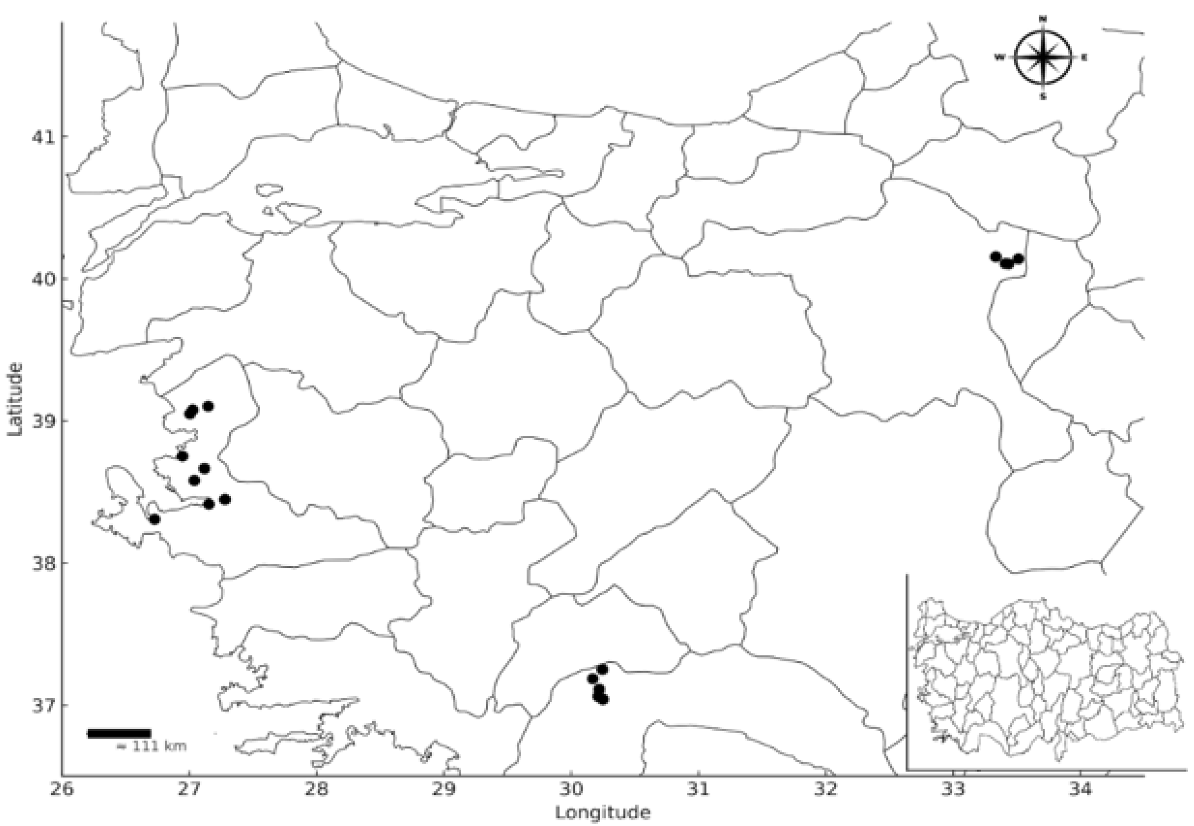
Figure 1. Origin of bacterial isolates that were collected.
Diversity 17 00757 g001
Figure 2. Results of the in vitro assay: (a) heatmap analysis derived from the in vitro data; (b) visual representations of the in vitro tests for the most successful bacterial strains.
Diversity 17 00757 g002
Figure 3. In vivo assay results: (a) mycelial growth of Agaricus bisporus; (b) comparative images of the applications (n.s. = not significant; **: p ≤ 0.01; ***: p ≤ 0.001).
Diversity 17 00757 g003
Figure 4. Field trial results under mushroom cultivation conditions: (a) A. bisporus field trial, characters with the same letter value are statistically insignificant according to the Tukey test, which was conducted with 95% confidence; (b1) control, (b2) control with Taa, (b3) treatment Pl 4/2 with Taa, (b4) treatment Pl 6/1 with Taa, (b5) treatment Pl 5/2 with Taa, and (b6) treatment Prochloraz with Taa.
Diversity 17 00757 g004
Table 1. Information regarding the trial design of the treatments.
Treatment NumberDescription
1Application of Pl 4/2
2Application of Pl 6/1
3Application of Pl 5/2
4Application of Prochloraz
5Application of Control Treatment
6Application of Pl 4/2 + T. aggressivum f. aggressivum
7Application of Pl 6/1 + T. aggressivum f. aggressivum
8Application of Pl 5/2 + T. aggressivum f. aggressivum
9Application of Prochloraz + T. aggressivum f. aggressivum
10Application of Control Treatment + T. aggresivum f. aggressivum
Table 2. Biochemical and In vitro Plant Growth Promotion Tests for Bacterial Strains.
No.Strain CodeLocationIn Vitro Assay
Biochemical TestPGP Test
Gr 1Pec 2HR 3Sid 4PhA 5IAA 6CA 7PA 8BioCo 9
377Pl 1Antalya6.7510.2546.6700.560
378Pl 2Antalya312.3886.6700.890
379Pl 3Antalya4.510165.33000
380Pl 4/1Antalya+3.2516.13158000
381Pl 4/2Antalya15.8814.88220.6701.160.5
384Pl 6/1İzmir+3101.411.852.1
385Pl 6/2İzmir4.388.387801.252.05
386Pl 6/3İzmir4.2515.13173.331.221.240.41
387Pl 5/1-1İzmir4.1312.5017200.470
388Pl 5/1-2İzmir3.511148.6700.911.88
389Pl 5/2İzmir+8.880181.422.401.09
390Pl 5/3İzmir4.511.516001.321.08
391Pl 5/4İzmir314.2585.3300.591.31
392Pl 7/1İzmir+3.757.2513001.910
393Pl 8/1Ankara6.2512.63000.350.15
394Pl 9/1Ankara4.7511.2536.6700.930
395Pl 9/2Ankara7.2511.75361.332.630.681.63
396Pl 9/3Ankara410.755300.661.28
1. Gr: Gram test, 2. Pec: pectolytic activity, 3. HR: hypersensitive reaction on tobacco, 4. Sid: siderophore production (mm), 5. PhA: phosphatase activity (mm), 6. IAA: indole acetic acid production (µg/mL), 7. CA: chitinolytic activity (mm), 8. PA: proteolytic activity (mm), 9. BioCo: in vitro biocontrol assay (mm).
Table 3. Results pertaining to the productivity of field trials conducted under the mushroom cultivation conditions.
TreatmentNumber of Fruiting Bodies of Mushrooms
PieceWeight of Mushrooms
gE%gE%
Pl 4/229.25 ± 22.86 a *24.47473.25 ± 411.31 ab *16.78
Pl 6/137.75 ± 8.99 a60.64598.50 ± 186.87 ab47.69
Pl 5/272.50 ± 26.91 a208.511268.00 ± 560.35 a212.89
Prochloraz37.00 ± 25.91 a57.45584.50 ± 388.66 ab44.23
Control28.00 ± 4.76 a19.15273.50 ± 76.30 b−32.51
Pl 4/2 + Taa33.25 ± 24.44 a41.49354.50 ± 198.88 ab−12.52
Pl 6/1 + Taa32.50 ± 24.79 a38.30485.50 ± 559.81 ab19.80
Pl 5/2 + Taa57.00 ± 22.19 a142.55854.50 ± 389.74 ab110.86
Prochloraz + Taa44.00 ± 17.49 a87.23675.50 ± 507.92 ab66.69
Control + Taa23.50 ± 15.15 a-405.25 ± 246.64 ab-
* Characters with the same letter value are statistically insignificant according to the Tukey test, which was conducted with 95% confidence.
Table 4. Molecular diagnosis and NCBI reference numbers of successful strains of bacteria.
No.Strain CodeBacteria SpeciesReference Similarity RateReference Access No.NCBI Access No.
381Pl 4/2Pseudomonas chlororaphis98.02CP027753PX069530
384Pl 6/1Bacillus wiedmannii98.26PV052600PX069532
389Pl 5/2Bacillus cereus98.01JX847613PX069538
Disclaimer/Publisher’s Note: The statements, opinions and data contained in all publications are solely those of the individual author(s) and contributor(s) and not of MDPI and/or the editor(s). MDPI and/or the editor(s) disclaim responsibility for any injury to people or property resulting from any ideas, methods, instructions or products referred to in the content.