Article

Phylogenetic Framework, a New Species, and Two New Species Records for China in Rhizopogon

1
College of Resources and Environment, Yunnan Agricultural University, Kunming 650201, China
2
College of Agriculture and Biological Science, Dali University, Dali 671003, China
3
Colegio de Postgraduados, Campus Montecillo, Edafología, Texcoco 56230, Mexico
4
Yunnan Key Laboratory for Fungal Diversity and Green Development & Yunnan International Joint Laboratory of Fungal Sustainable Utilization in South and Southeast Asia, Germplasm Bank of Wild Species, Kunming Institute of Botany, Chinese Academy of Sciences, Kunming 650201, China
*
Authors to whom correspondence should be addressed.
Diversity 2025, 17(10), 683; https://doi.org/10.3390/d17100683
Submission received: 25 August 2025 / Revised: 24 September 2025 / Accepted: 26 September 2025 / Published: 29 September 2025
(This article belongs to the Section Biodiversity Conservation)

Abstract

Rhizopogon, a globally distributed genus of edible ectomycorrhizal fungi, is the focus of this study. This study describes a new species, Rhizopogon qujingensis sp. nov., along with two newly recorded species of the genus Rhizopogon from China. The new species is distinguished from other known Rhizopogon species by its distinctly phylogenetic position and unique morphological characteristics, the species is characterized by comparatively smaller basidiomata, a peridium that exhibits yellow tinting after injury and contains red-brown pigment, along with smaller, narrowly oblong to elongate spores. Furthermore, R. rubescens and R. evadens are recorded for the first time in China, with their taxonomic status confirmed through integrated morphological and molecular analysis, thereby extending their known geographical distribution. Phylogenetic analysis was conducted to elucidate the relationships between Chinese Rhizopogon species and other taxa within the genus. This research contributes essential data for advancing the understanding of global Rhizopogon species diversity and biogeographic patterns.

1. Introduction

The genus Rhizopogon Fr. apud Fr. & Nordholm (Basidiomycota, Agaricomycetes, Boletales, Rhizopogonaceae) is characterized by hypogeous or semi-hypogeous basidiomes [1,2]. It forms ectomycorrhizal associations predominantly with Pinaceae (Pinus, Pseudotsuga, and Tsuga), and is among the most species-rich sequestrate fungal genera [3,4,5,6,7]. Rhizopogon spp. possesses considerable economic value as edible fungi; basidiomata are commercially marketed as “shoro” in Japan [8,9]. Harvesting and sale constitute significant income sources for local communities in the Yunnan and Sichuan Provinces in China [10]. Notably, Japan has achieved breakthroughs in mycorrhizal synthesis and large-scale plantations of Rhizopogon, successfully realizing the production of mushrooms on a large scale, establishing a technological paradigm for sustainable resource utilization [11,12].
Taxonomic exploration of Rhizopogon commenced with its initial description from Europe by Elias Magnus Fries in 1817. Subsequent studies have revealed substantial species diversity and a broad global distribution encompassing North America [2], North Africa [13], Europe [14,15], and Asia [10,16,17,18].
Taxonomic frameworks have evolved significantly with the application of molecular tools, particularly sequence analysis of nuclear ribosomal regions (nuc-ssu, nuc-lsu, ITS) and mitochondrial genes (atp6, mt-lsu) [19,20,21,22,23,24,25,26]. Smith and Zeller [3] established two subgenera: Rhizopogonella and Rhizopogon, the latter comprising sections Amylopogon, Fulviglebae, Rhizopogon, and Villosuli. Trappe [27] subsequently transferred subgenus the species of the subgenus Rhizopogonella to the genus Alpova. Current classification recognized five subgenera: Versicolores, Rhizopogon, Roseoli, Villosuli, and Amylopogon [21,22].
Although Rhizopogon has high diversity and wide geographical distribution in western North America and Europe [28,29], its species richness in China is still relatively low [30]. Historically, only ten species have been reported in China (including R. cylindrisporus, R. piceus, R. nigrescens, R. rubescens, and R. luteolus, etc.) with distributions documented in Shanxi, Yunnan, Guangdong, Sichuan, and Inner Mongolia [10,30,31,32,33,34,35,36]. Among them, R. rubescens was previously considered a synonym of R. roseolus [34,37], a taxon later reclassified as R. jiyaozi for populations in southwestern China [30]. Currently, only four species (R. jiyaozi, R. songmaodan, R. flavidus, and R. sinoalbidus) are accepted with robust morphological and molecular evidence, all exclusively associated with Pinus in southwestern China. Moreover, research on Rhizopogon in China lags significantly behind studies on other hypogeous genera such as the genus Tuber [38]. This discrepancy is largely due to limited attention given to Rhizopogon and a lack of molecular evidence supporting some of the reported species. As a result, there is a considerable gap in our understanding of the true diversity and distribution of this genus in China.
Systematic field surveys conducted over the past three years in northeast and southwest China have yielded numerous Rhizopogon specimens from the root zones of Pinus and Quercus. Preliminary integrated morphological and molecular analyses of these collections suggested the potential presence of a new species and two species previously unrecorded in China. However, these findings required formal description and rigorous taxonomic validation. This study aims to elucidate Rhizopogon diversity within China by (1) formally describing the novel species R. qujingensis sp. nov.; (2) confirming and documenting the two new records for China: R. rubescens and R. evadens. through integrated analysis; (3) clarifying the taxonomic status of these species through comparative analyses with closely related taxa; and (4) addressing critical knowledge gaps regarding the species richness and distribution of Chinese Rhizopogon. These findings are expected to provide a foundational framework for future systematic and evolutionary studies of this genus.

2. Materials and Methods

2.1. Morphological Characteristics Examination

Nine fresh basidiomata (specimens HKAS95544, HKAS95545, HKAS95546, HKAS95547, YNAU0608, YNAU0620, YNAU0630, YNAU1771, and YNAU2053) were collected from the Yunnan, Sichuan, and Heilongjiang Provinces. All voucher specimens are deposited in the Herbarium of Cryptogams at the Kunming Institute of Botany, Chinese Academy of Sciences (HKAS) and Yunnan Agricultural University. Specimens were field-dried using silica gel for morphological analysis, molecular studies, and permanent voucher preservation. Macroscopic features were documented from rehydrated material using a Leica S8APO stereomicroscope. For microscopic characterization, freehand sections were prepared using a razor blade. Sections were placed in 5% (w/v) aqueous KOH, stained with Congo red reagent, and examined using a Leica DMi8 (Leica Microsystems, Wetzlar, Germany) inverted electron microscope. Morphological structures were described and measured according to standard taxonomic protocols [10,16].

2.2. DNA Extraction, PCR Amplification, and Sequencing

Genomic DNA of basidiomata was extracted using the Aidlab™ (Aidlab Biotechnologies Co., Ltd., Beijing, China) Plant Genomic DNA extraction kit following the manufacturer’s protocol. The nuclear ribosomal internal transcribed spacer (ITS) region was amplified using the ITS1F and ITS4 [39,40]. The polymerase chain reaction (PCR) was performed in 25 μL volumes containing: 2.5 μL 10 × PCR buffer (with Mg2+), 1.5 μL dNTP mix (1 mM each), 1 μL bovine serum albumin (BSA; 0.1%), 1 μL of each primer (5 μM), 1.0 μL template DNA (diluted 1:25) extracts (obtained following the manufacturer’s protocol), 0.5 μL MgCl2 (25 mM), and 0.15 μL Taq DNA polymerase (1.5 U; Takara Biotechnology, Dalian, China) and nuclease-free water to volume. Thermocycling conditions comprised an initial denaturation at 94 °C for 5 min, followed by 35 cycles of 94 °C for 1 min, 50 °C for 1 min, and 72 °C for 1 min, with a final extension at 72 °C for 10 min. Three microliters of each PCR product were run on 1% (w/v) agarose gels and stained with ethidium bromide [10]. The PCR products were purified and subjected to bidirectional sanger sequencing using the amplification primers at TsingKe Biological Technology (Kunming, China) ITS1F and ITS4. Sequences were edited manually using Sequencher™ 4.1.4 (Gene Codes Corporation, Ann Arbor, MI, USA). Preliminary taxonomic identification was performed via BLASTn queries against the NCBI GenBank database. Sequences generated in this study were deposited in GenBank (accession numbers provided in Table 1).

2.3. Phylogenetic Analyses

The ITS sequence from the studied specimen was compiled, together with sequences from references taxa curated in GenBank (http://www.ncbi.nlm.nih.gov/) (accessed on 12 August 2025) (Detailed information provided in Table A1). The combined sequences were aligned using MAFFT v.7.0 with the online server implementation. The resulting multiple sequence alignment was refined using Gblocks 0.91b to eliminate ambiguously aligned regions under default parameters, followed by manual adjustment in Bioedit v.7.2.5 [41]. The optimal nucleotide substitution model was determined using Modeltest-NG v.2.0 under the Akaike information criterion (AIC) [42]. Maximum likelihood (ML) analysis was conducted in RAxML v.7.2.8 [43] using the selected model. Branch support was assessed via 1000 non-parametric bootstrap replicates. Bayesian inference (BI) was performed using MrBayes v.3.1.2 [44] with two parallel runs of four Markov Chain Monte Carlo (MCMC) chains each, sampling every 100 generations for 1 million generations. Stationarity was confirmed when the average standard deviation of split frequencies fell below 0.01, with the initial 25% of samples discarded as burn-in. Posterior probabilities (PPs) were calculated from the remaining trees.
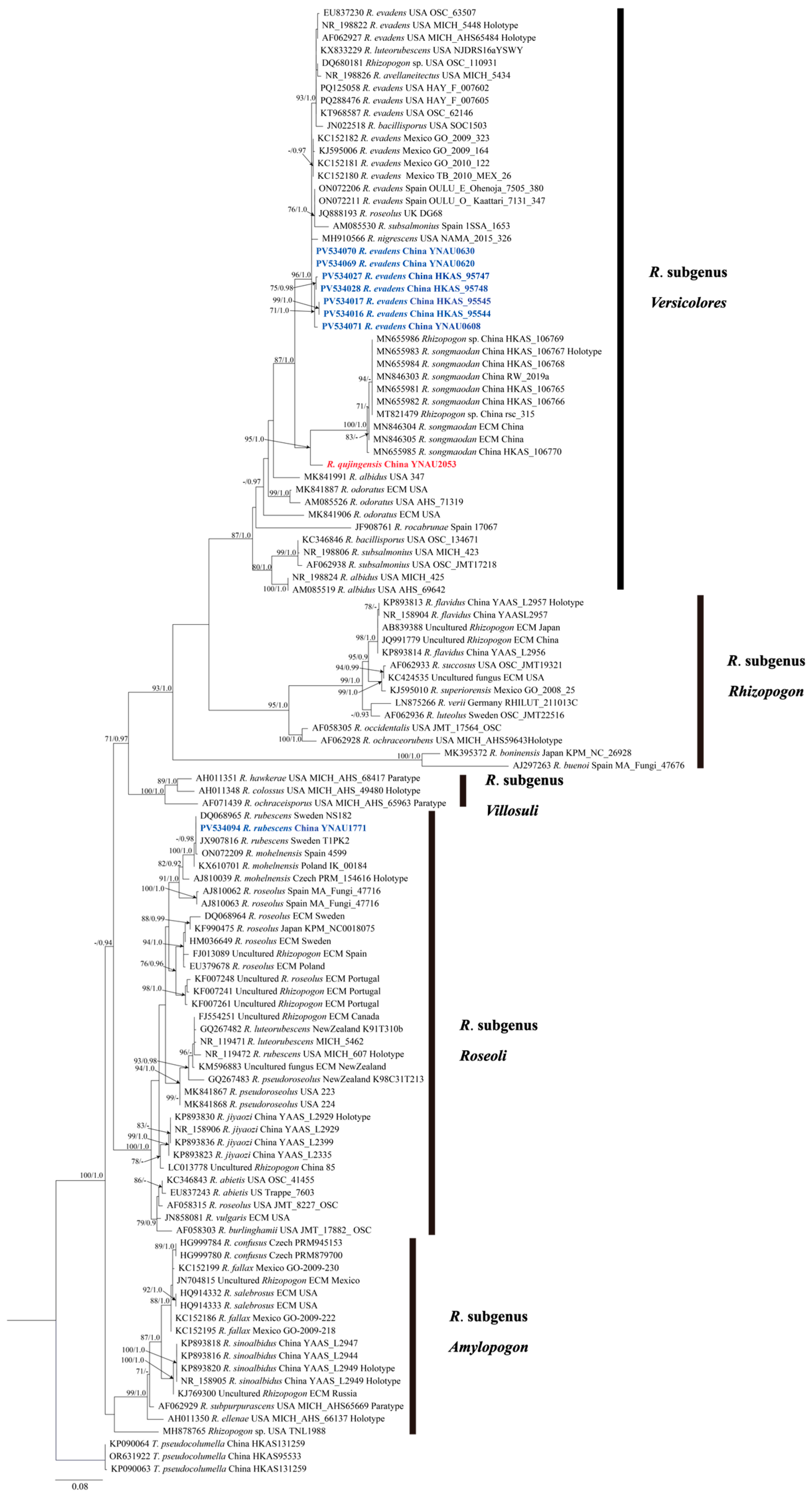
Figure 1. Phylogenetic reconstruction of based on ITS sequence data, inferred using RAxML maximum likelihood analysis. Branch support values are indicated adjacent to corresponding nodes: bootstrap support (BS) values ≥ 70% derived from maximum likelihood (ML) analysis and posterior probabilities (PP) ≥ 0.90 obtained from Bayesian inference. Red labels denote the newly described species; blue labels indicate species newly recorded in China.
Diversity 17 00683 g001

3. Results

3.1. Molecular Phylogenetics

A total of 117 ITS nrDNA sequences were analyzed, with three sequences of Truncocolumella pseudocolumella designated as the outgroup. This dataset included nine newly generated sequences from Chinese collections. Following alignment and exclusion of ambiguously aligned regions, the final matrix comprised 899 bp. Both maximum likelihood (ML) and Bayesian inference (BI) analyses resulted in similar phylogenetic tree topologies. The ML phylogram (Figure 1) is presented, with nodes exhibiting strong statistical bootstrap support from ML and posterior probability values from BI, corroborating the robustness of the inferred phylogeny.

3.2. Taxonomy

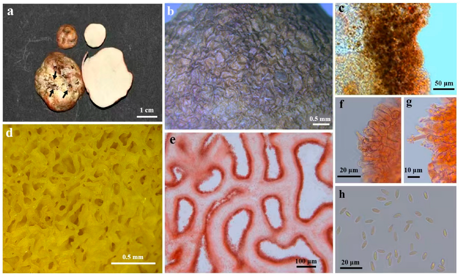
Rhizopogon qujingensis S. P. Wan, H. W. Zhang & F. Q. Yu sp. nov. (Figure 2).
MycoBank: 858797
Etymology: The epithet qujingensis refers to Qujing, the locality of the type collection.
Type: CHINA, Yunnan Province, Qujing city, in humus soil beneath litter under a monospecific forest of P. armandii Franch., 25.763234° N, 104.060502° E, elevation 2178 m, 2 December 2023, YNAU2053 (holotype).
Diagnosis: R. qujingensis is distinguished by a white peridium exhibiting rose-pink discoloration upon cutting or bruising and becoming yellow-brown upon drying; peridium thin (40.0–220.0 μm), composed of pigmented, interwoven hyphae. Gleba yellowish brown at maturity, with small, labyrinthine chambers. Basidiospores narrowly oblong to elongate, smooth, hyaline, 4.3–9.5 × 2.3–4.4 μm.
Description: Basidiomata subglobose to irregular, 1.3 × 1.1 cm, compliant to slightly soft to rubbery. Fresh coloration, white when young, maturing to yellow-brown; surfaces discolor rose-pink when cut or bruised (Figure 2a); dried specimens yellow-brown, shrunken, often exhibiting depressed areas (Figure 2b). Growth solitary, occurring in humus-rich soils. Peridial surface white to yellow (Figure 2c), bearing rhizomorphs and sparse hyphae, white to yellow (Figure 2d). Odor indistinct, faintly fungal. Peridium readily separable, thin (40.0–220.0 μm thick), comprising interwoven, septate hyphae, 4.0–8.0 μm in diam, hyaline with red-brown pigmentation, becoming rose-pink when cut. Gleba initially white when, maturing yellowish to yellowish-brown; chambers small, numerous, labyrinthine; xtrama composed of interwoven, hyaline and gelatinized hyphae, 4.4–6.1 μm diam, thin-walled (Figure 2e). Hymenium lining chamber surfaces, composed of basidia and brachybasidioles embedded in a pith of interwoven, hyaline, gelatinized hyphae (Figure 2f). Basidia predominantly four-spored, smooth, clavate to cylindrically clavate, thin-walled (Figure 2g). Brachybasidioles ellipsoid to clavate, 11.8–22.0 × 3.7–7.4 μm (Figure 2h). Basidiospores narrowly oblong to elongate, smooth, 4.3–9.5 × 2.3–4.4 μm, mean 6.90 × 3.35 μm (SD: 1.30 [length], 0.53 [width]), Q = 1.0–4.1 (Figure 2i).
Distribution: The species is currently known only from a single collection site in Qujing City, Yunnan Province, China.
Notes: Phylogenetic analysis places R. qujingensis within subgenes Versicolores, forming a distinct lineage sister to R. songmaodan. While sharing same peridia discoloration patterns and glebal configuration with R. songmaodan, R. qujingensis has a markedly thinner peridium and more slender basidiospores.
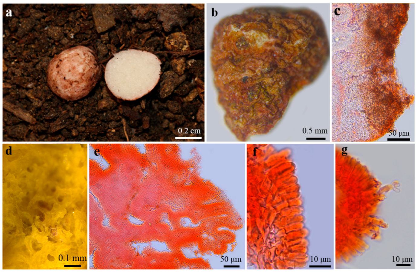
Rhizopogon evadens [3] (Figure 3).
Description: Basidiomata subglobose to irregular, 1.2–3.0 × 1.1–2.5 cm. Surface smooth, bearing a shallow depression; coloration white to brownish-yellow when fresh (Figure 3a), becoming rugose and tan upon drying (Figure 3b). Odor inconspicuous. Peridium constituting the outermost layer of the basidiomata, readily abraded; pigmentation tan; in some species staining pink to red where bruised or cut; thickness 38.0–200.0 μm thick (Figure 3c). Surface bearing adherent hyaline hyphae, 5.0–8.0 μm wide, septate, unbranched. Gleba initially white or off-white in immature specimens, maturing to light yellow to dark yellow; labyrinthine, exhibiting numerous small pores; composed of interwoven, hyaline hyphae, becoming pasty at maturity (Figure 3d). Hymenium lining the cavities. Central issue (pith) consisting of interwoven, hyaline, gelatinized hyphae (Figure 3e). Hymenium composed of basidia and brachybasidioles (Figure 3f,g). Basidia 4–8-spored, smooth-walled, cylindrical to clavate, 5.8–8.3 × 2.8–3.5 μm. Brachybasidioles ellipsoid to clavate, 10.0–19.6 × 4.8–6.9 μm (Figure 3g). Basidiospores ellipsoid to ovoid, smooth-walled, 11.8–15.0 (–19.0) × 4.3–6.1 (–8.7) μm, mean 13.40 × 5.20 μm (SD: 0.80 [length], 0.45 [width]), Q = 1.9–3.5 (Figure 3h). Trama composed of laxly interwoven, hyaline, gelatinized hyphae, 4.8–6.9 μm in diameter, predominantly unbranched.
Ecology and Distribution: Hypogeous to subhypogeous occurring in symbiotic association with P. densata Mast. Distribution encompasses Europe, America, and Asia. Within China, primarily recorded from the Yunnan and Sichuan Provinces.
Additional specimen examined:
China, Sichuan Province, Ganzi Tibetan Autonomous Prefecture, in forests of P. densata with associated Q. pannosa, 29.15695851° N, 99.92065430° E, elevation 3387 m, 4 October 2021, wsp1492 (YNAU0620); China, Yunnan Province, Lijiang City, symbiotic host P. yunnanensis Franch. Surrounded by Rhododendron sp. and Quercus sp., 27.00.03.35° N, 100.10.51.06° E, elevation 3260 m, 13 July 2015, wsp400 (HKAS95544); China, Sichuan Province, Ganzi Tibetan Autonomous Prefecture, forest of Q. guyavifolia H. Lév., 29.03163016° N, 99.70884830° E, elevation 3557 m, 4 October 2021, wsp1480 (YNAU0608); China, Yunnan Province, Chuxiong Prefecture, under Quercus sp. in mixed forests with P. yunnanensis, 25.18.58.8° N, 101.22.50.4° E, alt. 2519 m, 29 August 2015, wsp673 (HKAS95747); China, Yunnan Province, Lijiang City, Jade Dragon Snow Mountain Ecological Station, 13 July 2015, wsp401 (HKAS95545); China, Yunnan Province, Chuxiong Prefecture, under Quercus sp. in mixed forests with P. yunnanensis, 25.18.58.8° N, 101.22.50.4° E, elevation 2519 m, 29 August 2015, wsp674 (HKAS95746).
Rhizopogon rubescens [8] (Figure 4).
Description: Basidiomata subglobose to irregular, 0.4 × 0.5 cm, surface smooth; coloration pale pink to light brown when fresh (Figure 4a), becoming rugose and reddish brown upon drying, exhibiting subtle color gradations (Figure 4b). Odor inconspicuous. Peridium, the outermost layer of the basidiomata, readily abraded; pigmentation reddish brown; 85.0–460.0 μm thick (Figure 4c). Surface sparsely adorned with adherent hyaline hyphae, 1.0–6.0 μm wide, septate, unbranched. Gleba in immature specimens white to off-white when immature, becoming light yellow to earthy yellow at maturity; structure labyrinthine, bearing numerous small pores; composed of interwoven hyaline hyphae, becoming pasty at maturity (Figure 4d). Hymenium lining cavity surfaces. Central tissue (pith) of interwoven, hyaline, gelatinized hyphae (Figure 4e). Hymenium comprising basidia and brachybasidioles (Figure 4f,g). Basidia predominantly two-spored, smooth-walled, cylindrical to clavate, 12.2 × 4.7 μm. Brachybasidioles ellipsoid to clavate, 1.9–2.1 × 0.9–1.1 μm (Figure 4g). Trama laxly interwoven, hyaline, gelatinized hyphae, 1.8–5.2 μm in diameter, septate, branched.
Ecology and Distribution: Hypogeous, occurring in soils of mixed forests dominated by P. sylvestris var. mongolica and forests of Acer truncatum Bunge in Heilongjiang, China. Previous reports document occurrence in Europe and North America.
Voucher Specimen examined: China, Heilongjiang Province, mixed forests of P. sylvestris var. mongolica and A. truncatum, 46.760187° N, 130.365395° E, elevation 108.7085 m, 1 September 2023, PV534094 (YNAU1771).

4. Discussion

The genus Rhizopogon exhibits substantial species diversity, with a well- documented research history in Europe and North America extending to the early 18th century [30]. In Asia, confirmed taxa included Japanese records: R. boninensis, R. roseolus, R. alpinus, R. nitidus, and R. yakushimensis sp. nov. and Chinese reports enumerate ten taxa: R. rubescens var. ochraceus, R. rubescens var. pallidimaculatus, R. rubescens var. rileyi, R. nigrescens, R. piceus, R. shanxiensis, R. jiyaozi, R. songmaodan, R. flavidus, and R. sinoalbidus. Molecular analyses support the recognition of four species: R. jiyaozi, R. songmaodan, R. flavidus, and R. sinoalbidus within established subgenera Roesoli, Versicolores, Rhizopogon, and Amylopongon, respectively [10,30].
In the present study, integrated morphological and molecular evidence supports the recognition of a new species: R. qujingensis. Phylogenetic analysis places this taxon within the subgenus Versicolores (Figure 1), demonstrating a sister relationship to R. songmaodan. Despite their relatively close phylogenetic relationship, the species diverge into distinct, strongly supported clades (ML/BS = 95%; BI/PP = 0.98), corroborating their independent evolutionary trajectories.
Phylogenetically, the subgenus Versicolores is closely related to the subgenus Rhizopogon yet exhibits striking differences in peridial structure; morphologically, it more closely resembles the subgenus Roseoli but is distinguished by the absence of the latter’s yellow pigmentation [22]. Within Versicolores subgenus, R. qujingensis is morphologically distinguished from related species by a unique combination of macro- and micromorphological features. Unlike R. subsalmonius [22] which shows no staining reaction and R. evadens [22] which stains red but possesses a white, non-yellow peridium unaffected by KOH, R. qujingensis exhibits immediate rose-pink discoloration when bruised or cut. R. qujingensis has a thinner peridium (40.0–220.0 μm) compared to R. rocabrunae (300.0–600.0 μm) and lacks the characteristic orange pigments and prominent squamules found in the latter [45]. Moreover, R. rocabrunae develops brown to black squamules and produces larger, elongated ellipsoid spores with higher Qm values (2.59–2.87 μm), R. qujingensis has smaller, narrowly oblong to elongate spores (4.3–9.5 × 2.3–4.4 μm). Compared to R. songmaodan [10], which shares similar rubbery gleba and staining reaction, R. qujingensis has narrower spores and distinct red-brown pigmentation in its peridial hyphae. Additionally, R. qujingensis differs from R. albidus in its lack of peach to salmon coloration and from R. odoratus [46] by lacking an aromatic odor. It differs ecologically from the conifer-associated R. subsalmonius (with Abies) and R. albidus (with Abies) [47] by growing solitary in humus-rich soils. These diagnostic characteristics—including its rose-pink staining reaction, thin peridium with red-brown pigmentation, narrow spore morphology, and humus-rich habitat—collectively differentiate R. qujingensis from all compared species.
Phylogenetic analyses confirmed that the seven specimens collected in this study (YNAU0608, YNAU0620, YNAU0630, HKAS95544, HKAS95545, HKAS95746, and HKAS95747) represent R. evadens, showing strong support (BS = 96%; PP = 1.0) for clustering with European and North American taxa. The ectomycorrhizal fungal genus Rhizopogon exhibits a global distribution and demonstrates a strong preference for, and high specificity towards, Pinaceae hosts [22]. Within China, its diversity is centered in the Sichuan and Yunnan provinces. Specimens examined in this study were collected under P. densata, P. yunnanensis, and Quercus spp. The latter observation diverges from established host patterns. This observation suggests that Rhizopogon has greater ecological flexibility and a wider range of host species than previously thought. Our recent research further supports this idea, as we have successfully identified and confirmed, through ITS molecular evidence, the formation of ectomycorrhizae between R. sinoalbidus and Q. franchetii (GenBank accession no.: PX368895). This discovery marks a departure from the strict Pinaceae specificity seen in nearly all previously studied Rhizopogon species from North America and Europe, indicating a unique ecological adaptation within Chinese taxa that deserves further exploration.
This finding emphasizes the necessity to reevaluate the host-range limitations of Rhizopogon and serves as an important reference for future research into the ecological adaptability of this genus.

Author Contributions

Conceptualization, S.W.; methodology, L.L. and C.Y.; software, H.Z.; validation, F.Y. and Y.L.; data analysis, H.Z. and Y.Z.; data curation, H.Z.; writing—original draft preparation, H.Z. and Y.Z.; writing—review and editing, S.W., F.Y. and J.P.-M.; visualization, L.L. and Y.L.; supervision, J.P.-M.; project administration, S.W.; funding acquisition, F.Y. All authors have read and agreed to the published version of the manuscript.

Funding

This work was financially supported by the Science and Technology Fundamental Resources Investigation Program, Ministry of Science and Technology of China (2023FY101303), the Yunnan Revitalization Talent Support Program (XDYC-QNRC-2023-0415), and the Yunnan Technology Innovation Program (202205AD160036).

Institutional Review Board Statement

Not applicable.

Data Availability Statement

Data are contained within the article.

Acknowledgments

We sincerely appreciate Yun Wang from the New Zealand Institute for Crop and Food Research Limited in Mosgiel, New Zealand, for his invaluable help in finding and providing access to several historic literature references. We also thank ZhiJia Gu of the Key Laboratory for Plant Diversity and Biogeography of East Asia, Chinese Academy of Sciences, for performing the scanning electron microscopy (SEM).

Conflicts of Interest

The authors declare no conflicts of interest.

Appendix A

Table A1. Collection details of voucher specimens and corresponding GenBank accession numbers for ribosomal ITS sequences analyzed in this study.
Species NameVoucher/IsolationOriginGenBank No. of ITSReferences
R. evadensGO-2009-164MexicoKJ595006Unpublished
Uncultured RhizopogonECMRussiaKJ769300[48]
R. superiorensisGO-2008-25MexicoKJ595010Unpublished
Uncultured R. roseolusECMPortugalKF007248Unpublished
Uncultured RhizopogonECMPortugalKF007241Unpublished
R. roseolusKPM-NC0018075JapanKF990475[49]
Uncultured RhizopogonECMPortugalKF007261Unpublished
R. roseolusMA-Fungi 47716SpainAJ810062[26]
R. roseolusMA-Fungi 47716SpainAJ810063[26]
Uncultured RhizopogonECMSpainFJ013089Unpublished
Uncultured RhizopogonECMCanadaFJ554251[50]
R. roseolusECMPolandEU379678Unpublished
R. abietisTrappe 7603USEU837243Unpublished
R. evadensOSC 63507USAEU837230Unpublished
R. roseolusECMSwedenHM036649[51]
R. pseudoroseolusK98C31T213New ZealandGQ267483[52]
R. luteorubescensK91T310bNew ZealandGQ267482[52]
R. abietisOSC: 41455USAKC346843Unpublished
R. fallaxGO-2009-222MexicoKC152186Unpublished
R. fallaxGO-2009-230MexicoKC152199Unpublished
R. fallaxGO-2009-218MexicoKC152195Unpublished
R. evadensGO-2010-122MexicoKC152181Unpublished
Uncultured fungusECMUSAKC424535[53]
R. luteorubescensNJDRS16aYSWYUSAKX833229[54]
R. jiyaoziYAAS-L2399ChinaKP893836[30]
R. sinoalbidusYAAS-L2947ChinaKP893818[30]
Uncultured Rhizopogon85ChinaLC013778[55]
R. vulgarisECMUSAJN858081[56]
R. bacillisporusSOC1503USAJN022518Unpublished
Uncultured RhizopogonECMMexicoJN704815Unpublished
R. salebrosusECMUSAHQ914333[57]
R. salebrosusECMUSAHQ914332[57]
Uncultured RhizopogonECMJapanAB839388[58]
Uncultured fungusECMNew ZealandKM596883Unpublished
R. roseolusDG68UKJQ888193[59]
Uncultured RhizopogonECMChinaJQ991779Unpublished
R. flavidusYAAS: L2957ChinaNR_158904[30]
R. sinoalbidusYAAS: L2949
(Holotype)
ChinaNR_158905[30]
R. jiyaoziYAASL: 2929ChinaNR_158906[30]
R. succosusOSC JMT19321USAAF062933[22]
R. luteolusOSC JMT22516SwedenAF062936[22]
R. veriiRHILUT_211013CGermanyLN875266[29]
R. songmaodanHKAS-106768ChinaMN655984[10]
R. songmaodanRW_2019aChinaMN846303[10]
R. nigrescensNAMA 2015-326USAMH910566Unpublished
R. pseudoroseolus223USAMK841867Unpublished
R. albidus347USAMK841991Unpublished
R. pseudoroseolus224USAMK841868Unpublished
Rhizopogon sp.Trappe 28417;
SC 110931
USADQ680181Unpublished
R. subsalmonius1SSA-1653SpainAM085530Unpublished
R. subsalmoniusOSC JMT17218USAAF062938[22]
R. albidusAHS 69642USAAM085519Unpublished
R. evadensMICH AHS 65484
(Holotype)
USAAF062927[22]
R. odoratusAHS 71319USAAM085526Unpublished
R. rocabrunae17067SpainJF908761[60]
R. occidentalisJMT 17564, OSCUSAAF058305[22]
R. ochraceorubensMICH AHS 59643
(Holotype)
USAAF062928[22]
R. flavidusYAAS-L2957
(Holotype)
ChinaKP893813[30]
R. flavidusYAAS-L2956ChinaKP893814[30]
R. subpurpurascensMICH AHS 65669
(Paratype)
USAAF062929[22]
R. ellenaeMICH AHS 66137
(Holotype)
USAAH011350[22]
R. sinoalbidusYAAS-L2944ChinaKP893816[30]
R. sinoalbidusYAAS-L2949
(Holotype)
ChinaKP893820[30]
R. colossusMICH AHS 49480
(Holotype)
USAAH011348[22]
R. hawkeraeMICH AHS 68417
(Paratype)
USAAH011351[22]
R. ochraceisporusMICH AHS 65963
(Paratype)
USAAF071439[22]
R. jiyaoziYAAS-L2335ChinaKP893823[30]
R. jiyaoziYAASL-2929
(Holotype)
ChinaKP893830[30]
R. roseolusJMT 8227, OSCUSAAF058315[22]
R. burlinghamiiJMT 17882, OSCUSAAF058303[22]
R. buenoiMA-Fungi 47676
(Holotype)
SpainAJ297263[61]
R. boninensisKPM-NC 26928JapanMK395372Unpublished
R. songmaodanHKAS-106767
(Holotype)
ChinaMN655983[10]
R. songmaodanHKAS-106766ChinaMN655982[10]
R. mohelnensisIK-00184PolandKX610701[62]
R. rubescensNS182SwedenDQ068965[63]
R. rubescensT1PK2SwedenJX907816[64]
R. mohelnensis4599SpainON072209[65]
R. albidusAHS 69642USAAM085519Unpublished
R. albidusMICH: 425USANR_198824Unpublished
R. evadensMICH: 5448
(Holotype)
USANR_198822Unpublished
R. avellaneitectusMICH: 5434USANR_198826Unpublished
R. subsalmoniusMICH: 423USANR_198806Unpublished
R. odoratusMycorrhizal rootUSAMK841906Unpublished
R. odoratusMycorrhizal rootUSAMK841887Unpublished
R. bacillisporusOSC: 134671USAKC346846Unpublished
Rhizopogon sp.TNL1988USAMH878765Unpublished
R. confususPRM945153CzechHG999784[2]
R. confususPRM879700CzechHG999780[2]
R. songmaodanrsc-315ChinaMT821479[10]
R. songmaodanSynthesized ECM of Pinus armandiiChinaMN846304[10]
R. songmaodanSynthesized ECM of P. armandiiChinaMN846305[10]
R. songmaodanHKAS-106770ChinaMN655985[10]
R. songmaodanHKAS-106769ChinaMN655986[10]
R. songmaodanHKAS-106765ChinaMN655981[10]
R. evadensHAY-F-007602USAPQ125058Unpublished
R. evadensOSC 62146USAKT968587Unpublished
R. evadensOULU: E.Ohenoja 7505-380SpainON072206[65]
R. evadensOULU: O.Kaattari 7131-347SpainON072211[65]
R. evadensTB-2010-MEX 26MexicoKC152180Unpublished
R. evadensGO-2009-323MexicoKC152182Unpublished
R. evadensHAY-F-007605USAPQ288476Unpublished
R. rubescensMICH: 607
(Holotype)
USANR_119472[26]
R. mohelnensisPRM 154616
(Holotype)
CzechAJ810039[26]
R. roseolusECMSwedenDQ068964[63]
R. luteorubescensMICH: 5462USANR_119471[26]
T. pseudocolumellaHKAS131259ChinaKP090064Unpublished
T. pseudocolumellaHKAS131259ChinaKP090063Unpublished
T. pseudocolumellaHKAS95533ChinaOR631922Unpublished

References

  1. Fries, E.M. Symbolae Gasteromycorum; Ex Officina Berlingiana: Lund, Sweden, 1817. [Google Scholar]
  2. Koukol, O.; Valda, S.; Gaisler, J.; Kunca, V.; Dowie, N.J. Rhizopogon confusus sp. nov., a correct name for a fungus previously recorded in Central Europe as the North American Rhizopogon salebrosus. Mycol. Prog. 2022, 21, 49. [Google Scholar] [CrossRef]
  3. Smith, A.H.; Zeller, S.M. A preliminary account of the North American species of Rhizopogon. Mem. N. Y. Bot. Gard. 1966, 14, 1–178. [Google Scholar]
  4. Massicotte, H.B.; Molina, R.; Luoma, D.L.; Smith, J.E. Biology of the ectomycorrhizal genus, Rhizopogon. 2. Patterns of host-fungus specificity following spore inoculation of diverse hosts grown in monoculture and dual culture. New Phytol. 1994, 126, 677–690. [Google Scholar]
  5. Molina, R.; Massicotte, H.B.; Trappe, J.M. Biology of the ectomycorrhizal genus, Rhizopogon.1. Host associations, host-specificity and pure culture syntheses. New Phytol. 1994, 126, 653–675. [Google Scholar]
  6. Trappe, J.M.; Molina, R.; Luoma, D.L.; Cázares, E.; Pilz, D.; Smith, J.E.; Castellano, M.A.; Miller, S.L.; Trappe, M.J. Diversity, Ecology, and Conservation of Truffle Fungi in Forests of the Pacific Northwest; US Department of Agriculture, Forest Service, Pacific Northwest Research Station: Corvallis, OR, USA, 2009; Volume 772, pp. 1–194. [Google Scholar]
  7. Bubriski, R.; Kennedy, P. A molecular and morphological analysis of the genus Rhizopogon subgenus Villosuli section Villosuli as a preface to ecological monitoring. Mycologia 2014, 106, 353–361. [Google Scholar] [CrossRef]
  8. Kawai, M.; Yamahara, M.; Ohta, A. Bipolar incompatibility system of an ectomycorrhizal basidiomycete, Rhizopogon rubescens. Mycorrhiza 2008, 18, 205–210. [Google Scholar] [CrossRef]
  9. Molina, R.; Trappe, J.M.; Grubisha, L.C.; Spatafora, J.W. Rhizopogon. In Ectomycorrhizal Fungi: Key Genera in Profile; Cairney, J.W.G., Chambers, S.M., Eds.; Springer: Berlin/Heidelberg, Germany, 1999; pp. 129–161. [Google Scholar]
  10. Wang, R.; Yu, F.Q.; Jesús, P.M.; Carlos, C. A new edible Rhizopogon species from Southwest China, and its mycorrhizal synthesis with two native pines. Mycorrhiza 2020, 30, 85–92. [Google Scholar] [CrossRef]
  11. Shimomura, N.; Matsuda, M.; Ariyoshi, K.; Aimi, T. Development of mycelial slurries containing surfactant for cultivation of the edible ectomycorrhizal mushroom Rhizopogon roseolus (syn. Rhizopogon rubescens). Botany 2012, 90, 839–844. [Google Scholar] [CrossRef]
  12. Wang, Y.; Hall, I.R.; Dixon, C.; Hance-Halloy, M.; Strong, G.; Brass, P. The cultivation of Lactarius deliciosus (saffron milk cap) and Rhizopogon rubescens (shoro) in New Zealand. In Proceedings of the Second International Conference on Edible Mycorrhizal Mushrooms, Christchurch, New Zealand, 3–6 July 2001; Edible Mycorrhizal Mushrooms and Their Cultivation. Hall, I.R., Wang, Y., Danell, E., Zambonelli, A., Eds.; New Zealand Institute for Crop & Food Research: Christchurch, New Zealand, 2002. [Google Scholar]
  13. Pacioni, G. Italian hypogeous fungi. Mycologia 1984, 76, 988–997. [Google Scholar]
  14. Pietras, M.; Kolanowska, M. Predicted potential occurrence of the North American false truffle Rhizopogon salebrosus (AH Sm.) in Europe. Fungal Ecol. 2019, 39, 225–230. [Google Scholar] [CrossRef]
  15. Martín, M.P.; Calonge, F.D. Rhizopogon sect. Fulvigleba in Europe: A taxonomic revision. Mycotaxon 2006, 95, 229–240. [Google Scholar]
  16. Sugiyama, Y.; Murata, M.; Nara, K. A new Rhizopogon species associated with Pinus amamiana in Japan. Mycoscience 2018, 59, 176–180. [Google Scholar] [CrossRef]
  17. Miyamoto, Y.; Maximov, T.C.; Sugimoto, A.; Nara, K. Discovery of Rhizopogon associated with Larix from northeastern Siberia: Insights into host shift of ectomycorrhizal fungi. Mycoscience 2019, 60, 274–280. [Google Scholar] [CrossRef]
  18. Kumar, A.; Tapwal, A.; Kumar, D.; Yadav, R. Ectomycorrhizas of Rhizopogon himalayensis on Cedrus deodara. J. Basic Microbiol. 2024, 64, 2300616. [Google Scholar] [CrossRef]
  19. Grubisha, L.C. Systematics of the Genus Rhizopogon Inferred from Nuclear Ribosomal DNA Large Subunit and Internal Transcribed Spacer Sequences. Master’s Thesis, Oregon State University, Corvallis, OR, USA, 1998. [Google Scholar]
  20. Martín, M.P.; Högberg, N.; Nylund, J.-E. Molecular analysis confirms morphological reclassification of Rhizopogon. Mycol. Res. 1998, 102, 855–858. [Google Scholar] [CrossRef]
  21. Grubisha, L.C.; Trappe, J.M.; Molina, R.; Spatafora, J.W. Biology of the ectomycorrhizal genus Rhizopogon. V. Phylogenetic relationships in the Boletales inferred from LSU rDNA sequences. Mycologia 2001, 93, 82–89. [Google Scholar] [CrossRef]
  22. Grubisha, L.C.; Trappe, J.M.; Molina, R.; Spatafora, J.W. Biology of the ectomycorrhizal genus Rhizopogon. VI. Re-examination of infrageneric relationships inferred from phylogenetic analyses of ITS sequences. Mycologia 2002, 94, 381–619. [Google Scholar] [CrossRef]
  23. Grubisha, L.C.; Trappe, J.M.; Beyerle, A.R.; Wheeler, D. NATS truffle and truffle-like fungi 12: Rhizopogon ater sp. nov. and R. brunsii sp. nov. (Rhizopogonaceae, Basidiomycota). Mycotaxon 2005, 93, 345–353. [Google Scholar]
  24. Kretzer, A.M.; Luoma, D.L.; Molina, R.; Spatafora, J.W. Taxonomy of the Rhizopogon vinicolor species complex based on analysis ofITS sequences and microsatellite loci. Mycologia 2003, 95, 480–487. [Google Scholar] [CrossRef]
  25. Binder, M.; Hibbett, D.S. Molecular systematics and biological diversification of Boletales. Mycologia 2006, 98, 971–981. [Google Scholar] [CrossRef]
  26. Martín, M.P.; García, M.A. How many species in the Rhizopogon roseolus group? Mycotaxon 2009, 109, 111–128. [Google Scholar] [CrossRef]
  27. Trappe, J.M. A revision of the genus Alpova with notes on Rhizopogon and the Melanogastraceae. Beih. Nova Hedwigia 1975, 51, 279–309. [Google Scholar]
  28. Martín, M.P. The Genus Rhizopogon in Europe; Edicions de la Universitat de Barcelona (BCG): Barcelona, Spain, 1996. [Google Scholar]
  29. Sulzbacher, M.A.; Grebenc, T.; Garcia, M.A.; Silva, B.D.B.; Silveira, A.; Antoniolli, Z.I.; Marinho, P.; Munzenberger, B.; Telleria, M.T.; Baseia, I.G.; et al. Molecular and morphological analyses confirm Rhizopogon verii as a widely distributed ectomycorrhizal false truffle in Europe, and its presence in South America. Mycorrhiza 2016, 26, 377–388. [Google Scholar] [CrossRef]
  30. Li, L.; Zhao, Y.C.; Zhou, D.Q.; Yu, F.Q.; Zheng, L.; Wang, Y.; Zhang, X.L.; Duan, Z.J.; Zhao, X.Y.; He, Z.H.; et al. Three new species of Rhizopogon from Southwest China. Phytotaxa 2016, 282, 151–163. [Google Scholar] [CrossRef]
  31. Liu, B. New species and new records of hypogous fungi from China I. Acta Mycol. Sin. 1985, 4, 84–89. [Google Scholar]
  32. Tao, K.; Chang, M.C. The hypogeous fungi on the Rhizopogon found in Shanxi (I). J. Shanxi Agric Univ. 1988, 8, 226–229. [Google Scholar]
  33. Yu, F.Q.; Liu, P.G. Species diversity of wild edible mushroom from Pinus yunnanensis forest and conservation strategies. Biodivers. Sci. 2005, 13, 58–69. [Google Scholar] [CrossRef]
  34. Dai, Y.C.; Yang, Z.L. A revised checklist of medicinal fungi in China. Mycosystema 2008, 27, 801–824. [Google Scholar]
  35. Dai, Y.C.; Zhou, L.W.; Yang, Z.L.; Wen, H.A.; Bau, T.; Li, T.H. A revised checklist of edible fungi in China. Mycosystema 2010, 29, 1–21. [Google Scholar]
  36. Shao, D.H.; Yang, X.P.; Zhang, X.D.; Bai, S.L.; Zheng, R.; Wang, J.G. Ectomycorrhiza formation on Pinus tabulaeformis through Rhizopogon luteolus infection. Chin. J. Ecol. 2013, 32, 78–81. [Google Scholar]
  37. Liu, P.G. Rhizopogon. In Flora Fungorum Sinicorum; Science Press: Beijing, China, 2005; Volume 17, pp. 1–183. [Google Scholar]
  38. Fan, L. Research Progress on Species Diversity and Phylogeny of Hypogeous Fungi in China. J. Fungal Res. 2023, 21, 65–81. [Google Scholar]
  39. White, T.J.; Bruns, T.; Lee, S.; Taylor, J.W. Amplification and direct sequencing of fungal ribosomal RNA genes for phylogenetics. In PCR protocols: A Guide to Methods and Applications; Innis, M.A., Gelfand, D.H., Sninsky, J.J., White, T.J., Eds.; Academic Press: San Diego, CA, USA, 1990; pp. 315–322. [Google Scholar]
  40. Gardes, M.; Bruns, T.D. ITS primers with enhanced specificity for basidiomycetes application to the identification of mycorrhizae and rusts. Mol. Ecol. 1993, 2, 113–118. [Google Scholar] [CrossRef] [PubMed]
  41. Hall, T.A. BioEdit: A user-friendly biological sequence alignment editor and analysis program for Windows 95/98/NT. Nucleic Acids Symp. Ser. 1999, 41, 95–98. [Google Scholar]
  42. Nylander, J.A.A. MrModeltest, version 2.3; Program Distributed by the Author; Evolutionary Biology Center, Uppsala University: Uppsala, Sweden, 2004. [Google Scholar]
  43. Stamatakis, A. RAxML-VI-HPC: Maximum-likelihood-based phylogenetic analyses with thousands of taxa and mixed models. Bioinformatics 2006, 22, 2688–2690. [Google Scholar]
  44. Ronquist, F.; Huelsenbeck, J.P. MrBayes 3: Bayesian phylogenetic inference under mixed models. Bioinformatics 2003, 19, 1572–1574. [Google Scholar] [CrossRef]
  45. Zotti, M.; Di Piazza, S.; Vizzini, A. First records of Rhizopogon rocabrunae and R. pumilionum(Boletales) from Italy. Mycotaxon 2010, 113, 291–300. [Google Scholar]
  46. Martín, M.P.; Calonge, F.D. Rhizopogon aromaticus (Basidiomycotina) a new species found in Spain. Mycotaxon 2000, 76, 289–292. [Google Scholar]
  47. Trappe, M.; Evans, F.; Trappe, J. Field Guide to North American Truffles: Hunting, Identifying, and Enjoying the World’s Most Prized Fungi; Ten Speed Press: Berkeley, CA, USA, 2007. [Google Scholar]
  48. Malysheva, V.F.; Malysheva, E.F.; Kovalenko, A.E.; Pimenova, E.A.; Gromyko, M.N.; Bondarchuk, S.N. Ectomycorrhizal fungal diversity of Pinus koraiensis in the forests of the Central Sikhote-Alin based on rDNA sequence analysis of mycorrhizal tips. Mycol. Phytopathol. 2014, 48, 372–385. [Google Scholar]
  49. Orihara, T.; Okada, T.; Daiguji, T.; Takagi, N. Occurrence of a Truffle-like Fungus, Rhizopogon roseolus (Rhizopogonaceae, Boletales) in Kanagawa Prefecture, Japan. Kanagawa Kenritsu Hakubutsukan Kenkyu Hokoku Shizen Kagaku, 2014; in press. [Google Scholar]
  50. Hartmann, M.; Lee, S.; Hallam, S.J.; Mohn, W.W. Bacterial, archaeal and eukaryal community structures throughout soil horizons of harvested and naturally disturbed forest stands. Environ. Microbiol. 2009, 11, 3045–3062. [Google Scholar] [CrossRef]
  51. Menkis, A.; Vasaitis, R. Fungi in Roots of Nursery Grown Pinus sylvestris: Ectomycorrhizal Colonisation, Genetic Diversity and Spatial Distribution. Microb. Ecol. 2011, 61, 52–63. [Google Scholar] [CrossRef]
  52. Walbert, K.; Ramsfield, T.D.; Ridgway, H.J.; Jones, E.E. Ectomycorrhizal species associated with Pinus radiata in New Zealand including novel associations determined by molecular analysis. Mycorrhiza 2010, 20, 209–215. [Google Scholar]
  53. Cowden, C.C.; Peterson, C.J. Annual and Seasonal Dynamics of Ectomycorrhizal Fungi Colonizing White Pine (Pinus strobus) Seedlings Following Catastrophic Windthrow in Northern Georgia, USA. Can. J. For. Res. 2013, forthcoming. [Google Scholar] [CrossRef]
  54. Dowie, N.J.; Grubisha, L.C.; Burton, B.A.; Klooster, M.R.; Miller, S.L. Increased phylogenetic resolution within the ecologically important Rhizopogon subgenus Amylopogon using 10 anonymous nuclear loci. Mycologia 2017, 109, 35–45. [Google Scholar] [CrossRef] [PubMed]
  55. Long, D.; Liu, J.; Han, Q.; Wang, X.; Huang, J. Ectomycorrhizal fungal communities associated with Populus simonii and Pinus tabuliformis in the hilly-gully region of the Loess Plateau, China. Sci. Rep. 2016, 6, 24336. [Google Scholar] [CrossRef] [PubMed]
  56. Peay, K.G.; Schubert, M.G.; Nguyen, N.H.; Bruns, T.D. Measuring Ectomycorrhizal Fungal Dispersal: Macroecological Patterns Driven by Microscopic Propagules. Mol. Ecol. 2012, 21, 4122–4136. [Google Scholar] [CrossRef]
  57. Dowie, N.J.; Hemenway, J.J.; Miller, S.L. Identity, genetic lineages and putative hybrids of an obligate mycobiont associated with the mycoheterotrophic plant Pterospora andromedea in the south-central Rocky Mountains. Fungal Ecol. 2012, 5, 137–146. [Google Scholar] [CrossRef]
  58. Huang, J.; Nara, K.; Zong, K.; Lian, C. Soil propagule banks of ectomycorrhizal fungi along forest development stages after mining. Microb. Ecol. 2015, 69, 768–777. [Google Scholar]
  59. Pickles, B.J.; Genney, D.R.; Anderson, I.C.; Alexander, I.J. Spatial analysis of ectomycorrhizal fungi reveals that root tip communities are structured by competitive interactions. Mol. Ecol. 2012, 21, 5110–5123. [Google Scholar] [CrossRef]
  60. Osmundson, T.W.; Robert, V.A.; Schoch, C.L.; Baker, L.J.; Smith, A.; Robich, G.; Mizzan, L.; Garbelotto, M.M. Filling Gaps in Biodiversity Knowledge for Macrofungi: Contributions and Assessment of an Herbarium Collection DNA Barcode Sequencing Project. PLoS ONE 2013, 8, e62419. [Google Scholar]
  61. Martín, M.P.; Calonge, F.D. Rhizopogon buenoi (Boletales, Basidiomycota) a new species from Spain. Mycotaxon 2001, 79, 101–105. [Google Scholar]
  62. Kalucka, I.L.; Jagodzinski, A.M.; Nowinski, M. Biodiversity of Ectomycorrhizal Fungi in Surface Mine Spoil Restoration Stands in Poland—First Time Recorded, Rare, and Red-Listed Species. Acta Mycol. 2016, 51, 1080. [Google Scholar] [CrossRef]
  63. Menkis, A.; Vasiliauskas, R.; Taylor, A.F.; Stenlid, J.; Finlay, R. Fungal communities in mycorrhizal roots of conifer seedlings in forest nurseries under different cultivation systems, assessed by morphotyping, direct sequencing and mycelial isolation. Mycorrhiza 2005, 16, 33–41. [Google Scholar] [CrossRef]
  64. Klavina, D.; Gaitnieks, T.; Menkis, A. Survival, Growth and Ectomycorrhizal Community Development of Container- and Bare-Root Grown Pinus sylvestris and Picea abies Seedlings Outplanted on a Forest Clear-Cut. Balt. For. 2013, 19, 39–49. [Google Scholar]
  65. Martín, M.P.; Huang, A.; Ortega, M.A. Additions to Rhizopogon (Boletales, Basidiomycota) in Finland and Europe based on molecular and morphological evidence. Ann. Bot. Fenn. 2023, 60, 67–70. [Google Scholar] [CrossRef]
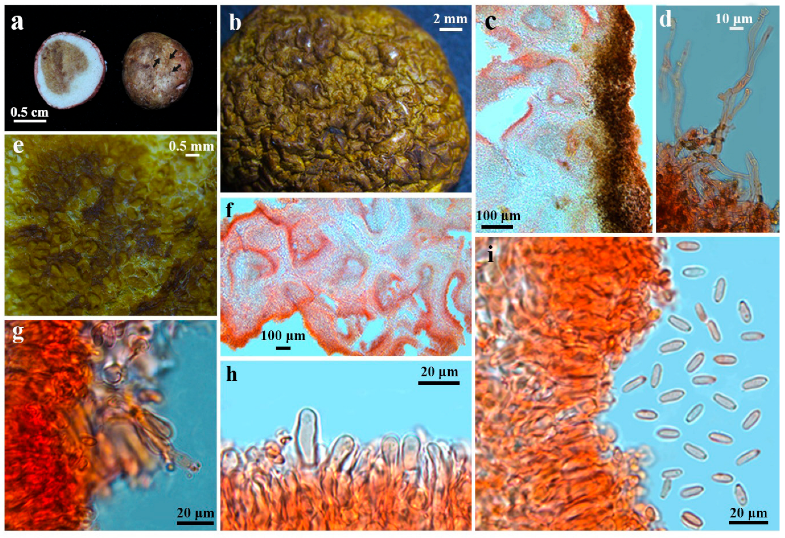
Figure 2. Morphological features of Rhizopogon qujingensis (voucher YNAU2053, type). (a) Fresh basidiomata (arrows indicate rhizomorphs). (b) Dried basidiomata. (c) Cross section of peridium. (d) Sparse hyphae on the peridial surface. (e) Cross section of gleba. (f) Cross section of irregular and sinuous glebal chambers. (g) Basidia and spores. (h) Clavate to cylindrical brachy-basidioles. (i) Hymenium and basidiospores.
Diversity 17 00683 g002
Figure 3. Morphological features of Rhizopogon evadens (voucher YNAU0630). (a) Fresh basidiomata (arrows indicate rhizomorphs). (b) Dried basidiomata. (c) Cross section of peridium. (d) Cross section of gleba. (e) Cross section of irregular and sinuous glebal chambers. (f) Clavate to cylindrical brachybasidioles. (g) Basidia and spores. (h) Basidiospores.
Diversity 17 00683 g003
Figure 4. Morphological features of Rhizopogon rubescens (voucher YNAU1771). (a) Fresh basidiomata. (b) Dried basidiomata. (c) Cross section of peridium. (d) Cross section of gleba. (e) Cross section of irregular and sinuous glebal chambers. (f) Clavate to cylindrical brachybasidioles. (g) Basidia and spores.
Diversity 17 00683 g004
Table 1. Collection details of self-collected voucher specimens in this study and corresponding GenBank accession numbers for ribosomal ITS sequences.
Species NameVoucher/IsolationOriginGenBank No. of ITS
R. qujingensisYNAU2053ChinaPV584203
R. evadensYNAU0620ChinaPV534069
R. evadensYNAU0630ChinaPV534070
R. evadensHKAS95544ChinaPV534016
R. evadensHKAS95545ChinaPV534017
R. evadensYNAU0608ChinaPV534071
R. evadensHKAS95747ChinaPV534027
R. evadensHKAS95746ChinaPV534028
R. rubescensYNAU1771ChinaPV534094
R. sinoalbidusEctomycorrhiza3117-5ChinaPX368895
Disclaimer/Publisher’s Note: The statements, opinions and data contained in all publications are solely those of the individual author(s) and contributor(s) and not of MDPI and/or the editor(s). MDPI and/or the editor(s) disclaim responsibility for any injury to people or property resulting from any ideas, methods, instructions or products referred to in the content.