Article

Early Metabarcoding Detection of Eukaryotic Putative Pathogens Nearby Wastewater Effluents of Ría de Vigo (NW Spain)

Ecology and Marine Biodiversity (EcoBioMar) Group, Institute of Marine Research, Spanish National Research Council (IIM-CSIC), 36208 Vigo, Spain
*
Author to whom correspondence should be addressed.
Diversity 2025, 17(10), 671; https://doi.org/10.3390/d17100671
Submission received: 8 July 2025 / Revised: 19 September 2025 / Accepted: 23 September 2025 / Published: 25 September 2025
(This article belongs to the Special Issue Applications on Environmental DNA in Aquatic Ecology and Biodiversity)

Abstract

From a One Health perspective, the Ría de Vigo (NW Spain) represents a complex socio-ecological system where urban, industrial, and aquaculture activities converge, creating vulnerabilities due to the interconnection between human, animal, and environmental health. This study evaluates the utility of a multi-marker environmental DNA (eDNA) metabarcoding approach as an early warning tool to detect potentially harmful eukaryotic pathogens near wastewater discharge points in three distinct municipalities of the Ría de Vigo. Water and sediment samples were analyzed using the V4 and V9 regions of the 18S rRNA gene and the ITS-2 region for fungi. Bioinformatics analysis was performed with DADA2 and taxonomic assignments were based on SILVA and UNITE databases. Eukaryotic diversity varied by site and marker, with the highest richness found in inner estuary sediments. The V9 region provided broader taxonomic coverage, while V4 produced higher read abundances. Putative pathogens, including human, zoonotic, and aquaculture-related taxa, were more prevalent in inner estuarine zones. Pathogens such as Rhodotorula mucilaginosa, Acanthamoeba sp., Cryptosporidium sp., and harmful algae (HA) were detected. The findings emphasize the importance of a multi-marker strategy, sediment inclusion, and landscape-scale variables to improve early pathogen detection, contributing to effective eDNA-based surveillance systems for coastal health management.

1. Introduction

Coastal ecosystems harbor an extensive diversity of eukaryotic microorganisms, each fulfilling distinct ecological roles and participating in biological interactions critical to maintaining ecosystem functionality. Furthermore, they provide services that benefit humanity, underpinning global economic stability by serving as resources for fisheries, raw materials, compounds with medical applications and recreational areas [1]. However, coastal zones are well-known as natural sinks for anthropogenic activities (e.g., inefficient wastewater discharges, industrial wastes, runoff of farms), leading to substantial environmental impacts, from habitat destruction and eutrophication to the release of potential pathogens, multidrug-resistant bacteria, and pharmaceutically active compounds (PhACs) [2,3,4]. Such pressures may reduce eukaryotic diversity, trigger pathogenic or harmful algal blooms, and disrupt trophic interactions, with cascading effects that compromise ecosystem functioning and services. In wastewater treatment plants (WWTPs), the microbial community is predominantly composed of prokaryotes, which have been extensively studied over the years compared to the non-bacterial community, which has been less investigated [5]. Within WWTPs and adjacent coastal zones, fungi contribute to organic matter degradation, protists act both as grazers that regulate bacterial populations and as potential pathogen vectors, and metazoans can influence microbial food webs. These groups thus represent both essential ecosystem players and potential health hazards, as some are pathogens with risks for human, animal, and environmental health [5,6]. In addition, WWTPs and coastal sediments can act as reservoirs and dissemination hubs for emerging pathogens, antibiotic resistance genes, and even invasive species, raising concerns for One Health frameworks. Therefore, a critical task in wastewater treatment is to monitor the human sewage microbiome to document its fate and effective removal [7]. While culture-dependent methods and molecular techniques (e.g., qPCR and in situ hybridization) are commonly used for monitoring, recent advances in next-generation sequencing (NGS) have enabled a more comprehensive exploration of microbial diversity [8].
There is an ongoing debate about the type of target gene chosen for sequencing on the efficiency of the metabarcoding eDNA technique. Examination of short hypervariable regions of the 18SrRNA gene (V9 and V4) or the full-length gene with universal primers involves trade-offs between sequencing cost, taxonomic resolution, and amplification bias [9]. Although fungi can be detected through the amplification of the 18S rRNA gene, this approach can lead to an underestimation of their proportion and diversity. Therefore, for fungal detection, the nuclear ribosomal internal transcribed spacer (ITS) regions are the universal DNA markers [10]. Beyond primer bias and incomplete databases, eDNA metabarcoding is also limited by DNA persistence, PCR inhibition, low taxonomic resolution, and its inability to distinguish viable from non-viable organisms.
This study focused on the Ría de Vigo (Galicia, NW Spain), an area bordered by nine municipalities with a combined population of approximately 413,000 inhabitants, distributed from the outer to the inner parts of the Ría. The region contains several WWTPs that discharge effluents into the Ría [11]. Additionally, this coastal ecosystem also maintains high fish and shellfish production values chains. The area has also experienced the presence of pathogens affecting the aquaculture sector and occurrences of potentially toxic harmful algal blooms (HABs) [12,13]. Most studies focused on describing biodiversity and detecting pathogens in the Ría de Vigo have relied on traditional methods, with limited application of high-throughput sequencing, which has mainly been used to explore microbial diversity and associations in general environments [14,15,16] rather than in discharge zones [17].
This study aimed to compare eukaryotic diversity through high-throughput amplicon sequencing in the effluent discharge areas of WWTPs from three municipalities in the Ría de Vigo located in the middle (Vigo, Cangas) and inner (Redondela) zones of the Ría. Given the mixing of effluents at discharge points, the study specifically focused on detecting DNA from anthropogenic putative pathogens, as well as those that may impact animal species and ecosystems. To enhance the detection of eukaryotic diversity and eukaryotic pathogens and minimize biases associated with specific target regions, the study employed a multi-marker approach using two commonly used regions for eukaryotic biodiversity assessment (V9 and V4) along with the universal fungal marker, the internal transcribed spacer (ITS). In this context, our work is framed as an environmental diagnosis, providing a timely snapshot of local community structure and putative pathogen diversity, while also laying the groundwork for future monitoring strategies in coastal ecosystems. By applying a multi-marker eDNA metabarcoding approach to WWTP discharge zones in the Ría de Vigo, this study provides one of the first comprehensive assessments of eukaryotic pathogen diversity in these environments, contributing to early detection capacity and advancing the application of high-throughput sequencing within a One Health framework.

2. Materials and Methods

2.1. Sampling

The diversity of micro-eukaryotic communities in both water and sediment was analyzed at three sampling sites within the Ría de Vigo (Galicia, NW Spain) in July 2023. Sampling was conducted once during this campaign with one site positioned near WWTPs outfall of each of the three municipalities: Vigo, Cangas, and Redondela. Notably, Vigo is the most populous city in the Ría de Vigo, whereas Cangas and Redondela are smaller towns. Cangas is located directly opposite Vigo, while Redondela is situated in the innermost part of the estuary, influenced by Alvedosa and Pexegueiro rivers, making it representative of the estuarine environment at the river’s mouth (Figure 1). Although the three selected sampling sites shared a common potential source of contamination (i.e., effluent discharges), they were also chosen to represent contrasting estuarine zones with markedly different environmental and ecological conditions, including riverine influence, hydrodynamics, and organic matter loads. These were one-off sampling locations selected for this diagnostic study rather than permanent monitoring stations.
At each site, one water sample (3 L) and one surface sediment sample (upper ~10 cm) were collected. In addition, in situ temperature and salinity of water were measured at each site, yielding the following average values: Cangas (21.35 °C, 19.36 PSU), Redondela (20.41 °C, 0.75 PSU), and Vigo (13.29 °C, 35.55 PSU). In total, six samples were obtained (three water and three sediment), each processed independently. For clarity, and hereafter throughout the manuscript, samples are referred to as WC and SC (Cangas water and sediment), WR and SR (Redondela water and sediment), and WV and SV (Vigo water and sediment). Water samples were pre-filtered through a 200 µm mesh to remove zooplankton and large particles and then filtered using sterile 0.22 µm pore size filters (Millipore, Merck, Burlington, MA, USA). Filters were stored at −20 °C until DNA isolation, while sediment samples were stored in sterile containers and kept frozen at −20 °C until further processing.

2.2. DNA Isolation, Amplification and Sequencing

DNA from sediments and water was isolated using DNeasy PowerSoil Pro and DNeasy PowerWater Kits (Qiagen, Hilden, Germany), respectively, following manufacturer’s indications. Negative isolation controls were performed to discard any contaminations in downstream analysis. DNA concentration and purity were verified with the DS-11FX Spectrophotometer/Fluorometer (Denovix, Wilmington, NC, USA).
The V9 and V4 regions of the 18S SSU rRNA gene, and the ITS-2 region of the nuclear internal transcribed spacer (ITS), were amplified using previously described primers: 1380F/1510R for V9 [18], TAReuk454FWD1/TAReukREV3 for V4 [19], and the fungal-specific primers ITS86F/ITS4 for ITS-2 [20]. Library preparation and paired-end sequencing (Illumina NovaSeq PE250, San Diego, CA, USA) were performed by AllGenetics (A Coruña, Spain) and Biology SL (Manilva, Spain). The resulting reads were deposited in the Sequence Read Archive (SRA) http://www.ncbi.nlm.nih.gov/sra (accessed on 15 May 2025) under BioProject accession number PRJNA1262121.

2.3. Bioinformatic Analisis

Raw paired-end reads were trimmed (adapter dimers removal) using Cutadapt v3.5 [21] and quality-checked with FastQC [22]. Bioinformatics analysis was conducted using the Microbial Genomics Module of Qiagen CLC Workbench 25 (Qiagen, https://digitalinsights.qiagen.com accessed on 3 March 2025).
Reads were quality filtered (Q > 20, quality limit = 0.05, maximum of 2 ambiguous nucleotides), trimmed to remove adapter and primer sequences, and length filtered (≤220 bp for V9; ≤250 bp for V4 and ITS-2). Reads with more than one expected error (maximum expected error = 1) were discarded. Amplicon Sequence Variants (ASVs) were inferred using the DADA2 algorithm [23], which includes dereplication of unique sequences, denoising (iterative estimation of a sample-specific error model and generation of candidate ASVs), chimera removal (consensus method), and merging of paired reads (minimum overlap of 12 bp, no mismatches allowed).
Taxonomic assignment was performed using the SILVA 99% SSU 18S database (v138.1) for V9/V4 ASVs [24] and the UNITE 99% database for ITS (v138.1) [25]. Unassigned fungal ASVs were further identified via BLASTn against the NCBI fungal ITS database. Singletons were excluded. Rarefaction plots were assessed from rarefied ASV samples.

2.4. Diversity and Community Composition

To compare the diversity and community composition across different locations and environmental compartments and for downstream analysis, ASV tables were subsampled to the sample with the lowest read count: 142,406 (V9), 267,595 (V4) and 40,124 (ITS-2). Alpha-diversity indices (Shannon–Wiener, Pielou and richness) were estimated using the Vegan R package 2.6.8 and UpSet plots were generated with UpSetR [26,27].
Putative pathogenic taxa were identified at the genus and/or species level, focusing on those associated with anthropogenic sources or marine environments. Classification was based on peer-reviewed scientific literature and two legal frameworks: Directive 2000/54/EC [28] and its Spanish transposition, Real Decreto 664/1997 [29]. Harmful algae were identified using the IOC-UNESCO Taxonomic Reference List of Harmful Micro Algae [30]. Taxonomic assignments were made using to the lowest possible rank based on sequence similarity. Identified genera were validated through BLASTn analysis, achieving sequence similarity values greater than 97%. The term “sp.” indicates assignment only at the genus level due to insufficient resolution, without implying detection of a single species. Additionally, several putative pathogens were detected at the species level and confirmed through BLASTn analysis, with similarity scores exceeding 99%. For V9 and V4 regions, species-level assignments were considered tentative (“closest match”) and only retained when supported by such high similarity values, providing greater confidence in those cases.

3. Results

3.1. Overall Sequencing Data

After sequencing, trimming and ASV detection, a total of 2,118,788 (V9), 2,472,531 (V4) and 499,846 (ITS-2) reads were obtained. These reads corresponded to 6039 (V9), 6679 (V4) and 4885 (ITS-2) taxonomically assigned ASVs. Following subsampling, 1,646,724 (V9) and 1,865,868 (V4) eukaryotic reads, along with 40,124 total fungal reads (ITS-2) were retained for further analysis (Table 1).

3.2. Microeukaryote Diversity

Rarefaction curves for the V9 and V4 regions reached a plateau in all samples, indicating that sequencing depth was sufficient to capture eukaryote diversity. The highest eukaryotic diversity was observed within the first 100,000 sequences for both V9 and V4 sequencing (Figure 2A). Differences in richness, Shannon and Pielou indices were detected between the V9 and V4 regions, with a higher number of ASVs generally observed in the V9 region, particularly at the Redondela sampling point. In terms of richness and evenness, the highest diversity indices were found in Redondela sediment samples, while the lowest diversity was detected in sediment of Cangas samples, where the V9 region showed higher diversity indices compared to the V4 region (Figure 2B). Additionally, rare taxa (i.e., those representing less than 1% of total reads) were better represented in the V9 region than in the V4 region.
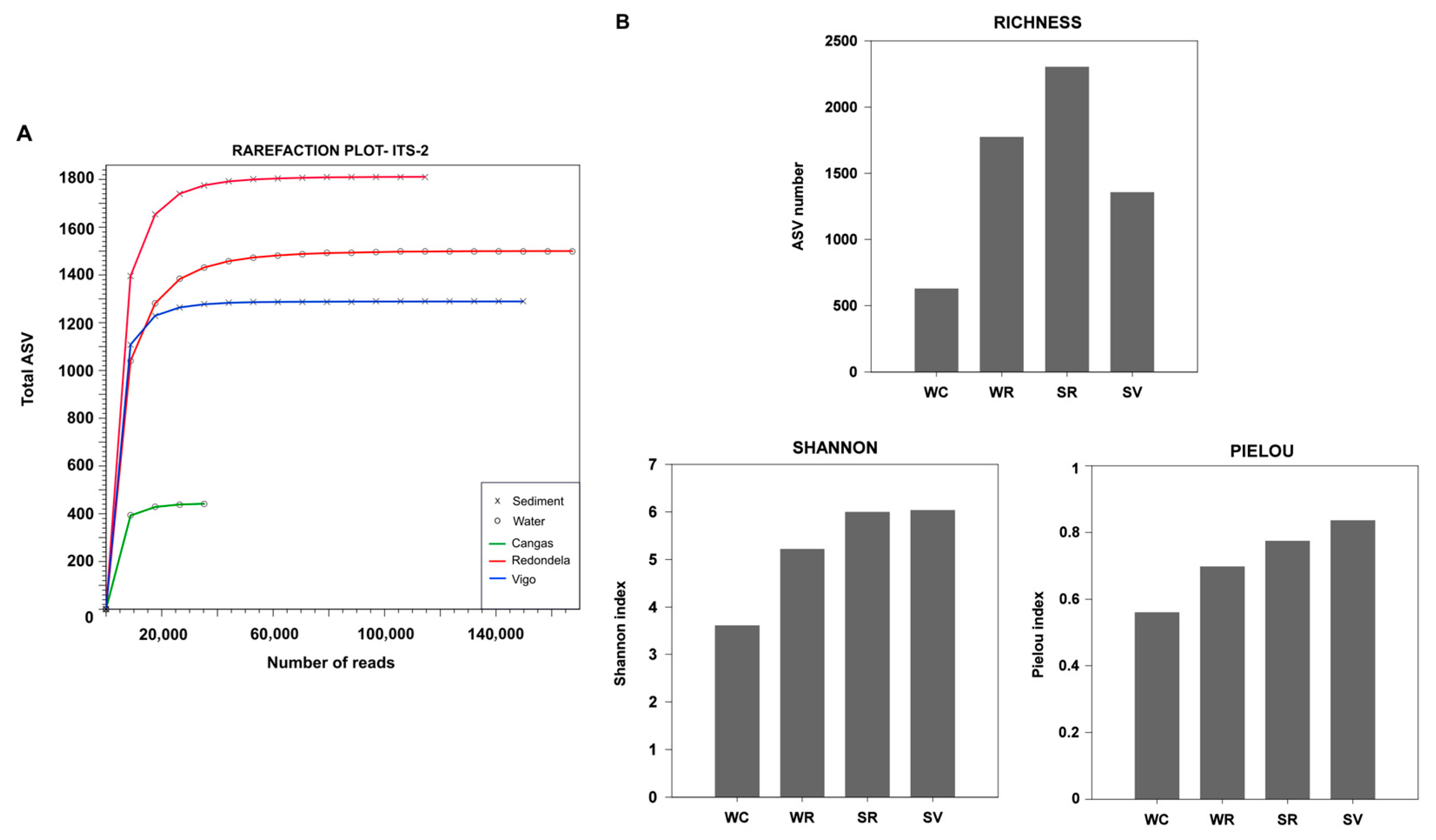
For the ITS-2 region, rarefaction curves revealed that samples from the sediment of Cangas and the water of Vigo revealed a low number of total reads. Including these samples in downstream analysis would have required rarefying the entire dataset to an impractically low threshold, leading to a substantial loss of information from other samples. Their low read counts may result from either technical limitation during sequencing or genuinely low microbial biomass in these environments. To ensure robust and comparable diversity analyses, we excluded these samples. For the remaining samples, rarefaction curves for the ITS-2 region reached a plateau across all sequencing efforts, confirming that sequencing depth was adequate to capture the full extent of fungal diversity. The initial 20,000 sequences yielded the highest fungal diversity (Figure 3A). Among the samples, sediment from Cangas (165 ASVs with 3325 total reads) and water from Vigo (120 ASVs with 6123 total reads) exhibited the lowest diversity, explaining their minimal representation in the rarefaction plot (Table 1, Figure 3A).
Diversity indices indicate that sediment from Redondela consistently had the highest species richness, diversity, and evenness, followed closely by sediment from Vigo. Water from Redondela exhibited moderate values across all diversity metrics, while water from Cangas ranked the lowest, reflecting lower species richness, reduced biodiversity and a more uneven species distribution. These findings highlight significant differences in fungal community structure among sites, with sediment from Redondela and sediment from Vigo supporting the most diverse and balanced fungal communities (Figure 3B).

3.3. Exclusive/Common ASVs

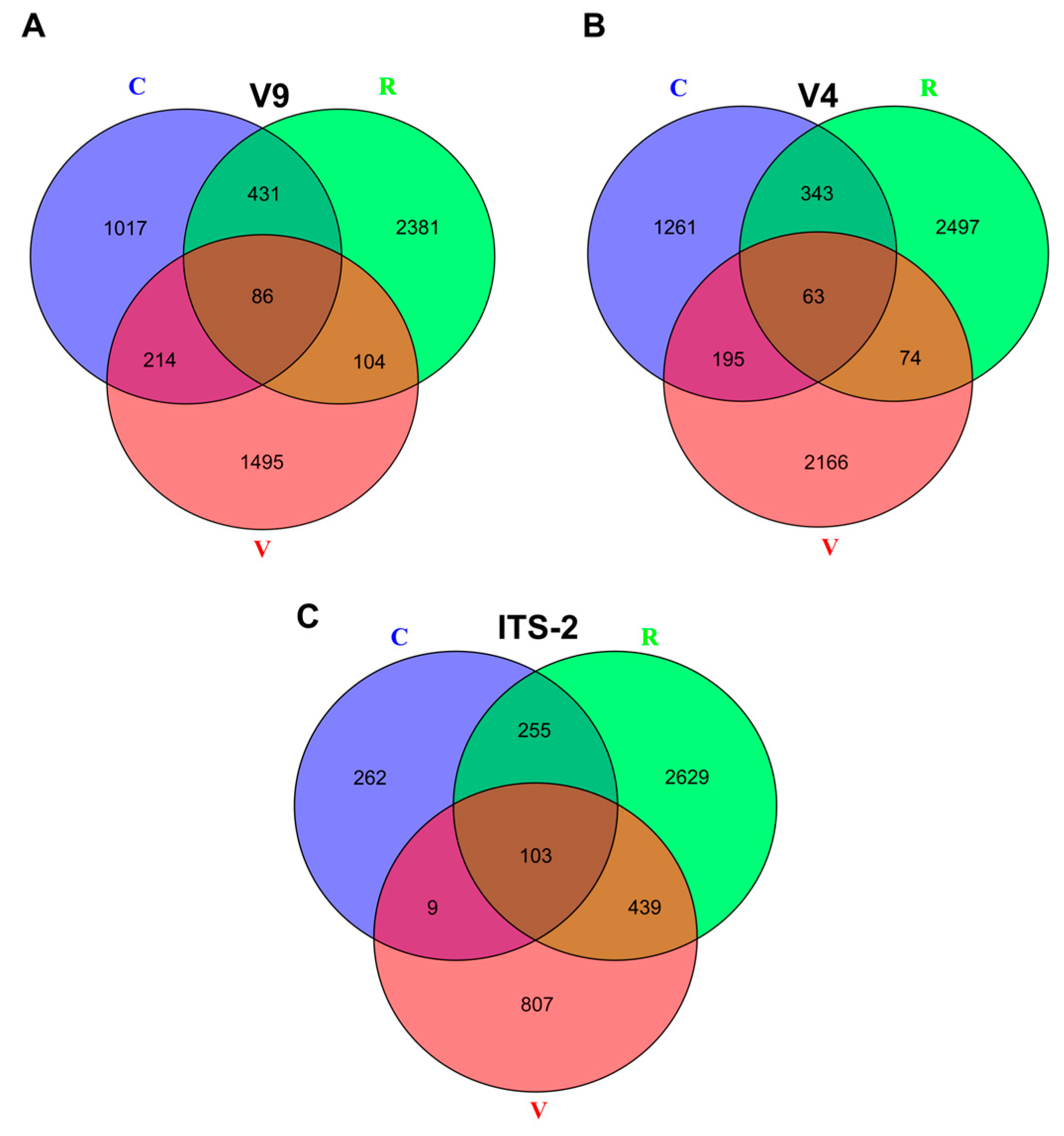
Analysis of the V9, V4, and ITS-2 gene regions showed a strong location-specific pattern in microeukaryotic diversity, with the majority of ASVs being exclusive to each sampling region. Across all markers, specific ASVs accounted for 58% to 87% of the total detected per location.
For the V9 region, 79%, 78%, and 58% of ASVs were unique to Redondela, Vigo, and Cangas, respectively. In contrast, only 86 ASVs were shared among the three sites, representing a small fraction of the total: 5% in Cangas, 3% in Redondela, and 4.5% in Vigo (Figure 4A). A similar pattern was observed for the V4 region, with high levels of site specificity: 84% of ASVs in Redondela, 87% in Vigo, and 67% in Cangas. The number of shared ASVs dropped to 63, comprising just 3% in Redondela, 2% in Vigo, and 3% in Cangas (Figure 4B). For the ITS-2 region, 76% of ASVs in Redondela, 59% in Vigo, and 41% in Cangas were found to be exclusive. A total of 103 ASVs were shared across all sites, accounting for 3% in Redondela, 7.6% in Vigo, and 16% in Cangas (Figure 4C).

3.4. Putative Biohazards

Using the multi-marker approach, we identified genetic material from a wide range of genera and species associated with putative human and zoonotic pathogens, as well as those affecting marine organisms, terrestrial plants, and HA. As species-level resolution was not always achieved (highest identity ≥ 99%), we reported genera with pathogenic species. Since not all species of the same genera are pathogenic, pathogenicity should be interpreted with caution and would require further confirmatory analyses.

3.4.1. Exclusive/Common Putative Biohazards

Across the three genetic regions analyzed (V9, V4, and ITS-2), the distribution of putative biohazards revealed both shared and unique taxa, with clear patterns of exclusivity evident not only between sampling sites but also across environmental fractions (water and sediment).
For the V9 region, a substantial number of putative biohazards were consistently detected across all sites, representing 42% of the total taxa in Cangas, 32% in Redondela, and 50% in Vigo (Figure 5A).
However, a considerable proportion of these taxa were exclusive to individual locations: 30% were found only in Redondela, 25% only in Vigo and 11% only in Cangas. Notable differences were also observed between fractions (sediment vs. water). For instance, ten genera/species exclusively found in sediment of Vigo, whereas Redondela’s sediment included 8 unique taxa (Figure 5A). Overall, only 3 putative biohazards were commonly detected in all samplings.
The V4 region displayed a similar pattern, although with slightly lower overall proportions. Putative biohazards were detected across all locations, comprising 20% of the total taxa in Cangas, 15% in Redondela, and 35.7% in Vigo. However, location-specific taxa were also observed: 40% were exclusive to Redondela, 43% to Vigo and 20% to Cangas. Environmental fractions again showed distinct profiles, with 18 unique genera/species in sediment of Redondela and 16 in sediment of Vigo (Figure 5B).
In contrast, the ITS-2 region exhibited a high level of overlap across locations, with most genera/species shared among them (Figure 5C).

3.4.2. Biohazards of Human/Zoonotic Origin

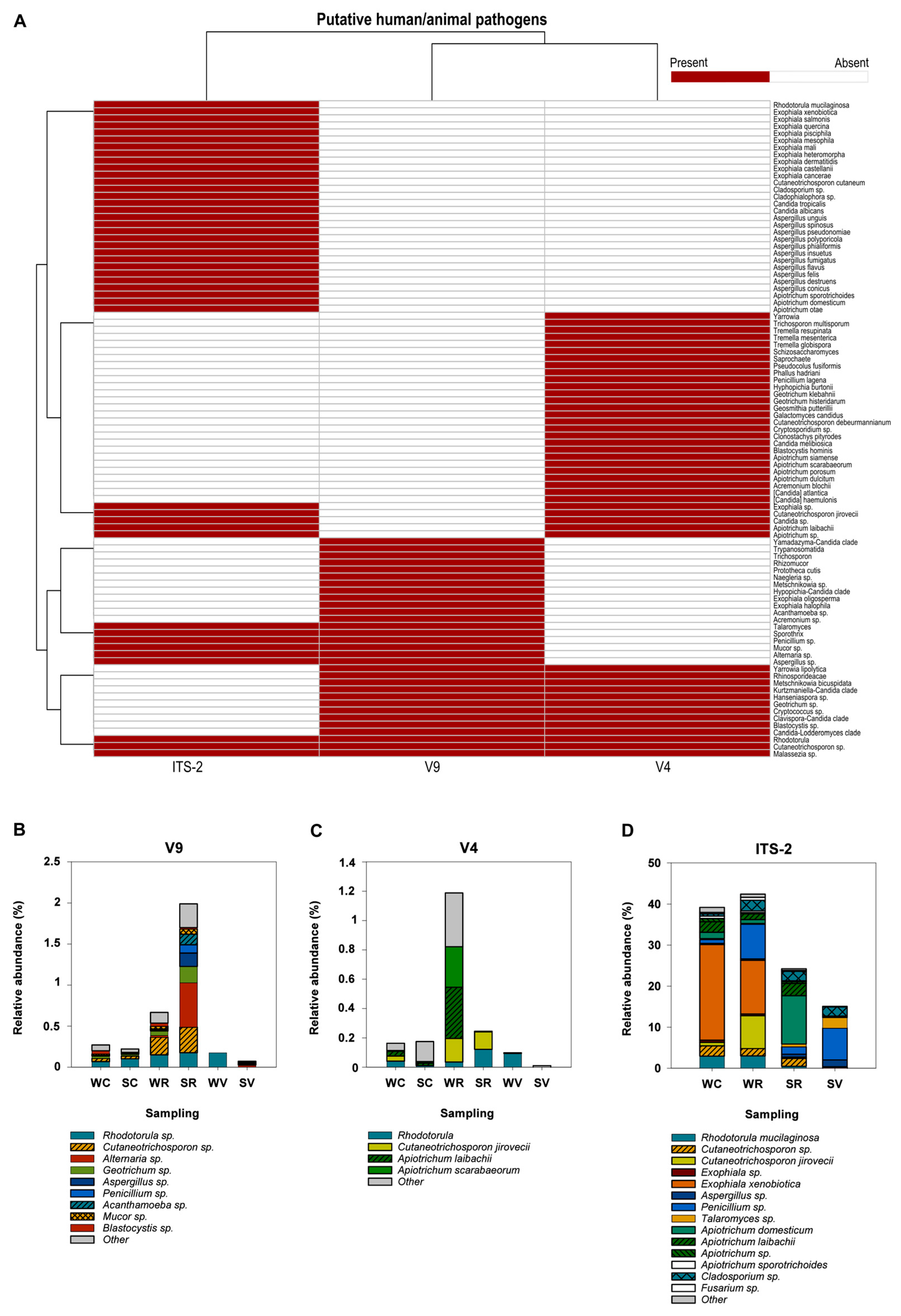
A total of 25 (V9), 31 (V4), and 26 (ITS-2) genera/species associated with human and/or zoonotic diseases were detected, with several taxa differentially identified across the three genetic markers. Figure 6A illustrates these differences, highlighting the distinct detection profiles of putative human/animal pathogens across ITS-2, V9, and V4. While some genera/species were consistently detected by all markers (e.g., Rhodotorula sp., Cutaneotrichosporon sp.), others were exclusive to one or two regions (e.g., Penicillium sp., Cladosporium sp.) (Figure 6A).
Using the V9 and V4 target regions, human and zoonotic putative pathogens represented only a small fraction of the total eukaryotic diversity, accounting for less than 0.2–2% of total reads across all analyzed samples (Figure 6B,C). In both regions, fungal pathogens were the most prevalent, followed by protists.
Analysis of the V9 region revealed significant local differences in the prevalence of human pathogens. In Redondela, putative human pathogens represented 0.67% of total reads in water samples and 2% in sediment samples. Comparatively, in Cangas, the proportions were 0.27% in water and 0.22% in sediment. Vigo exhibited the lowest prevalence, with 0.18% in water and 0.07% in sediment (Figure 6B). Redondela exhibited a diverse array of putative pathogen-associated genera in both sediment and water samples. Notable fungal genera included Rhodotorula (average 233 reads, 0.16% of total reads) and Cutaneotrichosporon (average 373 reads), which were detected in both matrices. Additionally, sediment samples were dominated by Alternaria (769 total reads), Geotrichum (285 reads), Aspergillus (232 reads), and Penicillium (143 reads), along with the putative pathogenic protist Acanthamoeba (183 reads). Several genera were exclusively identified in sediment from Redondela, including the fungi Exophiala, Malassezia, Cryptococcus, Acremonium, and Sporothrix, with less than 30 reads each (Figure 6B, Table S1). Other notable sediment-associated genera in Redondela included Naegleria (94 reads) and Talaromyces (80 reads), which are known to contain putative pathogenic species. Pathogen-associated taxa such as Blastocystis (54 reads) and Trypanosomatida (53 reads) were primarily identified in water samples. In Cangas, the most prevalent potential pathogens included the fungal genera Rhodotorula and Cutaneotrichosporon and the protist Blastocystis. Notably, Blastocystis was exclusively detected in sediment samples, accounting for 57 total reads. Vigo demonstrated the lowest diversity of human and zoonotic putative pathogen-associated genera. Rhodotorula was the predominant genus detected in water samples, with a total of 252 reads. Minor contributions from genera such as Alternaria, Aspergillus, Penicillium, and Talaromyces were also observed in water, each represented by 11–30 reads (Figure 6B, Table S1).
Using the V4 region, a lower pathogen abundance was observed compared to the V9 region. However, Redondela remained the area with the highest abundance of putative pathogens. The overall pathogen distribution pattern in this region is related with water sampling point instead of sediment observed with the V9 region. As with the V9 region, the V4 region revealed a higher abundance of putative pathogens in Redondela, followed by Cangas and Vigo, with slightly lower overall abundances compared to V9 (Figure 6C). In Redondela, Rhodotorula continued to be detected in both water (94 reads) and sediment (326 reads). Additionally, the genus Cutaneotrichosporon jirovecii was identified, in water (430 reads) and sediment (319 reads). Unlike the V9 region, the V4 region detected multiple species of the genus Apiotrichum in sediment, primarily A. laibachii (935 total reads) and A. scarabaerum (741 total reads), as well as the genus Cryptosporidium (80 total reads). However, the V4 region did not detect the genera Alternaria, Penicillium, or Aspergillus (Table S1).
The ITS-2 region identified several putative pathogenic fungi that were either undetected or present at low levels when using the universal eukaryotic V9 and V4 regions. Notably, Exophiala xenobiotica, along with the genera Cladosporium and Penicillium, were detected. E. xenobiotica accounted for 23% and 13% of total fungal reads in Cangas (water) and Redondela (water), respectively, and was exclusively found at these sampling points. The genus Cladosporium was detected in all samples, representing 0.5% to 2% of total fungal reads (Figure 6D). Penicillium sp. was another dominant genus, primarily associated with water from Redondela (WR) and sediment from Vigo (SV), comprising 8% of total fungal diversity at both sites. Different species of the genus Apiotrichum were also detected, with A. domesticum being the most dominant, accounting for 11% of total fungal diversity (4718 reads). Additionally, minor genera such as Talaromyces sp., Candida sp., Aspergillus sp., Fusarium sp., Mucor sp., Malassezia sp., and Apiotrichum sp. were detected across nearly all sampling points (Figure 6D).

3.4.3. Pathogens of Aquatic Organisms

DNA originating from putative pathogens typically associated with aquatic organisms was identified, revealing marked differences in detection efficiency among the three genetic markers. The V4 region identified the greatest number of taxa (39 genera/species), followed by V9 with (33), while in ITS-2 none was detected. Remarkably, 19 taxa were exclusively detected by V9, and 25 were unique to V4, highlighting the complementary nature of these two markers (Figure 7A). Although some pathogens were metazoans or fungi, the majority were protists, predominantly within the TSAR supergroup.
In line with patterns observed for zoonotic and human-associated taxa, the greatest presence of putative pathogen DNA was recorded in the Redondela samples, followed by those from Cangas and Vigo. Using the V9 marker, the relative abundance of these organisms remained below 2% in both Cangas and Vigo. Conversely, in Redondela, putative pathogens comprised 35% of water sample reads (WR) and 10.4% in sediment (SR). With the V4 marker, relative abundance was below 5% in Redondela and Cangas, and under 2% in Vigo (Figure 7B). Among the most representative pathogenic taxonomic groups identified using both V9 and V4 regions were Rhogostoma sp. (Rhizaria), Amoebophyra sp. (Alveolata), Scuticociliata sp. (Alveolata), Monocystis sp. (Alveolata); Anurofeca sp. (Opistokonta), Aplanochytrium sp. (Stramenopila) and Chilodonella uncinata (Alveolata) (Figure 7A, Table 2). Rhogostoma sp. was predominantly detected in both water and sediment samples from Redondela, where it accounted for 35% (48,675 reads) of total eukaryotic reads in water using V9, compared to only 3% (6589 reads) with V4, which enabled species-level identification as Rhogostoma minus. The Scuticociliata (Philasterida) taxonomic group, was detected in water from Cangas (WC), representing 0.2% (V9) and 0.3% (V4) of total eukaryote reads. Other minority pathogens of the Philasterida order were detected: Miamiensis avidus, Pseudocohnilembus hargisi and Uronema sp., some of which were exclusively detected with V9, while other appeared only with V4 (Figure 7A, Table S2). The amphibian larval pathogen of the genus Anurofeca was also detected in Cangas and Redondela, predominantly in sediment from Redondela (SR), where it represented 7.5% (10,685 total reads) and 3.7% (10,005 total reads) of the total eukaryotic reads for V9 and V4, respectively (Table 2).
Selecting of genetic marker had a substantial impact on the detection of putative pathogens across environmental compartments and study areas. Clear discrepancies were observed between the V9 and V4 regions, highlighting the limitations of relying on a single molecular region for assessing microeukaryotic microbial diversity. For instance, Monocystis sp. was not detected in either sediment or water from Vigo when using the V9 region, while it was detected in water from the same location (WV: 1176 reads) using the V4 region. Conversely, in sediment from Cangas (SC), this genus was detected with V9 but not with V4 (Table 2). Aphanomyces sp. further illustrated this contrast, since it was detected in both water and sediment from Cangas and Vigo with the V4 region, but was not detected with V9 (Table 2). Similarly, Amoebophyra sp. showed high read counts in water from Cangas (2454 total reads) and sediment from Vigo (2330 reads) with V4, whereas with V9, it was detected only in water from Vigo (692 reads) (Table 2).
A number of putative aquatic pathogenic taxa were detected exclusively by either the V9 (19 ASV) or V4 (25 ASV) genetic marker, each exhibiting distinct taxonomic profiles and spatial patterns (Figure 7C). Using the V9 region, several taxa were prominent in terms of detection and site-specificity. Cryptomycota and Selenidium was among the few taxa consistently detected across multiple sites and compartments, indicating a broad environmental distribution. In contrast, some taxa displayed more restricted patterns, such as Eimeria sp. exclusive in sediment, and Lagenidium sp. mainly detected in water, both from Redondela. Other exclusively taxa detected with V9 included the mollusc pathogens Minchinia spp. and Haplosporidium spp., Achlya (Stramenopiles), Pythium (Stramenopiles), Neobodo spp. (Excavata) and Duboscquella sp. (Alveolata). Similarly, certain genera were detected exclusively with the V4 region, such as Rhogostoma minus (Rhizaria), Lecudina spp. (Apicomplexa). and Pelagodinium sp. (Alveolata), strongly represented in water samples from Redondela (WR), Cangas (WC) and Vigo (WV). The V4 region also enabled species-level resolution for several taxa only classified to genus level with V9, including Aplanochitrium minuta, Thraustochytrium kinnei, Selenidium pygospionis, and Rhogostoma minus (Figure 7C, Table 2). Other exclusive taxa detected with V4 included: Vermamoeba vermiformis and Kudoa alliaria.

3.4.4. Harmful Algae (HA)

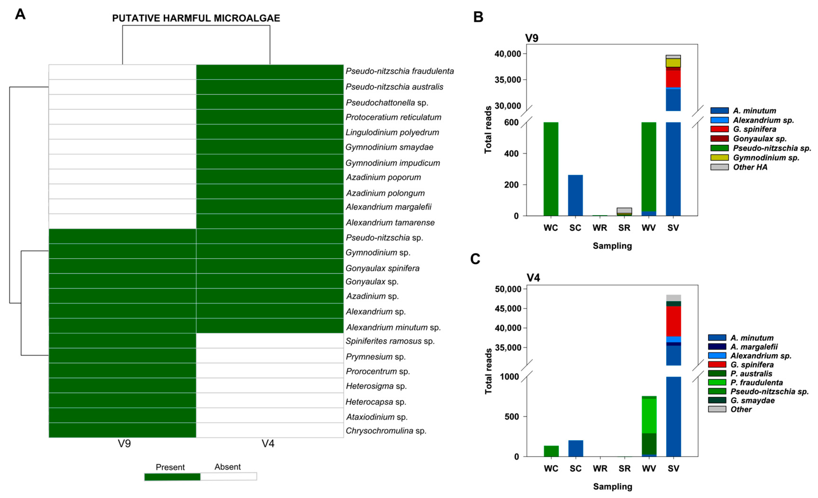
HA, as listed in the IOC-UNESCO Taxonomic Reference List of Harmful Micro Algae, were detected using both the V9 (14 genera) and V4 (18 genera) primer sets. Notably, 7 taxa were exclusively detected by V9, and 11 were unique to V4, highlighting the complementary nature of these two markers (Figure 8A).
Overall, in both regions, sediment samples from Vigo showed the highest diversity (number of genera and species) and relative abundance of HA. In this location, harmful taxa collectively accounted for 28% and 18% of total eukaryotic reads, based on the V9 and V4 regions, respectively. In the remaining samplings, HA represented only approximately 1% of total eukaryote reads, and in Redondela, they were scarcely detected. Several species of the genus Alexandrium (Dinophyceae) were identified with both the V9 and V4 regions, with Alexandrium minutum being the most dominant HA in the sediment of Vigo. This species accounted for 23% (33,232 reads) of total eukaryotic reads with the V9 region (Figure 8B) and 13.28% with the V4 region (Figure 8C). Other species, such as A. margaleffii and A. tamarense, were exclusively detected using the V4 region; however, they represented less than 0.5% of total eukaryote reads (Figure 8A,C; Table S3). Gonyaulax spinifera (Dinophyceae) and Gonyaulax sp. were the second most abundant HA in the sediment of Vigo. G. spinifera accounted for approximately 2% of total eukaryotic reads, with 3207 reads using the V9 region and 7730 reads using the V4 region (Figure 8B,C). Several Pseudo-nitzschia spp. (Bacillariaceae) were detected with both target genes, though notable differences were observed depending on the amplified region. Using the V9 region, the highest number of total reads of this genus was found in water from Cangas (1788 reads), followed by the water from Vigo (901 reads), although species-level classification was not achieved (Figure 8B). In contrast, the V4 region enabled species-level identification, revealing P. australis (266 total reads) and P. fraudulenta (428 total reads), both associated with water from Vigo (Figure 8C).
Discrepancies between amplicon sequencing results were also observed for Gymnodinium spp. and Azadinium spp., which were exclusively detected in sediment from Vigo using both regions Figure 8A. However, while the V4 region enabled the identification of two Gymnodinium species (G. impudicum, 380 reads; G. smaydae, 1286 reads) and two Azadinium species (A. poporum, 84 reads; A. polongum, 85 reads), the V9 region only allowed classification at the genus level (1644 reads) (Figure 8, Table S3). Other less abundant HA species were detected exclusively with the V9 region, including Heterosigma sp., Heterocapsa sp., and Chrysochromulina sp., while Lingulodinium sp. and Protoceratium reticulatum were identified with both target regions. Most of these genera were also dominant in the sediment of Vigo (Figure 8A, Table S3).

4. Discussion

4.1. Microeukaryotic Diversity

The analysis of microeukaryotic diversity provided essential baseline information on microbial communities in embayment impacted by wastewater effluents in the Ría de Vigo. The three sampling sites exhibited strikingly different environmental and ecological conditions, offering a valuable framework to examine how local factors modulate microbial eukaryotic community structure at the landscape scale.
Given the lack of consensus on optimal amplification targets and their associated taxonomic biases, we adopted a multi-marker approach (V9, V4, ITS2) for broad eukaryotic detection. This strategy captured complementary taxonomic profiles and revealed how marker choice influences biodiversity patterns, with V4 yielding more reads and V9 capturing more taxa [44,45]. Such discrepancies largely reflect primer bias and amplification efficiency rather than true biological absence or abundance, which is why the use of multiple markers was essential to obtain a more balanced and comprehensive picture of community diversity. Low fungal detection with ITS-2 suggest either true scarcity or technical limits, highlighting the need for greater sampling or sequencing depth.
Despite differences in sequencing outputs, all markers consistently identified the same sites as having the highest and lowest diversity, confirming the robustness of the spatial patterns detected. Redondela showed the highest richness and community uniqueness, linked to its intertidal estuarine setting with low hydrodynamics, high riverine inputs and muddy, organic-rich sediments [46]. Conversely, Cangas, with coarse, gravel-dominated sediments, exhibited the lowest diversity with all markers. These patterns were consistent with in situ measurements of temperature and salinity, which also revealed marked contrasts among sampling sites, tentatively suggesting that local physicochemical conditions may contribute to shaping community composition.
High proportions of site-exclusive ASVs (58–87%) across all sites, mainly in Redondela, indicate strong environmental filtering, with sediment type, nutrient availability, and hydrodynamic conditions shaping distinct communities despite their proximity.
This pattern shows that environmental filtering, shapes both taxonomic richness and spatial differentiation, and that fine-scale habitat heterogeneity within a single estuary can strongly structure microbial eukaryotic communities, underscoring the need to integrate microscale habitat features into biodiversity assessments.

4.2. Putative Pathogen Diversity

A diverse array of putative pathogenic taxa was recorded across sites and habitats in the Ría de Vigo, including eDNA signatures relevant to human, animal, and environmental health. It is important to note that DNA detection alone does not confirm organism viability or infectivity; therefore, all statements regarding potential human, animal, or aquaculture risk should be considered tentative. Nonetheless, these findings provide valuable baseline information to guide further investigations of these environments. Although less spatially exclusive than the overall microeukaryotic community, likely due to their lower relative abundance, distinct location-specific patterns emerged, with up to 45% of pathogenic taxa unique to a single site, indicating that local environmental conditions influence their distribution.
Using multiple markers, we detected 18 (V9) to 27 (ITS-2) fungal taxa, 3–5 protistan taxa, and one Ichthyosporean pathogen with zoonotic potential, some listed under the European Directive 2000/54/EC. Clinically relevant fungi, including the genera Rhodotorula, Cutaneotrichosporon and Exophiala are known to cause opportunistic infections [47,48,49], while toxin and allergen producing genera such as Aspergillus, Penicillium, Sporothrix (V9, ITS-2)) Alternaria, Talaromyces, and Cladosporium (ITS-2) were also found, several of them regulated under the European Directive 2000/54/EC [28]. Notable detections also included Apiotrichum domesticum (ITS-2, V4), associated with human summer-type hypersensitivity pneumonitis (SHP) [50]. Protozoan pathogens regulated under the European Directive 2000/54/EC were also identified. Acanthamoeba sp. (V9) can cause granulomatous amoebic encephalitis and keratitis [51], while Cryptosporidium sp. (V4) is associated with zoonotic cryptosporidiosis and waterborne outbreaks [52]. Blastocystis sp. (V4 and V9) is common in vertebrates including humans, with debated pathogenicity but growing evidence of waterborne transmission [53].
Detection patterns of zoonotic pathogens varied between markers, with some taxa appearing only in one or two targets. Certain markers also resolved multiple species within a genus, such as Rhodotorula mucilaginosa and Cutaneotrichosporon cutaneum with (V4 and ITS-2). This highlights the value of multi-marker approaches to improve coverage in pathogen detection. It is therefore likely that taxa detected exclusively by one marker reflect primer-specific bias rather than true absence, which reinforces the need for caution in interpreting marker-specific results. However, most findings were at genus level, and pathogenicity cannot be assumed for all members of a genus. Species-level confirmation with complementary techniques (e.g., targeted PCR, culturing, or sequencing of additional loci) is essential to validate pathogenic potential and avoid overestimation of health risks.
Pathogens of major concern for the aquaculture sector were detected, revealing the susceptibility of the Ría de Vigo to diseases affecting both bivalves and fish. The detection of Haplosporidium edule (V9) [39], exclusively in inner estuarine sediments (Redondela), highlights the potential for sediment to act as an environmental reservoir for benthic pathogens. This protozoan infects the cockle Cerastoderma edule, and its occurrence in muddy, low-hydrodynamic habitats suggests favourable conditions for maintaining infective stages near suitable hosts. Regarding fish pathogens, several taxa within the order Philasterida (Pseudochonilembus spp., Uronema sp., Uronemella sp., and Miamiensis avidus), all associated with scuticociliatosis, were recorded using both V9 and V4 regions [33,34], with Pseudochonilembus hargisi being unexpectedly prevalent. Although no outbreaks involving this species have been documented in Galicia, its repeated detection in metabarcoding surveys in Ría de Vigo [54] and its documented pathogenicity in Asian and American Aquaculture [34,35] suggest that its presence merits close monitoring. Pathogens associated with Nodular Gill Disease (NGD), including Vermamoeba vermiformis and Rhogostoma sp., were detected [40], with marker-specific differences (Rhogostoma sp., V9; Rhogostoma minus, V4) contributing to discrepancies in Rhizaria detection. Rhogostoma sp., common in WWTPs, and capable of harbouring Legionellales bacteria [55], and Vermamoeba vermiformis, a known host of pathogenic bacteria and viruses [56] were found exclusively in Cangas and Redondela, suggesting anthropogenic contamination. Other fish pathogens were detected at low abundance, including Chilodonella uncinata, which causes significant losses in freshwater fish farming [32], Goussia sp., a coccidian previously found in Galician waters, and Kudoa alliaria, a myxosporean parasite that infects the musculature of commercial fish.
Several marine pathogens with important ecological roles were identified, indicating that sediment type, hydrodynamics, and host availability shape pathogen persistence in the Ría de Vigo. The anuran larvae pathogen Anurofeca sp. [38], was highly abundant (7% relative abundance) in muddy low-energy sediments of Redondela, while gregarines (Selenidium spp., Lecudina tuzetae) and Cryptomycota (Rozella sp.) in Cangas and Redondela pointed to sedimentary “seed banks” of invertebrate parasites [57,58]. The detection of the genus Aphanomyces spp. in both sediment and water from Cangas highlight the potential for harmful oomycetes to persist across the habitats. This genus includes species of high ecological and economic impact, such as A. astaci, a notifiable pathogen under OIE regulation and causative agent of crayfish plague [43]. Although species-level resolution was not achieved in this study, our findings underscore the need for targeted, high-resolution monitoring at species-level in future analyses.
Harmful Algae Blooms are a recurring issue in the Galician Rías, impacting aquaculture, fisheries, and public health [14]. Our metabarcoding survey detected 14 harmful algal taxa listed in the IOC-UNESCO Taxonomic Reference List of Harmful Microalgae [30], predominantly in sediment samples from Vigo. Saxitoxin-producing Alexandrium spp. (A. minutum, A. margalefii, and A. tamarense), along with Gymnodinium spp. were especially relevant. Those HA are responsible for paralytic shellfish poisoning (PSP) in humans following the consumption of contaminated shellfish. Notably, A. minutum representing up to 20% of the total eukaryotic community. This high abundance likely reflects the accumulation of resistant cysts in muddy, aphotic sediments following the upwelling-driven bloom season. Such cyst beds are known to act as inoculant for future blooms, consistent with past events in Ría de Vigo [59] and the major bloom reported in the neighbouring Ría de Pontevedra in April 2023 [60] three months prior to sediment sampling. Other detected taxa included Pseudo-nitzschia spp., domoic acid producers, and Azadinium spp. responsible for amnesic shellfish poisoning (ASP) and azaspiracid shellfish poisoning (AZP), respectively [14,61]. While these HA taxa are not directly discharged from wastewater outfalls, nutrient enrichment from effluents may enhance bloom potential by fueling phytoplankton growth under favourable hydrodynamic conditions. Together, these findings reinforce the role of sediments as reservoirs of toxic microalgae and highlight the value of metabarcoding for early detection in integrated HAB monitoring strategies.
As a whole, this study provides valuable insights into the eukaryotic pathogen composition across three distinct outfall zones in the Ría de Vigo, encompassing outer, middle and inner sectors, and reflecting varying levels of urban pressure from small (Cangas, Redondela) to large (Vigo) municipalities. Surprisingly, Vigo recorded the lowest eukaryotic diversity and pathogens, despite the effluents from the WWTP of Vigo are responsible for 86% of the sewage dumping of a total of 12 WWTPs operating in the Ría. This finding agrees with that obtained from much less urbanized outer area in the Ría (Baiona) in which an efficient removal of prokaryotic putative pathogens from WWTP was confirmed in wastewater effluents and in mussels, acting as bioindicators of marine environment [17]. Conversely the inner estuarine zone of Redondela exhibited the highest overall eukaryotic diversity and the greatest abundance of putative pathogenic taxa, particularly in sediment samples. The spatial heterogeneity observed in pathogen distribution likely reflects difference in hydrodynamics and global circulation of water bodies and the continuous subsurface currents associated with river mouths, which affect the ecological processes and sediment typology in the Ría. Additionally, unlike other typical shallow estuaries, seasonal wind-driven coastal upwelling events promote short water renewal (3–4 days) in the Ría [62], mostly in outer-middle zones, therefore the suspended eukaryotic communities in the water column at those areas may be intermittently washed out or diluted. At this point, the strongest contribution to the eukaryotic putative pathogen community in the Ría was recorded in areas controlled by the land–sea interaction, in sediments of the inner estuarine area with a mixed impact of anthropogenic nitrogen-fertilization and biohazard inputs from wastewater and river mouths. This finding emphasizes the need to incorporate sediment analysis and integrate landscape characteristics into metabarcoding studies to achieve a more comprehensive assessment of environmental coastal health. In addition, this study was conceived as a diagnostic snapshot in a single summer sampling event, which provides a timely assessment of local community structure and putative pathogen diversity. However, this design does not capture potential seasonal or interannual variability, and future studies with temporal replication will be needed to validate and expand the spatial patterns observed.
This study demonstrates that a multi-marker eDNA metabarcoding approach is a powerful, non-invasive tool for earlier detection and simultaneously diagnosis of environmental pathogens in complex coastal socio-ecological systems. By improving taxonomic resolution, and capturing both abundant and rare taxa, this method expands the scope of marine pathogen diagnosis, despite inherent limitations such as genus-level resolution, PCR amplification, incomplete databases and the inability to distinguish viable from non-viable organisms. Given the incomplete depuration of wastewater withdrawal by the EU over the last years, both in large and small treatment plants, wastewater discharges were expected to be major sources of eukaryotic pathogens. Contrary to expectations, wastewater discharges accounted for only a small fraction of detected genera, with pathogen communities instead shaped by local biogeochemical conditions, particularly in inner estuarine zones where fluvial inputs and organic-rich sediments promoted up to tenfold higher pathogen eDNA concentrations. These findings highlight the need to integrate sediment analyses, landscape features, and temporal replication into One Health-oriented monitoring strategies to better identify contamination hotspots and manage health risks in dynamic coastal environments.

Supplementary Materials

The following supporting information can be downloaded at: https://www.mdpi.com/article/10.3390/d17100671/s1, Table S1: Relative abundance of total reads corresponding to human and/or zoonotic pathogens detected using the 18S rRNA gene (V9, V4) and ITS-2 markers across sampling sites. Sampling types included water and sediment from Cangas, Redondela, and Vigo Total reads: V9–142,406 reads; V4–267,592 reads; ITS-2–40,124 reads.; Table S2: Number of reads and relative abundance (%) of minor potential aquatic pathogens (defined as those with relative abundance < 0.1%) detected at each sampling point using the 18S rRNA gene (V9 and V4 regions). Sampling codes: WC: water from Cangas; SC: sediment from Cangas; WR: water from Redondela; SR: sediment from Redondela; WV: water from Vigo; SV: sediment from Vigo; NA: no data available. Total read counts per marker: V9 = 142,406; V4 = 267,592. Table S3: Number of reads and relative abundance (%) of HA across different sampling sites. Sampling codes: WC: water from Cangas; SC: sediment from Cangas; WR: water from Redondela; SR: sediment from Redondela; WV: water from Vigo; SV: sediment from Vigo; NA: no data available. Total, read counts per marker: V9 = 142,406; V4 = 267,592.

Author Contributions

Conceptualization, A.R., S.P. and E.A.; methodology, R.R.-C. and A.R.; formal analysis, R.R.-C.; investigation, A.R.; data curation, R.R.-C.; writing—original draft preparation, R.R.-C.; writing—review and editing, A.R., S.P. and E.A.; visualization, R.R.-C.; supervision, A.R.; funding acquisition, E.A., S.P. and A.R. All authors have read and agreed to the published version of the manuscript.

Funding

The authors acknowledge the support of the Galicia Marine Science Programme, part of the Complementary Science Plans for Marine Science of Ministerio de Ciencia, Innovación y Universidades included in the Recovery, Transformation and Resilience Plan (PRTR-C17.I1). Funded through Xunta de Galicia with NextGenerationEU and the European Maritime Fisheries and Aquaculture Fund. R. Ríos-Castro has been supported by the Postdoctoral Fellowship Program of the Xunta de Galicia (Regional Ministry of Education, Science, Universities and Vocational Training or the Galician Innovation Agency, (IN606B-2025/010)). A. Ramilo has been supported by a contract Technical Support Personnel (PTA) by the State Programme for the Promotion of Talent and its Employability in RandDand from Spanish Government (PTA2021-021206-I).

Institutional Review Board Statement

Not applicable.

Data Availability Statement

The resulting raw sequence reads were deposited in the Sequence Read Archive (SRA) http://www.ncbi.nlm.nih.gov/sra (accessed on 15 May 2025) under BioProject accession number PRJNA1262121.

Acknowledgments

We would like to thank Helena Rodríguez for its support in samplings and Jorge Hernández-Urcera for its support in visualization.

Conflicts of Interest

The authors declare no conflicts of interest.

References

  1. Landrigan, P.J.; Stegeman, J.J.; Fleming, L.E.; Allemand, D.; Anderson, D.M.; Backer, L.C.; Brucker-Davis, F.; Chevalier, N.; Corra, L.; Czerucka, D.; et al. Human health and ocean pollution. Ann. Glob. Health 2020, 86, 151. [Google Scholar] [CrossRef]
  2. Vidal-Dorsch, D.E.; Bay, S.M.; Maruya, K.; Snyder, S.A.; Trenholm, R.A.; Vanderford, B.J. Contaminants of emerging concern in municipal wastewater effluents and marine receiving water. Environ. Toxicol. Chem. 2012, 31, 2674–2682. [Google Scholar] [CrossRef] [PubMed]
  3. He, Q.; Silliman, B.R. Climate change, human impacts, and coastal ecosystems in the Anthropocene. Curr. Biol. 2019, 29, R1021–R1035. [Google Scholar] [CrossRef]
  4. Kang, M.; Yang, J.; Kim, S.; Park, J.; Kim, M.; Park, W. Occurrence of antibiotic resistance genes and multidrug-resistant bacteria during wastewater treatment processes. Sci. Total Environ. 2022, 811, 152331. [Google Scholar] [CrossRef]
  5. Ariyadasa, S.; Taylor, W.; Weaver, L.; McGill, E.; Billington, C.; Pattis, I. Nonbacterial microflora in wastewater treatment plants: An underappreciated potential source of pathogens. Microbiol. Spectr. 2023, 11, e00481-23. [Google Scholar] [CrossRef]
  6. Freudenthal, J.; Ju, F.; Bürgmann, H.; Dumack, K. Microeukaryotic gut parasites in wastewater treatment plants: Diversity, activity, and removal. Microbiome 2022, 10, 27. [Google Scholar] [CrossRef]
  7. Cai, L.; Ju, F.; Zhang, T. Tracking human sewage microbiome in a municipal wastewater treatment plant. Appl. Microbiol. Biotechnol. 2014, 98, 3317–3326. [Google Scholar] [CrossRef]
  8. Sánchez, O.; Garrido, L.; Forn, I.; Massana, R.; Maldonado, M.I.; Mas, J. Molecular characterization of activated sludge from a seawater-processing wastewater treatment plant. Microb. Biotechnol. 2011, 4, 628–642. [Google Scholar] [CrossRef]
  9. Geisen, S.; Vaulot, D.; Mahé, F.; Lara, E.; de Vargas, C.; Bass, D. A user guide to environmental protistology: Primers, metabarcoding, sequencing, and analyses. bioRxiv 2019. [Google Scholar] [CrossRef]
  10. Liu, J.; Yu, Y.; Cai, Z.; Bartlam, M.; Wang, Y. Comparison of ITS and 18S rDNA for estimating fungal diversity using PCR-DGGE. World J. Microbiol. Biotechnol. 2015, 31, 1387–1395. [Google Scholar] [CrossRef]
  11. Guerra, A.; Lens, S.; Rocha, F. Impacto del hombre sobre el ecosistema de la Ría de Vigo: Hacia una gestión integrada. In La Ría de Vigo: Una Aproximación Integral al Ecosistema Marino de la Ría de Vigo; González-Garcés Santiso, A., Vilas-Martín, F., Álvarez-Salgado, X.A., Eds.; Instituto de Estudios Vigueses: Vigo, Spain, 2002; pp. 51–84. [Google Scholar]
  12. Balseiro, P.; Montes, J.; Fernández Conchas, R.; Novoa, B.; Figueras, A. Comparison of diagnostic techniques to detect the clam pathogen Perkinsus olseni. Dis. Aquat. Organ. 2010, 90, 143–151. [Google Scholar] [CrossRef]
  13. Broullón, E.; López-Mozos, M.; Reguera, B.; Chouciño, P.; Doval, M.D.; Fernández-Castro, B.; Gilcoto, M.; Nogueira, E.; Souto, C.; Mouriño-Carballido, B. Thin layers of phytoplankton and harmful algae events in a coastal upwelling system. Prog. Oceanogr. 2020, 189, 102449. [Google Scholar] [CrossRef]
  14. Bellisario, B.; Fais, M.; Duarte, S.; Vieira, P.E.; Canchaya, C.; Costa, F.O. The network structure of intertidal meiofaunal communities from environmental DNA metabarcoding surveys in Northwest Iberia. Aquat. Sci. 2021, 83, 71. [Google Scholar] [CrossRef]
  15. Ríos-Castro, R.; Costas-Selas, C.; Pallavicini, A.; Vezzulli, L.; Novoa, B.; Teira, E.; Figueras, A. Co-occurrence and diversity patterns of benthonic and planktonic communities in a shallow marine ecosystem. Front. Mar. Sci. 2022, 9, 934976. [Google Scholar] [CrossRef]
  16. Costas-Selas, C.; Martínez-García, S.; Logares, R.; Hernández-Ruiz, M.; Teira, E. Role of bacterial community composition as a driver of the small-sized phytoplankton community structure in a productive coastal system. Microb. Ecol. 2023, 86, 777–794. [Google Scholar] [CrossRef]
  17. Ríos-Castro, R.; Cabo, A.; Teira, E.; Cameselle, C.; Gouveia, S.; Payo, P.; Novoa, B.; Figueras, A. High-throughput sequencing as a tool for monitoring prokaryote communities in a wastewater treatment plant. Sci. Total Environ. 2023, 861, 160531. [Google Scholar] [CrossRef]
  18. Amaral-Zettler, L.A.; McCliment, E.A.; Ducklow, H.W.; Huse, S.M. A method for studying protistan diversity using massively parallel sequencing of V9 hypervariable regions of small-subunit ribosomal RNA genes. PLoS ONE 2009, 4, e6372. [Google Scholar] [CrossRef]
  19. Stoeck, T.; Bass, D.; Nebel, M.; Christen, R.; Jones, M.D.; Breiner, H.W.; Richards, T.A. Multiple marker parallel tag environmental DNA sequencing reveals a highly complex eukaryotic community in marine anoxic water. Mol. Ecol. 2010, 19, 21–31. [Google Scholar] [CrossRef] [PubMed]
  20. Op De Beeck, M.; Lievens, B.; Busschaert, P.; Declerck, S.; Vangronsveld, J.; Colpaert, J.V. Comparison and validation of some ITS primer pairs useful for fungal metabarcoding studies. PLoS ONE 2014, 9, e97629. [Google Scholar] [CrossRef]
  21. Martin, M. Cutadapt removes adapter sequences from high-throughput sequencing reads. EMBnet J. 2011, 17, 10–12. [Google Scholar] [CrossRef]
  22. Andrews, S. FastQC: A Quality Control Tool for High Throughput Sequence Data. 2010. Available online: https://www.bioinformatics.babraham.ac.uk/projects/fastqc/ (accessed on 12 August 2025).
  23. Callahan, B.J.; McMurdie, P.J.; Rosen, M.J.; Han, A.W.; Johnson, A.J.; Holmes, S.P. DADA2: High-resolution sample inference from Illumina amplicon data. Nat. Methods 2016, 13, 581–583. [Google Scholar] [CrossRef] [PubMed]
  24. Quast, C.; Pruesse, E.; Yilmaz, P.; Gerken, J.; Schweer, T.; Yarza, P.; Peplies, J.; Glöckner, F.O. The SILVA ribosomal RNA gene database project: Improved data processing and web-based tools. Nucleic Acids Res. 2013, 41, D590–D596. [Google Scholar] [CrossRef]
  25. Abarenkov, K.; Nilsson, R.H.; Larsson, K.-H.; Taylor, A.F.S.; May, T.W.; Frøslev, T.G.; Pawlowska, J.; Lindahl, B.; Põldmaa, K.; Truong, C.; et al. The UNITE database for molecular identification and taxonomic communication of fungi and other eukaryotes: Sequences, taxa, and classifications reconsidered. Nucleic Acids Res. 2023, 52, D791–D797. [Google Scholar] [CrossRef]
  26. Conway, J.R.; Lex, A.; Gehlenborg, N. UpSetR: An R package for the visualization of intersecting sets and their properties. Bioinformatics 2017, 33, 2938–2940. [Google Scholar] [CrossRef]
  27. Oksanen, J.; Simpson, G.L.; Blanchet, F.G.; Kindt, R.; Legendre, P.; Minchin, P.R.; O′HAra, R.; Solymos, P.; Stevens, M.H.H.; Szoecs, E.; et al. vegan: Community Ecology Package, R package version 2.8-0. 2025. Available online: https://vegandevs.github.io/vegan/ (accessed on 3 April 2025).
  28. European Parliament and Council of the European Union. Directive 2000/54/EC of the European Parliament and of the Council of 18 September 2000 on the protection of workers from risks related to exposure to biological agents at work. Off. J. Eur. Communities 2000, L262, 21–45. Available online: https://eur-lex.europa.eu/legal-content/EN/TXT/?uri=CELEX:32000L0054 (accessed on 20 December 2024).
  29. Real Decreto 664/1997, de 12 de mayo, sobre la protección de los trabajadores contra los riesgos relacionados con la exposición a agentes biológicos durante el trabajo. Boletín Oficial del Estado 1997, 124, 15114–15119. Available online: https://www.boe.es/buscar/doc.php?id=BOE-A-1997-11144 (accessed on 2 February 2025).
  30. IOC-UNESCO. Taxonomic Reference List of Harmful Microalgae. Available online: https://www.marinespecies.org/hab/index.php (accessed on 5 March 2025).
  31. Velavan, T.P.; Schulenburg, H.; Michiels, N.K. Detection of multiple infections by Monocystis strains in a single earthworm host using ribosomal internal transcribed spacer sequence variation. Parasitology 2010, 137, 45–51. [Google Scholar] [CrossRef]
  32. Gomes, G.B.; Miller, T.L.; Vaughan, D.B.; Jerry, D.R.; McCowan, C.; Bradley, T.L.; Hutson, K.S. Evidence of multiple species of Chilodonella (Protozoa, Ciliophora) infecting Australian farmed freshwater fishes. Vet. Parasitol. 2017, 237, 8–16. [Google Scholar] [CrossRef] [PubMed]
  33. Iglesias, R.; Paramá, A.; Alvarez, M.F.; Leiro, J.; Fernández, J.; Sanmartín, M.L. Philasterides dicentrarchi (Ciliophora, Scuticociliatida) as the causative agent of scuticociliatosis in farmed turbot Scophthalmus maximus in Galicia (NW Spain). Dis. Aquat. Organ. 2001, 46, 47–55. [Google Scholar] [CrossRef]
  34. Kim, S.M.; Cho, J.B.; Lee, E.H.; Kwon, S.R.; Kim, S.K.; Nam, Y.K.; Kim, K.H. Pseudocohnilembus persalinus (Ciliophora: Scuticociliatida) is an additional species causing scuticociliatosis in olive flounder Paralichthys olivaceus. Dis. Aquat. Organ. 2004, 62, 239–244. [Google Scholar] [CrossRef] [PubMed]
  35. Jones, S.R.; Prosperi-Porta, G.; LaPatra, S.E. First isolation of Pseudocohnilembus persalinus (Ciliophora: Scuticociliatida) from freshwater-reared rainbow trout, Oncorhynchus mykiss. J. Parasitol. 2010, 96, 1014–1016. [Google Scholar] [CrossRef]
  36. Chen, T.; Liu, Y.; Xu, S.; Song, S.; Li, C. Variation of Amoebophrya community during bloom of Prorocentrum donghaiense Lu in coastal waters of the East China Sea. Estuar. Coast. Shelf Sci. 2020, 243, 106887. [Google Scholar] [CrossRef]
  37. Gleason, F.H.; Carney, L.T.; Lilje, O.; Glockling, S.L. Ecological potentials of species of Rozella (Cryptomycota). Fungal Ecol. 2012, 5, 651–656. [Google Scholar] [CrossRef]
  38. Baker, G.C.; Beebee, T.J.C.; Ragan, M.A. Prototheca richardsi, a pathogen of anuran larvae, is related to a clade of protistan parasites near the animal-fungal divergence. Microbiology 1999, 145, 1777–1784. [Google Scholar] [CrossRef]
  39. Azevedo, C.; Conchas, R.F.; Montes, J. Description of Haplosporidium edule n. sp. (Phylum Haplosporidia), a parasite of Cerastoderma edule (Mollusca, Bivalvia) with complex spore ornamentation. Eur. J. Protistol. 2003, 39, 161–165. [Google Scholar] [CrossRef]
  40. Dyková, I.; Kostka, M.; Wortberg, F.; Nardy, E.; Pecková, H. New data on aetiology of nodular gill disease in rainbow trout, Oncorhynchus mykiss. Folia Parasitol. 2010, 57, 157–163. [Google Scholar] [CrossRef]
  41. FioRito, R.; Leander, C.; Leander, B. Characterization of three novel species of Labyrinthulomycota isolated from ochre sea stars (Pisaster ochraceus). Mar. Biol. 2016, 163, 8. [Google Scholar] [CrossRef]
  42. Mendoza, L.; Taylor, J.W.; Walker, E.D.; Vilela, R. Description of three novel Lagenidium (Oomycota) species causing infection in mammals. Rev. Iberoam. Micol. 2016, 33, 83–91. [Google Scholar] [CrossRef]
  43. Svoboda, J.; Mrugała, A.; Kozubíková-Balcarová, E.; Petrusek, A. Hosts and transmission of the crayfish plague pathogen Aphanomyces astaci: A review. J. Fish Dis. 2017, 40, 127–140. [Google Scholar] [CrossRef]
  44. Maritz, J.M.; Rogers, K.H.; Rock, T.M.; Liu, N.; Joseph, S.; Land, K.M.; Carlton, J.M. An 18S rRNA workflow for characterizing protists in sewage, with a focus on zoonotic trichomonads. Microb. Ecol. 2017, 74, 923–936. [Google Scholar] [CrossRef]
  45. Choi, J.; Park, J.S. Comparative analyses of the V4 and V9 regions of 18S rDNA for the extant eukaryotic community using the Illumina platform. Sci. Rep. 2020, 10, 6519. [Google Scholar] [CrossRef] [PubMed]
  46. Vilas Martín, F.; Rey García, D.; Rubio Armesto, B.; Bernabeu Tello, A.; Méndez Martínez, G.; Duran Gallego, R.; Mohamed Falcón, K. Los fondos de la Ría de Vigo: Composición, distribución y origen del sedimento. In La Ría de Vigo: Una Aproximación Integral al Ecosistema Marino de la Ría de Vigo; González-Garcés Santiso, A., Vilas Martín, F., Álvarez-Salgado, X.A., Eds.; Instituto de Estudios Vigueses: Vigo, Spain, 2002; pp. 17–50. [Google Scholar]
  47. Ramos, A.; Cabero, M.; Orden, B.; Ángel-Moreno, A.; Forés, R. Fungemia por Rhodotorula mucilaginosa. Presentación de dos casos. Rev. Esp. Quimioter. 2012, 25, 76–78. [Google Scholar]
  48. Yoo, I.Y.; Heo, W.; Kwon, J.A.; Lee, M.; Park, Y.J. Identification of the rare yeast Cutaneotrichosporon (Trichosporon) debeurmannianum from diabetic foot infection. J. Clin. Lab. Anal. 2022, 36, e24785. [Google Scholar] [CrossRef]
  49. Morio, F.; Berre, J.Y.; Garcia-Hermoso, D.; Najafzadeh, M.J.; de Hoog, S.; Benard, L.; Michau, C. Phaeohyphomycosis due to Exophiala xenobiotica as a cause of fungal arthritis in an HIV-infected patient. Med. Mycol. 2012, 50, 513–517. [Google Scholar] [CrossRef]
  50. Li, Q.; Xiao, W.; Wu, P.; Zhang, T.; Xiang, P.; Wu, Q.; Zou, L.; Gui, M. The first two mitochondrial genomes from Apiotrichum reveal mitochondrial evolution and different taxonomic assignment of Trichosporonales. IMA Fungus 2023, 14, 7. [Google Scholar] [CrossRef]
  51. Connelly, L.; Anijeet, D.; Tole, D.; Alexander, C.L. Acanthamoeba keratitis: Molecular typing of Acanthamoeba species directly from ocular tissue. Curr. Res. Parasitol. Vector Borne Dis. 2023, 4, 100141. [Google Scholar] [CrossRef]
  52. Ryan, U.; Fayer, R.; Xiao, L. Cryptosporidium species in humans and animals: Current understanding and research needs. Parasitology 2014, 141, 1667–1685. [Google Scholar] [CrossRef] [PubMed]
  53. Hublin, J.S.Y.; Maloney, J.G.; Santin, M. Blastocystis in domesticated and wild mammals and birds. Res. Vet. Sci. 2021, 135, 260–282. [Google Scholar] [CrossRef]
  54. Ríos-Castro, R.; Romero, A.; Aranguren, R.; Pallavicini, A.; Banchi, E.; Novoa, B.; Figueras, A. High-throughput sequencing of environmental DNA as a tool for monitoring eukaryotic communities and potential pathogens in a coastal upwelling ecosystem. Front. Vet. Sci. 2021, 8, 765606. [Google Scholar] [CrossRef]
  55. Pohl, N.; Solbach, M.D.; Dumack, K. The wastewater protist Rhogostoma minus (Thecofilosea, Rhizaria) is abundant, widespread, and hosts Legionellales. Water Res. 2021, 203, 117566. [Google Scholar] [CrossRef] [PubMed]
  56. Delafont, V.; Rodier, M.H.; Maisonneuve, E.; Cateau, E. Vermamoeba vermiformis: A free-living amoeba of interest. Microb. Ecol. 2018, 76, 991–1001. [Google Scholar] [CrossRef]
  57. John, A.W.G.; Reid, P.C. Possible resting cysts of Dissodinium pseudolunula Swift ex Elbrächter et Drebes in the Northeast Atlantic and the North Sea. Br. Phycol. J. 1983, 18, 61–67. [Google Scholar] [CrossRef]
  58. Desportes, I.; Schrével, J. Treatise on Zoology—Anatomy, Taxonomy, Biology: The Gregarines; Brill: Leiden, The Netherlands, 2013. [Google Scholar]
  59. Rodríguez, F.; Escalera, L.; Reguera, B.; Nogueira, E.; Bode, A.; Ruiz-Villarreal, M.; Rossignoli, A.E.; Ben-Gigirey, B.; Rey, V.; Fraga, S. Red tides in the Galician rías: Historical overview, ecological impact, and future monitoring strategies. Environ. Sci. Process. Impacts 2024, 26, 16–34. [Google Scholar] [CrossRef]
  60. Nogueira, E.; Bravo, I.; Montero, P.; Díaz-Tapia, P.; Calvo, S.; Ben-Gigirey, B.; Figueiras, R.I.; Garrido, J.L.; Ramilo, I.; Lluch, N.; et al. HABs in coastal upwelling systems: Insights from an exceptional red tide of the toxigenic dinoflagellate Alexandrium minutum. Ecol. Indic. 2022, 137, 108790. [Google Scholar] [CrossRef]
  61. Trainer, V.; Pitcher, G.; Reguera, B.; Smayda, T. The distribution and impacts of harmful algal bloom species in eastern boundary upwelling systems. Prog. Oceanogr. 2010, 85, 33–52. [Google Scholar] [CrossRef]
  62. Barton, E.D.; Largier, J.L.; Torres, R.; Sheridan, M.; Trasviña, A.; Souza, A.; Pazos, Y.; Valle-Levinson, A. Coastal upwelling and downwelling forcing of circulation in a semi-enclosed bay: Ría de Vigo. Prog. Oceanogr. 2015, 134, 173–189. [Google Scholar] [CrossRef]
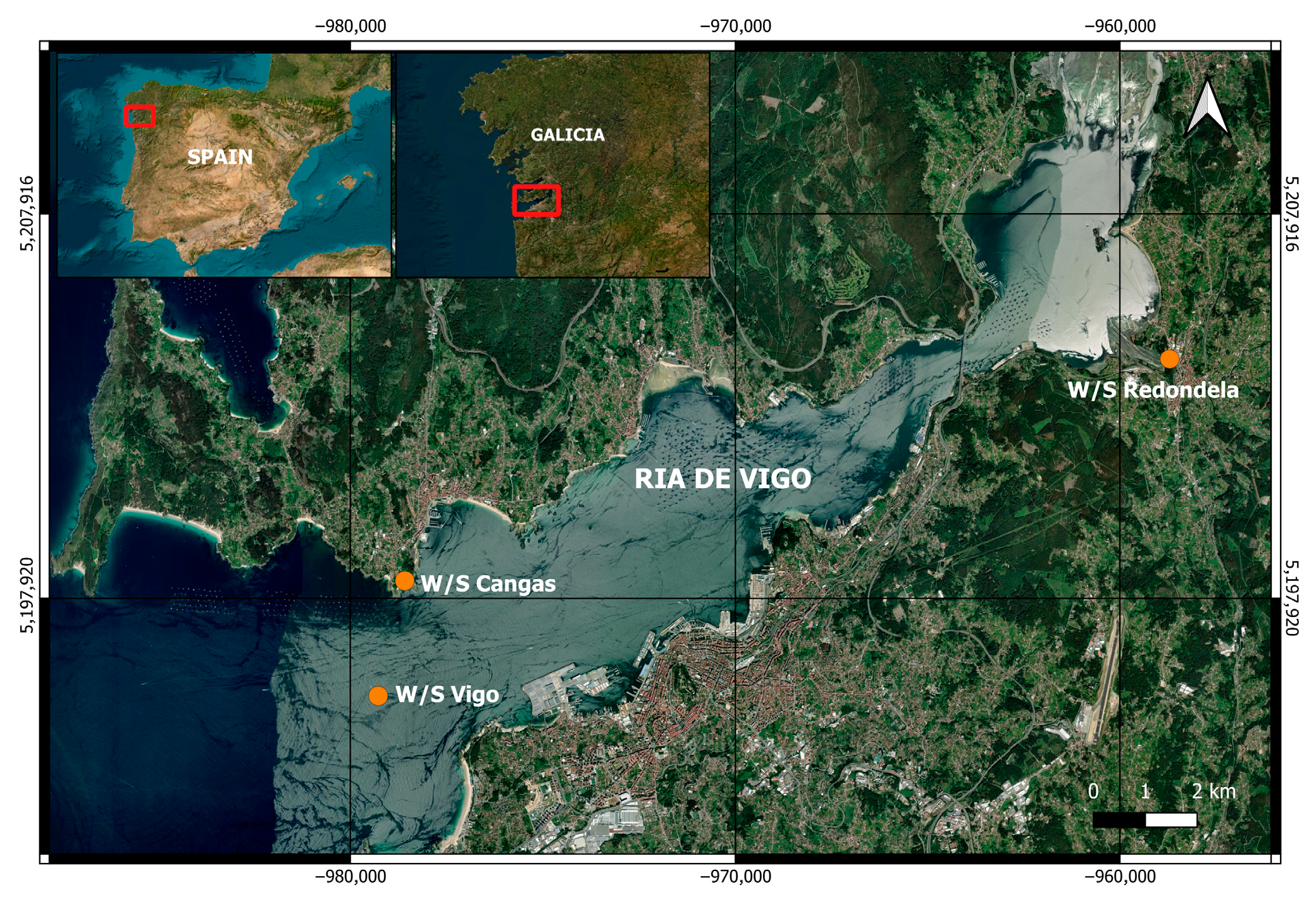
Figure 1. Geographical location of the sampling sites within the Ría de Vigo (Galicia, NW Spain). Water (W) and sediment (S) samples were collected at three stations: Vigo, Cangas, and Redondela. Sampling coordenates were as follows. Redondela: 42.28752° N, −8.61216° W; Cangas: 42.2491° N, −8.79076° W; Vigo, 42.21108° N, −8.77636° W. The red frames indicate the sampling locations.
Diversity 17 00671 g001
Figure 2. Eukaryotic diversity patterns based on multi-marker metabarcoding using V9 and V4 regions. (A) Rarefaction plots for each sample sequenced with the V9 and V4 regions, obtained for each environmental compartment across the sampling locations: Cangas, Redondela, and Vigo. (B) Alpha diversity indices (richness, Shannon, and Pielou) derived from eukaryotic sequencing of the V9 and V4 regions.
Diversity 17 00671 g002
Figure 3. Fungal diversity overview obtained with the ITS-2 sequencing region in each sampling location and environmental compartment. (A) showing sequencing depth (total reads) and number of amplicon sequence variants (ASVs) detected. (B) alpha diversity indices (richness, Shannon and Pielou). Abbreviations: WC: water of Cangas, WR: water of Redondela, SR: sediment of Redondela, SV: sediment of Vigo.
Diversity 17 00671 g003
Figure 4. Venn diagrams illustrating the number of shared and unique ASVs among Cangas (C), Redondela (R), and Vigo (V), based on: (A) the V9 region of the 18S rRNA gene, (B) the V4 region of the 18S rRNA gene, (C) the nuclear marker internal transcribed spacer 2 (ITS2).
Diversity 17 00671 g004
Figure 5. Venn diagrams illustrating the number of exclusive and shared putative biohazard ASVs among the three sampling regions: Cangas (C), Redondela (R), and Vigo (V). An accompanying Upset plot shows the number of exclusive and shared ASVs across both sampling locations and environmental compartments. Results are presented for: (A) V9 region, (B) V4 region, and (C) ITS-2 region. Abbreviations: WC: water from Cangas; SC: sediment from Cangas; WR: water from Redondela; SR: sediment from Redondela; WV: water from Vigo; SV: sediment from Vigo.
Diversity 17 00671 g005
Figure 6. Detection and relative abundance of putative human and zoonotic pathogens across sampling sites and genetic markers. (A) Presence/absence heatmap of putative human and/or zoonotic pathogens detected across three genetic markers (ITS-2, V9, and V4). Relative abundance (%) of different potential human pathogens taxa across sampling sites based on (B) V9 region of 18S rRNA gene, (C) V4 region of 18S rRNA gene, and (D) Internal Transcribed Spacer 2. Taxa with a relative abundance below 0.01% are grouped under “Other putative pathogens”. Abbreviations: WC: water from Cangas; SC: sediment from Cangas; WR: water from Redondela; SR: sediment from Redondela; WV: water from Vigo; SV: sediment from Vigo.
Diversity 17 00671 g006
Figure 7. Detection and marker-specific distribution of putative aquatic pathogens across sampling sites. (A) Heatmap showing the presence or absence of putative pathogens affecting aquatic organisms, detected using three genetic markers: V9, V4, and ITS-2. (B) Relative abundance (%) of these putative pathogens at each sampling point. (C) Dot plot representing the number of sequencing reads corresponding to putative pathogens exclusively detected using the V9 and V4 regions of the 18S rRNA gene. Abbreviations: WC: water from Cangas; SC: sediment from Cangas; WR: water from Redondela; SR: sediment from Redondela; WV: water from Vigo; SV: sediment from Vigo.
Diversity 17 00671 g007
Figure 8. (A) Heatmap showing the presence or absence of HA, detected using V9 and V4 markers. Read counts and distribution of HA taxa across sampling sites based on two genetic markers: (B) V9 and (C) V4 region of the 18S rRNA gene. Abbreviations: WC: water from Cangas; SC: sediment from Cangas; WR: water from Redondela; SR: sediment from Redondela; WV: water from Vigo; SV: sediment from Vigo.
Diversity 17 00671 g008
Table 1. Summary of sequencing results for each sample and target region (V9 and V4 18S rRNA gen and ITS-2). The table includes the number of reads before (BT) and after trimming (AT), average read length before and after trimming, percentage of reads retained, number of reads used in ASV (Amplicon Sequence Variant) generation, number of unique sequences, ASVs assigned taxonomy, and the total reads in ASVs with taxonomy. The number of reads after subsampling were also included. C-neg-W and C-neg-S refer to negative control samples of water and sediment, respectively. Sampling codes: WC: water from Cangas; SC: sediment from Cangas; WR: water from Redondela; SR: sediment from Redondela; WV: water from Vigo; SV: sediment from Vigo. NA: not applicable.
TargetNameReads BTAvg. Length BTReads ATRetained Reads (%)Avg. Length ATReads in ASVsUnique SequencesASV with TaxonomyReads in ASV with TaxonomyTotal ASV with TaxonomyTotal Reads ASV
V9WC724,544250720,14299.39133657,26810,3042447328,63460391,646,724
SC762,576250757,77499.37136711,93670431031355,968
WR1,302,6362501,289,87699.021321,152,14612,9083095576,073
SR681,264250672,75698.75133616,40214,0734342308,201
WV620,302250616,64899.41135550,78092811597275,390
SV615,656250612,12699.43134548,90888342002274,454
C-neg-W402503485.00137285314NA
C-neg-S17825014078.65138108121054
Total4,707,196NA4,669,496NANA4,237,57662,460NA2,118,788
V4WC1,068,6082501,067,38699.89231714,46026,6432388357,23066791,865,868
SC885,248250884,20499.88231698,97816,031614349,489
WR1,779,2982501,778,56499.962311,469,39434,2312363734,697
SR968,022250966,65299.86231754,38228,8803162377,191
WV961,382250960,27099.88231685,73829,2112090342,869
SV865,048250864,22899.91231621,95625,6441945310,978
C-neg-W682505682.35237364318NA
C-neg-S25425024496.062471189859
Total6,527,928NA6,521,604NANA4,945,062160,653NA2,472,531
ITS-2WC481,222250480,38099.83227.79368,624987763040,1244885 40,124
SC618,584250616,79099.85229.08496,13886671653325NA
WR2,153,7282502,147,65899.86228.391,766,63825,3751915169,67340,124
SR658,958250657,28899.87227.34509,19416,8492462123,820
WV502,398250501,20699.88229.27330,59474041206173
SV594,614250593,59699.83228.46461,18614,2231401156,731NA
C-neg-W306250306100229.482141913107NA
C-neg-S6825068100230.04623331
Total5,009,878NA4,000,320NANA3,932,65082,4176709499,846
Table 2. Representative Putative Aquatic-Origin Pathogens Detected by V9 and V4 Regions of the 18S rRNA gene. Values include the number of reads and their corresponding relative abundance (%) calculated from total eukaryotic reads per sampling site. Taxonomic classifications are shown by group, phylum/order, and genus/species. Known hosts and relevant references are provided for each taxon. WC: water from Cangas; SC: sediment from Cangas; WR: water from Redondela; SR: sediment from Redondela; WV: water from Vigo; SV: sediment from Vigo.
GroupPhylum/
Order
Genus/
Specie
V9 Number of Reads (%)V4 Number of Reads (%)HostReferences
WCSCWRSRWVSVWCSCWRSRWVSV
AlveolataGregarinasinaMonocystis sp.NA350 (0.24)67 (0.05)1369 (0.96)NANA1880 (0.70)NA636 (0.24)68 (0.03)1176 (0.44)NAEarthworm [31]
ChlamydodontidaChilodonella
uncinata
54 (0.04)NA147 (0.10)NANANA155 (0.05)NA66 (0.02)NANANAFarmed freshwater fishes [32]
PhilasteridaMiamiensis
avidus
16 (0.01)NANANANANA7 (0.002)NANANANANAFarmed fishes [33,34,35]
Pseudocohnilembus hargisiNANA266 (0.18)12 (0.01)NANANANA422 (0.16)6 (0.002)NANA
Pseudocohnilembus persalinusNANANANANA NA79 (0.03)NANANANA
Scuticociliatia sp. 476 (0.33)NA45 (0.03)NANANA613 (0.23)NA49 (0.02)NANANA
SyndinialesAmoebophrya sp.37 (0.03)NANANA692 (0.48)12 (0.01)2454 (0.91)59 (0.02)NANANA2330 (0.87)Dinophyceae [36]
OpisthokontaFungiCryptomycota sp.12 (0.01)16 (0.01)33 (0.0232)1053 (0.73)NA18 (0.01)NANANANANANAPhytoplankton [37]
IchthyophonidaAnurofeca sp.897 (0.63)367 (0.26)202 (0.14)10,685 (7.5)NA20 (0.01)97 (0.04)570 (0.21)265 (0.1)10,005 (3.73)NA78 (0.03)Anuran larvae [38]
RhizariaHaplosporidaHaplosporidium eduleNANA5 (0.003)172 (0.12)NA2 (0.01)NANANANANANABivalves (C. edule) [39]
ThecofiloseaRhogostoma sp.900 (0.63)8 (0.006)48,675 (34.18)1319 (0.93)NANA39 (0.01)NA444 (0.16)33 (0.01)NANAFish. NGD [40]
Rhogostoma minusNANANANANANA99 (0.037)NA6589 (2.46)200 (0.07)NANA
StramenopilesLabirinthulomycetesAplanochytrium sp.31 (0.02)4 (0.003)16 (0.01)58 (0.04)273 (0.19)171 (0.12)918 (0.340)49 (0.02)36 (0.01)17 (0.01)72 (0.02)543 (0.2)Seastar [41]
LagenidialesLagenidium sp.NANA676 (0.47)6 (0.004)NANANANANANANANAMammals [42]
OomycetesAphanomyces sp.NANA6 (0.004)6 (0.004)NANA234 (0.09)124 (0.05)NA78 (0.03)32 (0.01)NACrustaceans [43]
Disclaimer/Publisher’s Note: The statements, opinions and data contained in all publications are solely those of the individual author(s) and contributor(s) and not of MDPI and/or the editor(s). MDPI and/or the editor(s) disclaim responsibility for any injury to people or property resulting from any ideas, methods, instructions or products referred to in the content.