Article

First Investigation of the Marine Gastrotrich Fauna from the Waters of North Tunisia, with the Description of a New Species of Halichaetonotus (Gastrotricha, Chaetonotida) †

1
Laboratory of Ecology, Biology and Physiology of Aquatic Organisms (LR18ES41), Faculty of Sciences of Tunis (FST), University of Tunis El Manar (UTM), Tunis 2092, Tunisia
2
Department of Earth and Marine Sciences, University of Palermo, Viale delle Science, 90128 Palermo, Italy
3
National Biodiversity Future Center (NBFC), Piazza Marina 61, 90133 Palermo, Italy
4
Department of Life Sciences, University of Modena and Reggio Emilia, Via Giuseppe Campi, 213/D, 41125 Modena, Italy
*
Author to whom correspondence should be addressed.
Diversity 2025, 17(1), 17; https://doi.org/10.3390/d17010017
Submission received: 18 November 2024 / Revised: 12 December 2024 / Accepted: 25 December 2024 / Published: 27 December 2024
(This article belongs to the Special Issue 2024 Feature Papers by Diversity’s Editorial Board Members)

Abstract

:
Gastrotrichs (hairy bellies) are microscopic, acoelomate worms that live in freshwater and marine environments and represent one of the significant components of the meiobenthic communities. There are over 520 marine species, and several of them are also found in the Mediterranean Sea. However, data on the gastrotrich fauna from Tunisia is negligible, with nearly zero annotated records. We have initiated a series of investigations along the Tunisian shores to address this gap. Herein, we report data from four collecting sites on the northern Tunisian coastline. Sandy samples from 2 m water depth yielded 21 species, 11 in the order Macrodasyida and 10 in Chaetonotida. One chaetonotidan new to science is formally described, while morphometric data and photomicrographs are provided for all the others. Incidentally, we report on the longest gastrotrich ever found. Information about the microhabitat’s characteristics, including the water’s salinity and temperature and granulometric parameters of the sediment, are also reported. Finally, we provide information on Tunisian species previously discovered by our team and involved in phylogenetic studies for which morphological data and details about their findings have never been published. We explicitly address and correct the errors in GenBank concerning the attribution of gene sequences for these species.

1. Introduction

The phylum Gastrotricha consists of small, benthic metazoans that are closely related to Platyhelminthes flatworms [1]. They are found in both freshwater and marine habitats. The group is highly diverse, comprising approximately 890 formally recognized species, distributed across 72 genera, 18 families, and two orders [2,3]: Macrodasyida, primarily found in marine environments, often with a worm-like shape and no outer cuticular ornamentations, and Chaetonotida, mostly found in freshwater, characterized by a tenpin shape and spiny features [2]. Gastrotrichs are abundant in various aquatic environments, especially in sandy marine habitats [2,4,5,6], where they play a significant role in interstitial meiofaunal communities. They can also be found in inland water bodies, where they may exist as epibenthic, periphytic, and even semi-planktonic organisms [7,8]. The majority of gastrotrich species are microscopic, ranging from 80 to 3500 µm in body length. Chaetonotidans are usually smaller than Macrodasyidans [2,8]. Members of both orders are microphagous and detritivores, feeding on bacteria, fungi, microalgae, and protists, which they ingest mainly by suction due to their strongly muscularized (myoepithelial) pharynxes. In turn, gastrotrichs are preyed upon by large protozoans and carnivorous meio- and macrofauna [7,9,10]. Their defensive strategies may involve the cuticular armor made of scales and/or spines, as well as (repellent) exudates from numerous epidermal glands [11,12].
The classification of Gastrotricha has traditionally relied on morphological traits. However, recent advancements in methodologies such as high-resolution microscopy and gene sequencing have allowed for innovative systematics. This often involves an integrated approach using both morphological and genetic data, which has revitalized interest in the biodiversity, taxonomy, and phylogeny of the group (see Kånneby and Todaro [13]; Todaro et al. [14,15,16,17]; Leasi and Todaro [18]; Schuster et al. [19]; Fromm et al. [20]; Garraffoni et al. [21]; Bosco et al. [22]; Campos et al. [23]; Martinez et al. [24]; Kieneke and Todaro [25]; Kolicka [26,27]; Kolicka et al. [28]; Minowa and Garraffoni [29]; Rataj Križanová and Vďačný [30,31]; Salgado et al. [32]; Araújo [33]; Saponi et al. [34]).
Advanced methodologies have also significantly contributed to our knowledge of the neuro-muscular organization (see Hochberg and Litvaitis [35,36,37,38]; Leasi and Todaro [39]; Leasi et al. [40]; Rothe et al. [41,42]; Kieneke and Ostman [43]; Münter and Kieneke [44]), reproductive biology (see Guidi et al. [45,46,47,48]; Cesaretti et al. [49]), as well as the biogeography of Gastrotricha (see Curini-Galletti et al. [50,51]; Kieneke et al. [52]; Magpali et al. [53]; Macher et al. [54]).
Many of the above-reported studies have been conducted on gastrotrich species from the Mediterranean region, which is globally recognized as being the best-studied area, particularly in terms of marine organisms [55]. Todaro et al. [56] summarized information on the biodiversity and distribution of Gastrotricha in the Mediterranean and its connected seas in 2003. Later works [57,58,59,60] were included in 2010 in an informal document by Hummon [61], which also reports the author’s unpublished findings from research conducted in the Eastern Mediterranean basin (Israel, Cyprus, and Greece). For those seeking additional data and more recent information, several other papers [16,17,24,25,49,50,62,63,64,65,66,67] are available.
Currently, based on the sources mentioned above, the Mediterranean and the Black Sea are home to 197 described marine Gastrotricha species, which account for approximately 38% of all known marine species worldwide (522 species, WoRMS [68]). The existing knowledge about the Mediterranean gastrotrich fauna is mostly based on research from northern countries such as Italy and France. However, the coasts of African nations have been less explored, with the exception of the Egyptian coast [56,61]. The Moroccan coast has not been studied, and the available information for Algeria is limited to a few sites in the western part of the country [69]. The Tunisian coasts have records of seven species, but these reports are not from focused, comprehensive faunistic surveys, being derived from a paper focusing on the interstitial fauna of a single beach [70] or from a phylogenetic study involving also species found in Tunisia [71].
A recent research program agreement between the University of Tunis El Manar (Tunisia) and the University of Modena and Reggio Emilia (Italy) aims to shed light on the Tunisian meiofauna biodiversity. As part of this agreement, a series of investigations have been initiated along the Tunisian shores to address the gap in knowledge of Tunisian marine gastrotrich biodiversity. In this report, we present the results of a faunistic survey conducted along the country’s northern coastline. Additionally, we provide morphometric and other information on four Tunisian species (Dactylopodola typhle (Remane, 1927) [72], Mesodasys littoralis Remane, 1951 [73], Tetranchyroderma esarabdophorum Tongiorgi & Balsamo, 1984 [74], and Xenotrichula intermedia Remane, 1934 [75] that were previously collected by our team. These species have been involved in phylogenetic studies, but the details about their findings have never been published [71].

2. Materials and Methods

2.1. New Samples

On 16 September 2023, a sampling campaign was carried out by skin diving along the coasts of Bizerte, north of Tunisia. Samples of sandy material were collected at a depth of 2 m from four stations: Cap Angela (St1), Les Grottes (St2), Rimmel Beach (St3), and Rimmel Epave (St4) (Table 1, Figure 1). We started our sampling in the northern area for logistical reasons, as it is the most accessible region to us. The selected beaches reflect the physiographic characteristics of the northern coast of Tunisia and are located relatively close to one another, allowing us to conduct sampling within a single day. The limited number of sampling stations is designed to facilitate an in-depth in vivo study of the fauna within a reasonable timeframe (see below). At each station, sand was manually dredged from the top 10 cm layer of sediment using two 500 mL jars for each station. The collected material was transported to Modena, Italy the next day. In the laboratory, the sediment was analyzed for fauna within 10 days. Gastrotrichs were extracted from the sand using the narcotization decantation technique. In short, a spoonful of sand from a single jar was transferred each time to a beaker, and a 7% magnesium chloride solution was added to cover the sand. After 5 min, the supernatant was poured into 5 cm Petri dishes and scanned for specimens under a Wild M8 stereomicroscope [2]. Upon detection, gastrotrichs were carefully suctioned out using a handheld micropipette and whole mounted in a drop of MgCl2 solution on a microscope slide. The morphological survey was conducted on living, (mostly) relaxed individuals using a light microscope equipped with differential interference contrast optics (Nikon Eclipse Ni-U, Tokyo, Japan) and a high-resolution digital camera (Nikon Fi3) for photographic recording. Measurements were taken from the photomicrographs using the Nikon NIS D v.4 software.

2.2. Old Samples

Sampling campaign was conducted on 13 March 2008, using the procedure described above. Sandy samples containing Xenotrichula intermedia and Tetranchyroderma esarabdophorum were collected from Mahdia (b), and those that yielded Dactylopodola typhle and Mesodasys littoralis were collected from Bouficha (a) (Table 1, Figure 1). The collected material was transported the next day to Modena and processed as described above except for the following differences: taxonomic survey and photographic recording were conducted under a Nikon Eclips 90i microscope equipped with DIC optics and a DS-5Mc Nikon digital camera, driven by the UCT-2U v.3 Nikon software.

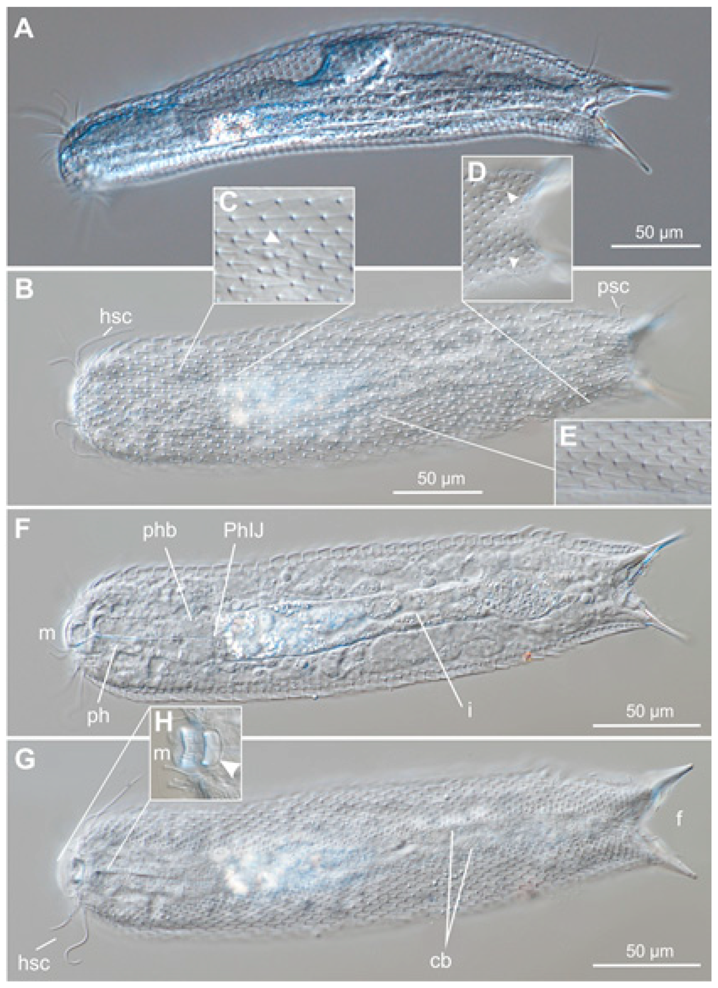
2.3. Species Description

The species description adheres to the standards set by Hummon et al. [76]. The position of morphological traits along the body is given in percentage of unit (U) relative to the total body length, from front to rear. Abbreviations used in the text are as follows: PhIJ—pharyngo-intestinal junction; TbA—anterior adhesive tube; TbD—dorsal adhesive tube; TbDL—dorsolateral adhesive tube; TbL—lateral adhesive tube; TbP—posterior adhesive tube; TbV—ventral adhesive tube; TbVL—ventrolateral adhesive tubes; TL—total body length.

2.4. Granulometric Analysis

The sediment’s grain size distribution was analyzed using dry sieving as described in Todaro et al. [17,57]. The mean grain size, sorting coefficient, kurtosis, and skewness were computed using a computer program [77] based on the equation developed by Seward-Thompson and Hail [78].

3. Results and Discussion

3.1. Sampling Stations

The water temperature and salinity of the sampling stations in 2023 were higher than those in 2008 (Table 1). This difference is clearly linked to the time of year when the sampling took place: early spring in 2008 and late summer in 2023.

3.2. Granulometric Analysis

The sediment’s granulometric characteristics varied slightly across the sampled stations. It mainly consisted of medium, moderately well-sorted, or sorted sand. The exception was the sediment at two locations, Rimel Beach (St3) and Mahdia (b), which was made up of fine sand (Table 2).

3.3. Gastrotrich Fauna

The faunistic analysis of the sandy samples collected in 2023 from the four locations along the north Tunisian coast (Bizerte) yielded 21 putative species and a total of 31 records (species x location) or a mean of 7.8 ± 2.2 SD species per location (Table 3). Cap Angela (St1) displayed the highest species richness with eleven species (35% of total species), followed by Rimel Beach (St3) with eight species (26%). Rimel Epave (St4) and Les Grottes (St2) have the lowest species richness with seven (23%) and five (16%) species. It is noteworthy that there is a low overlap in fauna among the investigated stations (also see Tables S1 and S2). The maximum overlap observed is just three species that are shared between stations st1 and st3, which are characterized by distinctly different sediment types (st1 has medium sand, while st3 has fine sand). In this framework, it is somewhat paradoxical that station st1 has a higher species richness due to a greater number of Chaetonotida, whereas station st3 is richer in Macrodasyida. This finding contrasts with most literature, which suggests that Chaetonotida typically prefer finer sediments (see [5,79]). Future studies, ideally involving higher sampling efforts and quantitative approaches, should aim to confirm or challenge our preliminary findings.
Eleven species in ten genera and six families belong to the order Macrodasyida, and ten species in six genera and two families belong to the order Chaetonotida (Table 3).
Out of the 21 species found, sixteen are known and have been previously reported in the Mediterranean Sea, see [56,79]. One species of the genus Halichaetonotus is represented by specimens with a unique set of morphological traits, suggesting that it may be a new species. This new species is formally named and described below. To accurately determine whether the remaining four species are already classified or warrant descriptions as new species, more comprehensive data are essential. Therefore, they have been provisionally named Dolichodasys sp1, Heteroxenotrichula sp1, Heteroxenotrichula sp2, and Megadasys sp1 (Table 3).
Morphometric information along with photomicrographs as vouchers are provided for all recorded species under the taxonomic account section below.

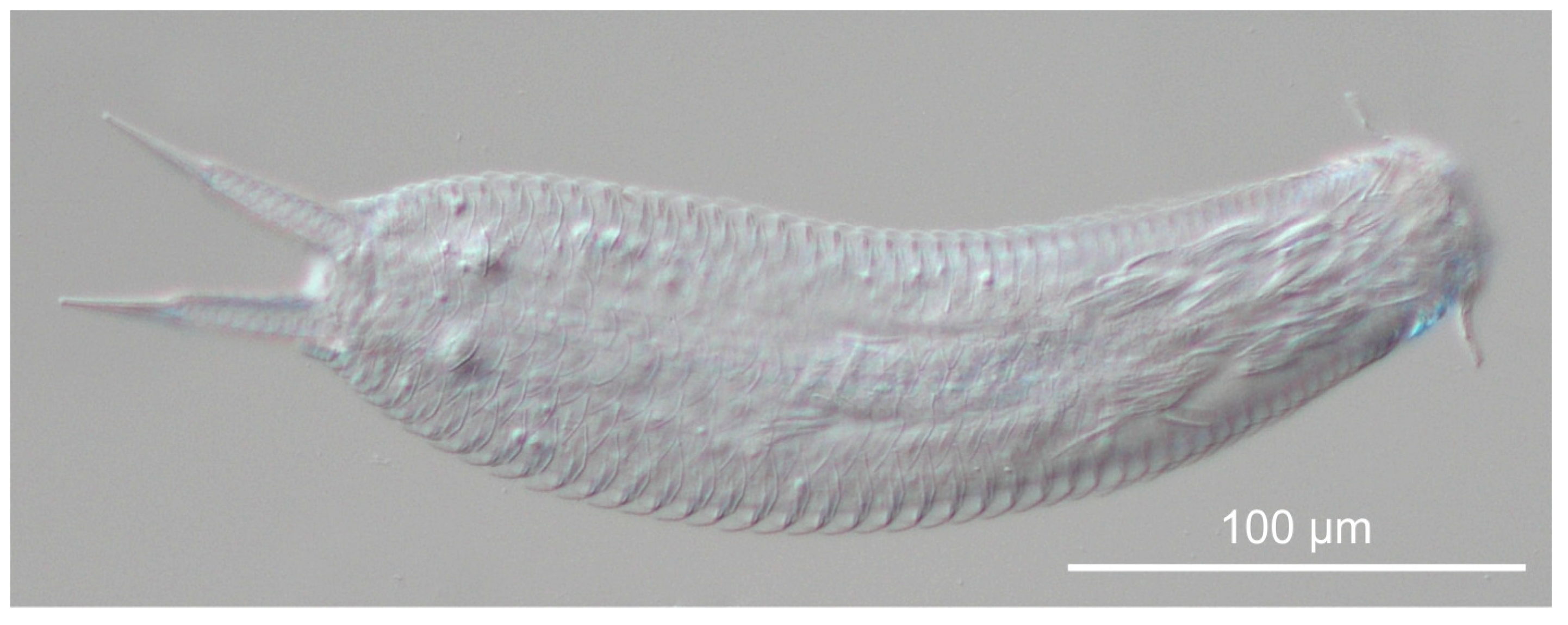
3.4. Taxonomic Account

Order Macrodasyida Remane, 1925 [80] [Rao & Clausen, 1970] [81]
Family Cephalodasyidae Hummon & Todaro, 2010 [82]
Genus Dolichodasys Gagne, 1977 [83]
Dolychodasys sp1
Material recorded: A single specimen from Rimel Epave (St4).
Description: The single, extremely elongated specimen measures 2626 µm in TL (Figure 2). Unfortunately, the specimen was damaged during the mounting and observation; therefore, not all the morphological traits could be recorded for complete identification. However, we were able to observe the following traits: two TbA, with one on each side (Figure 2C), the posterior end was not clearly lobed, and there were several TbP along its margin (four visible, seven estimated) (Figure 2B).
Remarks: Among macrodasyidan Gastrotricha, only specimens of two genera, Megadasys and Dolichodasys, may reach the total body length of the observed specimen. Although severely damaged, the few visible traits allow us to identify the studied gastrotrich as a species of Dolichodasys. In fact, the presence of a single TbA on each side, the posterior end being not clearly lobed, and very few TbP along its margin are diagnostic traits for Dolichodasys and, at the same time, exclude it from being a member of the genus Megadasys.
Genus Mesodasys Remane, 1951 [73]
Mesodasys littoralis Remane, 1951 [73]
Material recorded: Five specimens from Bouficha, collected in 2008.
Description: The following morphometric information is derived from an adult specimen reaching 980 µm in TL (Figure 3). The body is strap-shaped with parallel sides that narrow posteriorly, then widen slightly towards the caudal lobe. The head is indistinct, carrying several sensorial cilia, some arranged in lateral clusters. Piston pits and eye spots are absent (Figure 3E). The TbA are arranged in three to four transverse rows ventrally behind the mouth, counting nine to ten pairs (Figure 3E). For TbVL, there were 43 per side, with 22 along the pharyngeal region; TbL were present but difficult to count; no TbD were seen; and for TbP, there were eight tubes at the edge of the caudal lobe (Figure 3B). The mouth, measuring 21.4 µm in diameter, opens anteriorly and leads into a cup-like buccal cavity (31.6 × 38 µm); the pharynx is 352 µm long and bears pores near the base (U38); the PhIJ is at U40; the intestine is sac-like, wider at the front than at the rear; and the anus opens ventrally at U84. This organism is a hermaphrodite, possessing paired testes that begin at the PhIJ (Figure 3D), with sperm ducts connecting to a small copulatory organ situated posteriorly (U79) (Figure 3C). The ovary is unpaired, and a developing egg, 86 × 77 µm, is located dorsally to the gut, at approximately half the length of the body.
Remarks: M. littoralis was initially described from Cuma, Italy [73]. The species has repeatedly been found along the western shores of Italy [56,84,85], other Mediterranean coastlines [56,61,69], and along the North Sea and the Atlantic coasts of Europe [61]. However, despite the numerous records, comprehensive information regarding the anatomical traits of the specimens found is minimal. The morphometric characteristics of the Tunisian specimens align with literature data on the species. In particular, the large size of the current specimens is most similar to the size of the Italian specimens reported by Wilke [85] and those from Israel that were video-recorded by W.D. Hummon http://www.gastrotricha.unimore.it/moviegallery.htm” (accessed on 24 November 2024). It is essential to highlight that a specimen of M. littoralis from Bouficha was utilized by Todaro et al. [71] to obtain nucleotide sequences of the nuclear 18S and 28S rRNA genes. These sequences are available in GenBank under the following accession codes: JF357658.1 for 18S and JF357706.1 for 28S. A partial sequence of the mitochondrial COI gene was obtained from the same specimen (voucher MT45) and is available in GenBank under the accession code JF432044. However, due to a submission error, this sequence is incorrectly labeled as belonging to a different specimen (voucher MT50). The repository has not responded to requests for correction [86].
Family Dactylopodolidae Strand, 1929 [87]
Genus Dactylopodola Strand, 1929 [87]
Dactylopodola typhle (Remane, 1927) [72]
Material recorded: Three specimens, two from Rimel Beach (St3) and one from Rimel Epave (St4).
Description: All the specimens found were in the juvenile stage, meaning they did not have visible sperm or ova inside. The largest specimen was about 200 µm in TL (Figure 4). It had a distinct pentagonal head, a noticeable neck constriction, and a bilobed caudum originating from a stalked posterior region. The adhesive apparatus consisted of a pair of TbA on each side located ventrally under the head, three pairs of TbL along the trunk region (arranged as 1 and 1 + 1 tubes on each side), and a pair of TbP on each side at the end of the bilobed caudum.
Remarks: The identification of gastrotrich species should be based on adult specimens, as the metric and meristic characteristics vary during the ontogenetic development [2,14,88]. Consequently, the morphometric characteristics of the studied specimens alone could not allow a precise species determination; however, the reported presence of D. typhle at nearby Bouficha and the record of juveniles with matching morphology in a large population of D. typhle in Italy [89] reasonably suggest that the present specimens are members of the same taxon (i.e., D. typhle), especially considering that D. typhle has been recorded in several Mediterranean locations [61]. A specimen of D. typhle from Bouficha was used by Todaro et al. [71] to obtain nucleotide sequences for the nuclear 18S and 28S rRNA genes, as well as the Folmer region of the mitochondrial COI gene. These sequences, which are designated as voucher MT37, are available in GenBank under the following accession codes: JF357652.1, JF357700.1, and JF432037.1.
Family Macrodasyidae Remane, 1924 [90]
Genus Urodasys Remane, 1926 [91]
Urodasys viviparus Wilke, 1954 [85]
Material recorded: A single specimen from Rimel Epave (St4).
Description: The specimen that was examined measures 368 µm in TL and has a maximum trunk width of 56 µm. The tail is approximately 1550 µm long (Figure 5). The pharynx measures 162 µm in length. Along the trunk region, there is a pair of TbA and approximately five pairs of TbL. Additionally, one pair of TbD is observed in the mid-pharyngeal region and another in the posterior trunk region. Two relatively large, highly refractive glands are present in the anterior region just posterior to the piston-pit organs. Furthermore, the observed specimen contains an internal embryo at an early developing stage (egg cleavage) (Figure 5B).
Remarks: The extremely long tail is the distinctive characteristic of the genus Urodasys [92]. However, while most congeneric species are hermaphroditic, U. viviparus lacks testes and accessory reproductive structures [85] while retaining the paired ovaries. The maturing eggs are retained inside the body, where they develop by parthenogenesis, and the offspring are eventually delivered by rupture of the body wall (ovoviparity) [49,93,94]. U. viviparus has a cosmopolitan distribution (see Todaro and Rocha [95]; Hummon [61]; Hochberg et al. [93]) and in the Mediterranean Sea, it has been recorded in France, Croatia, and Greece and is widespread along the Italian coastlines [61,96,97,98]. Morphometrics of the U. viviparus specimen from Rimel Epave agrees with the literature data on the species [85,93,96].
Family Planodasyidae Rao & Clausen, 1970 [81]
Genus Megadasys Schmidt, 1974 [99]
Megadasys sp1
Material recorded: Four specimens, three adults, and a subadult from Rimel Epave (St4).
Description: The adult examined specimens range from 2773 µm to 3867 µm in TL (Figure 6). The body is naked and highly elongated, with an indistinct head and a rounded caudal lobe (70 µm wide × 40 µm long). The adhesive apparatus consists of four pairs of TbA as a continuation of the ventrolateral tubes, with at least twenty-four pairs of TbVL (Figure 6C); TbL and TbD are present but are difficult to count due to the twisting of the animals. There are at least 20 TbP distributed along the margin of the caudal lobe. Ventral locomotory cilia are arranged in two bands spanning almost the entire animal length. The mouth is terminal and 23–25 µm in diameter, leading into a shallow, almost cylindrical buccal cavity and then to a 555–830 µm long pharynx. The PhIJ is at about U22. The pharyngeal pores are located slightly away from the pharyngeal base at U17. The adults are simultaneous hermaphrodites; the paired testes originate well past the PhIJ, with the deferens filled with sperm and leading back along the intestine’s sides, joining on the right side anterior to the mature egg (about U52) (Figure 6F,G). The paired ovaries have a mature egg centered at U60. The caudal organ is bullet-shaped, measuring 206 µm long × 48 µm wide, and is almost equally divided into an anterior glandular region and a posterior muscular, hollow portion. The frontal organ, per se, was not observed, but a bundle of allosperm (>230 µm long) (Figure 6E) was visible on the right side, just posterior to the ovary (U65). The subadult measured about 1200 µm in length and showed only three TbA and eight TbP (Figure 6B).
Remarks: The genus Megadasys includes three species: M. pacificus Schmidt, 1974 [99]; M. sterreri (Boaden, 1974) [100], and M. minor Kisielewski, 1987 [101]. The last two species have also been found in the Mediterranean Sea [24,79]. Hummon [61] reports M. pacificus from the Egyptian coast of the Red Sea and along the Suez Canal region. The large size of our specimens rules out the possibility of them being M. minor. However, the differences in some characteristics with the other two species (see a lower number of TbA and TbP) do not conclusively place the Tunisian species into either category. It is also possible that the Tunisian specimens belong to the undescribed species reported from Israel by Hummon [61]. On the other hand, the notable difference in the overall length of the examined adults (greater than 1 mm) may suggest that the specimens belong to two separate species. Due to these uncertainties, we prefer to keep the status of the current specimens undetermined. We hope to obtain additional information on this species as our investigation along the Tunisia coasts continues. Meanwhile, we highlight that one of the current specimens exceeds the 3.5 mm length of M. pacificus by more than 350 µm, making it the longest gastrotrich ever recorded.
Family Thaumastodermatidae Remane, 1927 [102]
Sub-family Diplodasyinae Ruppert, 1978 [103]
Genus Acanthodasys Remane, 1927 [102]
Acanthodasys aculeatus Remane, 1927 [102]
Material recorded: Two specimens, an adult from Cap Angela (St1) and a juvenile from Rimel Beach (St3).
Description: The fully grown specimen measures 637 µm in TL, with a maximum trunk width of 112 µm (Figure 7).
The body is covered with uniancres, which are longer on the dorsal side (measuring 8 to 13 µm) than on the ventral side (measuring 4 to 5 µm). The adhesive apparatus is made up of three pairs of TbA located behind the mouth, seven pairs of TbVL located along the pharyngeal and trunk regions, at least three TbD along the mid-line of the posterior pharyngeal and anterior trunk region, and six TbP, two at the end of each caudal foot and one at the inner margin of the foot. The TbVL are the longest, ranging from 21 to 25 µm. The frontal mouth, which is 58 µm in diameter, leads to a 198 µm long pharynx. The reproductive system matches the genus’s structure, but the rosette pore and allosperm in front of the largest oocyte were absent in the examined specimen.
Remarks: The morphometric data of the examined specimen is consistent with the original description [102]. The TL falls within the range observed in previous studies (500–800 µm) in the Mediterranean Sea [96,97]. In the Adriatic Sea, specimens have been recorded with a TL of 988 µm [98].
Genus Diplodasys Remane, 1927 [102]
Diplodasys sanctimariae Hummon & Todaro, 2009 [60]
Material recorded: A single sub-adult specimen found at Les Grottes (St2).
Description: The examined specimen measures 226 µm in TL (Figure 8). It has a large ovoid anterior mouth with a rounded oral hood (Figure 8A). The dorsal side is covered with five columns of 22 flat scales that have numerous fine openings (Figure 8B), and there are 32 lateral spines on each side, from U13 to U92. The adhesive apparatus consists of 10 TbA arranged in two rows, 22 TbV on each side, and 12 TbP, two of which are located at the end of each caudal pedicle (Figure 8C).
Remarks: The species was first found at Santa Maria di Leuca, Italy, and later at Cyprus and the Red Sea [60]. The general morphology and cuticular covering match the original description of D. sanctimariae. The metrics of the examined specimen approach those of the sub-adult found in Cyprus [60].
Sub-family Thaumastodermatinae Remane, 1927 [102]
Genus Pseudostomella Swedmark, 1956 [104]
Pseudostomella etrusca Hummon, Todaro & Tongiorgi, 1993 [76]
Material recorded: A single adult specimen found at Rimel Beach (St3).
Description: The specimen measures 343 µm in TL (Figure 9). Its body features well-developed preoral palps and posterior caudal pedicles. The preoral palps curve ventromedially, with the dorsal border extending slightly beyond the ventral side. On the dorsal surface, there are five papillae arranged in a 2 + 1 + 2 pattern along the inner border of the palps, with the largest papillae positioned laterally and smaller ones medially (Figure 9B). On the ventral side, there are six to eight recognizable papillae, approximately the same size, symmetrically arranged towards the center (Figure 9C). The cuticular armature comprises relatively robust five-spined hooks (pentancres) covering the dorsal and ventrolateral aspects. The adhesive apparatus consists of four pairs of TbA on each side: three are located laterally, and one is positioned at the ventral border of the mouth (Figure 9C). Additionally, there are ten pairs of TbL along the trunk region on each side, three TbP (two at the ends of each caudal pedicle and one medially), and two TbDL, one on the trailing surface of each oral palp (Figure 9C) and one near the base of each caudal pedicle. The reproductive system is hermaphroditic. There is a single testis on the right side of the body, which leads to an extended structure that connects to a pear-shaped, glandular-muscular caudal organ, which is in turn connected to a vesicular frontal organ. Small, developing oocytes are present in the posterior trunk region.
Remarks: The species is endemic to the Mediterranean Sea and is widespread in both the western and eastern basins [61,76,105]. The morphometric data of the studied species agree with the original description, except for the lower number of TbL (10 pairs vs. 14 pairs). The difference may be an intraspecific variation due to the difficulty of recognizing smaller additional tubes in our specimen [76].
Genus Tetranchyroderma Remane, 1926 [91]
Tetranchyroderma esarabdophorum Tongiorgi & Balsamo, 1984 [74]
Material recorded: Two specimens, a subadult and a juvenile from Mahdia collected in 2008.
Description: The subadult specimen measures 252 µm in total length (Figure 10). The body is covered by pentancres and features three pairs of rod-like tubes on its dorsal side, which give the species its name. One pair is situated in the pharyngeal region (U28), while the other two pairs are located along the trunk (U47 and U64) (Figure 10A). There are four pairs of epidermal glands. The head has dorsal rod-like tentacles, sometimes referred to as antennae, and lateral knob-like sensory organs (Figure 10B). The caudal region consists of two short caudal pedicles. The adhesive apparatus includes, on each side, three TbA: a medial one located behind the mouth rim and two lateral ones. Additionally, there are 11 TbVL; the first is found in the anterior pharyngeal region (U20) (Figure 10C) and the others extend along the trunk. There is one TbDL in the posterior trunk region (U89) and four TbP, with three at the end of each caudal pedicle and one tube along the inner margin of the pedicle.
Remarks: T. esarabdophorum was initially described from the northern Adriatic Sea, specifically at Pesaro [74]. It has since been reported in various locations throughout eastern and western Italian coastlines [79] and is also known from the western Mediterranean Sea [56,61]. Unlike our subadult specimen, adult T. esarabdophorum can reach a total length (TL) of up to 350 µm and typically possess four pairs of TbA and up to 22 pairs of ventral tube appendages (TbVL). Nonetheless, the distinct pentancrous covering, the head furnished of rod- and knob-like tentacles, and the three pairs of dorsal rod-like tubes clearly indicate that the specimens from Madhia belong to T. esarabdophorum. Notably, a specimen collected in 2008 at Madhia has yielded nucleotide sequences for the nuclear 18S and 28S rRNA genes, as well as the mitochondrial COI gene [71]. These sequences, designated as voucher MT07, are available in GenBank under the following accession codes: JF357627.1, JF357675.1, and JF432022.1.
Tetranchyroderma heterotubulatum Hummon, Todaro & Tongiorgi, 1993 [76]
Material recorded: Four specimens, two from Les Grottes (St2) and two from Rimel Beach (St3).
Description: The following morphometric information is derived from the largest specimen found at Les Grottes, which measures 323 µm in TL (Figure 11). The head is bilobed and features a subterminal mouth that is 33 µm wide. The pharynx measures 93 µm in length, with pharyngeal pores located near its base. The pharyngeal intestinal junction (PhIJ) is situated at U36. Approximately 11 pairs of epidermal glands are scattered along the sides of the body, which are covered with robust tetrancres. The adhesive apparatus includes three pairs of TbA (Figure 11B), eight pairs of TbVL, inserted from U42 to U78, two pairs of TbD at U55 and U98 (Figure 11A), and five pairs of TbP. The latter comprises two tubes at the end of each caudal pedicle, two along the medial side of the pedicle, and one on its lateral side (Figure 11C). The glandular-muscular caudal organ is pear-shaped, while the vesicular frontal organ is roughly spherical and contains a bundle of sperm inside (Figure 11D).
Remarks: The species exhibits an amphiatlantic distribution. Originally described in Italy, it has also been found in northern Europe and along the Atlantic coast of the USA [61]. In the Mediterranean Sea, it is relatively common in both the western and eastern basins [56,106]. The metric and meristic data of specimens from Tunisia fall within the range of morphometric information known for the species (see Hummon et al. [76]).
Tetranchyroderma papii Gerlach, 1953 [107]
Material recorded: A single juvenile specimen from Cap Angela (St1).
Description: The specimen examined is a juvenile measuring 219 µm in TL (Figure 12). Its body is covered with pentancres and shows four pairs of epidermal glands. The head features a pair of rod-shaped tentacles on the dorsal side, each measuring approximately 11 µm in length, along with a pair of elongated spatiform tentacles on either side of the mouth measuring 26.5 µm in length. The adhesive apparatus consists of six TbA, which are located ventrally behind the mouth rim, along with three pairs of TbVL along the mid trunk, a pair of TbDL located in the posterior trunk region, eight TbP, three tubes arranged at the end of each caudal pedicle, and two tubes in between the pedicles (Figure 12B). A pair of lateral cirrata tubes are along the pharyngeal region between the first two epidermal glands (Figure 12B).
Remarks: The morphometric characteristics of the examined specimens do not allow for precise determination at the species level; however, identification can still be inferred. A combination of rod-like and spatiform tentacles is observed in only three species of Tetranchyroderma: T. papii, T. bunti (Thane-Fenchel, 1970) [108], and T. parapapii (Hummon, 2009) [109]. T. bunti features a cuticular armature composed of tetrancres and is primarily distributed along the Atlantic and Gulf coasts of the USA [61,110]. T. parapapii, described as the North American counterpart of T. papii, shares a covering of pentancres. However, T. parapapii is found only along the Atlantic coast of the USA, from Maine to South Carolina, whereas T. papii is distributed throughout the Mediterranean Sea, with an additional record from southern Portugal [110]. Given this information, it is reasonable to identify the studied species as T. papii, particularly since T. papii has previously been reported in Tunisia by Westheide [70]. In a wider taxonomic context, the morphometric information reported may aid studies that illuminate the ontogenetic morphometric variation occurring in species of the genus Tetranchyroderma and beyond.
Family Turbanellidae Remane, 1926 [102]
Genus Paraturbanella Remane, 1927 [102]
Paraturbanella teissieri Swedmark, 1954 [111]
Material recorded: Nine specimens, seven from Cap Angela (St1) and two from Rimel Beach (St3).
Description: The following description is based mainly on an adult specimen 504 µm in TL and 58 µm in width, found at Rimel Beach (Figure 13). The body is ribbon-shaped, with a recognizable head and a noticeable cone between the slender caudal lobes. The head is 35 µm wide and is characterized by a marked anterior hexagonal portion that includes the anterior mouth (10 µm in diameter) and the heavily cuticularized buccal cavity (19 × 13 µm). Lateral sensory pits and ventral papillae are present, past the perioral swellings (Figure 13B,D). The pharynx is 139 µm in length and shows the pharyngeal pores near the base (U31); PhIJ is at U35. The intestine is straight and not clearly regionalized; the anus opens ventrally at U91. The adhesive apparatus consists of TbA, with six per side originating from a fleshy base, with the inner tube being the smallest (6 µm vs. 9 µm) (Figure 13E), and TbP, with six per side occurring along the inner and distal margin of each caudal lobe. The medial cone is 11 µm long. The paired accessory adhesive tubes (dohrni/Seitenfüsschen) are located at U16, the longest tube is 32 µm, and the shortest is 18 µm (Figure 13C). The hermaphroditic reproductive system features paired testes that extend posteriorly from U36; the sperm ducts curve toward the front at U55 and empty into the exterior through the male pore located at U39. The ovaries are also paired, with oocytes maturing from the posterior to the anterior. In most specimens a large egg, measuring approximately 129 × 45 μm, is visible in the mid-gut region centered at U57. The vesicular frontal organ is located dorsal to the intestine, centered at around U63; in some specimens, it contained bunches of agglutinated sperm along with secretory material.
Remarks: Initially described in northern Europe, Paraturbanella teissieri has been regarded as a senior synonym of P. microptera Wilke, 1954 [85], which was described from the Gulf of Naples in Italy [101,112]. In the Mediterranean Sea, this species is widely distributed [56]. The anatomical characteristics of the Tunisian specimens align with those documented by Tongiorgi [113] and Balsamo et al. [114] for specimens found in the Italian seas.
Genus Turbanella Schultze, 1853 [115]
Turbanella bocqueti Kaplan, 1958 [116] sensu Boaden, 1974 [100]
Material recorded: Two specimens, an adult from Les Grottes (St2) and a juvenile from Rimel Epave (St4).
Description: The studied adult specimen measures circa 1025 µm in TL and 110 µm in width (Figure 14). The body is vermiform, with a clearly defined head, trunk widest in the midgut region, tapering gradually to the caudal base; the caudum is bilobed, bearing a small median cone. The head is furnished with lateral, cone-like tentacles (18 µm long) and is separated from the rest of the body by a marked neck constriction. The mouth opens anteriorly, leading into a sallow buccal cavity and then to a muscular pharynx (230 µm in length) bearing pores near the base (U19). The PhIJ is at U22; the intestine is straight with refringent material in the anterior half; the ventral anus opens at U94. The adhesive apparatus includes, on each side, 11 TbA, originating from a fleshy base (Figure 14C); the tubes are all the same size (8 µm) except the innermost one, which is shorter (5 µm); a total of 32 TbL are visible along the body side (14–18 µm), and there are 11 TbP (6–10 µm) inserting along the inner and distal margin of the caudal lobe (Figure 14B). TbD are present but difficult to count. The reproductive apparatus is hermaphrodite, but only the male organs were seen in detail in the examined specimen. The paired testes originate past the mid-trunk (U56) and extend posteriorly to U69, where sperm ducts curve toward the front and join anteriorly on the midline at U29.
Remarks: Turbanella bocqueti was first described in Northern Europe [100,116,117] and is also found in the Mediterranean Sea, as well as along the Atlantic and Gulf coasts of the USA [61]. In the Mediterranean Sea, it is widely distributed in both the western and eastern basins [56]. The size and general morphology of the Tunisian population align with data reported for both North European and Mediterranean populations [100,113]. However, there is a notable difference in the location of the testes between populations. In the Tunisian population, the testes originate along the mid-trunk, while in the North European population, they are situated near the pharyngo-intestinal junction [117]. Conversely, the position of the male pore is similar in both populations. Since the arrangement of the reproductive system has phylogenetic significance [16,45,118], the highlighted difference, if confirmed, could suggest the existence of a species complex. Future studies should also focus on examining the female component of the reproductive system, as there is currently no information regarding for example the frontal organ of this species.
Order CHAETONOTIDA Remane, 1925 [76] [Rao & Clausen, 1970] [77]
Sub-order Paucitubulatina d’Hondt, 1971 [119]
Family Chaetonotidae Gosse, 1864 [120] sensu Leasi & Todaro, 2008 [18]
Sub-family Chaetonotinae Kisielewski, 1991 [121]
Genus Aspidiophorus Voigt, 1903 [122]
Aspidiophorus mediterraneus Remane, 1927 [123]
Material recorded: Two specimens, an adult and a subadult from Cap Angela (St1).
Description: The adult examined specimen is about 170 µm in TL (Figure 15); it has a unilobed head and well-recognizable furca.
The head features the cephalion and hypostomion (2 µm long × 7.5 µm wide) (Figure 15H). The furca is 23 µm long, including the terminal adhesive tubes (10–12 µm). The widths at the head/neck/trunk and caudal base are 31 µm/37 µm/40 µm and 22 µm measured at U05/U26/U61 and U91, respectively. The cuticular armature comprises pedunculated, ovoid scales (3.7 × 2 µm) bearing a noticeable median keel throughout the length (Figure 15E). Scales are arranged in 43 longitudinal columns (23 dorsal, 10 + 10 ventral); the median column comprises about 65–70 scales. The sensory cilia (10–24 µm long) are arranged in two tufts on the dorsolateral side of the head and as a paired element on the dorsolateral sides along the pharyngeal region (U22) and on the posterior trunk region (U86); the latter two pairs originate from small papillae (Figure 15B–D). Locomotory cilia are arranged into two bands wider under the head and neck region, narrowing while approaching the intestinal region but never joining (Figure 15G). The mouth (5.5 µm in diameter) is subterminal; the pharynx is 38.6 µm in length, showing a noticeable posterior swelling (10 µm wide); the PhIJ is at U24; the intestine is straight; the anus opens ventrally at U88.
Remarks: A. mediterraneus was first described by Remane [123] from samples collected in the Gulf of Naples, Italy. Currently, this species is known from several regions around the world [61], including various Mediterranean locations such as Marseille [104], Naples [85], the coasts of Tuscany [124], and many other Italian locations [79]. The morphometric characteristics of the specimens found in Tunisia align with those reported for specimens from the Tuscany coasts by Luporini et al. [124]. However, the number of longitudinal columns observed in the Tunisian specimens (43 columns) differs from those found on Elba Island in Italy (60 columns, Balsamo et al. [125]) and from specimens collected in North America (20 columns, Hummon [126]). These notable differences suggest that further investigations at the molecular genetic level are needed to explore potential phenomena of cryptic speciation within this taxon.
Aspidiophorus paramediterraneus Hummon, 1974 [126]
Material recorded: Three specimens, one from Cap Angela (St1) and two from Rimel Epave (St4).
Description: The adult specimen from Rimel Epave is 298 µm in TL (Figure 16); it features a single-lobed head and a relatively short furca. The head bears a small dorsal cephalion and obvious ventral hypostomion (5 long × 13 µm wide) (Figure 16E); the furca is 34 µm long, including the terminal adhesive tubes (14 µm). The widths at head/neck/trunk and caudal base are 35 µm/54 µm/66 µm and 32 µm measured at U10/U24/U58 and U88, respectively. The cuticular armature comprises pedunculated, elliptic scales (about 4.5 long × 2.5 µm wide) bearing a noticeable median keel throughout their length (Figure 16B). Scales are arranged in 62 longitudinal columns (40 dorsal, 11 + 11 ventral); the median column comprises about 120 scales. The sensory cilia (15–21 µm long) are arranged in two tufts on the dorsolateral side of the head and as a paired element on the dorsal side of the posterior trunk region (Figure 16A). Locomotory cilia are arranged ventrally in two bands that narrow in an antero-posterior direction and that join anteriorly to mid-trunk to form a single band (Figure 16D). The mouth is subterminal (10 µm in diameter), the pharynx is 66 µm in length and features a noticeable posterior bulb (17 µm wide); the PhIJ is at U26, the intestine is straight and slightly larger in front; the anus opens ventrally at U86, surrounded by a pair of elongated, keeled perianal scales (9 µm long × 2 µm wide) (Figure 16F). An egg occupies most of the trunk region (Figure 16C).
Remarks: A. paramediterraneus was first described from Beaufort, North Carolina, USA by Hummon [126]. Moreover, it is noted from several areas around the world, in the Mediterranean Sea, it has been recorded from Cyprus, Greece, Egypt, France, and Italy [61]. The Tunisian examined specimen matches the original description in terms of body shape and size, the shape of the pedunculated scales, and the arrangement of the ciliary band. However, it possesses a much higher number of columns of scales and scale per column. The cuticular covering (62 longitudinal columns, and 120 scales in the median column) corresponds to the Italian specimen described from Tuscany Archipelago (65 longitudinal columns, 33 dorsal) [125]. The difference in the number of columns and scales per column between the Mediterranean and the American populations may be due to the different methods used for their computation [77]. Nonetheless, such differences in the morphological patterns found in species from geographically distant locations need to go through molecular genetic analyses to test speciation patterns.
Genus Chaetonotus Ehrenberg, 1830 [127]
Chaetonotus dispar Wilke, 1954 [85]
Material recorded: Two specimens, one adult from Cap Angela (St1) and a juvenile from Rimel Epave (St4).
Description: The adult specimen from Cap Angela is 134 µm in LT (Figure 17); it features a tri-lobed head and a relatively short furca. The head bears dorsal cephalion and small ventrolateral pleurae; the hypostomion is not seen; the furca is 27 µm in length, including the terminal adhesive tubes (17 µm long). The widths at the head/neck/trunk and furcal base are 24.5 µm/16.7 µm/30 µm and 16.5 µm measured at U09/U30/U60 and U84, respectively. The cuticular armature comprises 11 longitudinal columns (7 dorsal, 2 + 2 ventrolateral) of separated three-lobed scales, with each carrying a robust spine increasing in length from front to the rear; the median column comprises about 17–18 scales; spines of the dorsal columns bear two accessory points at about 3/4 of their length and a notched tip (Figure 17A); ventrolateral spines have only the two accessory points along their lengths. The ventral locomotory cilia are arranged in two longitudinal bands that are wider in the front but never merge. The interciliary field is covered with tiny, round, keeled scales arranged in 4–6 longitudinal columns from U35 to U72. The ventral terminal scales are elongated and furnished with a keel that continues in a short, notched spine (Figure 17C). On the ventral side of the furcal base, there are two pairs of small trilobate, keeled scales, each carrying a spine similar to the ventrolateral ones; the spines of the medial pair protrude from the furcal indentation. The subterminal mouth is 6.5 µm in diameter and leads into a 32.5 µm long pharynx, which shows gentle swelling at both ends; the PhIJ is at U27; the intestine (70 µm in length) is straight and lacks evident differentiation; the anus opens ventrally at U81 (Figure 17B).
Remarks: C. dispar is a cosmopolitan species that was first described from the Gulf of Naples [61,85] and has been frequently reported in the Mediterranean Sea [56]. The morphometric data of the specimen from Cap Angela is consistent with the original description, and with data reported for the specimens from the Tuscan Archipelago [125] and those from the US Virgin Islands [128].
Genus Halichaetonotus Remane, 1936 [129]
Halichaetonotus bizertae sp. nov.
Etymology:bizertae” refers to Bizerte, the city where the species was first found.
Diagnosis: Small-sized Halichaetonotus, 127 ± 2.2 µm in TL; the head, neck, trunk, and furcal appendages are well-defined. The head is slightly five-lobed with a frontal cephalion and lateral pleurae but lacks the hypostomion; there is a medium-long furca (25.3 ± 1.7 µm) projecting from the posterior of the trunk. The body is covered dorsally by seven longitudinal columns of alternating, keeled scales. The median dorsal column is composed of 18–20 scales; the scales are shield-shaped on the head and trunk and round on the neck; in general, the keel extends beyond the edge of the scales as a spiny process. In the posterior trunk region, there are two round, double-keeled scales carrying sensorial bristles and a pair of scales with spines protruding into the furcal indentation; there are keeled, spiny scales on the dorsal and ventral base of the furca. Laterally and ventrolaterally, there are 2 + 2 columns of hydrofoil scales with spine-bearing lamellae of varying length; ventrally, there are 2 + 2 additional columns of small, round scales; locomotory cilia are arranged in two longitudinal bands; the interciliary field is naked, ending with two pair of perianal, elliptical keeled scales of similar size, with each having a keel extending in a spiny process. The mouth is of medium size (6.4 ± 0.2 µm in diameter), opening into a 29.9 ± 0.5 long pharynx, showing swellings at both ends; the anterior bulb is provided with pharyngeal teeth; the intestine is sack-like with a terminal ventral anus. All specimens are parthenogenetic, sometimes with a single large egg in the dorsal to mid-intestine region.
Type locality: Tunisia, Bizerte: Les Grottes (37°19′60″ N; 09°50′38″ E; Figure 19 and Figure 20). The specimens were found in medium, moderately well-sorted sand. The characteristics of the site are shown in (Table 2). Additional site: Cap Angela (37°19′53″ N; 09°46′10″ E).
Type material: Holotype: The adult specimen shown in Figure 19 and Figure 20, no longer extant, was collected by AS on 16 September 2023 (International Code of Zoological Nomenclature 19, Articles 73.1.1 and 73.1.4; also see the recommendation 73 G–J of Declaration 45—Addition of Recommendations to Article 73) [130,131]. Additional studied specimens: Two specimens from Cap Angela (St1), showing maturing eggs inside. All specimens were examined in vivo and were destroyed during the microscopical survey.
Description: The description is mainly based on the holotypic adult specimen measuring a length of 129 µm in TL (Figure 19 and Figure 20).
The body is tenpin-shaped, featuring a marginally penta-lobed head consisting of two pairs of loose tufts of sensorial cilia, a shallow cephalion (10 µm wide), and small pleurae but lacks a hypostomion and eye spots; the neck constriction is well-defined, and the trunk is sac-like, terminating in a furcate caudum. The body widths at the head/neck/trunk and furcal base are 19 µm/15.5 µm/25 µm and 14 µm measured at U01/U25/U36 and U78, respectively. The caudal appendages are of medium length (27 μm), with paired laterally divergent adhesive tubes (15 μm in length) and a slightly inflated base (6 μm) covered by scales (Figure 18 and Figure 19A,D).
Cuticular armature: The cuticular armature extends over the entire body surface, except for the ventral interciliary field. The head, neck, and trunk covered dorsally and lateroventrally by alternating columns (seven dorsal, 1 + 1 lateral hydrofoil, and 1 + 1 ventrolateral hydrofoil, 2 + 2 ventral) of 18–20 keeled scales, barely overlapping. On the dorsal side (Figure 18A and Figure 20A–D), the head and trunk scales are shield-shaped (3 × 5 µm on the head, Figure 20B; 5 × 8 µm on the trunk Figure 20D); on the neck, the scales have round heads (3 µm in diameter, Figure 20C). In general, the keel on the dorsal scales extends beyond the edge of scales as a spiny process; the spines are about 5 µm long on the head, 3 µm on the neck, and up to 10 µm on the trunk (Figure 18A and Figure 20B–D). On the posterior trunk region, there are two round, double-keeled scales (about 4 μm in diameter), each anchoring a sensorial bristle at U78, and a couple of scales (4 × 3 μm) bearing spines (5 μm long) protruding into the furcal indentation, along with a pair of spiny scales on the furcal branches (Figure 18A and Figure 19A). The lateral and ventrolateral hydrofoil scales are similar in shape and size to the dorsal scales. They have spines that bear flattened lamellae, most of which taper into a pointed process. The lamellae-bearing spines of the lateral scales are more protracted than related ventrolateral ones (up to 10 µm vs. up to 5 µm), while the lamellae of a column are longest at the mid-trunk (Figure 18 and Figure 19B–D). On the ventral side, there are two columns of rounded keeled scales with a short spine (3–4 µm long) on each side between the ciliary band and the hydrofoil scales; the innermost column consists of scales smaller than the outer one; on the posterior ventral region, up to five keeled scales, 3–4 μm long, cover the fleshy portion of each furcal branch (Figure 18B and Figure 19E,F). The interciliary field appears naked except for two pairs of elliptical, keeled scales in the perianal region; the scales of the anterior pair are slightly larger (7 × 4 μm) than the posterior ones (6.0 × 3 μm); the keel of these scales continues in a short spine process (Figure 18B and Figure 19E,F).
Ventral ciliation: Paired longitudinal bands extend from U03 to approximately U73. These bands are broadly club-shaped at the front but narrow significantly toward the posterior pharyngeal region. They come close together just behind the mouth but remain separate along their entire length. The individual cilia measure between 9 and 12 μm in length (Figure 18B, Figure 19B and Figure 21B).
Digestive tract: The subterminal mouth is of medium size (7 μm in diameter), projecting very slightly ventrally and leading progressively into a 30 μm long pharynx featuring swellings at both ends; the anterior bulb is slightly larger (11 μm vs. 9 μm in diameter) and is provided with pharyngeal teeth; the pharyngeal-intestinal junction is at U26; the intestine is straight, narrowing posteriorly; the anus opens ventrally at U77 (Figure 18B, Figure 20E and Figure 21A).
Reproductive tract: All the examined specimens were in the parthenogenetic phase (i.e., no spermatozoa were observed); the holotype had a mature egg (41.5 × 21 µm) located dorsally to the intestine, filling almost the trunk region.
Variability and remarks. The general appearance and characteristics of the other two specimens studied are very similar to those of the holotype (Figure 21 and Table 4).
Negligible differences were observed among the specimens regarding the following major traits: (i) total body length ranging from 124 to 129 µm; (ii) pharynx length fluctuating between 29 and 30 µm; (iii) furca length spanning from 23 to 27 µm; and (iv) adhesive tube length varying from 12 to 15 µm. The measurements of these traits are generally correlated with the size of the animals (Table 4).
Taxonomic affinities: The marine genus Halichaetonotus comprises 31 known species, showcasing a significant diversity in their cuticular coverings. However, the specimens found in Tunisia share certain characteristics with five other species: they possess a naked intraciliary field and the keel on the dorsal scale that extends into a spiny process. The species sharing these traits are H. bataceus Evans, 1992 [132], H. etrolomus Hummon, Balsamo & Todaro, 1992 [133], H. genatus Balsamo, Fregni & Tongiorgi, 1995 [97], H. sanctaeluciae Todaro, Dal Zotto, Bownes & Perissinotto, 2011 [134], and H. euromarinus Hummon & Todaro, 2010 [82]. The specimens we are studying can be easily distinguished from H. bataceus, H. etrolomus, and H. genatus by the presence of small round scales on the ventral side, located between the ciliary bands and the hydrofoil scales, a feature that the latter three species lack. In contrast, the Tunisian specimens share this trait with H. sanctaeluciae and H. euromarinus. Additionally, all three are characterized by having two pairs of large scales in the perianal area.
Despite their similarities, these three species can be easily distinguished by various prominent morphological features. For instance, H. euromarinus has only ventrolateral columns of hydrofoil scales, while H. sanctaeluciae and the Tunisian specimens have lateral columns as well. Furthermore, the specimens being studied can be differentiated from H. sanctaeluciae because they do not possess the three dorsal posterior scales, one median and two laterals, which have long, robust spines extending beyond the end of the trunk. In contrast, the Tunisian specimens have a couple of scales in the posterior region that feature spines protruding into the furcal indentation, which are absent in H. sanctaeluciae. Based on these distinctions, it is clear that the specimens under study are unique enough to be classified as a new species, which we propose to name Halichaetonotus bizertae sp. nov.
Halichaetonotus euromarinus Hummon & Todaro, 2010 [82]
Syn. Halichaetonotus spinosus Mock, 1979 [135]
Material recorded: A single specimen from Cap Angela (St1).
Description: The specimen studied measures 133 µm in TL and features a roughly rounded head with a distinct furca (Figure 22).
The head includes a frontal cephalion and small, ventrolateral pleurae, while the hypostomion is not visible. The furca measures 21 µm in length, including the terminal adhesive tubes, which are 14 µm long. The widths at the head/neck/trunk and furcal base are 27 µm (16.3 µm/34.4 µm/and 15 µm, respectively, measured at U12/U30/U58 and U82. The cuticular armature consists of nine dorsal columns of slightly overlapping, keeled scales. The scales on the head and trunk regions are shield-shaped, measuring approximately 8 µm by 4 µm on the trunk, while those on the neck are roughly round. In all cases, the keel extends into a short spiny process (Figure 22B). The dorsal medial column contains 17–18 scales, with the posterior-most scale’s spiny process protruding from the furcal indentation. Additionally, each side features two ventrolateral columns of scales. The lateral column contains scales similar to those on the dorsal side but has much higher keels that bulge upward and backward, lacking the spiny process found on the dorsal scales. The more ventral column consists of roughly circular scales that bear spinous lamellae (hydrofoil scales) (Figure 22C). In addition, on each side, between the ciliary band and the hydrofoil scales, there are three to four columns of small, rounded spined scales. The ventral locomotory cilia are arranged in two longitudinal bands that are wider at the front but never merge. The interciliary field is bare, and in the perianal area, there are two pairs of elliptical, weakly keeled scales, which are flanked on each side by three to four smaller scales (Figure 22D). The subterminal mouth has a diameter of 5.5 µm and leads into a 27 µm long pharynx, which displays conspicuous swellings at both ends. The anterior bulb contains pharyngeal teeth; the PhIJ is located at U24. The intestine is straight and broader in the anterior half, while the anus opens ventrally at U82.
Remarks: Halichaetonotus euromarinus has an amphi-Atlantic distribution; described initially from the North Sea [135], it has been reported from the Atlantic and Gulf coasts of the USA [5,61]. The occurrence of the species in the Mediterranean Sea was reported first by Todaro and Balsamo [136], who found it in Sicily (see also Balsamo et al. [125]). Since then, the species has been found frequently in western and eastern basins [56]. The Tunisian specimens match the original description of the species.
Halichaetonotus thalassopais Hummon, Balsamo & Todaro, 1992 [133]
Material recorded: Seven specimens, five from Cap Angela (St1) and two from Les Grottes (St2).
Description: The following information is based mainly on an adult specimen from Cap Angela measuring 114 µm in TL (Figure 23). The specimen shows a tri-lobed head and a relatively short furca. The head bears dorsal cephalion, small ventrolateral pleurae, and a sub-rectangular hypostomion; the furca is about 20 µm in length, including the terminal adhesive tubes (11 µm long). The widths at the head/neck/trunk and furcal base are 20 µm/15 µm/36 µm and 14 µm measured at U12/U36/U65 and U87, respectively. The cuticular armature consists of 15 longitudinal columns (nine dorsal, 2 + 2 ventrolateral, and 1 + 1 ventral) of overlapping ovoidal, keeled scales (Figure 23B); the dorsal medial column contains 17 scales; the ventrolateral scales have higher keels that rise more as the columns progress ventrally, bulging upward and backward, protruding past the posterior end of the scale; the scales of the ventral columns bear delicate spinous lamellae (hydrofoils scales). Ventral locomotory cilia are organized in paired longitudinal rows, each forming a broad patch anteriorly that meets medially behind the hypostomion. The interciliary field is bare. On the posterior ventral surface, there are six or seven pairs of ovoid keeled scales; the perianal scales are the largest and the only ones with a keel that extends posteriorly as a spiny process (Figure 23D). The mouth is subterminal, about 6 µm in diameter, leading into a 35 µm long pharynx showing strong anterior and posterior swellings (9 µm in diameter). The anterior bulb is armed with robust pharyngeal teeth; the PhIJ is at U29 (Figure 23C); the intestine is straight and lacks evident differentiation; the anus opens ventrally at U82. A mature egg occupies most of the trunk region (Figure 23A).
Remarks: Halichaetonotus thalassopais was first described in Tuscany, Italy [133]. Since then, it has been reported in several other locations across the Italian peninsula, as well as in Greece and along the Mediterranean coasts of Israel and Egypt [56,61,79]. The metric and meristic characteristics of the specimens from Tunisia align with the original description [133]. We are presenting microphotographs of this species here for the first time.
Genus Heterolepidoderma Remane, 1927 [72]
Heterolepidoderma loricatum Schrom, 1972 [137]
Material recorded: Two adult specimens, one from Cap Angela (St1) and one from Rimel Beach (St3).
Description: The following information is primarily based on an adult specimen from Rimel Beach, measuring 110 µm in TL (Figure 24). The body is relatively stout and features a roughly trilobed head and distinct furca. The head includes a frontal cephalion, noticeable ventrolateral pleurae, and a broad hypostomion. The furca measures 18 µm in length, including the 11 µm long terminal adhesive tubes. The widths at the head/neck/trunk and furcal base are 22 µm/21 µm/31 µm, and 20 µm, respectively, measured at U08/U23/U65/and U86. The cuticular armature consists of keeled scales arranged in 13 longitudinal, imbricated columns, with 9 columns covering the dorsal side. There are 18 scales in the medial column (Figure 24A). The scales in the ventral-most column on each side feature spines with lamellae (hydrofoil scales), rather than just a keel. Ventral locomotory cilia are arranged in two longitudinal bands that are broader at the front but do not merge. The interciliary field is covered by rounded keeled scales from U37 to U88, and there are three terminal pairs of elliptical, keeled scales (Figure 24B). The mouth, which is 4 µm in diameter, opens subterminally and leads to a 28 µm long pharynx that has swellings at both ends. The anterior bulb is armed with pharyngeal teeth; the PhIJ is located at U33. The intestine is straight and slightly broader in the anterior portion, with the anus opening ventrally at U86 (Figure 24C). The specimen is in the parthenogenetic phase and shows a large egg that fills most of the trunk.
Remarks: Heterolepidorma loricatum is an amphiatlantic species [5]. It was originally described from the northern Adriatic Sea [137] and has since been recorded in several other locations in Italy and the Mediterranean Sea (see Balsamo et al. [138]; Todaro et al. [56]). The morphometric data of H. loricatum from Rimel Beach align with the published information on this species. Specimens that possess hydrofoil scales have frequently been observed, leading Todaro [77] to suggest that these features may have been overlooked by Schrom [137] during the species’ initial description due to their elusive nature.
Family Xenotrichulidae Remane, 1927 [139]
Sub-family Draculiciterinae Ruppert, 1979 [140]
Genus Draculiciteria Hummon, 1974 [141]
Draculiciteria tesselata (Renaud-Mornant, 1968) [142]
Synonym Polymerurus tesselatus Renaud-Mornant, 1968 [142]
Material recorded: a single subadult specimen from Rimel Beach (St3).
Description: The specimen studied measures 155 µm in TL (Figure 25) and features a distinct, roughly pentagonal head and relatively long furca (47 µm in length). The terminal, lateral spines at the furcal base are 8 µm long. The widths at the head/neck/trunk and furcal base are 33.5 µm/17 µm/30.5 µm and 15.5 µm measured at U08/U22/U55 and U75, respectively. Due to the specimen’s poor condition, the details of the cuticular ornamentations, such as shape and arrangement, could not be detected. The mouth is 7 µm in diameter and leads to a 52 µm long cylindrical pharynx; the PhIJ is at U35; the intestine is straight and lacks evident differentiation; the anus opens ventrally at U70.
Remarks: Draculicitera tesselata is a cosmopolitan species. Described initially as Polymerurus tesselatus from Ischia Island, Italy [142] (see also Hummon [141]), it is known to occur in different areas of the World (see Hummon [61]). In the Mediterranean Sea, it is relatively common [56]. The general morphology of the Tunisian specimens aligns with literature data on the species; the metric data are in line with those of the subadult specimens recorded along the Italian coastlines [79].
Sub-family Xenotrichulinae Remane, 1927 [139]
Genus Heteroxenotrichula Wilke, 1954 [85]
Heteroxenotrichula sp1
Material recorded: A single adult specimen from Cap Angela (St1).
Description: The specimen surveyed is 188 µm in TL (Figure 26) and features a tenpin-shaped body, which has a distinct, trapezoidal head and a pedunculated, V-shaped furca. The head carries two pairs of dorsal sensory cirri and one pair of relatively short lateral tentacles/auricles (9 µm); the furca is 53 µm in length (measured from the indentation to the tip), including the 15 µm long adhesive tubes. The widths of the heads at the auricles/brain commissure/neck/trunk and furcal base are 26 µm/30 µm/37 µm/51 µm and 18 µm measured at U4.5/U10.5/U21/U48 and U68, respectively. The body is covered dorsolaterally with nine longitudinal columns (seven dorsal, 1 + 1 lateral) of overlapping scales (Figure 26A); dorsal columns made up of squamosa-type scale; lateral columns composed of hydrofoil scales (Figure 26B). The anterior locomotory cirri are of two sizes, the smaller confined under the head to U11, the larger distributed between U11 and U30; the paired tuft of posterior cirri (trunk cirri) is located at U46 (Figure 26B); each tuft is made up of three cirri of equal size. The mouth is about 3 µm in diameter, leading to a 35 µm long pharynx featuring an anterior swelling armed with pharyngeal teeth; the PhIJ is at U21; the intestine is straight without evident differentiation; the anus opens ventrally at U69. The reproductive apparatus is hermaphrodite; the male pore opens ventrally at U23, and the mature egg is centered at U45.
Remarks: Most of its morphometric characteristics approach the Tunisian specimens to the cosmopolitan H. squamosa Wilke, 1954 [85], which is widely distributed also in Mediterranean [56,61,79,140,143]. However, the V-shaped furca and especially the much shorter distal adhesive tube compared to the proximal, scaled portion suggest it may belong to a different, undescribed species. Given the uncertainties, we prefer to keep the status of the current specimen as undetermined. We hope to obtain more information about this species as our investigation along the coasts of Tunisia continues.
Heteroxenotrichula sp2
Material recorded: A single adult specimen from Rimel Beach (St3).
Description: The surveyed specimen is 308 µm in TL (Figure 27) and features an elongate tenpin-shaped body furnished with a distinct, trapezoidal head and a slightly pedunculated, U-shaped furca. The head carries two pairs of dorsal sensorial cirri and one pair of relatively short lateral tentacles/auricles (15 µm); the furca is 57 µm in length (measured from the indentation to the tip), including the 25 µm long adhesive tubes. The widths of the heads at the auricles/head at the brain commissure/neck/trunk and furcal base are 36 µm/43 µm/57 µm/71 µm and 27 µm measured at U2.5/U9/U23/U58 and U79, respectively. The body is covered dorsolaterally with nine (seven dorsal, 1 + 1 lateral) longitudinal columns of overlapping scales; dorsal columns made up of squamosa-type scale, lateral columns composed of hydrofoil scales. The anterior locomotory cirri are of two sizes, the smaller confined under the head to U9.5 and the more oversized inserted between U9.5 and U23; the paired tuft of posterior cirri (trunk cirri) is located at U47; each tuft is made up of three cirri of equal size. The mouth is about 4 µm in diameter, leading to a 60 µm long pharynx featuring an anterior swelling showing pharyngeal teeth; the PhIJ is at U21; the intestine is straight without evident differentiation; the anus opens ventrally at U78. The reproductive apparatus is probably hermaphrodite; however, only sparse spermatozoa were seen.
Remarks: The distinctive shape and large size of the Tunisian specimen set it apart from the 11 species of Heteroxenotrichula that have been described to date [68,140,144,145]. However, the morphometric data collected from this single specimen are insufficient to provide a modern description of a new species. Therefore, we prefer to classify the current specimen as undetermined. We look forward to gathering more information about this species as our investigation along the Tunisian coasts progresses.
Genus Xenotrichula Remane, 1927 [139]
Xenotrichula intermedia Remane, 1934 [75]
Material recorded: Two specimens, an adult and a subadult collected at Mahdia in 2008.
Description: The adult specimen is 208 µm in TL (Figure 28). The body is tenpin-shaped and includes a unilobed head and U-shaped furca. The head bears two pairs of dorsal sensorial cirri and lack of auricles; the furca is 30 µm in length including the 15 µm long adhesive tubes. The width at head/neck/trunk and caudal furca are 36 µm/30 µm/52 µm and 23 µm at U6/U24/U54 and U86, respectively. The cuticular covering is composed of intermedia-type stalked scales. On the dorsal side, scales are organized in 17 columns with 55 scales in the median column (Figure 28A). The anterior mouth is 4 µm in diameter and leads into a 47 µm long cylindrical pharynx; the PhIJ is at U25; the intestine is straight, narrowing in the rear; the anus opens ventrally at about U74 (Figure 28B). The reproductive apparatus is hermaphrodite.
Remarks: X. intermedia was first described from the North Sea [75] and has since been reported in various oceans and connected seas [61]. However, there is increasing evidence suggesting that populations located far apart are actually distinct, cryptic species, which can be identified through genetic analysis and/or muscle architecture [39,44,146]. The morphometric data for the Tunisian specimen aligns with findings reported for Italian populations. It is important to note that a specimen of X. intermedia from Mahdia was used by Todaro et al. [71] to obtain nucleotide sequences of the nuclear 18S and 28S rRNA genes, which are accessible in GenBank under the following accession codes: JF357664.1 (18S) and JF357712.1 (28S). Additionally, from the same specimen (voucher MT71), a partial sequence of the mitochondrial COI gene was obtained, available in GenBank under the accession code JF432048.1. However, due to an error during submission, this sequence is mistakenly labeled as belonging to a different specimen (voucher MT82). So far, the repository has not responded to requests to correct this error [86].

4. Conclusions

Gastrotrichs are a vital part of marine meiobenthos found in sandy habitats [4,147]. This study provides, for the first time, a faunistic overview, though not comprehensive, of this group along the northern coast of Tunisia. In a larger framework, the research expands the known marine gastrotrich species in Tunisia from 6 (5 Macrodasyida and 1 Chaetonotida) to 24 species (14 Macrodasyida and 10 Chaetonotida). The average number of species per location is 7.8 ± 2.5 and aligns with the data for Italy, where the average is 8.5 ± 2.5 species per site [79]. This is notable considering the vicinity of the sampled locations and the small quantity of sandy material collected from each location. As anticipated, most of the species identified in this research were already known from the Mediterranean Sea. However, the discovery of Halichaetonotus bizertae sp. nov. and likely at least two other new species (Heteroxenotrichula sp1 and sp2) indicates that our understanding of Mediterranean gastrotrichs is still incomplete. This suggests a pressing need for further research, particularly along the southern shores of the Mare Nostrum. Our research underscores a critical gap in the literature: the scarcity of morphometric data and photographs, even for the most commonly reported species. This shortfall greatly limits our ability to compare distant populations and increases the risk of misidentification. In today’s scientific landscape, where molecular genetic data forms the backbone of many analyses, it is imperative that we prioritize thorough morphological vouchering of species. Such efforts will enhance accuracy and reliability in our research, paving the way for more robust scientific conclusions.

Supplementary Materials

The following supporting information can be downloaded at: https://www.mdpi.com/article/10.3390/d17010017/s1, Table S1. Number of species shared among the four Tunisian stations sampled in 2023; Table S2. Pairwise Dice-Sørensen similarity coefficient [1,2] calculated for the four Tunisian stations sampled in 2023. References [148,149] are cited in the supplementary materials.

Author Contributions

Conceptualization, A.S., M.G. and M.A.T.; methodology and data acquisition, all authors; resources, M.G., M.E.C. and M.A.T.; writing—original draft preparation, A.S. and M.A.T.; writing—review and editing, all authors. All authors have read and agreed to the published version of the manuscript.

Funding

A.S. holds a scholarship from the Ministry of Higher Education and Scientific Research in Tunisia—University of Tunis El Manar (UTM)—Faculty of Sciences of Tunis (FST). This project is partially funded under the National Recovery and Resilience Plan (NRRP), Mission 4 Component 2 Investment 1.4—Call for tender number 3138 of 16 December 2021, rectified by Decree number 3175 of 18 December 2021 of the Italian Ministry of University and Research funded by the European Union—NextGenerationEU. Project Code CN_00000033, Concession Decree number 1034 of 17 June 2022 adopted by the Italian Ministry of University and Research, CUP E93C22001090001, Project title ‘National Biodiversity Future Center—NBFC’.

Institutional Review Board Statement

Not applicable.

Data Availability Statement

The data that support this study are available in the article and in the Supplementary Materials.

Acknowledgments

We would like to express our gratitude to the Faculty of Sciences of Tunis (FST) and the University of Tunis el Manar (UTM) for partially funding this publication research, which is part of Aicha Souid’s doctoral thesis. A.S. extends special thanks to A. Cesaretti for her invaluable assistance with the granulometric analysis and to A. Souid for his vital support during field sampling.

Conflicts of Interest

The authors declare no conflicts of interest.

References

  1. Gammuto, L.; Serra, V.; Petroni, G.; Todaro, M.A. Molecular phylogenetic position and description of a new genus and species of freshwater Chaetonotidae (Gastrotricha: Chaetonotida: Paucitubulatina), and the annotation of its mitochondrial genome. Invertebr. Syst. 2024, 38, IS23059. [Google Scholar] [CrossRef] [PubMed]
  2. Todaro, M.A.; Sibaja-Cordero, J.A.; Segura-Bermúdez, O.A.; Coto-Delgado, G.; Goebel-Otárola, N.; Barquero, J.D.; Cullell-Delgado, M.; Dal Zotto, M. An introduction to the study of Gastrotricha, with a taxonomic key to families and genera of the Group. Diversity 2019, 11, 117. [Google Scholar] [CrossRef]
  3. Saponi, F.; Todaro, M.A. Status of the Italian freshwater Gastrotricha biodiversity, with the creation of an interactive GIS-based web map. Diversity 2023, 16, 17. [Google Scholar] [CrossRef]
  4. Coull, B. Long-term variability of estuarine meiobenthos: An 11 Year Study. Mar. Ecol. Prog. Ser. 1985, 24, 205–218. [Google Scholar] [CrossRef]
  5. Todaro, M.A.; Fleeger, J.W.; Hummon, W.D. Marine Gastrotrichs from the sand beaches of the northern gulf of Mexico: Species list and distribution. Hydrobiologia 1995, 310, 107–117. [Google Scholar] [CrossRef]
  6. Hochberg, R. Spatiotemporal size-class distribution of Turbanella mustela (Gastrotricha: Macrodasyida) on a northern California beach and its effect on tidal suspension 1. Pac. Sci. 1999, 53, 50–60. [Google Scholar]
  7. Balsamo, M.; Artois, T.; Smith, J.P.S.; Todaro, M.A.; Guidi, L.; Leander, B.S.; Van Steenkiste, N.W.L. The curious and neglected soft-bodied meiofauna: Rouphozoa (Gastrotricha and Platyhelminthes). Hydrobiologia 2020, 847, 2613–2644. [Google Scholar] [CrossRef] [PubMed]
  8. Kieneke, A.; Schmidt-Rhaesa, A. 1. Gastrotricha. In Gastrotricha and Gnathifera; Schmidt-Rhaesa, A., Ed.; De Gruyter: Berlin, Germany, 2014; pp. 1–134. ISBN 978-3-11-027381-6. [Google Scholar]
  9. Balsamo, M.; Todaro, M.A. Gastrotricha. In Freshwater Meiofauna: Biology and Ecology; Bachhuys Publishers: Oegstgeest, The Netherlands, 2002; pp. 45–61. [Google Scholar]
  10. Todaro, M.A.; Luporini, P. Not too big for its mouth: Direct evidence of a Macrodasyidan Gastrotrich preyed in nature by a dileptid ciliate. Eur. Zool. J. 2022, 89, 785–790. [Google Scholar] [CrossRef]
  11. Ruppert, E.E. Gastrotricha. In Microscopic Anatomy of Invertebrates: Aschelminthes; Wiley-Blackwell: Hoboken, NJ, USA, 1991; pp. 44–109. [Google Scholar]
  12. Schnier, J.; Ahlrichs, W.H.; Gruhl, A.; Schulbert, C.; Teichert, S.; Kieneke, A. Ultrastructure of the epidermal gland system of Tetranchyroderma Suecicum Boaden, 1960 (Gastrotricha: Macrodasyida) indicates a defensive function of its exudate. Zoomorphology 2019, 138, 443–462. [Google Scholar] [CrossRef]
  13. Kånneby, T.; Todaro, M.A. The phylogenetic position of Neogosseidae (Gastrotricha: Chaetonotida) and the origin of planktonic Gastrotricha. Org. Divers. Evol. 2015, 15, 459–469. [Google Scholar] [CrossRef]
  14. Todaro, M.A.; Leasi, F.; Hochberg, R. A new species, genus and family of marine Gastrotricha from Jamaica, with a phylogenetic analysis of Macrodasyida based on molecular data. Syst. Biodivers. 2014, 12, 473–488. [Google Scholar] [CrossRef]
  15. Todaro, M.A.; Dal Zotto, M.; Leasi, F. An integrated morphological and molecular approach to the description and systematisation of a novel genus and species of Macrodasyida (Gastrotricha). PLoS ONE 2015, 10, e0130278. [Google Scholar] [CrossRef]
  16. Todaro, M.A.; Dal Zotto, M.; Kånneby, T.; Hochberg, R. Integrated data analysis allows the establishment of a new, cosmopolitan genus of marine Macrodasyida (Gastrotricha). Sci. Rep. 2019, 9, 7989. [Google Scholar] [CrossRef]
  17. Todaro, M.A.; Cesaretti, A.; Dal Zotto, M. Marine Gastrotrichs from Lanzarote, with a description of a phylogenetically relevant species of Urodasys (Gastrotricha, Macrodasyida). Mar. Biodivers. 2019, 49, 2109–2123. [Google Scholar] [CrossRef]
  18. Leasi, F.; Todaro, M.A. The muscular system of Musellifer delamarei (Renaud-Mornant, 1968) and other chaetonotidans with implications for the phylogeny and systematization of the Paucitubulatina (Gastrotricha). Biol. J. Linn. Soc. 2008, 94, 379–398. [Google Scholar] [CrossRef]
  19. Schuster, J.; Atherton, S.; Todaro, M.A.; Schmidt-Rhaesa, A.; Hochberg, R. Redescription of Xenodasys riedli (Gastrotricha: Macrodasyida) based on SEM analysis, with first report of population density data. Mar. Biodivers. 2018, 48, 259–271. [Google Scholar] [CrossRef]
  20. Fromm, B.; Tosar, J.P.; Aguilera, F.; Friedländer, M.R.; Bachmann, L.; Hejnol, A. Evolutionary implications of the microRNA- and piRNA complement of Lepidodermella squamata (Gastrotricha). Non-Coding RNA 2019, 5, 19. [Google Scholar] [CrossRef]
  21. Garraffoni, A.R.S.; Araújo, T.Q.; Lourenço, A.P.; Guidi, L.; Balsamo, M. Integrative taxonomy of a new Redudasys species (Gastrotricha: Macrodasyida) sheds light on the invasion of fresh water habitats by Macrodasyids. Sci. Rep. 2019, 9, 2067. [Google Scholar] [CrossRef]
  22. Bosco, I.; Lourenço, A.P.; Guidi, L.; Balsamo, M.; Hochberg, R.; Garraffoni, A.R.S. Integrative description of a new species of Acanthodasys Remane, 1927 (Gastrotricha, Macrodasyida, Thaumastodermatidae) based on four distinct morphological techniques and molecular data. Zool. Anz. 2020, 286, 31–42. [Google Scholar] [CrossRef]
  23. Campos, A.; Todaro, M.A.; Garraffoni, A.R.S. A New species of Paraturbanella Remane, 1927 (Gastrotricha, Macrodasyida) from the Brazilian coast, and the molecular phylogeny of Turbanellidae Remane, 1926. Diversity 2020, 12, 42. [Google Scholar] [CrossRef]
  24. Martínez, A.; Eckert, E.M.; Artois, T.; Careddu, G.; Casu, M.; Curini-Galletti, M.; Gazale, V.; Gobert, S.; Ivanenko, V.N.; Jondelius, U.; et al. Human access impacts biodiversity of microscopic animals in sandy beaches. Commun. Biol. 2020, 3, 175. [Google Scholar] [CrossRef]
  25. Kieneke, A.; Todaro, M.A. Discovery of two ‘chimeric’ Gastrotricha and their systematic placement based on an integrative approach. Zool. J. Linn. Soc. 2021, 192, 710–735. [Google Scholar] [CrossRef]
  26. Kolicka, M. Gastrotricha—Not only in sediments: New epiphytic species of Chaetonotida from the Jubilee greenhouse of the botanical garden in Kraków. Eur. J. Taxon. 2019, 511, 1–100. [Google Scholar] [CrossRef]
  27. Kolicka, M. From saline to salty? Heterolepidoderma sinus (Chaetonotida, Chaetonotidae) from subsaline coal mine settling ponds. Zootaxa 2019, 4559, 568–572. [Google Scholar] [CrossRef]
  28. Kolicka, M.; Dabert, M.; Olszanowski, Z.; Dabert, J. Sweet or salty? The origin of freshwater Gastrotrichs (Gastrotricha, Chaetonotida) revealed by molecular phylogenetic analysis. Cladistics 2020, 36, 458–480. [Google Scholar] [CrossRef] [PubMed]
  29. Minowa, A.K.; Garraffoni, A.R.S. New data on Brazilian semiplanktonic Gastrotrichs (Gastrotricha: Chaetonotida). Zootaxa 2022, 5209, 45–68. [Google Scholar] [CrossRef]
  30. Rataj Križanová, F.; Vďačný, P. A huge undescribed diversity of the subgenus Hystricochaetonotus (Gastrotricha, Chaetonotidae, Chaetonotus) in central Europe. Eur. J. Taxon. 2022, 840, 1–93. [Google Scholar] [CrossRef]
  31. Rataj Križanová, F.; Vďačný, P. A Heterolepidoderma and Halichaetoderma gen. nov. (Gastrotricha: Chaetonotidae) riddle: Integrative taxonomy and phylogeny of six new freshwater species from Central Europe. Zool. J. Linn. Soc. 2023, 200, zlad079. [Google Scholar] [CrossRef]
  32. Salgado, K.F.A.; Minowa, A.K.; Garraffoni, A.R.S. New records, neotype designation and DNA sequences of three species of Chaetonotus (Gastrotricha: Chaetonotidae) from Brazil. Zootaxa 2022, 5213, 101–129. [Google Scholar] [CrossRef]
  33. Araújo, T.Q. A description of a new species of Cephalodasys (Macrodasyida: Gastrotricha) from Florida, USA using an integrative morphological approach. Zootaxa 2024, 5463, 581–597. [Google Scholar] [CrossRef]
  34. Saponi, F.; Kosakyan, A.; Cesaretti, A.; Todaro, M.A. A contribution to the taxonomy and phylogeny of the genus Chaetonotus (Gastrotricha, Paucitubulatina, Chaetonotidae), with the description of a new species from Italian inland waters. Eur. Zool. J. 2024, 91, 1078–1092. [Google Scholar] [CrossRef]
  35. Hochberg, R.; Litvaitis, M.K. A muscular double helix in Gastrotricha. Zoologischer 2001, 240, 61–68. [Google Scholar] [CrossRef]
  36. Hochberg, R.; Litvaitis, M. The muscular system of Dactylopodola Baltica and other Macrodasyidan Gastrotrichs in a functional and phylogenetic perspective. Zool. Scr. 2001, 30, 325–336. [Google Scholar] [CrossRef]
  37. Hochberg, R.; Litvaitis, M.K. Organization of muscles in Chaetonotida Paucitubulatina (Gastrotricha). Meiofauna Mar. 2003, 12, 47–58. [Google Scholar]
  38. Hochberg, R.; Litvaitis, M.K. Ultrastructural and immunocytochemical observations of the nervous systems of three macrodasyidan gastrotrichs. Acta Zool. 2003, 84, 171–178. [Google Scholar] [CrossRef]
  39. Leasi, F.; Todaro, M.A. Meiofaunal cryptic species revealed by confocal microscopy: The case of Xenotrichula Intermedia (Gastrotricha). Mar. Biol. 2009, 156, 1335–1346. [Google Scholar] [CrossRef]
  40. Leasi, F.; Rothe, B.H.; Schmidt-Rhaesa, A.; Todaro, M.A. The musculature of three species of gastrotrichs surveyed with confocal laser scanning microscopy (CLSM). Acta Zool. 2006, 87, 171–180. [Google Scholar] [CrossRef]
  41. Rothe, B.H.; Kieneke, A.; Schmidt-Rhaesa, A. The nervous system of Xenotrichula intermedia and X. velox (Gastrotricha: Paucitubulatina) by means of immunohistochemistry (IHC) and TEM. Meiofauna Mar. 2011, 19, 71–88. [Google Scholar]
  42. Rothe, B.H.; Schmidt-Rhaesa, A.; Kieneke, A. The nervous system of Neodasys chaetonotoideus (Gastrotricha: Neodasys) revealed by combining confocal laserscanning and transmission electron microscopy: Evolutionary comparison of neuroanatomy within the Gastrotricha and basal protostomia. Zoomorphology 2011, 130, 51–84. [Google Scholar] [CrossRef]
  43. Kieneke, A.; Ostmann, A. Structure, function and evolution of somatic musculature in Dasydytidae (Paucitubulatina, Gastrotricha). Zoomorphology 2012, 131, 95–114. [Google Scholar] [CrossRef]
  44. Münter, L.; Kieneke, A. Novel myo-anatomical insights to the Xenotrichula Intermedia species complex (Gastrotricha: Paucitubulatina): Implications for a pan-European species and reconsideration of muscle homology among Paucitubulatina. Proc. Biol. Soc. Wash. 2017, 130, 166–186. [Google Scholar] [CrossRef]
  45. Guidi, L.; Todaro, M.A.; Ferraguti, M.; Balsamo, M. Reproductive system and spermatozoa ultrastructure support the phylogenetic proximity of Megadasys and Crasiella (Gastrotricha, Macrodasyida). Contrib. Zool. 2014, 83, 119–131. [Google Scholar] [CrossRef]
  46. Guidi, L.; Garraffoni, A.R.S.; Semprucci, F.; Balsamo, M. Spermatozoa ultrastructure, spermatogenesis and reproductive system of Acanthodasys australis (Gastrotricha, Macrodasyida). Zool. Anz. 2020, 286, 108–116. [Google Scholar] [CrossRef]
  47. Guidi, L.; Balsamo, M.; Ferraguti, M.; Todaro, M.A. Reproductive organs and spermatogenesis of the peculiar spermatozoa of the genus Kryptodasys (Gastrotricha, Macrodasyida), with an appraisal of the occurrence and origin of the tail-less spermatozoa in Gastrotricha. J. Zool. Syst. Evol. Res. 2021, 59, 1673–1688. [Google Scholar] [CrossRef]
  48. Guidi, L.; Balsamo, M.; Grassi, E.; Semprucci, F.; Todaro, M.A. New data on reproductive system and spermatozoa confirm Macrodasys as a model in comparative reproductive analysis in Macrodasyida (Gastrotricha). Water 2022, 14, 3085. [Google Scholar] [CrossRef]
  49. Cesaretti, A.; Leasi, F.; Todaro, M.A. Confocal laser scanning microscopy applied to a new species helps understand the functioning of the reproductive apparatus in stylet-bearing Urodasys (Gastrotricha: Macrodasyida). Water 2023, 15, 1106. [Google Scholar] [CrossRef]
  50. Curini-Galletti, M.; Artois, T.; Delogu, V.; Smet, W.H.D.; Fontaneto, D.; Jondelius, U.; Leasi, F.; Martínez, A.; Meyer-Wachsmuth, I.; Nilsson, K.S.; et al. Patterns of diversity in soft-bodied meiofauna: Dispersal ability and body size matter. PLoS ONE 2012, 7, e33801. [Google Scholar] [CrossRef]
  51. Curini-Galletti, M.; Artois, T.; Di Domenico, M.; Fontaneto, D.; Jondelius, U.; Jörger, K.M.; Leasi, F.; Martínez, A.; Norenburg, J.L.; Sterrer, W.; et al. Contribution of soft-bodied meiofaunal taxa to Italian marine biodiversity. Eur. Zool. J. 2020, 87, 369–384. [Google Scholar] [CrossRef]
  52. Kieneke, A.; Martínez Arbizu, P.M.; Fontaneto, D. Spatially structured populations with a low level of cryptic diversity in European marine Gastrotricha. Mol. Ecol. 2012, 21, 1239–1254. [Google Scholar] [CrossRef] [PubMed]
  53. Magpali, L.; Machado, D.R.P.; Araújo, T.Q.; Garraffoni, A.R.S. Long distance dispersal and pseudo-cryptic species in Gastrotricha: First description of a new species (Chaetonotida, Chaetonotidae, Polymerurus) from an oceanic island with volcanic rocks. Eur. J. Taxon. 2021, 746, 62–93. [Google Scholar] [CrossRef]
  54. Macher, J.-N.; Martínez, A.; Çakir, S.; Cholley, P.-E.; Christoforou, E.; Curini Galletti, M.; van Galen, L.; García-Cobo, M.; Jondelius, U.; de Jong, D.; et al. Enhancing metabarcoding efficiency and ecological insights through integrated taxonomy and DNA reference barcoding: A case study on beach meiofauna. Mol. Ecol. Resour. 2024, 24, e13997. [Google Scholar] [CrossRef] [PubMed]
  55. Coll, M.; Piroddi, C.; Steenbeek, J.; Kaschner, K.; Ben Rais Lasram, F.; Aguzzi, J.; Ballesteros, E.; Bianchi, C.N.; Corbera, J.; Dailianis, T.; et al. The biodiversity of the Mediterranean Sea: Estimates, patterns, and threats. PLoS ONE 2010, 5, e11842. [Google Scholar] [CrossRef] [PubMed]
  56. Todaro, M.A.; Matinato, L.; Balsamo, M.; Tongiorgi, P. Faunistics and zoogeographical overview of the Mediterranean and Black Sea marine Gastrotricha. Biogeographia 2003, 24, 131–160. [Google Scholar] [CrossRef]
  57. Todaro, M.A.; Leasi, F.; Bizzarri, N.; Tongiorgi, P. Meiofauna densities and gastrotrich community composition in a Mediterranean Sea cave. Mar. Biol. 2006, 149, 1079–1091. [Google Scholar] [CrossRef]
  58. Todaro, M.A.; Guidi, L.; Leasi, F.; Tongiorgi, P. Morphology of Xenodasys (Gastrotricha): The first species from the Mediterranean sea and the establishment of Chordodasiopsis gen. nov. and Xenodasyidae fam. nov. J. Mar. Biol. Assoc. U. K. 2006, 86, 1005–1015. [Google Scholar] [CrossRef]
  59. Marotta, R.; Todaro, M.A.; Ferraguti, M. The unique gravireceptor organs of Pleurodasys helgolandicus (Gastrotricha: Macrodasyida). Zoomorphology 2008, 127, 111–119. [Google Scholar] [CrossRef]
  60. Hummon, W.D.; Todaro, M.A. Italian marine Gastrotricha: VI. seven new species of Macrodasyida. Zootaxa 2009, 2278, 47–68. [Google Scholar] [CrossRef]
  61. Hummon, W.D. Global Distribution of Marine Gastrotricha. Available online: http://www.gastrotricha.unimore.it/checklist.htm (accessed on 18 October 2024).
  62. Hummon, W.D. Marine Gastrotricha of the Near East: 1. fourteen new species of Macrodasyida and a redescription of Dactylopodola agadasys Hochberg, 2003. Zookeys 2011, 94, 1–59. [Google Scholar] [CrossRef]
  63. Dal Zotto, M.; Ghiviriga, S.; Todaro, M.A. A new Tetranchyroderma (Gastrotricha, Thaumastodermatidae) with triancres from the Mediterranean sea. Meiofauna Mar. 2010, 18, 41–48. [Google Scholar]
  64. Leasi, F.; Todaro, M.A. The gastrotrich community of a North Adriatic sea site, with a redescription of Musellifer profundus (Chaetonotida: Muselliferidae). J. Mar. Biol. Assoc. U. K. 2010, 90, 645–653. [Google Scholar] [CrossRef]
  65. Sergeeva, N.; Ürkmez, D.; Todaro, M.A. Significant occurrence of Musellifer profundus Vivier, 1974 (Gastrotricha, Chaetonotida) in the Black sea. Check List 2019, 15, 219–224. [Google Scholar] [CrossRef]
  66. Saponi, F.; Rebecchi, C.; Cesaretti, A.; Souid, A.; Todaro, M.A. Nuovi dati sui Gastrotrichi marini della Sicilia. Biol. Mar. Mediterr. 2024, 28, 133–136. [Google Scholar]
  67. Todaro, M.A.; Rebecchi, C. Gastrotricha from the gulf of Squillace and the gulf of Sant’Eufemia (Calabria, Italy). In Proceedings of the 52 SIBM Congress, Rome, Italy, 12–15 June 2024. [Google Scholar]
  68. WoRMS—World Register of Marine Species. Available online: https://www.marinespecies.org/aphia.php?p=stats (accessed on 18 October 2024).
  69. d’Hondt, J.-L. Contribution a l’étude de la microfaune interstitielle des plages de l’ouest Algérien. Vie Milieu 1973, XXIII, 227–241. [Google Scholar]
  70. Westheide, W. Räumliche und zeitliche differenzierungen im verteilungsmuster der marinen interstitialfauna. Verhandlungsbericht Dtsch. Zool. 1972, 65, 23–32. [Google Scholar]
  71. Todaro, M.A.; Kånneby, T.; Dal Zotto, M.; Jondelius, U. Phylogeny of Thaumastodermatidae (Gastrotricha: Macrodasyida) inferred from nuclear and mitochondrial sequence data. PLoS ONE 2011, 6, e17892. [Google Scholar] [CrossRef]
  72. Remane, A. Beitrage zur systematik der susswassergastrotrichen. Zool. Jahrbücher Abt. Syst. Geogr. Und Biol. Tiere 1927, 53, 268–320. [Google Scholar]
  73. Remane, V.A. Mesodasys, Ein neues genus der Gastrotricha Macrodasyoidea aus der Kieler Bucht. Kiel. Meeresforsch 1951, 8, 102–105. [Google Scholar]
  74. Tongiorgi, P.; Balsamo, M. A new Tetranchyroderma species (Gastrotricha, Macrodasyoidea) from the Adriatic coast. Boll. Zool. 1984, 51, 335–338. [Google Scholar] [CrossRef]
  75. Remane, A. Die gastrotrichen des küstengrundwassers von schilksee. Schriften Naturwissenschaftlichen Ver. Schleswig-Holst. 1934, 20, 473–478. [Google Scholar]
  76. Hummon, W.D.; Todaro, M.A.; Tongiorgi, P. Italian marine Gastrotricha: II. one new genus and ten new species of Macrodasyida. Boll. Zool. 1993, 60, 109–127. [Google Scholar] [CrossRef]
  77. Todaro, M.A. Contribution to the study of the Mediterranean meiofauna: Gastrotricha from the island of Ponza, Italy. Boll. Zool. 1992, 59, 321–333. [Google Scholar] [CrossRef]
  78. Seward-Thompson, B.L.; Hails, J.R. An Appraisal of the computation of statistical parameters in grain size analysis. Sedimentology 1973, 20, 161–169. [Google Scholar] [CrossRef]
  79. Todaro, M.A.D.; Hummon, W.D.; Balsamo, M.; Fregni, E.; Tongiorgi, P. Inventario dei gastrotrichi marini italiani: Una checklist annotata. Atti Della Soc. Toscana Sci. Nat. Mem. Ser. B 2001, 107, 75–137. [Google Scholar]
  80. Remane, A. Organisation und systematische stellung der aberranten Gastrotrichen. Verhandlungen Dtsch. Zool. Ges. 1925, 30, 121–128. [Google Scholar]
  81. Rao, G.C.; Clausen, C. Planodasys marginalis gen. et sp. nov. and Planodasyidae fam. nov. (Gastotricha Magrodasyoidea). Sarsia 1970, 42, 73–82. [Google Scholar] [CrossRef]
  82. Hummon, W.D.; Todaro, M.A. Analytic taxonomy and notes on marine, brackish-water and estuarine Gastrotricha. Zootaxa 2010, 2392, 1–32. [Google Scholar] [CrossRef]
  83. Gagne, G.D. Dolichodasys elongatus n.g., n.sp., a new macrodasyid gastrotrich from New England. Trans. Am. Microsc. Soc. 1977, 96, 19. [Google Scholar] [CrossRef]
  84. Todaro, M.A.; Rossaro, S.; Dal Zotto, M.; Giere, O. Marine gastrotrichs from west-Mediterranean calcareous sediments. In Proceedings of the 12th International Meiofauna Conference, Ravenna, Italy, 11–16 July 2004. [Google Scholar]
  85. Wilke, U. Mediterrane Gastrotrichen. Zoologische Jahrbücher Abteilung für Systematik. Geogr. Biol. Tiere 1954, 82, 497–550. [Google Scholar]
  86. Todaro, M.A. (Department of Life Sciences, University of Modena and Reggio Emilia, Modena, Italy). Personal communication, 2024.
  87. Strand, E. Zoological and Palaeontological Nomenclatorical Notes; Arbeiten aus dem Systematisch-Zoologischen Institut der Lettländischen Universität: Riga, Latvia, 1929. [Google Scholar]
  88. Todaro, M.A.; Hummon, W. An overview and a dichotomous key to genera of the phylum Gastrotricha. Meiofauna Mar 2008, 16, 3–20. [Google Scholar]
  89. Todaro, M.A. (Department of Life Sciences, University of Modena and Reggio Emilia, Modena, Italy). Unpublished work. 2024.
  90. Remane, A. Neue aberrante Gastrotrichen. I. Macrodasys buddenbrocki nov. gen. nov. spec. Zool. Anz. 1924, 61, 289–297. [Google Scholar]
  91. Remane, A. Morphologie und verwandtschaftsbeziehungen der aberranten Gastrotrichen I. Z. Morphol. Okol. Tiere 1926, 5, 625–754. [Google Scholar] [CrossRef]
  92. Schoepfer-Sterrer, C. Five new species of Urodasys and remarks on the terminology of the genital organs in Macrodasyidae (Gastrotricha). Cah. Biol. Mar. 1974, 15, 229–259. [Google Scholar]
  93. Hochberg, R.; Atherton, S.; Kieneke, A. Marine Gastrotricha of Little Cayman island with the description of one new species and an initial assessment of meiofaunal diversity. Mar. Biodivers. 2014, 44, 89–113. [Google Scholar] [CrossRef]
  94. Cesaretti, A.; Kosakyan, A.; Saponi, F.; Todaro, M.A. Gaining and losing on the way: The evolutionary scenario of reproductive diversification in genus Urodasys (Macrodasyida, Gastrotricha) inferred by multi-gene phylogeny. Zool. J. Linn. Soc. 2024, 202, zlae148. [Google Scholar] [CrossRef]
  95. Todaro, M.A.; Rocha, C.E.F. Further data on marine Gastrotrichs from the state of São Paulo and the first records from the state of Rio de Janeiro (Brazil). Meiofauna Mar. 2005, 14, 27–31. [Google Scholar]
  96. Todaro, M.A.; Balsamo, M.; Tongiorgi, P. Marine gastrotrichs from the Tuscan Archipelago (Tyrrhenian Sea): I. Macrodasyida, with description of three new species. Boll. Zool. 1992, 59, 471–485. [Google Scholar] [CrossRef]
  97. Balsamo, M.; Fregni, E.; Tongiorgi, P. Marine Gastrotricha from the coasts of Sardinia (Italy). Bolletino Zool. 1995, 62, 273–286. [Google Scholar] [CrossRef]
  98. Fregni, E.; Faienza, M.G.; De Zio Grimaldi, S.; Tongiorgi, P.; Balsamo, M. Marine gastrotrichs from the Tremiti Archipelago in the Southern Adriatic Sea, with the description of two new species of Urodasys. Ital. J. Zool. 1999, 66, 183–194. [Google Scholar] [CrossRef]
  99. Schmidt, P. Interstitielle Fauna von Galapagos, IV.Gastrotricha. Mikrofauna Meeresbod. 1974, 26, 1–76. [Google Scholar]
  100. Boaden, P.J.S. Three new thiobiotic Gastrotricha. Cah. Biol. Mar. 1974, 15, 267–378. [Google Scholar]
  101. Kisielewski, J. New records of marine Gastrotricha from the French coasts of Manche and Atlantic I. Macrodasyida, with descriptions of seven new species. Bull. Muséum Natl. D’histoire Nat. 1987, 9, 837–877. [Google Scholar] [CrossRef]
  102. Remane, A. Neue Gastrotricha Macrodasyoidea. Zool. Jahrbuecher Abt. Syst. Oekologie Geogr. Tiere 1927, 54, 203–242. [Google Scholar]
  103. Ruppert, E.E. The reproductive system of gastrotrichs. III. Genital organs of Thaumastodermatinae subfam. n. and Diplodasyinae subfam. n. with discussion of reproduction in Macrodasyida. Zool. Scr. 1978, 7, 93–114. [Google Scholar] [CrossRef]
  104. Swedmark, B. Etude de la microfaune des sables marins de la région de Marseille. Arch. Zool. Expérimentale Générale 1956, 2, 70–95. [Google Scholar]
  105. Todaro, M.A.; Balsamo, M.; Tongiorgi, P. Marine gastrotrich fauna in Corsica (France), with a description of a new species of the genus Tetranchyroderma (Macrodasyida, Thaumastodermatidae). Sarsia 2002, 87, 248–257. [Google Scholar] [CrossRef]
  106. Todaro, M.A.; Balsamo, M.; Tongiorgi, P. Gastrotricha. In Check list della flora e della fauna dei mari Italiani:(Parte I) (ed. G. Relini). Biol. Mar. Mediterr. 2009, (Suppl. S15), 160–169. [Google Scholar]
  107. Gerlach, S.A. Gastrotrichen aus dem Küstengrundwasser des Mittelmeeres. Zool. Anz. 1953, 150, 203–211. [Google Scholar]
  108. Thane-Fenchel, A. Interstitial gastrotrichs in some south Florida Beaches. Ophelia 1970, 7, 113–137. [Google Scholar] [CrossRef]
  109. Hummon, W.D. Tetranchyroderma parapapii n. sp.(Gastrotricha, Thaumastodermatidae), a North American analog to the european T. papii, with a redescription of the latter. Meiofauna Mar. 2009, 17, 121–132. [Google Scholar]
  110. Todaro, M.A. (Department of Life Sciences, University of Modena and Reggio Emilia, Modena, Italy). Unpublished work. 2020.
  111. Swedmark, B. Description de Paraturbanella teissieri, n.sp. (Gastrotriche Macrodasyoide). Bull. Sociéte Zool. Fr. 1954, 79, 46–49. [Google Scholar]
  112. Todaro, M.A. Paraturbanella solitaria a new psammic species (Gastrotricha: Macrodasyida), from the coast of California. Proc. Biol. Soc. Wash. 1995, 108, 553–559. [Google Scholar]
  113. Tongiorgi, P. Two interesting Macrodasyoidea (Gastrotricha) from the coast of Tuscany. Boll. Zool. 1975, 42, 275–278. [Google Scholar] [CrossRef]
  114. Balsamo, M.; Ferraguti, M.; Guidi, L.; Todaro, A.M.; Tongiorgi, P. Reproductive system and spermatozoa of Paraturbanella teissieri (Gastrotricha, Macrodasyida): Implications for sperm transfer modality in Turbanellidae. Zoomorphology 2002, 121, 235–241. [Google Scholar] [CrossRef]
  115. Schultze, M. Über Chaetonotus und Ichthydium (Ehrb.) und eine ueue verwandte gattung Turbanella. Müller’s Arch. Anat. Physiol. Wiss. Med. 1853, 6, 241–254. [Google Scholar]
  116. Kaplan, G. Premières observations sur les Gastrotriches psammophiles des côtes du Calvados. Arch. Zool. Exp. Gén. 1958, 1, 27–37. [Google Scholar]
  117. Hummon, W.D. Gastrotricha of the North Atlantic Ocean: 1. Meiofauna Mar. 2008, 16, 117–174. [Google Scholar]
  118. Dal Zotto, M.; Leasi, F.; Todaro, M.A. A new species of Turbanellidae (Gastrotricha, Macrodasyida) from Jamaica, with a key to species of Paraturbanella. ZooKeys 2018, 734, 105–119. [Google Scholar] [CrossRef] [PubMed]
  119. d’Hondt, J.L. Gastrotricha. Oceanogr. Mar. Biol. Annu. Rev. 1971, 9, 141–192. [Google Scholar]
  120. Gosse, P.H. The natural history of the hairy-backed animalcules (Chaetonotidae). Intellect. Obs. 1864, 5, 387–406. [Google Scholar]
  121. Kisielewski, J. Inland-water Gastrotricha from Brazil. Ann. Zool. 1991, 43, 1–168. [Google Scholar]
  122. Voigt, M. Eine neue Gastrotrichenspecies (Chaetonotus arquatus) aus dem Schlossparkteiche Zu Plön. Plöner Forschungsberichte 1903, 10, 90–93. [Google Scholar]
  123. Remane, A. Gastrotricha. In Die Tierwelt der Nord-und Ostsee; Grimpe, G., Ed.; Akademische Verlagsgesellschaft: Leipzig, Germany, 1927. [Google Scholar]
  124. Luporini, P.; Magagnini, G.; Tongiorgi, P. Contribution à la connaissance des gastrotriches des côtes de Toscane. Cah. Biol. Mar. 1971, 12, 433–455. [Google Scholar]
  125. Balsamo, M.; Todaro, M.A.; Tongiorgi, P. Marine gastrotrichs from the Tuscan Achipelago (Tyrrhenian Sea): II. Chaetonotida, with description of three new species. Boll. Zool. 1992, 59, 487–498. [Google Scholar] [CrossRef]
  126. Hummon, W.D. Gastrotricha from Beaufort, North Carolina, USA. Cah. Biol. Mar. 1974, 15, 85–92. [Google Scholar]
  127. Ehrenberg, C.G. Organisation, Systematik, Und Geographisches Verhältnis Der Infusionstierchen. In Druckerei dei Koningliche Akademie der Wissenschaften.t. I–VIII; F. Dümmler: Berlin, Germany, 1830; 108p. [Google Scholar]
  128. Hummon, W.D. Marine Gastrotricha of the Caribbean sea: A review and new descriptions. Bull. Mar. Sci. 2010, 86, 661–708. [Google Scholar]
  129. Remane, A. Gastrotricha und Kinorhyncha. In Klassen und Ordnungen des Tierreichs; Akademische Verlagsgesellschaft: Leipzig, Germany, 1935–1936; Band IV, Abt. 2, Buch 1,Teil 2, Lieferung 1: pp. 1–160, 2: pp. 161–242 + Appendix, pp. 373–385. [Google Scholar]
  130. ICZN. International Code of Zoological Nomenclature, 4th ed.; The Natural History Museum, The International Trust for Zoological Nomenclature: London, UK, 1999. [Google Scholar]
  131. ICZN. Declaration 45—Addition of recommendations to article 73 and of the term “specimen, preserved” to the glossary. Bull. Zool. Nomencl. 2017, 73, 96–97. [Google Scholar] [CrossRef]
  132. Evans, W.A. Five new species of marine Gastrotricha from the Atlantic coast of Florida. Bull. Mar. Sci. 1992, 51, 315–328. [Google Scholar]
  133. Hummon, W.D.; Balsamo, M.; Todaro, M.A. Italian marine Gastrotricha: I. Six new and one redescribed species of Chaetonotida. Boll. Zool. 1992, 59, 499–516. [Google Scholar] [CrossRef]
  134. Todaro, M.A.; Dal Zotto, M.D.; Bownes, S.J.; Perissinotto, R. First records of Gastrotricha from South Africa, with description of a new species of Halichaetonotus (Chaetonotida, Chaetonotidae). ZooKeys 2011, 1, 1–13. [Google Scholar] [CrossRef] [PubMed]
  135. Mock, H. Chaetonotoidea (Gastrotricha) der Nordseeinsel Sylt. Mikrofauna Meeresbod. 1979, 78, 1–107. [Google Scholar]
  136. Todaro, M.A.; Balsamo, M. Marine gastrotrichs from Sicily (Italy). Biol. Gallo-Hell. 1995, 22, 291–292. [Google Scholar]
  137. Schrom, H. Nordadriatische Gastrotrichen. Helgol. Wiss. Meeresunters. 1972, 23, 286–351. [Google Scholar] [CrossRef]
  138. Balsamo, M.; Fregni, E.; Tongiorgi, P. Marine Gastrotricha from Sicily with the description of a new species of Chaetonotus. Ital. J. Zool. 1996, 63, 173–183. [Google Scholar] [CrossRef]
  139. Remane, A. Xenotrichula velox nov. gen. nov. spec., ein Chaetonotoides Gastrotrich mit Männlichen Geschlechtsorganen. Zool. Anz. 1927, 71, 289–294. [Google Scholar]
  140. Ruppert, E. Morphology and systematics of the Xenotrichulidae. Mikrofauna Meeresbod. 1979, 76, 5–56. [Google Scholar]
  141. Hummon, W.D. Some taxonomic revisions and nomenclatural notes concerning marine and brackish-water Gastrotricha. Trans. Am. Microsc. Soc. 1974, 93, 194. [Google Scholar] [CrossRef]
  142. Renaud-Mornant, J. Présence du Genre Polymerurus en Milieu Marin, Description de Deux Espèces Nouvelles (Gastrotricha, Chaetonotoidae); Pubblicazioni della Stazione Zoologica di Napoli: Naples, Italy, 1968. [Google Scholar]
  143. Luporini, P.; Magagnini, G.; Tongiorgi, P. Chaetonotoid gastrotrichs of the Tuscan Coast. Boll. Zool. 1973, 40, 31–40. [Google Scholar] [CrossRef]
  144. Araújo, T.Q.; Wieloch, A.H.; Hochberg, R.; Garraffoni, A.R.S. Description of Xenotrichula tropicalis sp. nov. (Gastrotricha: Chaetonotida) and new records of Xenotrichulidae species from Brazil and USA. Ann. Zool. 2022, 72, 167–185. [Google Scholar] [CrossRef]
  145. Todaro, M.A. Gastrotricha World Portal: Marine Species. Available online: http://www.gastrotricha.unimore.it/marine.htm (accessed on 13 November 2024).
  146. Todaro, M.A.; Fleeger, J.W.; Hu, Y.P.; Hrincevich, A.W.; Foltz, D.W. Are meiofaunal species cosmopolitan? Morphological and molecular analysis of Xenotrichula intermedia (Gastrotricha: Chaetonotida). Mar. Biol. 1996, 125, 735–742. [Google Scholar] [CrossRef]
  147. Todaro, M.A.; Dal Zotto, M.; Segura-Bermúdez, O.A.; Cambronero-Bolaños, R.; Vargas, J.A.; Sibaja-Cordero, J.A. Biodiversity and distribution of marine Gastrotricha along the Pacific coast of Costa Rica. Estuar. Coast. Shelf Sci. 2025, 313, 109097. [Google Scholar] [CrossRef]
  148. Dice, L.R. Measures of the amount of ecologic association between Species. Ecology 1954, 26, 297–302. [Google Scholar] [CrossRef]
  149. Sørensen, T. A method of establishing groups of equal amplitude in plant sociology based on similarity of species and its application to analyses of the vegetation on Danish commons. Kong. Dan. Vidensk. Selsk. Biol. Skr. 1948, 5, 1–34. [Google Scholar]
Figure 1. Maps of the Tunisian locations where marine Gastrotricha were sampled: (A) General map displaying Tunisia’s position in the Mediterranean Sea area; (B) satellite map of Tunisia with a close-up on the four locations sampled in September 2023 (1—Cap Angela St1; 2—Les Grottes St2; 3—Rimel Beach St3, 4—Rimel Epave St4). In the same map (B), the yellow dot indicates the area around Tunis investigated by Westheide [70], while the cyan dots mark the locations from which the four species reported in Todaro et al. [71] were sampled (a—Bouficha; b—Mahdia). See text and Table 1 for further details.
Diversity 17 00017 g001
Figure 2. Photomicrographs of the adult specimen of Dolichodasys sp1 found at Rimel Epave: (A) habitus; (B) posterior end showing the posterior adhesive tubes (arrows); (C) anterior end, ventral view, showing one of the anterior adhesive tubes and the mouth. m-mouth; TbA—anterior adhesive tubes; TbP—posterior adhesive tubes. Differential interference contrast microscopy (Nomarski).
Diversity 17 00017 g002
Figure 3. Photomicrographs of Mesodasys littoralis found at Bouficha: (A) habitus; (B) close-up of posterior region, ventral view showing the adhesive tubes around the caudal lobe (arrows); (C) close-up of trunk region, internal view, showing the copulatory organ (arrow); (D) close-up of the internal view at the pharyngo-intestinal junction (arrowhead) showing the sperm-filled testes (arrow); (E) close-up of the anterior region, ventral view, showing the anterior adhesive tubes (arrows). cop—copulatory organ; m—mouth; ph—pharynx; PhIJ—pharyngo-intestinal junction. Differential interference contrast microscopy (Nomarski).
Diversity 17 00017 g003
Figure 4. Photomicrographs of a juvenile of Dactylopodola typhle found at Rimel Beach: (A) habitus, dorsal view; (B) habitus, ventral view. C—bilobed caudum; TbA—anterior adhesive tubes; TbL—lateral adhesive tubes. Differential interference contrast microscopy (Nomarski).
Diversity 17 00017 g004
Figure 5. Photomicrographs of Urodasys viviparus found at Rimel Epave: (A) habitus; (B) close-up of the posterior trunk region showing an early stage (egg cleavage) of the internal embryo. Em—embryo. Differential interference contrast microscopy (Nomarski).
Diversity 17 00017 g005
Figure 6. Photomicrographs of Megadasys sp1, found at Rimel Epave: (A) habitus of the 3867 µm long specimen; (B) close-up of the caudal lobe of the subadult specimen, showing the TbP (arrow); (C) anterior region, ventral view, of an adult specimen showing the TbA (arrow); (D) another adult specimen showing the internal reproductive structures; (E) close-up of the bundle of allosperm (arrow); (F) close-up of the sperm-filled deferens (arrow); (G) close-up of the deferens confluence (arrow). as—allosperm; de—deferens; co—caudal organ; mo—mature ovum; ov—ovary; sd—deferens splice. Differential interference contrast microscopy (Nomarski).
Diversity 17 00017 g006
Figure 7. Photomicrographs of the adult specimen of Acanthodasys aculeatus found at Cap Angela: (A) habitus, ventral view; (B) habitus, dorsal view. e—egg; m—mouth; PhIJ—pharyngo-intestinal junction; TbVL—ventrolateral adhesive tubes; TbP—posterior adhesive tubes. Differential interference contrast microscopy (Nomarski).
Diversity 17 00017 g007
Figure 8. Photomicrographs of Diplodasys sanctimariae found at Les Grottes: (A) habitus; (B) anterior end, dorsal view, showing the cuticular scales; (C) posterior end, ventral view, showing the posterior adhesive tubes (arrows). Differential interference contrast microscopy (Nomarski).
Diversity 17 00017 g008
Figure 9. Photomicrographs of Pseudostomella etrusca found at Rimel Beach: (A) habitus, with indicated pre-oral palps and the caudal pedicles; (B) anterior region, dorsal view, showing the cuticular scales and the dorsal papillae of the palps; (C) anterior region, ventral view; showing the anterior and dorsolateral adhesive tubes and the ventral papillae on the palps. cp—caudal pedicles; dpp—dorsal papillae of the palps; pp—preoral palps; TbA—anterior adhesive tube; TbDL—dorsolateral adhesive tube; vpp—ventral papillae of the palps Differential interference contrast microscopy (Nomarski).
Diversity 17 00017 g009
Figure 10. Photomicrographs of a subadult specimen of Tetranchyroderma esarabdophorum found at Mahdia in 2008: (A) habitus, dorsal view, showing the head rod-like tentacles (arrowhead) and the dorsal rod-like tubes (arrows); (B) anterior region, dorsal view, showing the head knob-like tentacles (arrow); (C) anterior region, ventral view, showing the TbA (arrows). eg—epidermal glands; TbDL—dorsolateral adhesive tubes; TbVL—ventrolateral adhesive tubes. Differential interference contrast microscopy (Nomarski).
Diversity 17 00017 g010
Figure 11. Photomicrographs of an adult specimen of Tetranchyroderma heterotubulatum found at Les Grottes: (A) habitus, with indicated the dorsal adhesive tubes; (B) anterior region, ventral view, showing the anterior adhesive tube; (C) posterior region, ventral view showing the ventro-lateral and posterior adhesive tubes; (D) posterior region showing the frontal- and the caudal organ. co—caudal organ; cp—caudal pedicule; fo—frontal organ; TbA—anterior adhesive tube; TbD—dorsal adhesive tube; TbP—posterior adhive tube; TbVL—ventrolateral adhesive tube. Differential interference contrast microscopy (Nomarski).
Diversity 17 00017 g011
Figure 12. Photomicrographs of the juvenile specimen of Tetranchyroderma papii found at Cap Angela: (A) habitus, dorsal view, showing the cuticular covering made up of pentancres, the head tentacles, and the epidermal glands; (B) habitus, ventral view, showing the head tentacles and the adhesive apparatus. cp—caudal pedicles; ct—cirrata tube; eg—epidermal gland; rt—rod-like tentacle; st—spatiform tentacle; TbA—anterior adhesive tubes; TbDL—dorso-lateral adhesive tube; TbP—posterior adhesive tube; TbVL—ventro-lateral adhesive tube. Differential interference contrast microscopy (Nomarski).
Diversity 17 00017 g012
Figure 13. Photomicrographs of an adult specimen of Paraturbanella teissieri found at Rimel Beach: (A) habitus; (B) close-up of the anterior region showing the piston pit; (C) posterior region showing the posterior adhesive tubes and the caudal cone; (D) anterior region, ventral view showing the ventral papillae; (E) anterior region, ventral view showing the anterior adhesive tubes. cc—caudal cone; ps—piston pit; TbA—anterior adhesive tubes; TbP—posterior adhesive tubes; vp—ventral papillae. Differential interference contrast microscopy (Nomarski).
Diversity 17 00017 g013
Figure 14. Photomicrographs of the adult specimen of Turbanella bocqueti found at les Grottes: (A) habitus; (B) close-up of the posterior region, ventral view, showing the posterior adhesive tubes; (C) close-up of the anterior region, ventral view, showing the anterior adhesive tubes. TbA—anterior adhesive tubes; TbP—posterior adhesive tubes. Differential interference contrast microscopy (Nomarski).
Diversity 17 00017 g014
Figure 15. Photomicrographs of Aspidiophorus mediterraneus found at Cap Angela: (A) habitus of the adult specimen; (BG) subadult specimen; (B) habitus, dorsal view; (C) close-up of the neck region showing the sensory cilia (arrowhead); (D) close-up of the posterior trunk region showing the sensorial cilia (arrowheads); (E) close-up of the dorsal scales; (F) habitus, showing the internal anatomy; (G) habitus, ventral view; (H) details of the perioral region, showing the mouth and hypostomion (arrowhead). cb—ciliary band; f—furca; hsc—head sensorial cilia; i—intestine; m—mouth; ph—pharynx; phb—pharynx bulb; PhIJ—pharyngo-intestinal junction; psc—posterior sensorial cilia. Differential interference contrast microscopy (Nomarski).
Diversity 17 00017 g015
Figure 16. Photomicrographs of Aspidiophorus paramediterraneus found at Rimel Epave: (A) habitus, dorsal view; (B) close-up of the dorsal scales; (C) close-up of the posterior sensorial cilia (arrow); (D) habitus, ventral view; (E) habitus, internal view; (F) close-up of the perioral area showing the hypostomion (arrowhead); (G) close-up of perianal scales (arrows). cb—ciliary bands; e—egg; f—furca; i—intestine; m-mouth; ph—pharynx; phb—pharynx bulb; PhIJ—pharyngo-intestinal junction; sc—sensory cilia; scb—single ciliary band. Differential interference contrast microscopy (Nomarski).
Diversity 17 00017 g016
Figure 17. Photomicrographs of Chaetonotus dispar found at Cap Angela: (A) habitus, dorsal view, arrowhead show the two accessory points along the spines; (B) habitus, internal anatomy; (C) habitus, ventral view. icf—interciliary field, ifs—intrafurcal spines; lc—locomotor cilia; m—mouth; ph—pharynx, PhIJ—pharyngo-intestinal junction; vls—ventrolateral spine. Differential interference contrast microscopy (Nomarski).
Diversity 17 00017 g017
Figure 18. Schematic drawings of Halichaetonotus bizertae sp. nov. (A) dorsal view showing scales and spines pattern; (B) ventral view, showing also the pharynx and intestine (dotted lines).
Diversity 17 00017 g018
Figure 19. Photomicrographs of the holotype of Halichaetonotus bizertae sp. nov. found at Les Grottes: (A) habitus, showing the intrafurcal spines (arrows); (B) close-up of the trunk hydrofoil scales (C) close-up of the neck hydrofoil scales; (D) habitus, ventral view, showing the hydrofoil scales of the ventrolateral columns (arrow) and of the lateral columns (arrowhead), (E) posterior region, ventral view, showing the first pair of the terminal scales (arrows); (F) posterior region, ventral view, showing the second pair of terminal scales (arrows). m-mouth; f—furca. (A) bright field microscopy; (BF) Differential interference contrast microscopy (Nomarski).
Diversity 17 00017 g019
Figure 20. Photomicrographs of Halichaetonotus bizertae sp. nov. holotype found at Les Grottes. (A) habitus, dorsal view; (B) close-up of the head scales; (C) close-up of the neck scales; (D) close-up of the trunk scales; (E) habitus, showing the internal anatomy. ce—cephalion; i—intestine; ph—pharynx; PhIJ—pharyngo-intestinal junction; pht—pharyngeal teeth. Differential interference contrast microscopy (Nomarski).
Diversity 17 00017 g020
Figure 21. Photomicrographs of Halichaetonotus bizertae sp. nov., another adult specimen found at Cap, Angela. (A) habitus, showing the internal anatomy; (B) habitus, ventral view. cb—ciliary bands; ce—cephalion; f—furca; hysc—hydrofoil scale; i—intestine; m—mouth; ph—pharynx; PhIJ—pharyngo-intestinal junction; pht—pharyngeal teeth; sc—sensorial cilia. Differential interference contrast microscopy (Nomarski).
Diversity 17 00017 g021
Figure 22. Photomicrographs of Halichaetonotus euromarinus found at Cap Angela: (A) habitus, dorsolateral view; (B) insert showing the dorsal scales bearing the keels extending in a spiny process; (C) habitus, ventrolateral view; (D) posterior region, ventral view showing the two pairs of perianal scales (arrows). hsc—hydrofoil scales. Differential interference contrast microscopy (Nomarski).
Diversity 17 00017 g022
Figure 23. Photomicrographs of Halichaetonotus thalassopais found at Cap Angela: (A) habitus; (B) close-up of the dorsal scales; (C) anterior region, internal view, showing the pharynx; (D) posterior region of different specimen, showing the high keel of the ventro-lateral scales and the lamellae of the hydrofoil scales. e—egg; hsc—hydrofoil scales; ph—pharynx; pht—pharyngeal teeth; vlk—keels of the ventrolateral scales; vtsc—ventral scales of the posterior region. Differential interference contrast microscopy (Nomarski).
Diversity 17 00017 g023
Figure 24. Photomicrographs of Heterolepidoderma loricatum found at Cap Angela: (A) habitus, dorsal view; (B) habitus, ventral view, showing among others, the hydrofoil scales and the terminal scales (arrows); (C) habitus, internal anatomy. c—cilia; i—intestine; hsc—hydrofoil scales; hy—hypostomion; icsc—scales of the interciliary field; m—mouth; ph—pharynx; pht—pharyngeal teeth. Differential interference contrast microscopy (Nomarski).
Diversity 17 00017 g024
Figure 25. Photomicrograph of Draculiciteria tessalata found at Rimel Beach. tlsp—terminal lateral spines. Differential interference contrast microscopy (Nomarski).
Diversity 17 00017 g025
Figure 26. Photomicrographs of Heteroxenotrichula sp1 found at Cap Angela: (A) habitus, dorsal view; (B) habitus, ventral view. a—auricules; ahsc—anterior head sensory cirri; tlc—trunk locomotory cirri; dlhsc—dorsolateral head sensory cirri; e—egg; f—furca; fp—furca peduncle; lac—large anterior cirri; m—mouth; mp—male pore; sac—small anterior cirri; sp—spermatozoa. Differential interference contrast microscopy (Nomarski).
Diversity 17 00017 g026
Figure 27. Photomicrographs of Heteroxenotrichula sp2 found at Cap Angela: habitus, ventral view. Differential interference contrast microscopy (Nomarski).
Diversity 17 00017 g027
Figure 28. Photomicrographs of Xenotrichula intermedia found at Mahdia: (A) habitus, dorsal view; (B) habitus, ventral view. an—anus; i—intestine; ph—pharynx; PhIJ—pharyngo-intestinal junction; sc—sensorial cirri. Differential interference contrast microscopy (Nomarski).
Diversity 17 00017 g028
Table 1. Characteristics of the new and old sampling locations along the Tunisian coast. Stations, geographic coordinates, salinity, temperature, and date of sampling.
LocationStationLatitudeLongitudeSalinity
(‰)
Temperature
(°C)
Date of
Sampling
Cap AngelaSt137°19′53″ N09°46′10″ E402516 September 2023
Les GrottesSt237°19′60″ N09°50′38″ E402516 September 2023
Rimel BeachSt337°15′34″ N09°54′12″ E402516 September 2023
Rimel EpaveSt437°15′11″ N09°56′35″ E402516 September 2023
Boufichaa36°16′46″ N10°29′37″ E38.51513 March 2008
Mahdiab35°31′17″ N11°02′37″ E38.51513 March 2008
Table 2. Granulometric characteristics of the four stations sampled in 2023 (St1–St4) and two stations sampled in 2008 (a, b).
StationMean Grain Size (phi)Size ClassSortingSorting ClassKurtosisSkewness
St11.96Medium sand0.61Moderately well-sorted2.29−0.17
St21.94Medium sand0.62Moderately well-sorted2.40−0.17
St32.05Fine sand0.63Moderately well-sorted2.68−0.54
St41.71Medium sand0.77Moderately sorted2.58−0.24
a1.37Medium sand0.72Moderately sorted3.08−0.21
b2.33Fine sand0.83Moderately sorted6.50−1.68
Table 3. Gastrotricha species found (+) at the four locations along the northern coast of Tunisia (Bizerte) investigated in September 2023.
TaxonSt1St2St3St4
MACRODASYIDA
Cephalodasyidae
Dolychodasys sp1---+
Dactylopodolidae
Dactylopodola typhle--++
Macrodasyidae
Urodasys viviparus---+
Planodasyidae
Megadasys sp1---+
Thaumastodermatidae
Acanthodasys aculeatus+-+-
Diplodasys sanctimariae-+--
Pseudostomella etrusca--+-
Tetranchyroderma heterotubulatum-++-
Tetranchyroderma papii+---
Turbanellidae
Paraturbanella teissieri+-+-
Turbanella bocqueti-+-+
Number of macrodasyidan species
by locations
3355
CHAETONOTIDA
Chaetonotidae
Aspidiophorus mediterraneus+---
Aspidiophorus paramediterraneus+--+
Chaetonotus dispar+--+
Halichaetonotus bizertae sp. nov.++--
Halichaetonotus thalassopais++--
Halichaetonotus euromarinus+---
Heterolepidoderma loricatum+-+-
Xenotrichulidae
Draculiciteria tesselata--+-
Heteroxenotrichula sp1+---
Heteroxenotrichula sp2--+-
Number of chaetonotidan species
by locations
8232
Total gastrotrich species
by locations
11587
Table 4. Halichaetonotus bizertae sp. nov. main taxonomic characters and measurements (in µm) of the specimens found. Specimen 1 is the holotype.
TraitSpecimenMean ± SD
123
Total length129128124127.0 ± 2.2
Pharynx length30302929.9 ± 0.5
Furca length27262325.3 ± 1.7
Adhesive tubes length15151213.9 ± 1.4
Head width25231922.5 ± 2.4
Neck width15151414.8 ± 0.7
Trunk width30252326.1 ± 2.6
Furcal base width13141413.7 ± 0.3
Anterior pharynx width11101010.1 ± 0.3
Mid pharynx width6666.1 ± 0.3
Posterior pharynx width9999.0 ± 0.2
Head scale length5554.7 ± 0.0
Head scale width3443.6 ± 0.3
Head spine length5--5.2 ± 0.0
Neck scale length3343.5 ± 0.6
Neck scale width3343.4 ± 0.4
Neck spine length-3-3.0 ± 0.0
Trunk scale length87-7.2 ± 0.4
Trunk scale width54-4.6 ± 0.2
Trunk spine length108-9.1 ± 0.0
Terminal interciliary scale length7777.0 ± 0.2
Terminal interciliary scale width4443.6 ± 0.1
Spine length8888.0 ± 0.3
Cephalion length1121.3 ± 0.2
Cephalion width10888.5 ± 0.8
Mouth diameter7666.4 ± 0.2
Bristle scale length4544.3 ± 0.5
Bristle scale width3433.3 ± 0.5
Egg length-42-41.5 ± 0.0
Egg width-21-21.0 ± 0.0
Disclaimer/Publisher’s Note: The statements, opinions and data contained in all publications are solely those of the individual author(s) and contributor(s) and not of MDPI and/or the editor(s). MDPI and/or the editor(s) disclaim responsibility for any injury to people or property resulting from any ideas, methods, instructions or products referred to in the content.

