Review

Fungal Bioluminescence: Past, Present, and Future

by
Wenhua Lu
1,2,
Alviti Kankanamalage Hasith Priyashantha
2,
Mahesh C. A. Galappaththi
3,
Saowaluck Tibpromma
1,
Dong-Qin Dai
1,
Nimesha M. Patabendige
4,
Bhagya M. Premarathne
5,
Dinesh M. Kulasuriya
6,
Aseni N. Ediriweera
7,
Thilina S. Nimalrathna
8,9,10,11,
Nakarin Suwannarach
2,12,
Saisamorn Lumyong
2,12,
Alvin Tang
13,
Shi-Cheng Shao
8,* and
Samantha C. Karunarathna
1,*
1
Center for Yunnan Plateau Biological Resources Protection and Utilization, College of Biological Resource and Food Engineering, Qujing Normal University, Qujing 655011, China
2
Department of Biology, Faculty of Science, Chiang Mai University, Chiang Mai 50200, Thailand
3
Harry Butler Institute, Murdoch University, Perth, WA 6150, Australia
4
School of Medical, Molecular and Forensic Sciences, Murdoch University, Murdoch, WA 6150, Australia
5
National Institute of Fundamental Studies (NIFS), Hanthana, Kandy 20000, Sri Lanka
6
Postgraduate Institute of Science (PGIS), University of Peradeniya, Peradeniya 20400, Sri Lanka
7
Centre for Mountain Futures, Kunming Institute of Botany, Chinese Academy of Sciences, Kunming 650201, China
8
CAS Key Laboratory of Tropical Forest Ecology, Xishuangbanna Tropical Botanical Garden, Chinese Academy of Sciences, Xishuangbanna 666303, China
9
Southeast Asia Biodiversity Research Institute, Center for Integrative Conservation, Xishuangbanna Tropical Botanical Garden, Chinese Academy of Sciences, Xishuangbanna 666303, China
10
Yunnan International Joint Laboratory of Southeast Asia Biodiversity Conservation, Yunnan Key Laboratory for Conservation of Tropical Rainforests and Asian Elephants, Xishuangbanna 666303, China
11
University of Chinese Academy of Sciences, Beijing 100101, China
12
Center of Excellence in Microbial Diversity and Sustainable Utilization, Chiang Mai University, Chiang Mai 50200, Thailand
13
Continuing and Professional Education Division, School of Continuing Education, The Hong Kong Baptist University, Hong Kong 999077, China
*
Authors to whom correspondence should be addressed.
Diversity 2024, 16(9), 539; https://doi.org/10.3390/d16090539
Submission received: 25 July 2024 / Revised: 25 August 2024 / Accepted: 27 August 2024 / Published: 2 September 2024

Abstract

:
The complex and diverse phenomenon of fungal bioluminescence has captured human curiosity. Nevertheless, in the field of studies, there are not many attempts made particularly to reveal the new species of these interesting fungi. This study comprehensively reviews the diversity, distribution, evolution, bioluminescence mechanisms, ecological roles, and potential applications of these fungi. Most importantly, we also present an updated list of the reported bioluminescent fungi (122) so far identified from five distinct evolutionary lineages worldwide—Armillaria, Eoscyphella, Lucentipes, Mycenoid, and Omphalotus—mainly in tropical and subtropical areas. Bioluminescent fungi are descended from the last common ancestor of the Mycenoid and the Marasmioid clades of Agaricales, which have been maintained for at least 160 million years of evolution. We underscore the potential for future research to understand the ecological role of bioluminescent fungi, inspiring hope and optimism for the future of this field.

1. Introduction

1.1. Bioluminescence as a Common Term

Bioluminescence, a captivating natural phenomenon, is found in various living organisms. ‘Bio’ (in Greek) stands for life, while ‘lumen’ (in Latin) means light. Bioluminescence is the emission of light from living organisms, a type of chemiluminescence that produces light without heat due to chemical reactions (luciferase-catalyzed oxidation reaction of luciferin) accompanied by energy stores, enzymes, substrates, and other molecules [1,2]. It is thought to represent the distribution of bioluminescent organisms throughout the Tree of Life across approximately 17 phyla and over 700 genera [3,4].
Further, bioluminescence manifests independently in at least 94 origins in the Tree of Life [5]. Throughout it, researchers have identified 40 distinct bioluminescent systems, with 13 pairs of luciferin–luciferase being the most thoroughly studied, understood, and practically applied [2,6,7]. However, the full extent of bioluminescent diversity is yet to be uncovered.

1.2. Brief History of Studies on Bioluminescent Organisms

Looking back at the historical records of bioluminescence, Harvey’s early documents traced bioluminescent organisms back to ancient Greece and Rome [8,9]. Aristotle (384–322 BCE) stood out as a pioneer, making groundbreaking observations and recognizing the self-luminosity of these organisms. His detailed records included observations on dead fish, bioluminescent bacteria in the flesh of fish, and the bioluminescence of fireflies and worms [10,11].
Pliny the Elder’s Naturalis Historia (23–79 CE) provided the first specific and comprehensive record of bioluminescent organisms [9]. Despite lacking independent verification, beliefs in the existence of bioluminescent birds persisted for over a thousand years [10,11]. The Dark Ages in Europe (500 CE) witnessed a scientific slowdown, but literature described ocean ‘phosphorescence’ and mentioned the Chinese ‘candle fly’ [12].
The Renaissance period witnessed a significant revival of learning, with reports of the ‘burning sea’ and mysterious lights at sea by Christopher Columbus. Oviedo (1478–1557) documented bioluminescent organisms and Sir Francis Drake (1540–1596) observed tropical fireflies, marking a significant advancement in bioluminescence studies [10]. In the 16th century, Conrad Gestner focused studies on luminous animals, plants, and stones, further contributing to our understanding of bioluminescence [9,11].
Three landmark bioluminescence studies were conducted during the scientific revolution in the 17th century. Philosophers like Francis Bacon (1561–1626) and René Descartes laid bioluminescence foundational principles, leading to a surge of interest in luminescent phenomena. The study of Robert Boyle (1627–1691) emphasized the importance of interrogating nature through experimentation [13].
Expanding research in the field perspective globally, evidence of bioluminescent species in Eastern countries, particularly China and Japan, became apparent. Records from the Chinese Tang and Liang dynasties trace bioluminescence back centuries [12]. The first appearance of bioluminescent fungi in Japanese literature was in ancient tales in Japan’s Heian Period (6–12th century) [14]. The 19th century brought a renewed focus, with significant contributions by Dubois and Harvey, shedding light on the mechanisms of bioluminescence. Since the 20th century, more precise identifications and research have been carried out thanks to methodological and technological advancements. The blooming of molecular approaches over the past 20 years has brought significant changes in fungal taxonomy, and several databases have been launched to the public, e.g., Index Fungorum [15] and MycoBank [16]. The Amsterdam Declaration on Fungal Nomenclature “One fungus = one name” [17], ITS designated as a universal barcode for fungi, and the NCBI RefSeq Targeted Loci project for ITS has initiated [18,19], obligate registration for the valid publication of new fungal names [20]. Remarkably, the progression of molecular phylogeny has unveiled an unprecedented spectrum of fungal diversity, connecting researchers worldwide. Incorporating culture-independent techniques, notably high-throughput amplicon sequencing, has substantially escalated the enumeration of fungal operational taxonomic units. Further, throughout the last two decades, numerous innovative taxa encompassing novel divisions, classes, orders, and families have been methodically established. Molecular phylogenetics, in particular, has been instrumental in morphologically similar species, thereby advancing the understanding of fungi. Correspondingly, this genomic revolution has similarly contributed to discovering and characterizing new bioluminescent mushrooms.
Today, bioluminescent fungi (e.g., Armillaria, Mycena, and Roridomyces) [21,22,23] and bacteria (e.g., Photobacterium and Vibrio) [24,25] are the most recognized microorganisms, while animals such as fishes (e.g., Lanternfish) [26] and insects (e.g., Cheguevaria and Photuris) are also among the popularly studied groups [27,28]. Recently, the continuous identification of bioluminescent fungi has drawn enormous attention from many research groups worldwide, leading to the discovery of many novel species [29,30]. This ongoing study trend is the driving force behind this review, which aims to provide a comprehensive overview of the current state of bioluminescent research.

1.3. Aspects of Bioluminescent Fungi

Bioluminescent fungi, also known as glowing fungi, can be spotted in nature by emitting a green light (delayed fluorescence), generally growing on the base of dead bamboo, tree trunks, roots, decaying wood, and fallen leaves [31]. Visible at nightfall, bioluminescence can be observed in living cultures and fruiting bodies for at least days or even a week. However, in a dense forest’s darkest place, they are best observed with the naked eye at nighttime. Significant progress has been made in unraveling the mysteries surrounding these bioluminescent fungi, yet certain aspects remain unresolved. Recent taxonomic studies have shown that many works have been attempted on the taxonomy and evolution of bioluminescent mushrooms, reporting more than 40 bioluminescent mushroom species in the last decade [2,22,32,33,34,35].
This review addresses the recent surge of interest in bioluminescent fungi, particularly their diversity, worldwide distribution, evolution, glowing mechanism, and ecological significance. Furthermore, this review explores the potential applications of these fascinating organisms, offering a glimpse into the exciting future of bioluminescent research. Our study meticulously screened 35 papers to compile the species list, including reviews and original articles. We conducted an extensive literature survey across various platforms and databases, such as Scopus, National Center for Biotechnology Information (NCBI), Google Scholar, and China National Knowledge Infrastructure (CNKI), using the keywords ‘bioluminescence’, ‘bioluminescence mushroom’, and ‘light fungi’. We also gathered gray literature through Google’s general platform. The papers were chosen based on relevance, recent publication dates, peer-review status, and citation count, ensuring a rigorous and comprehensive literature review. Note that each scientific name was cross-checked in Index Fungorum (http://www.indexfungorum.org/) (accessed on 5 June 2024) and Mycobank (https://www.mycobank.org/) http://www.indexfungorum.org/) (accessed on 5 June 2024).

2. Diversity and Distribution of Bioluminescent Fungi

A team of fungal experts [36] recently assessed the fungal diversity in the world using four main academic pathways, viz. scaling laws, fungus/plant ratios, actual versus previously known number of species, and DNA-based studies; according to them, there are likely to be 2–3 million species of fungi, with a best estimate of 2.5 million. Nevertheless, the findings of these magnificent organisms are far behind; as of 2024, around 155,000 species have only been recorded and described by taxonomists, which is comparatively lower than other particular types of fungi. For example, more than 800 genera of endophytic fungi [37] and 50,000 species of mycorrhizal fungi [38] have been recognized. Currently, over 2500 species of novel fungi are named yearly; if this continues at the current rate, it will take 750–1000 years to name the remaining unknown species [36]. According to the most recent report by Stefani et al. [39], over 125 bioluminescent fungi have been highlighted. However, this study identified the presence of 122 species (see Table 1). Different parts of fungi may glow based on the species: comparatively, 37 species (30.3%) have been reported to have both fruiting body and mycelium bioluminescence, 38 species (44.7% of known bioluminescent fruiting bodies) display undetermined mycelium bioluminescence, 36 species (29.5%) present only mycelium bioluminescence, one species shows an undetermined fruiting body bioluminescence, and 48 species (39.3%) present only fruiting body bioluminescence. Furthermore, 14 species (29.1%) have not been specified where they emit light. Figure 1 shows the global distribution of all these bioluminescent fungi. Despite the regional study bias, according to the available findings, bioluminescent fungi are mainly documented in Asia, North America, and South America.
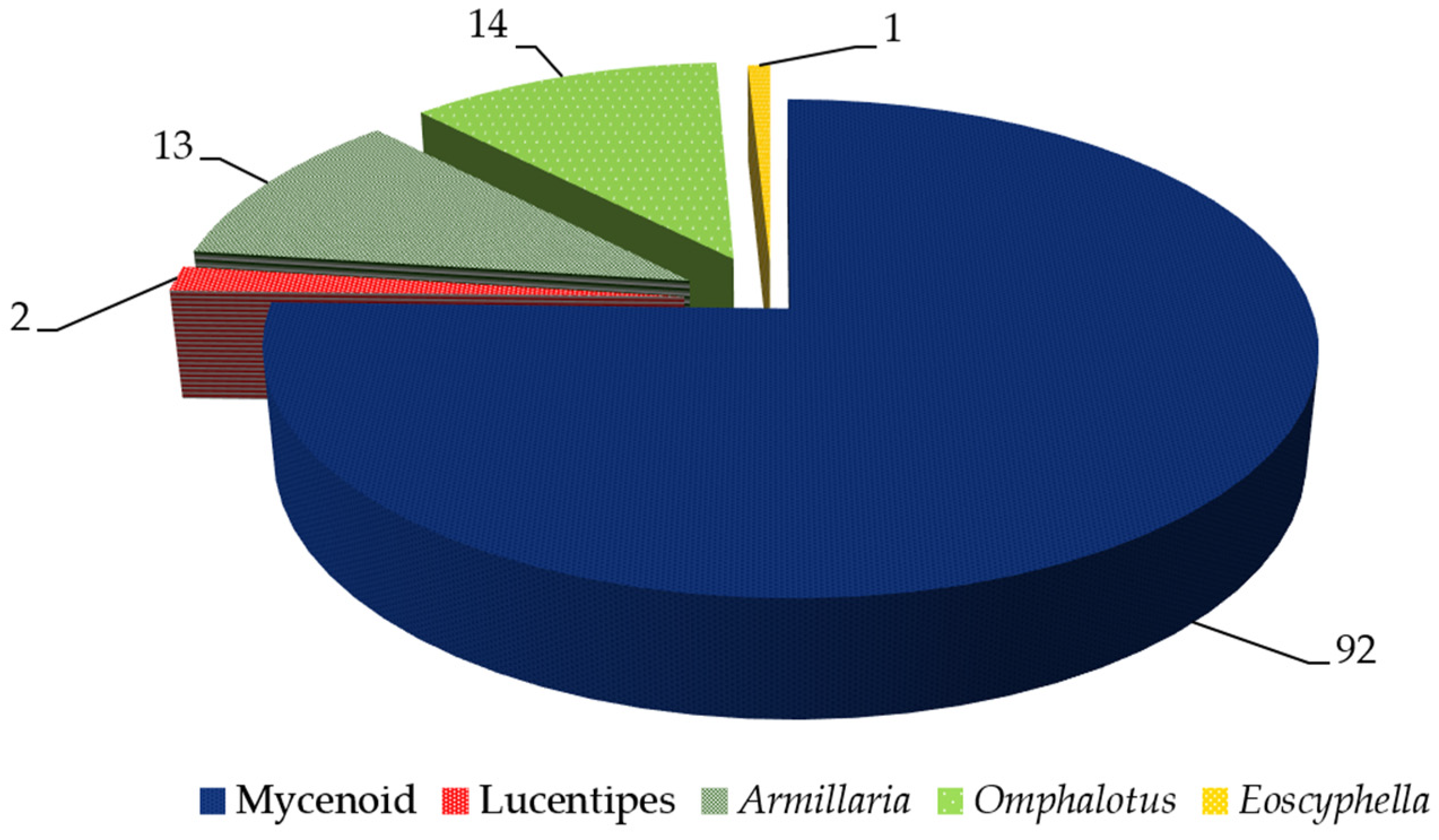
All bioluminescent fungi records belong to the Basidiomycota division except for Xylaria hypoxylon (L.) Grev., which falls under the Ascomycota [56,74]. It is worth noting that despite analyses of multiple specimens of Xylaria hypoxylon, differing conclusions have been drawn regarding its bioluminescence [73]; Ludwig and Gueguen reported the detection of bioluminescence in X. hypoxylon, whereas Molisch cultivated pure cultures for four years without observing any bioluminescent properties (for more information see [73]). This conclusion may be attributed to variations in geographical distribution and cultivation conditions. Therefore, further investigation is warranted to determine its luminescent properties conclusively [73]. Figure 2 shows five lineages that comprise all known species of bioluminescent fungi. They are part of the Mycenoid (92 species), Armillaria (13 species), Omphalotus (14 species), Lucentipes (two species), and Eoscyphella (one species) lineages [47].
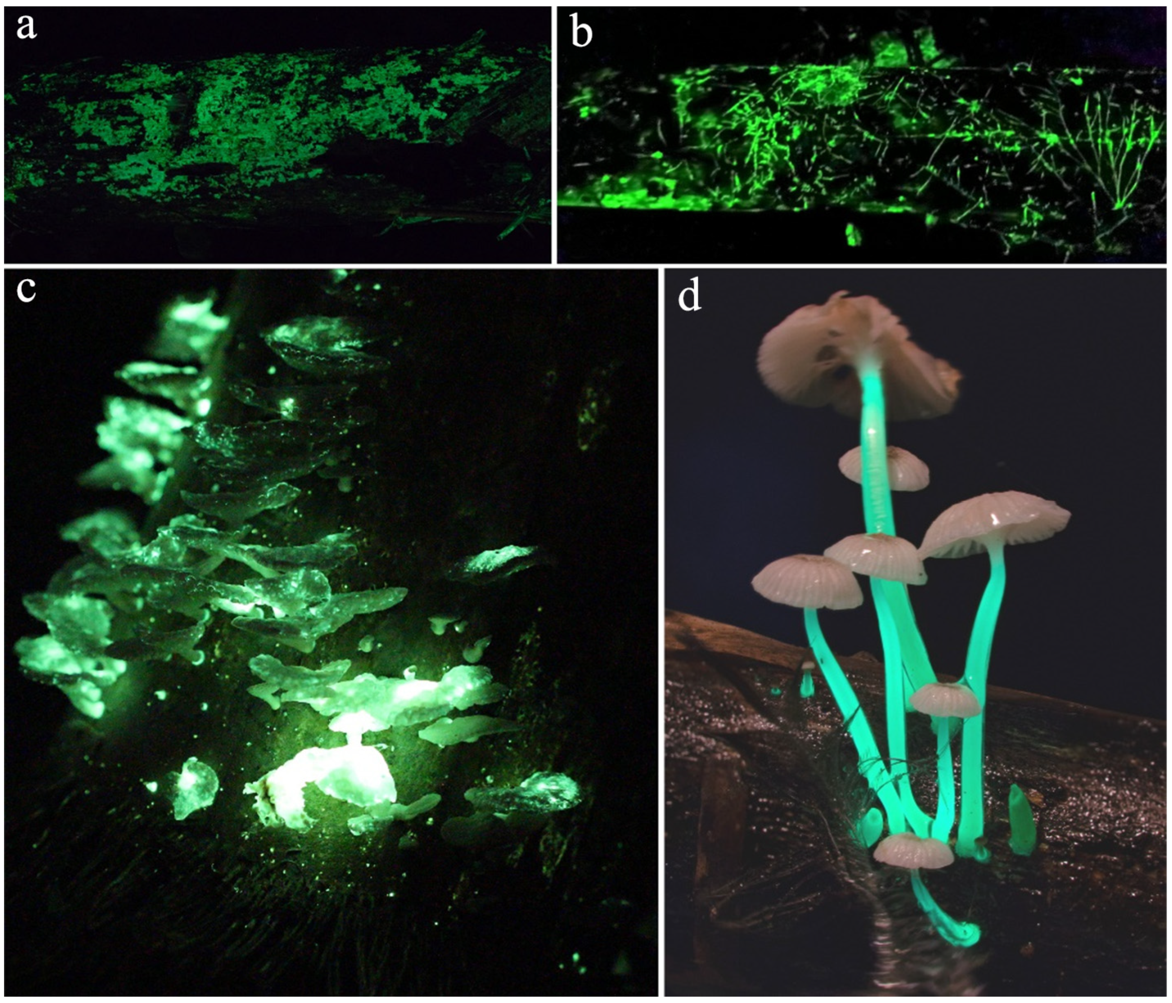
Mycena is the main genus that exhibits bioluminescence in fungi and is distributed worldwide [41,42,75]. Furthermore, accounting for the species level, for example, Mycena chlorophos, M. inclinata, and Neonothopanus nambi show a wide distribution worldwide [4,56,76]. Meanwhile, species like Favolaschia xtbgensis and Roridomyces viridiluminus show a restricted habitat, particularly in some places in Southwestern China (Figure 3) [48,69].
Bioluminescent fungi are not confined to a single region; their distribution spans the globe. For instance, Gerronema viridilucens was reported only in Brazil [77], and Neonothopanus gardneri from the states of Maranhão, Piauí, and Tocantins in Brazil [78]. Interestingly, some species, like Pannellus stipticus, are naturally found in different countries, showcasing the global nature of bioluminescent fungi. However, they did not show bioluminescence in all the recorded places. For instance, P. stipticus shows bioluminescence grown in North America but not in Eurasian [79]. In addition, Armillaria mellea, Mycena chlorophos, M. deeptha, Nothopanus eugrammus, Omphalotus olearius, O. olivascens, and Roridomyces cf. phyllostachydis have been reported from India [22,59,80,81]. In contrast, Filoboletus manipularis, Mycena chlorophos, and Panellus luxfilamentus have been found in Sri Lanka [56].
In an early study, Desjardin et al. [42] presented a revised list of bioluminescent fungi with 64 species in their distribution. Desjardin et al. [56] reported seven new luminescent fungi species two years after publication. Later, Aravindakshan et al. [59] and Shih et al. [82] reported two additional novel species from India and the Taiwan Province of China, respectively. In addition, Chew et al. [53,56] disclosed 15 bioluminescent fungi from Peninsular Malaysia, where eight were reported for the first time. Mihail [41] detected the bioluminescent mycelia of five Armillaria species for the first time.
In a most recent study, Terashima et al. [50] identified another eight new species of glowing mushrooms from Japan, thus bringing the country’s total reported number up to 25 [14]. Seven species of bioluminescent fungi were recorded from the cloud forests in Mexico, where six species have been identified as a new species of Mycena, whereas M. globulispora made a new distribution record for the country [61]. Furthermore, several other new species have been reported from the Taiwan Province (M. jingyinga, M. luguensis and M. venus) and Yunnan Province of China (Favolaschia xtbgensis and Roridomyces viridiluminus) and India (R. phyllostachydis) [22,34,48,69]. All known bioluminescent mushrooms form gilled or poroid basidiomata within the order Agaricales. However, the latest bioluminescent species Eoscyphella luciurceolata represents a new lineage with a reduced cyphelloid form [47]. The discovery of new bioluminescent cyphelloid basidiomata challenges existing biological classification systems and deepens this study’s understanding of bioluminescent diversity within the fungal kingdom. There is a wealth of undiscovered species, particularly in unexplored ecosystems such as forest floors, tropical regions, and polar areas, where diverse bioluminescent mushroom species may exist and represent a biodiversity hotspot for these organisms [47]. These discoveries have the potential to significantly enhance our understanding of bioluminescence mechanisms, evolutionary adaptations, and contributions to ecosystem stability.

3. Evolution and Mechanisms of Bioluminescent Fungi

3.1. Evolution

Understanding the development of bioluminescence in fungi is a challenging and fascinating subject. Bioluminescence is most likely the result of ancient beginnings, convergent evolution, and potentially horizontal gene transfer [83]. Numerous fungal lineages have distinctive characteristics that have recently been investigated concerning the genetic and environmental factors that have influenced them [83]. Nonetheless, two main hypotheses have been proposed to explain the scattered phylogenetic distribution and lesser occurrence of bioluminescence in fungi. These hypotheses can be summarized as the genes associated with bioluminescence shared by the common ancestor were missing in some branches and multiple convergent evolutions of bioluminescence in fungi [2]. Like other bioluminescent organisms, bioluminescent fungi have independent evolutionary occurrences, converging multiple times. Genomic analysis shows that this fragmented phylogenetic position may be another case. These findings are significant as they contribute to our understanding of the evolutionary origins and genetic mechanisms of bioluminescence in fungi, advancing mycology.
Oliveira et al. [21] uncovered a significant revelation in their research, suggesting that the origin of fungal bioluminescence can be traced back to a single evolutionary ancestry. Their evidence, demonstrated by successful light production from cross-reactions between the luciferins and luciferases of distant lineages, sheds new light on this fascinating phenomenon.
Recent studies by Kotlobay et al. [33] and Ke et al. [83] have reached a consensus, concluding that bioluminescence in fungi can be traced back to the last common ancestor of the Mycenoid and Marasmioid clades of Agaricales. This consensus echoed in recent surveys by Ke and colleagues [84], provides a solid foundation for our understanding of fungal bioluminescence.
Ke et al. [83] revisited the evolutionary dynamics of the luciferase cluster previously studied by Kotlobay et al. [33] and noted that the ancestral luciferase cluster on the same chromosome contains the genes luciferase (Luz), hispidin-3-hydroxylase (H3H), cytochrome P450 (CYP450), hispidin synthase (HispS), and caffeylpyruvate hydrolase (CPH). Their study corroborates and extends upon earlier findings, providing additional insights into the genes’ genomic organization and evolutionary history.
Further studies revealed that gene clusters frequently undergo either deletions or retention due to differences in genomic plasticity, which explains the frequent loss of the bioluminescence property of Mycenaean fungi [2,83,85]. The conservation of the gene cluster during the process of evolution signifies that, unlike other groups of bioluminescent organisms, bioluminescence evolves once in fungi with Luz, H3H, and HispS genes generated through gene duplications [33]. Moreover, the species phylogenetic tree and reconstructed phylogenetic trees of Luz, H3H, and HispS genes of the family Agaricaceae reveal the evolution of bioluminescent cascades in fungi. The primary Luz enzyme formed through a gene duplication at the base of Agaricales, followed by the duplication of H3H and HipS a few million years later [33]. These findings open up new avenues for future research, particularly in understanding the genetic mechanisms and evolutionary origins of bioluminescence in fungi.

3.2. Mechanisms

As previously mentioned, bioluminescent fungi, including mushrooms, have been discovered worldwide in a wide range of terrestrial environments; nevertheless, fungal bioluminescence mechanisms remain the least studied [86]. In general, bioluminescence occurs through the chemical oxidation of luciferin, catalyzed by the luciferase enzyme in the presence of oxygen [87,88]. The molecular oxygen reacts with luciferin, forming a high-energy intermediary whose decomposition emits sufficient energy to generate the emitter ‘oxyluciferin’ in the singlet, which is electronically excited. This excited metabolite’s fluorescence property results in the emission of visible light used in nature for illumination [2,35,89].
First, it is interesting to understand how this bioluminescence mechanism has been revealed throughout history. In an early study, Dubois [90] used an in vitro luciferin/luciferase system with a mixture of heated substrate and cold enzyme–water extracts to demonstrate the first light emission experiment. In his experiment, he utilized extracts (cold and hot) from the light-emitting organs of the beetle Pyrophorus noctilucus. In this experimental setup, the cold extract process contained a heat-labile enzyme called luciferase that was needed to emit light. The hot extract was the thermo-stable fraction that was named luciferin. Further, it was determined that the luminescence of the mixture formed with two extracts resulted from the substrate/enzyme reaction [78].
In a study that supported Dubois [90], Airth and McElroy [91] used an in vitro setup made up of cold and hot extracts from bioluminescent fungi to confirm the role and nature of the enzymatic reaction. Later experiments by Airth and Foerster [92] explained that adding DPNH (the obsolete name for NADH) or NADPH to the cold and hot extracts activates the light emission. Furthermore, the proteinaceous cold extracts could be separated into two fractions, a pellet (insoluble) and a supernatant (soluble), by ultracentrifugation, which is necessary for the light emission in luminous fungi. Thus, the essential enzymes in each fraction for light generation postulated a two-step mechanism of enzymatic reaction [92]. Note that basidiomycetous fungi emit a green light with a maximum intensity in the 520–530 nm range [42]. Returning to Airth and Foerster’s study [92], they proposed the following two-step mechanism for fungal bioluminescence:
Luciferin + NAD ( P ) H + H +   R e d u c t a s e Reduced   luciferin + NAD ( P ) +
Reduced   luciferin + O 2   L u c i f e r a s e Oxyluciferin + H 2 O + Hv   ( light )
In the first step, luciferin, a molecule that acts as an electron acceptor, is involved in the process. Here, reductase, an enzyme that catalyzes the reduction of other substances, is present in the liquid part of the mixture (supernatant). In contrast, luciferase, an enzyme that catalyzes the oxidation of luciferin, is found in the solid part (pellet). During the first step, a dark chemical reaction that does not produce a visible change occurs between NAD(P)H, a coenzyme involved in cellular respiration, luciferin, and the soluble enzyme in the supernatant. In the second step, the reduced form of luciferin reacts with molecular oxygen, catalyzed by the enzyme (luciferase) in the re-suspended pellet, producing visible light [92].
These initial findings led to understanding the chemistry behind this scenario; however, along with the development of technologies, more questions were raised, such as the in-depth aspects of the specific roles of the enzymes in bioluminescence [31,42,93]. Oliveira and Stevani [31] attempted to find the answer using an enzyme-mediated reaction by mixing a hot extract containing heat-stable substrate/luciferin with a cold extract containing the enzyme luciferase. Later, it was demonstrated that the substrates combined with the enzymes extracted from the mycelia of different bioluminescent species (Armillaria mellea, Gerronema viridilucens, Mycena luxperpetua, and Neonothopanus gardneri), and these results strongly suggest that all known bioluminescent fungi share similar types of luciferins/luciferases in bioluminescent systems [21]. Purtov et al. [94] identified the structure of fungal luciferin and its precursor as 3-hydroxyhispidin and hispidin in extracts from four diverse genera of bioluminescent fungi, namely, Armillaria borealis, Mycena citricolor, Neonothopanus nambi, and Panellus stipticus. Kaskova et al. [32] conducted an in-depth study of the mechanisms of fungal bioluminescence and color modulation and reported the structure of fungal oxyluciferin to investigate the mechanism of fungal bioluminescence. Hispidin is produced through the enzymatic activity of HispS. Subsequently, the resulting hispidin undergoes hydroxylation mediated by H3H, leading to the formation of 3-hydroxyhispidin, also known as fungal luciferin. Then, it is oxidized by O2, generating a high-energy intermediary that decomposes in CO2 and the excited oxyluciferin. Light emission produces the ground-state oxyluciferin and hydrolyzes enzymatically into caffeic acid [32].
Kotlobay et al. [33] identified the fungal Luz and three other key enzymes, HispS, H3H, and CPH, in Neonothopanus nambi that jointly form the biosynthetic cycle of the fungal luciferin from caffeic acid. Fungal luciferin can be biosynthesized and recycled within this proposed mechanism. Caffeic acid is transformed to hispidin owing to HispS activity and is hydroxylated by H3H, producing 3-hydroxyhispidin fungal luciferin. The luciferase adds molecular oxygen, producing an endoperoxide (a high-energy intermediate) through decomposition that produces oxyluciferin (caffeoyl pyruvate) and light. Oxyluciferin can be recycled to caffeic acid by CPH [33].
In a study, Wang and Liu [95] revealed cross-reactions among four lineages of luminescent fungi, indicating that they shared a common bioluminescence mechanism, and described the bioluminescence process at the molecular level and electronic state by using multireference and density functional theory calculations. The findings revealed that fungal bioluminescence began with the cycloaddition of O2 to luciferin and that formed a high-energy intermediate called α-pyrone endoperoxide. This oxygenation can be explained by a charge transfer followed by a spin inversion mechanism. The high-energy intermediate thermolysis produces S1-Oxyluciferin (S1-singlet excited state) via a zwitterion intermediate. De-excitation of S1-Oxyluciferin can be a light emitter [94]. Nevertheless, according to Ke et al. [83], the complete cluster of genes involved in the bioluminescence process is still unknown to science.
In fungi, the genes that code for the enzymes that produce secondary metabolites are frequently grouped in the fungus genome [96]. In most bioluminescent fungi (e.g., Armillaria fuscipes, A. mellea, A. ostoyae, A. gallica, Mycena citricolor, M. chlorophos, Neonothopanus nambi, N. gardneri, Omphalotus olearius, and Panellus stipticus), it has been demonstrated that the aforesaid genes are generally found to be located adjacent to each other forming a cluster [33]. Rokas et al. [97] also revealed that genes in the primary and secondary metabolic pathways of fungi are often physically connected on fungal chromosomes, creating metabolic gene clusters, thus hypothesizing that this might be the reason for the formation of enzymes in the bioluminescent cascade, as this is thought to be conserved among bioluminescent fungi. In addition to the four key genes (Luz, H3H, HispH, and CPH) coding for four enzymes, CYP450 is inside the cluster in all bioluminescent Armillaria and Mycena genomes [83]. Thus, there is controversy about the involvement of other enzymes or regulators in the bioluminescence process. On the other hand, few studies have reported the possible involvement of other genes in fungus bioluminescence [98]. In another study, Oliveira et al. [99] investigated the circadian rhythm in Neonothopanus gardneri. They found that the bioluminescence of the mycelium is controlled by a temperature-compensated circadian clock and the result of cycles in content/activity between the luciferase, reductase, and luciferin that comprise the bioluminescence system. Ke et al. [83] determined the regulation of bioluminescence in Mycena kentingensis during its development. They identified 57 gene-bearing expressions correlated to Luz, H3H, and HispS, agreeing with the bioluminescence mechanism discussed by Kotlobay et al. [33].

4. Importance of Bioluminescent Fungi in Ecology

The ecological importance and the underlying phenomenon of bioluminescent fungi continue to be a subject of intense debate and exploration among researchers. There are several proposed hypotheses, including dispersed spores by attracting phototactic insects, deterring negative phototrophic fungivores, and potentially aposematic signals [4,100], which are a testament to the complexity of these organisms. Initially, Sivinski [101] proposed that bioluminescence in fungi serves as a warning signal to repel nocturnal fungivores or as an attractant for fungivore predators. However, Sivinski’s theories challenge hypotheses suggesting that animals are primarily attracted to the aroma or odor of fungal fruiting bodies rather than their bioluminescent properties. Electroretinography is suggested to clarify whether invertebrates are attracted explicitly to bioluminescent fungi due to emitted light, although such studies are still on the bench [64].
Bioluminescent fungi have also been studied for their ability to attract insects at night for spore dispersal. Research on Neonothopanus gardneri has shown that the bioluminescence mechanism is regulated by circadian rhythms, involving cycles in luciferase, reductase, and luciferin activity [4,99]. This conclusion is further strengthened as researchers found that beetles bathe with the fruiting bodies of N. gardneri [100]. In a recent study, Karunarathna et al. [22] explained that the members of Roridomyces inhabiting humid environments co-evolved with some insects aiding spore dispersal. Bechara [102] demonstrated that insects such as ants, beetles, flies, wasps, and bugs are attracted to the green light emitted by bioluminescent fungi, facilitating nocturnal spore transfer in forests with minimal wind and high humidity. However, this hypothesis is challenged in species where bioluminescence emanates only from the stipe or mycelium [69,84]. Furthermore, some bioluminescent fungi may utilize their light as a warning signal to deter potential predators, signaling the presence of toxins or unpalatability [69,102].
Conversely, some view bioluminescence in fungi as a mere metabolic by-product devoid of ecological benefits [103,104]. Weinstein et al. [103] indicated that bioluminescence in fungi, exemplified by Omphalotus nidiformis, is a metabolic by-product without evident selective advantage. They suggest that the role of bioluminescence may vary among fungal lineages and environmental conditions affecting spore dispersal dynamics, such as wind patterns and insect abundance. However, the potential evolutionary advantages of bioluminescence in fungi continue to intrigue researchers, driving them to seek answers to why fungi exhibit bioluminescence. They also explore its multifaceted roles and ecological significance in different fungal lineages and environmental contexts [14,103].

5. Application of Fungal Bioluminescence

Fungal bioluminescence carries both historical significance and contemporary scientific potential. As aforementioned, folk stories and historical reports show that different tribes or local people, particularly in India and Indonesia, use glowing mushrooms to find their way through the dense forests [100,105,106]. In contrast, Aboriginal people in Australia consider glowing mushrooms related to the spirit [107]. In modern scientific contexts, bioluminescence has revolutionized plant biology and inspired experiments and research in biochemistry, cell biology, evolution, and photochemistry. Bioluminescence is also applied in scientific research, including several aspects such as biological sensors in environmental monitoring, effectors, hygiene control, preservation of artworks, gene assays, the detection of protein-protein interactions, bioluminescence-based imaging and photodynamic therapy, neuron treatments, and high-throughput screening in drug discovery [6,108,109].
Intriguingly, scientists are now looking for ways to switch to green light instead of light generated through electricity [100]. Recent achievements include the genetic engineering of tobacco plants (Nicotiana tabacum and N. benthamiana, see [35]) to autonomously emit light through the conversion of caffeic acid into luciferin, enabling applications in environmental assessments and potentially auto-luminescent plants [89]. The successful expression of fungal bioluminescence has also been reported in Arabidopsis thaliana, Catharanthus roseus, Dahlia pinnata, Petunia hybrida, Rosa rubiginosa, and Solanum lycopersicum [89,110,111].
Environmental bioassays can be performed using fungi’s natural bioluminescent enzyme reaction [78]. Eukaryotic bioluminescent fungi are a more suitable research organism for soil toxicology than luminescent bacteria. However, the mechanism of toxicity and its specific impact on the fungal bioluminescence response is not yet fully understood. The uncoupling of oxidative phosphorylation and the depolarization of mitochondrial membranes by toxic compounds can be possible pathways; they would possibly indirectly affect the NADH availability that is involved in the bioluminescent reaction [78]. Additionally, it is still feasible to use organisms in a terrestrial setting, including bioluminescent fungi, without further engineering marker labeling [2], even if known uses include marker-labeled bacteria and marine bioluminescent Vibrio species [112,113]. Since 2002, toxicity tests have been developed using bioluminescent fungi, particularly with Armillaria mellea and Mycena citricolor. These studies were based on globular mycelia grown on liquid media with varying concentrations of heavy metals (copper and zinc) or chemical compounds (chlorophenol). Several bioassays with bioluminescent basidiomycetes have been developed using Pannellus stipticus and Omphalotus olearius [78]. More recently, the toxicity of the other bioluminescent lineages of Gerronema viridilucens and N. gardneri was assessed [114,115]. Bioluminescence technology plays a pivotal role in in vivo bioimaging, broadly applied in the study of diseases and assessing therapeutic interventions in animal models. Researchers can monitor and analyze dynamic biological processes like tumor progression and inflammatory responses in real-time by employing bioluminescent markers such as the luciferase system. This capability facilitates the evaluation of treatment efficacy and safety profiles. The noninvasive nature, high spatiotemporal resolution, and ability to quantify drug distribution and metabolism make bioluminescence imaging indispensable for personalized medicine and innovative drug discovery [116,117,118]. Beyond advancing the frontiers of biomedical research, bioluminescence imaging accelerates the refinement and development of therapeutic strategies.
On the one hand, the reconstruction of the fungal bioluminescence pathway in an organism, making it autonomous luminescence, is beneficial for detecting the status of its various growth stages. It could also facilitate the development of the next generation of organic architecture, modified light-emitting plants in buildings, and urban infrastructure [33]. On the other hand, fungal bioluminescence can be indicated in agriculture when crops need water or nutrients. Due to this autonomous bioluminescence, plants can warn early about illnesses and pest attacks that could harm harvests. Furthermore, bioluminescence paves the path for eco-friendly house/street lighting, health applications, and food industries [100,105,106]. The alterations of these technologies will drive massive growth in bioluminescence in the coming future [75]. Bioluminescent mushrooms offer significant potential in both horticulture and tourism. In horticultural landscape design, they create a unique nighttime ambiance, enhancing the visual appeal of gardens through their natural glow in flowerbeds, pathways, and lawns. These mushrooms also provide educational opportunities by providing practical examples of bioluminescence in horticultural institutions. In tourism, they serve as distinctive attractions, potentially forming mushroom gardens or designated viewing areas and contributing to local economies. Incorporating bioluminescent mushrooms into eco-tourism activities, such as nocturnal ecological tours, offers visitors novel nature interactions while promoting an understanding of biodiversity and ecosystems. Overall, bioluminescent mushrooms enrich aesthetic and educational aspects and present promising opportunities for tourism development. Bioluminescent mushrooms hold tremendous promise for both horticulture and tourism. In horticultural design, these mushrooms create a magical nighttime atmosphere, adding a natural glow that enhances the beauty of gardens, whether nestled in flowerbeds, lining pathways, or dotting lawns. Beyond their aesthetic value, they also serve as practical tools in educational settings, vividly illustrating the wonders of bioluminescence in horticulture. In the realm of tourism, bioluminescent mushrooms become unique attractions, capable of forming enchanting mushroom gardens or designated viewing spots that captivate visitors and potentially bolster local economies. Integrating these mushrooms into eco-tourism initiatives, such as nocturnal ecological tours, offers guests an extraordinary opportunity to engage with nature in new and profound ways while fostering a deeper appreciation for biodiversity and ecosystems. Ultimately, bioluminescent mushrooms enrich the visual and educational dimensions and open exciting avenues for developing tourism experiences.

6. Conclusions and Future Directions

According to our understanding, 122 species of bioluminescent fungi have been reported. These fungi are primarily categorized within the Basidiomycota, distributed across four established phylogenetic lineages (Armillaria, Lucentipes, Mycenoid, and Omphalotus), with the recent addition of a novel fifth lineage, Eoscyphella. Despite phylogenetic diversity, all bioluminescent fungi share conserved bioluminescent mechanisms rooted in luciferin oxidation catalyzed by luciferase, which involves two enzymatic steps. However, detailed studies elucidating the genetic regulation of this process remain a critical area for future investigation. Recent advancements highlight the possible diverse applications of fungal bioluminescence. Bioluminescent fungi hold immense promise across a wide spectrum of disciplines, including ecology, agriculture, art, medicine, and education. They offer potential as bioindicators for environmental monitoring and innovative strategies for crop health management through genetic modification. Furthermore, their aesthetic allure has inspired creative designs such as luminous gardens and interactive art installations. In medicine, these fungi offer exciting avenues for developing new therapeutic agents and diagnostic tools. In summary, realizing the full potential of bioluminescent fungi necessitates ongoing interdisciplinary collaboration. However, it is crucial to stress that continued research efforts are needed to expand the understanding of fungal biodiversity, ecological roles, and practical applications in various fields as the field constantly evolves.

Author Contributions

S.C.K. and S.T. conceptualized the topic; W.L. and A.K.H.P. wrote the first draft of the manuscript; M.C.A.G., N.M.P., A.N.E., B.M.P. and D.M.K. helped with the literature searches and drafted the chemistry part of the manuscript; T.S.N. prepared the map; S.T., S.-C.S., D.-Q.D., A.K.H.P., A.T., N.S., S.L. and S.C.K. edited the manuscript; S.T. and S.C.K. supervised the work; D.-Q.D., N.S., S.L., S.-C.S. and S.C.K. provided funding for the research. All authors have read and agreed to the published version of the manuscript.

Funding

This work was supported by the National Natural Science Foundation of China (Grant Nos. 32260004, 31760013, and 32171655), the West Light Talent Program of the Chinese Academy of Sciences (Grant No. E1XB011B01), and Chiang Mai University and Kaomai Lanna Res., Thailand (Grant No. R66IN00132). Samantha C. Karunarathna, Dong-Qin Dai, and Saowaluck Tibpromma thank the High-Level Talent Recruitment Plan of Yunnan Province (“High-End Foreign Expert” and “Young Talents” Programs) for their support.

Data Availability Statement

The data that support this study are available on request.

Acknowledgments

The authors thank the Key Laboratory of the Yunnan Provincial Department of Education of the Deep-Time Evolution on Biodiversity from the Origin of the Pearl River, for their support.

Conflicts of Interest

The authors declare no conflicts of interest.

References

  1. Pandey, A.; Sharon, M. Bioluminescent organisms. BAOJ Chem. 2017, 3, e029. [Google Scholar]
  2. Ke, H.M.; Tsai, I.J. Understanding and using fungal bioluminescence-Recent progress and future perspectives. Curr. Opin. Green Sustain. Chem. 2022, 33, 100570. [Google Scholar] [CrossRef]
  3. Waldenmaier, H.E.; Oliveira, A.G.; Stevani, C.V. Thoughts on the diversity of convergent evolution of bioluminescence on earth. Int. J. Astrobiol. 2012, 11, 335–343. [Google Scholar] [CrossRef]
  4. Vieira, M.B.B.; Oliveira, I.C.; de Oliveira, M.D.D.A.; da Costa Júnior, J.S.; dos Santos, T.D.J.A.; Feitosa, C.M.; Rai, M.; Lima, N.M.; da Costa Silva, D. A review on bioluminescent fungus Neonothopanus gardneri. Res. Soc. Dev. 2022, 11, e16811528009. [Google Scholar] [CrossRef]
  5. Lau, E.S.; Oakley, T.H. Multi-level convergence of complex traits and the evolution of bioluminescence. Biol. Rev. Camb. Philos. Soc. 2020, 96, 673–691. [Google Scholar] [CrossRef] [PubMed]
  6. Syed, A.J.; Anderson, J.C. Applications of bioluminescence in biotechnology and beyond. Chem. Soc. Rev. 2021, 50, 5668–5705. [Google Scholar] [CrossRef] [PubMed]
  7. Delroisse, J.; Duchatelet, L.; Flammang, P.; Mallefet, J. Leaving the dark side? Insights into the evolution of luciferases. Front. Mar. Sci. 2021, 8, 673620. [Google Scholar] [CrossRef]
  8. Harvey, E.N. Bioluminescence; Academic Press: New York, NY, USA, 1952; pp. 1–649. [Google Scholar]
  9. Harvey, E.N. A History of Luminescence: From the Earliest Times until 1900; The American Philosophical Society: Philadelphia, PA, USA, 1957; pp. 1–692. [Google Scholar] [CrossRef]
  10. Lee, S.M.L. The status of bioluminescent fungal species in Singapore. Nat. Singap. 2022, 1, e2022124. [Google Scholar] [CrossRef]
  11. Mahish, P.K.; Chandrawanshi, N.K.; Kunjam, S.; Jadhav, S.K. Opportunities in the living lights: Special reference to bioluminescent fungi. Energy Cris. Chall. Solut. 2021, 10, 191–207. [Google Scholar]
  12. Lu, D. 71 species of macrofungi that bioluminescence. Edible Med. Mushrooms 2011, 19, 55–57. (In Chinese) [Google Scholar]
  13. Boyle, R. Observations and tryals about the resemblances and differences between a burning coal and shining wood. Phil. Trans. 1667, 2, 605–612. [Google Scholar] [CrossRef]
  14. Oba, Y.; Hosaka, K. The luminous fungi of Japan. J. Fungi 2023, 9, 615. [Google Scholar] [CrossRef] [PubMed]
  15. Kirk, P.M. World catalogue of 340K fungal names on-line. Mycol. Res. 2000, 104, 516–517. [Google Scholar]
  16. Crous, P.W.; Gams, W.; Stalpers, J.A.; Robert, V.; Stegehuis, G. MycoBank: An online initiative to launch mycology into the 21st century. Stud. Mycol. 2004, 50, 19–22. [Google Scholar]
  17. Taylor, J.W. One Fungus = One Name: DNA and fungal nomenclature twenty years after PCR. IMA Fungus 2011, 2, 113–120. [Google Scholar] [CrossRef] [PubMed]
  18. Schoch, C.L.; Seifert, K.A.; Huhndorf, S.; Robert, V.; Spouge, J.L.; Levesque, C.A.; Chen, W.; Fungal Barcoding Consortium; Fungal Barcoding Consortium Author List; Bolchacova, E.; et al. Nuclear ribosomal internal transcribed spacer (ITS) region as a universal DNA barcode marker for Fungi. Proc. Natl. Acad. Sci. USA 2012, 109, 6241–6246. [Google Scholar] [CrossRef] [PubMed]
  19. Schoch, C.L.; Robbertse, B.; Robert, V.; Vu, D.; Cardinali, G.; Irinyi, L.; Meyer, W.; Nilsson, R.H.; Hughes, K.; Miller, A.N.; et al. Finding needles in haystacks: Linking scientific names, reference specimens and molecular data for Fungi. Database 2014, 2014, bau061. [Google Scholar] [CrossRef] [PubMed]
  20. McNeill, J.; Barrie, F.R.; Buck, W.R.; Demoulin, V.; Greuter, W.; Hawksworth, D.L.; Herendeen, P.S.; Knapp, S.; Marhold, K.; Prado, J.; et al. International Code of Nomenclature for algae, fungi, and plants. Regnum Veg. 2012, 154. Available online: http://www.iapt-taxon.org/nomen/main.php (accessed on 25 July 2024).
  21. Oliveira, A.G.; Desjardin, D.E.; Perry, B.A.; Stevani, C.V. Evidence that a single bioluminescent system is shared by all known bioluminescent fungal lineages. Photochem. Photobiol. Sci. 2012, 11, 848–852. [Google Scholar] [CrossRef] [PubMed]
  22. Karunarathna, S.C.; Mortimer, P.E.; Tibpromma, S.; Dutta, A.K.; Paloi, S.; Hu, Y.; Baurah, G.; Axford, S.; Marciniak, C.; Luangharn, T.; et al. Roridomyces phyllostachydis (Agaricales, Mycenaceae), a new bioluminescent fungus from Northeast India. Phytotaxa 2020, 459, 155–167. [Google Scholar] [CrossRef]
  23. Cortés-Pérez, A.; Guzmán-Dávalos, L.; Ramírez-Cruz, V.; Villalobos-Arámbula, A.R.; Ruiz-Sanchez, E.; Ramírez-Guillén, F. New Species of Bioluminescent Mycena Sect. Calodontes (Agaricales, Mycenaceae) from Mexico. J. Fungi 2023, 9, 902. [Google Scholar] [CrossRef]
  24. Labella, A.M.; Arahal, D.R.; Castro, D.; Lemos, M.L.; Borrego, J.J. Revisiting the genus Photobacterium: Taxonomy, ecology and pathogenesis. Int. Microbiol. 2017, 20, 1–10. [Google Scholar]
  25. Burtseva, O.; Kublanovskaya, A.; Baulina, O.; Fedorenko, T.; Lobakova, E.; Chekanov, K. The strains of bioluminescent bacteria isolated from the White Sea finfishes: Genera Photobacterium, Aliivibrio, Vibrio, Shewanella, and first luminous Kosakonia. J. Photoch. Photobio. B 2020, 208, 111895. [Google Scholar] [CrossRef]
  26. de Busserolles, F.; Marshall, N.J. Seeing in the deep-sea: Visual adaptations in lanternfishes. Philos. Trans. R. Soc. Lond. B Biol. Sci. 2017, 372, 20160070. [Google Scholar] [CrossRef] [PubMed]
  27. Al-Handawi, M.B.; Polavaram, S.; Kurlevskaya, A.; Commins, P.; Schramm, S.; Carrasco-López, C.; Lui, N.M.; Solntsev, K.M.; Laptenok, S.P.; Navizet, I.; et al. Spectrochemistry of firefly bioluminescence. Chem. Rev. 2022, 122, 13207–13234. [Google Scholar] [CrossRef]
  28. Owens, A.; Van den Broeck, M.; De Cock, R.; Lewis, S.M. Behavioral responses of bioluminescent fireflies to artificial light at night. Front. Ecol. Evol. 2022, 10, 946640. [Google Scholar] [CrossRef]
  29. Elkhateeb, W.A.; Daba, G.M. Bioluminescent Mushrooms: Boon for environmental health. Environ. Sci. Arch. 2022, 1, 88–97. Available online: https://www.envsciarch.com/_files/ugd/4b6a78_9155754cbd4e4412b6bab4385f8ef2e1.pdf (accessed on 14 October 2022).
  30. Adams, C.A.; Donald, M.L.; Swearingen, C.; Escudero, E.; Sourell, S.; Landrein, S.; Seas, C.; Mueller, G.; Chaverri, P. Let there be nightlights: The ecological role of bioluminescence in a Costa Rican mushroom. bioRxiv 2023. [Google Scholar] [CrossRef]
  31. Oliveira, A.G.; Stevani, C.V. The enzymatic nature of fungal bioluminescence. Photochem. Photobiol. Sci. 2009, 8, 1416. [Google Scholar] [CrossRef] [PubMed]
  32. Kaskova, Z.M.; Dörr, F.A.; Petushkov, V.N.; Purtov, K.V.; Tsarkova, A.S.; Rodionova, N.S.; Mineev, K.S.; Guglya, E.B.; Kotlobay, A.; Baleeva, N.S.; et al. Mechanism and color modulation of fungal bioluminescence. Sci. Adv. 2017, 3, e1602847. [Google Scholar] [CrossRef]
  33. Kotlobay, A.A.; Sarkisyan, K.S.; Mokrushina, Y.A.; Marcet-Houben, M.; Serebrovskaya, E.O.; Markina, N.M.; Gonzalez, S.L.; Gorokhovatsky, A.Y.; Vvedensky, A.; Purtov, K.V.; et al. Genetically encodable bioluminescent system from fungi. Proc. Natl. Acad. Sci. USA 2018, 115, 12728–12732. [Google Scholar] [CrossRef] [PubMed]
  34. Chang, C.C.; Chen, C.Y.; Li, W.W.; Ka, W. Mycena jingyinga, Mycena luguensis, and Mycena venus: Three new species of bioluminescent fungi from Taiwan. Taiwania 2020, 65, 396–406. Available online: https://taiwania.ntu.edu.tw/pdf/tai.2020.65.396.pdf (accessed on 27 July 2020).
  35. Mitiouchkina, T.; Mishin, A.S.; Somermeyer, L.G.; Markina, N.M.; Chepurnyh, T.V.; Guglya, E.B.; Karataeva, T.A.; Palkina, K.A.; Shakhova, E.S.; Fakhranurova, L.I.; et al. Plants with genetically encoded autoluminescence. Nat. Biotechnol. 2020, 38, 944–946. [Google Scholar] [CrossRef] [PubMed]
  36. Niskanen, T.; Lücking, R.; Dahlberg, A.; Gaya, E.; Suz, L.M.; Mikryukov, V.; Liimatainen, K.; Druzhinina, I.; Westrip, J.R.; Mueller, G.M.; et al. Pushing the frontiers of biodiversity research: Unveiling the global diversity, distribution, and conservation of fungi. Annu. Rev. Environ. Resour. 2023, 48, 149–176. [Google Scholar] [CrossRef]
  37. Rashhmi, M.; Kushveer, J.S.; Sarma, V.V. A worldwide list of endophytic fungi with notes on ecology and diversity. Mycosphere 2019, 10, 798–1079. [Google Scholar] [CrossRef]
  38. Van Der Heijden, M.G.; Martin, F.M.; Selosse, M.A.; Sanders, I.R. Mycorrhizal ecology, and evolution: The past, the present, and the future. New Phytol. 2015, 205, 1406–1423. [Google Scholar] [CrossRef] [PubMed]
  39. Stevani, C.V.; Zamuner, C.K.; Bastos, E.L.; de Nóbrega, B.B.; Soares, D.M.; Oliveira, A.G.; Bechara, E.J.; Shakhova, E.S.; Sarkisyan, K.S.; Yampolsky, I.V.; et al. The living light from fungi. J. Photochem. Photobiol. C Photochem. Rev. 2024, 58, 100654. [Google Scholar] [CrossRef]
  40. Vydryakova, G.A.; Gusev, A.A.; Medvedeva, S.E. Effect of organic and inorganic toxic compounds on luminescence of luminous fungi. Appl. Biochem. Microbiol. 2011, 47, 293–297. [Google Scholar] [CrossRef]
  41. Mihail, J.D. Bioluminescence patterns among North American Armillaria species. Fungal Biol. 2015, 119, 528–537. [Google Scholar] [CrossRef]
  42. Desjardin, D.E.; Oliveira, A.G.; Stevani, C.V. Fungi bioluminescence revisited. Photochem. Photobiol. Sci. 2008, 7, 170–182. [Google Scholar] [CrossRef]
  43. Tan, Z.J.; Xie, D.P.; Wang, Z.; Li, W.G.; Liu, S. The study on bioluminescence condition of Amillaria mellea. Acta Laser Biol. Sinica 2001, 10, 1007–7146. (In Chinese) [Google Scholar]
  44. Patil, S.R.; Yadav, S.V. Photographic record of Armillaria mellea a bioluminescent fungus from Lonavala in western Ghats, India. J. Threat. Taxa 2022, 14, 20692–20694. [Google Scholar] [CrossRef]
  45. BITB. Bioluminescence in the Bush: Glow in the Dark Mushrooms in Stewart Island. Available online: https://www.myconeer.com/p/bioluminescence-in-the-bush-glow (accessed on 24 April 2024).
  46. Ainsworth, M. Searching for luminous mushrooms of the marsh fungus Armillaria ectypa. Field Mycol. 2004, 5, 142–144. [Google Scholar] [CrossRef]
  47. Silva-Filho, A.G.; Mombert, A.; Nascimento, C.C.; Nóbrega, B.B.; Soares, D.M.; Martins, A.G.; Domingos, A.H.; Santos, I.; Della-Torre, O.H.; Perry, B.A.; et al. Eoscyphella luciurceolata gen. and sp. nov. (Agaricomycetes) shed light on Cyphellopsidaceae with a new lineage of bioluminescent fungi. J. Fungi 2023, 9, 1004. [Google Scholar] [CrossRef] [PubMed]
  48. Nimalrathna, T.; Tibpromma, S.; Nakamura, A.; Galappaththi, M.C.A.; Xu, J.; Mortimer, P.E.; Karunarathna, S.C. The case of the missing mushroom: A novel bioluminescent species discovered within Favolaschia in Southwestern China. Phytotaxa 2022, 539, 244–256. [Google Scholar] [CrossRef]
  49. Liu, P.G. Luminescent fungi. Chin. Biodivers. 1995, 3, 109–112. (In Chinese) [Google Scholar]
  50. Terashima, Y.; Neda, H.; Hiroi, M. Luminescent intensity of cultured mycelia of eight basidiomycetous fungi from Japan. Mushroom Sci. Biotechnol. 2016, 24, 176–181. [Google Scholar]
  51. Terashima, Y.; Takahashi, H.; Taneyama, Y. The Fungal Flora in Southwestern Japan: Agarics and Boletes; Tokai Daigaku: Kanagawa, Japan, 2016; p. 349. [Google Scholar]
  52. Liu, P.G.; Yang, Z.L. Studies of classification and geographic distribution of Laschia-complex from the Southern and Southeastern Yunnan, China. Acta Bot. Yunnanica 1994, 16, 47–52. (In Chinese) [Google Scholar]
  53. Chew, A.L.; Desjardin, D.E.; Tan, Y.S.; Musa, M.Y.; Sabaratnam, V. Bioluminescent fungi from peninsular Malaysia- A taxonomic and phylogenetic overview. Fungal Divers. 2014, 70, 149–187. [Google Scholar] [CrossRef]
  54. Yan, J.J.; Liu, X.R.; Xie, B.G.; Deng, Y.J. Isolation, identification, and characterization of Neonothopanus nambi (Basidiomycota, Fungi), a new record from China. Bull. Microbiol. 2015, 42, 1703–1709. [Google Scholar]
  55. Desjardin, D.E.; Perry, B.A.; Lodge, D.J.; Stevani, C.V.; Nagasawa, E. Luminescent Mycena: New and noteworthy species. Mycologia 2010, 102, 459–477. [Google Scholar] [CrossRef] [PubMed]
  56. Chew, A.L.; Tan, Y.S.; Desjardin, D.E.; Musa, M.Y.; Sabaratnam, V. Four new bioluminescent taxa of Mycena sect. Calodontes from Peninsular Malaysia. Mycologia 2014, 106, 976–988. [Google Scholar] [CrossRef]
  57. Oliveira, J.J.; Vargas-Isla, R.; Cabral, T.S.; Cardoso, J.S.; Andriolli, F.S.; Rodrigues, D.P.; Ikeda, T.; Clement, C.R.; Ishikawa, N.K. The amazonian luminescent Mycena cristinae sp. nov. from Brazil. Mycoscience 2021, 62, 395–405. [Google Scholar] [CrossRef]
  58. Heinzelmann, R.; Baggenstos, H.; Rudolf, A. Is the bioluminescence in many Mycena species overlooked?—A case study from M.crocata in Switzerland. Mycoscience 2024, 65, MYC633. [Google Scholar] [CrossRef]
  59. Aravindakshan, D.M.; Kumar, T.K.A.; Manimohan, P. A new bioluminescent species of Mycena sect. Exornatae from Kerala State, India. Mycosphere 2012, 3, 556–561. [Google Scholar] [CrossRef]
  60. Desjardin, D.E.; Perry, B.A.; Stevani, C.V. New luminescent Mycenoid fungi (Basidiomycota, Agaricales) from Sao Paulo state, Brazil. Mycologia 2016, 108, 1165–1174. [Google Scholar] [CrossRef] [PubMed]
  61. Cortés-Pérez, A.; Desjardin, D.E.; Perry, B.A.; Ramírez-Cruz, V.; Ramírez-Guillén, F.; Villalobos-Arámbula, A.R.; Rockefeller, A. New species, and records of bioluminescent Mycena from Mexico. Mycologia 2019, 111, 319–573. [Google Scholar] [CrossRef]
  62. Soares, C.C.; Cabral, T.S.; Vargas-isla, R.U.B.Y.; Cardoso, J.S.; Rodrigues, D.P.; Ishikawa, N.K.; Oliveira, J.J. Mycena lamprocephala, a new luminescent species from the Brazilian Amazon. Phytotaxa 2024, 634, 187–203. [Google Scholar] [CrossRef]
  63. Alves, M.H.; do Nascimento, C.C. Mycena margarita (Murrill) Murrill, 1916 (Basidiomycota: Agaricales: Mycenaceae): A bioluminescent agaric first recorded in Brazil. Check List 2014, 10, 239–243. [Google Scholar] [CrossRef]
  64. Desjardin, D.E.; Capelari, M.; Stevani, C. Bioluminescent Mycena species from São Paulo, Brazil. Mycologia 2007, 99, 317–331. [Google Scholar] [CrossRef] [PubMed]
  65. Corner, E.J.H. Descriptions of two luminous tropical agarics (Dictyopanus and Mycena). Mycologia 1950, 42, 423–431. [Google Scholar] [CrossRef]
  66. Corner, E.J.H. The agaric genus Panellus Karst. (including Dictyopanus Pat.) in Malaysia. Gard. Bull. 1986, 39, 103–147. Available online: https://biostor.org/reference/177690 (accessed on 25 July 2024).
  67. Horak, E. Mycena rorida (Fr.) Quél. and related species from the Southern Hemisphere. Berichte 1978, 88, 20–29. [Google Scholar] [CrossRef]
  68. Tolgor, B. Notes on Basidiomycetes of Jilin province (VIII). J. Fungal Res. 2007, 5, 72–74. [Google Scholar]
  69. Dauner, L.A.P.; Karunarathna, S.C.; Tibpromma, S.; Xu, J.; Mortimer, P.E. Bioluminescent fungus Roridomyces viridiluminus sp. nov. and the first Chinese record of the genus Roridomyces from Southwestern China. Phytotaxa 2021, 487, 233–250. [Google Scholar] [CrossRef]
  70. Yang, Z.L.; Feng, B. The genus Omphalotus (Omphalotaceae) in China. Mycosystema 2013, 32, 545–556. [Google Scholar]
  71. Li, J.Z.; Hu, X.W. A new species of Lampteromyces from Hunan. Acta Sci. Nat. Univ. Norm. Hunan 1993, 16, 2. [Google Scholar]
  72. OSSJ. Omphalotus Subilludens—Southern Jack O’ Lantern. Available online: https://www.texasmushrooms.org/en/omphalotus_subilludens.htm (accessed on 1 June 2024).
  73. Buller, A.H.R. Book: Researches on Fungi; Longmans: London, UK, 1958; Volume III, pp. 416–419. Available online: https://archive.org/details/researchesonfung03bull/page/418/mode/2up (accessed on 30 April 2008).
  74. Foerster, G.E.; Behrens, P.Q.; Airth, R.L. Bioluminescence and other characteristics of Collybia velutipes. Am. J. Bot. 1965, 52, 487–495. [Google Scholar] [CrossRef] [PubMed]
  75. Kushwaha, V.; Hajirnis, S. A review on bioluminescent fungi: A torch of curiosity. Int. J. Life Sci. 2016, A7, 107–110. Available online: https://oaji.net/articles/2017/736-1518519635.pdf (accessed on 1 December 2016).
  76. Wassink, E.C. Observations on the luminescence in fungi I, including a critical review of the species mentioned as luminescent in literature. Recl. Des Trav. Bot. Néerlandais 1948, 4, 150–212. [Google Scholar]
  77. Desjardin, D.E.; Capelari, M.; Stevani, C.V. A new bioluminescent Agaric from São Paulo, Brazil. Fungal Divers. 2005, 18, 9–14. [Google Scholar]
  78. Stevani, C.V.; Oliveira, A.G.; Mendes, L.F.; Ventura, F.F.; Waldenmaier, H.E.; Carvalho, R.P.; Pereira, T.A. Current status of research on fungal bioluminescence: Biochemistry and prospects for ecotoxicological application. Photochem. Photobiol. 2013, 89, 1318–1326. [Google Scholar] [CrossRef] [PubMed]
  79. Peterson, R.H.; Bermudes, D. Intercontinental compatibility in Panellus stypticus with a note on bioluminescence. Persoonia 1992, 14, 457–463. [Google Scholar]
  80. Arya, C.P.; Ratheesh, S.; Pradeep, C.K. New record of luminescent Mycena chlorophos (Mycenaceae) from Western Ghats of India. Stud. Fungi 2021, 6, 507–513. [Google Scholar] [CrossRef]
  81. Dutta, A.; Gupta, S.; Roy, J.K.; Ahmed, M.F. New distribution record of Roridomyces cf. phyllostachydis (Agaricales: Mycenaceae), a bioluminescent fungus from Namdapha National Park, Arunachal Pradesh, India. J. Threat. Taxa 2023, 15, 22920–22923. [Google Scholar] [CrossRef]
  82. Shih, Y.S.; Chen, C.Y.; Lin, W.W.; Kao, H.W. Mycena kentingensis, a new species of luminous mushroom in Taiwan, with reference to its culture method. Mycol. Prog. 2014, 13, 429–435. [Google Scholar] [CrossRef]
  83. Ke, H.M.; Lee, H.H.; Lin, C.I.; Liu, Y.C.; Lu, M.R.; Hsieh, J.A.; Chang, C.C.; Wu, P.H.; Lu, M.J.; Li, J.Y.; et al. Mycena genomes resolve the evolution of fungal bioluminescence. Proc. Natl. Acad. Sci. USA 2020, 117, 31267–31277. [Google Scholar] [CrossRef]
  84. Ke, H.M.; Lu, M.R.; Chang, C.C.; Hsiao, C.; Ou, J.H.; Taneyama, Y.; Tsai, I.J. Evolution and Diversity of Bioluminescent Fungi. In Evolution of Fungi and Fungal-Like Organisms; Springer International Publishing: Cham, Switzerland, 2023; pp. 275–294. [Google Scholar]
  85. Palkina, K.A.; Balakireva, A.V.; Belozerova, O.A.; Chepurnykh, T.V.; Markina, N.M.; Kovalchuk, S.I.; Tsarkova, A.S.; Mishin, A.S.; Yampolsky, I.V.; Sarkisyan, K.S. Domain truncation in hispidin synthase orthologs from non-bioluminescent fungi does not lead to hispidin biosynthesis. Int. J. Mol. Sci. 2023, 24, 1317. [Google Scholar] [CrossRef]
  86. Kahlke, T.; Umbers, K.D.L. Bioluminescence. Curr. Biol. 2016, 26, R313–R314. [Google Scholar] [CrossRef]
  87. Wilson, T.; Hastings, J.W. Bioluminescence: Living Lights, Lights for Living; Harvard University Press: Cambridge, MA, USA, 2013; p. 176. [Google Scholar] [CrossRef]
  88. Liu, X.; Wang, M.; Liu, Y. Chemistry in fungal bioluminescence: Theoretical studies on biosynthesis of luciferin from caffeic acid and regeneration of caffeic acid from oxidized luciferin. J. Fungi 2023, 9, 369. [Google Scholar] [CrossRef] [PubMed]
  89. Khakhar, A.; Starker, C.G.; Chamness, J.C.; Lee, N.; Stokke, S.; Wang, C.; Swanson, R.; Rizvi, F.; Imaizumi, T.; Voytas, D.F. Building customizable auto-luminescent luciferase-based reporters in plants. eLife 2020, 9, e52786. [Google Scholar] [CrossRef]
  90. Dubois, R. Note sur la fonction photogénique chez la Pholas dactylus. C. R. Seances Soc. Biol. 1887, 39, e564. [Google Scholar]
  91. Airth, R.L.; McElroy, W.D. Light emission from extract of luminous fungi. J. Bacteriol. 1959, 77, 249–250. Available online: https://journals.asm.org/doi/pdf/10.1128/jb.77.2.249-250.1959 (accessed on 25 July 2024). [CrossRef]
  92. Airth, R.L.; Foerster, G.E. The isolation of catalytic components required for cell-free fungal bioluminescence. Arch. Biochem. Biophys. 1962, 97, 567–573. [Google Scholar] [CrossRef] [PubMed]
  93. Shimomura, O. Bioluminescence: Chemical Principles and Methods; World Scientific Publishing Co. Pte. Ltd.: Singapore, 2006; p. 500. [Google Scholar] [CrossRef]
  94. Purtov, K.V.; Petushkov, V.N.; Baranov, M.S.; Mineev, K.S.; Rodionova, N.S.; Kaskova, Z.M.; Tsarkova, A.S.; Petunin, A.I.; Bondar, V.S.; Rodicheva, E.K.; et al. The chemical basis of fungal bioluminescence. Angew. Chem. Int. Ed. 2015, 54, 8124–8128. [Google Scholar] [CrossRef] [PubMed]
  95. Wang, M.Y.; Liu, Y.J. Chemistry in fungal bioluminescence: A theoretical study from Luciferin to light emission. J. Org. Chem. 2021, 86, 1874–1881. [Google Scholar] [CrossRef] [PubMed]
  96. Keller, N.P.; Turner, G.; Bennett, J.W. Fungal secondary metabolism-from biochemistry to genomics. Nat. Rev. Microbiol. 2005, 3, 937–947. [Google Scholar] [CrossRef] [PubMed]
  97. Rokas, A.; Wisecaver, J.H.; Lind, A.L. The birth, evolution and death of metabolic gene clusters in fungi. Nat. Rev. Microbiol. 2018, 16, 731–744. [Google Scholar] [CrossRef]
  98. Kim, J.; Park, M.J.; Shim, D.; Ryoo, R. De novo genome assembly of the bioluminescent mushroom Omphalotus guepiniiformis reveals an Omphalotus-specific lineage of the luciferase gene block. Genomics 2022, 114, e110514. [Google Scholar] [CrossRef] [PubMed]
  99. Oliveira, A.G.; Stevani, C.V.; Waldenmaier, H.E.; Viviani, V.; Emerson, J.M.; Loros, J.J.; Dunlap, J.C. Circadian control sheds light on fungal bioluminescence. Curr. Biol. 2015, 25, 964–968. [Google Scholar] [CrossRef] [PubMed]
  100. Mishara, M.; Srivastava, D. Bioluminescent fungi: Reviewing nature’s riddle. J. Mycopathol. Res. 2021, 59, 199–206. [Google Scholar]
  101. Sivinski, J.M. Arthropods attracted to luminous fungi. Psyche A J. Entomol. 1981, 88, 383–390. [Google Scholar] [CrossRef]
  102. Bechara, E.J.H. Bioluminescence: A fungal nightlight with an internal timer. Curr. Biol. 2015, 25, R283–R285. [Google Scholar] [CrossRef] [PubMed]
  103. Weinstein, P.; Delean, S.; Wood, T.; Austin, A.D. Bioluminescence in the ghost fungus Omphalotus nidiformis does not attract potential spore dispersing insects. IMA Fungus 2016, 7, 229–234. [Google Scholar] [CrossRef]
  104. Lingle, W.L. Bioluminescence and ligninolysis during secondary metabolism in the fungus Panellus stypticus. J. Biolum. Chemilum. 1993, 8, e100. [Google Scholar]
  105. Jabr, F. The Secret History of Bioluminescence. Hakai Magazine. 2016. Available online: https://hakaimagazine.com/features/secret-history-bioluminescence/ (accessed on 14 July 2023).
  106. Jain, N. In Meghalaya, Scientists Discover Glowing Mushrooms, Used by Locals as Natural Torches. 2020. Available online: https://scroll.in/article/978927/in-meghalaya-scientists-discover-glowing-mushrooms-used-by-locals-as-natural-torches (accessed on 14 July 2023).
  107. Lepp, H. Aboriginal Use of Fungi. 2013. Available online: https://www.anbg.gov.au/fungi/aboriginal.html (accessed on 14 July 2023).
  108. Araújo-Gomes, N.; Zambito, G.; Johnbosco, C.; Calejo, I.; Leijten, J.; Löwik, C.; Karperien, M.; Mezzanotte, L.; Teixeira, L.M. Bioluminescence imaging on-chip platforms for non-invasive high-content bioimaging. Biosens. Bioelectron. 2023, 237, e115510. [Google Scholar] [CrossRef] [PubMed]
  109. Davies, K.A.; Welch, S.R.; Jain, S.; Sorvillo, T.E.; Coleman-McCray, J.D.; Montgomery, J.M.; Spiropoulou, C.F.; Albariño, C.; Spengler, J.R. Fluorescent and bioluminescent reporter mouse-adapted Ebola viruses maintain pathogenicity and can be visualized in vivo. J. Infect. Dis. 2023, 228, S536–S547. [Google Scholar] [CrossRef] [PubMed]
  110. Li, B.; Chen, R.; Zhu, C.; Kong, F. Glowing plants can light up the night sky? A review. Biotechnol. Bioeng. 2021, 118, 3706–3715. [Google Scholar] [CrossRef]
  111. Zheng, P.; Ge, J.; Ji, J.; Zhong, J.; Chen, H.; Luo, D.; Li, W.; Bi, B.; Ma, Y.; Tong, W.; et al. Metabolic engineering and mechanical investigation of enhanced plant autoluminescence. Plant Biotechnol. J. 2023, 21, 1671–1681. [Google Scholar] [CrossRef] [PubMed]
  112. Steinberg, S.M.; Poziomek, E.J.; Engelmann, W.H.; Rogers, K.R. A review of environmental applications of bioluminescence measurements. Chemosphere 1995, 30, 2155–2197. [Google Scholar] [CrossRef]
  113. Gianfreda, L.; Rao, M.A. Interactions between xenobiotics and microbial and enzymatic soil activity. Crit. Rev. Environ. Sci. Technol. 2008, 3, 269–310. [Google Scholar] [CrossRef]
  114. Ventura, F.F.; Mendes, L.F.; Oliveira, A.G.; Bazito, R.C.; Bechara, E.J.; Freire, R.S.; Stevani, C.V. Evaluation of phenolic compound toxicity using a bioluminescent assay with the fungus Gerronema viridilucens. Environ. Toxicol. Chem. 2020, 39, 1558–1565. [Google Scholar] [CrossRef] [PubMed]
  115. Ventura, F.F.; Soares, D.M.; Bayle, K.; Oliveira, A.G.; Bechara, E.J.; Freire, R.S.; Stevani, C.V. Toxicity of metal cations and phenolic compounds to the bioluminescent fungus Neonothopanus gardneri. Environ. Adv. 2021, 4, 100044. [Google Scholar] [CrossRef]
  116. Endo, M.; Ozawa, T. Advanced bioluminescence system for in vivo imaging with brighter and red-shifted light emission. Int. J. Mol. Sci. 2020, 21, 6538. [Google Scholar] [CrossRef]
  117. Chen, M.; Zhou, K.; Dai, S.Y.; Tadepalli, S.; Balakrishnan, P.B.; Xie, J.; Rami, F.E.; Dai, T.; Cui, L.; Idoyaga, J.; et al. In vivo bioluminescence imaging of granzyme B activity in tumor response to cancer immunotherapy. Cell Chem. Biol. 2022, 29, 1556–1567. [Google Scholar] [CrossRef] [PubMed]
  118. Zhang, Q.; Song, B.; Xu, Y.; Yang, Y.; Ji, J.; Cao, W.; Lu, J.; Ding, J.; Cao, H.; Chu, B.; et al. In vivo bioluminescence imaging of natural bacteria within deep tissues via ATP-binding cassette sugar transporter. Nat. Commun. 2023, 14, 2331. [Google Scholar] [CrossRef] [PubMed]
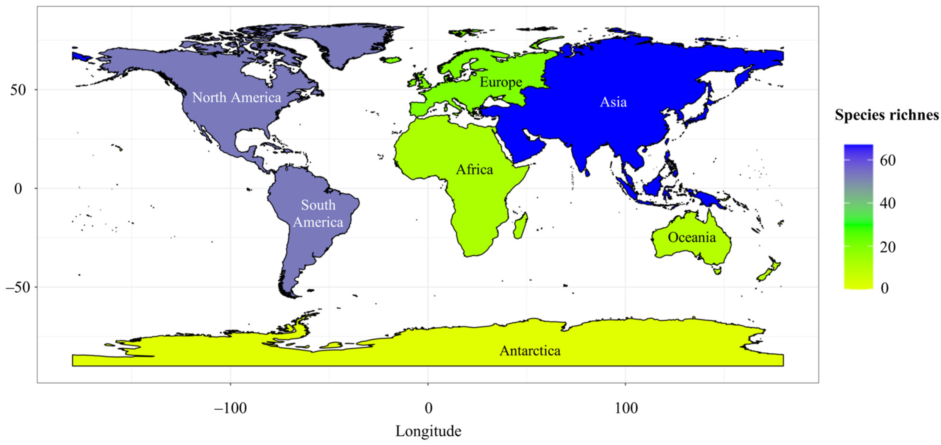
Figure 1. The global distribution of bioluminescent fungi is based on the available literature; as the study was conducted, the richest fungi species were reported in Asia, followed by North and South America.
Diversity 16 00539 g001
Figure 2. Species percentage based on species counts reported from each lineage of bioluminescent fungi.
Diversity 16 00539 g002
Figure 3. Glowing piece of wood (a). Glowing mycelia on a wood in the Xishuangbanna Tropical Botanical Garden of China (b). The whole fruiting body and spores glowing in Favolaschia xtbgensis in the Xishuangbanna Tropical Botanical Garden of China (c). Stipe glowing in Roridomyces phyllostachydis from Northeast India (d) (photo credit: Stephen Axford).
Diversity 16 00539 g003
Table 1. The list of bioluminescent fungi reported worldwide. Bioluminescence can be produced by the entire fungus or, sometimes, only through mycelia, fruiting bodies, or spores.
Fungal TaxaDistributionGlowing PartReferences
MyceliumFruiting BodiesCapStipeSpores
Armillaria LineageArmillaria borealisRussia+////[40]
Armillaria calvescensThe USA+////[41]
Armillaria cepistipesThe USA+////[41]
Armillaria fuscipesMalaysia+////[42]
Armillaria gallicaEurope and the USA+////[42]
Armillaria geminaThe USA+////[42]
Armillaria melleaChina, Europe, India, and the USA+////[42,43,44]
Armillaria nabsnonaThe USA+////[41]
Armillaria novae-zelandiaeNew Zealand+////[45]
Armillaria ostoyaeEurope and USA+////[42]
Armillaria sinapinaThe USA+////[41]
Desarmillaria ectypaEurope+++//[46]
Desarmillaria tabescensEurope and the USA++///[42]
Eoscyphella LineageEoscyphella luciurceolataBrazil?+???[47]
Lucentipes LineageMycena lucentipesSouth America and ++?+/[42]
Gerronema viridilucensSouth America++++/[42]
Mycenoid LineageCruentomycena orientalisJapan++++/[14]
Dictyopanus foliicolaJapan++///[42]
Favolaschia xtbgensisChina+++++[48]
Favolaschia tonkinensisChina?+++/[49]
Favolaschia peziziformisJapan?+++/[50,51]
Filoboletus manipularisAfrica, China, Sri Lanka, Thailand, and ?+/+/[52,53,54]
Filoboletus hanedaeJapan?+/+/[42]
Filoboletus pallescens?+???[42]
Filoboletus yunnanensisChina?+??/[52,54,55]
Gerronema glutinipesAfrica and China?+///[52]
Mycena abieticolaBrazil?+++/[55]
Mycena aspratilisBrazil and Puerto Rico/+/+/[55]
Mycena asterinaSouth America+++//[42]
Mycena cahaya++++/[56]
Mycena chlorophosChina, Japan, the Pacific Islands, Sri Lanka, and ++++/[42,53]
Mycena citricolorSouth America and the USA+////[42]
Mycena coralliformis+////[53]
Mycena cristinaeBrazil++///[57]
Mycena crocataSwitzerland+////[58]
Mycena daisyogunensisJapan?+??/[42]
Mycena deepthaIndia+////[59]
Mycena deformisBrazil+////[60]
Mycena discobasisAfrica and South America?+++/[42]
Mycena epipterygiaEurope, the USA, and Japan+////[42]
Mycena feraSouth America?+++/[42]
Mycena flammiferaJapan++++/[50,51]
Mycena fulgorisMexico/+/+/[61]
Mycena galopusEurope, the USA, and Japan+////[42]
Mycena globulisporaBrazil and Mexico?+/+/[60,61]
Mycena gombakensis++++/[53]
Mycena guzmaniiMexico++++/[61]
Mycena haematopusChina, Europe, the USA, Japan, and South America+++//[42]
Mycena illuminansJapan and ?++//[42,53]
Mycena inclinataAfrica, China, Europe, and the USA+////[42]
Mycena jingyingaChina+////[34]
Mycena kentingensisChina+++//[61]
Mycena lacrimansSouth America?+/+/[42]
Mycena lamprocephalaBrazil++++?[62]
Mycena lazulinaJapan++++/[50,51]
Mycena luceataMexico?++/?[23]
Mycena luciferinaMexico?++/?[23]
Mycena lucinieblaeMexico+///?[23]
Mycena luguensisChina+////[34]
Mycena luminaMexico++++/[61]
Mycena luxaeternaBrazil++/+/[55]
Mycena luxarboricolaBrazil?+++/[55]
Mycena lux-coeliJapan?+++/[42]
Mycena luxfoliataJapan+////[50,51]
Mycena luxfoliicolaMexico++++/[61]
Mycena luxmanantlanensisMexico+++/?[23]
Mycena luxperpetuaPuerto Rico++++/[42]
Mycena maculataAfrica, Europe, and the USA+////[42]
Mycena margaritaBelize, Dominican Republic, Jamaica, Puerto Rico, and Brazil/+++/[42,63]
Mycena nebulaMexico?+++/[61]
Mycena nocticaelum+++//[53]
Mycena noctilucensPacific Islands and ?+++/[42,53]
Mycena oculisnymphaeBrazil/+++/[60]
Mycena olivaceomarginataEurope and the USA+////[42]
Mycena perlaeMexico/++//[61]
Mycena polygrammaChina, Europe, the USA, Japan, and Africa++///[42]
Mycena pseudostylobatesJapan+???/[42]
Mycena puraChina, Europe, the USA, Japan, and South America+////[42]
Mycena roseaEurope+////[42]
Mycena roseoflavaNew Zealand++/+/[45]
Mycena sanguinolentaChina, Europe, the USA, and Japan+////[42]
Mycena seminau+++//[56]
Mycena silvaelucens?+++/[42,56]
Mycena sinar++++/[56]
Mycena singeriSouth America and ?+++/[42]
Mycena sophiaeMexico+///?[23]
Mycena sp. (PDD 80772)New Zealand?+///[42]
Mycena sp. (SP #380150)South America++///[42,64]
Mycena sp. (SP #380281)South America?+///[42,64]
Mycena stellarisJapan++++/[50,51]
Mycena stylobatesAfrica, China, Europe, the USA, and Japan+////[42]
Mycena tintinnabulumEurope+////[42]
Mycena venusChina+////[34]
Mycena zephirusEurope+////[42]
Panellus luminescens++++?[65,66]
Panellus luxfilamentusSri Lanka and +////[56]
Panellus pusillusAfrica, Australasia, China, Japan, the USA, South America, and ?+??/[42,49]
Panellus stipticusAfrica, Australasia, China, Europe, Japan, the USA, and South America+++//[42]
Resinomycena fulgensJapan?+++/[50,51]
Resinomycena petarensisBrazil+////[60]
Roridomyces irritansAustralasia/++/?[42]
Roridomyces lamprosporusBrazil, Ceylon, Malaysia, and Papua New Guinea,
Singapore, and Trinidad
/+//+[67]
Roridomyces phyllostachydisIndia?+/+/[10,22]
Roridomyces pruinosoviscidusAustralasia and ++++?[42,53]
Roridomyces roridusChina, Europe, the USA, South America, and Japan+////[42,68]
Roridomyces sublucensIndonesia and /+++/[42]
Roridomyces viridiluminusChina++++/[69]
Omphalotus LineageMarasmiellus venosusJapan++///[50,51]
Marasmiellus lucidusJapan?+++/[50,51]
Neonothopanus gardneriSouth America?+++/[42]
Neonothopanus nambiAustralasia, China, South America, Thailand, , and ?+++/[42,53,54]
Nothopanus noctilucensJapan?+///[42]
Omphalotus guepiniiformisChina and Japan++///[42,54,70]
Omphalotus illudensEurope and the USA+++//[42]
Omphalotus mangensisChina?++//[42,54,71]
Omphalotus nidiformisAustralasia?+++/[42]
Omphalotus oleariusChina and Europe++++/[42,54]
Omphalotus olivascensThe USA/+///[42]
Omphalotus subilludensThe USA?+//?[72]
Pleurotus decipiens?+///[42]
Pleurotus nitidusJapan?+///[50,51]
AscomycotaXylaria hypoxylonEurope?+??/[73]
Note: For Armillaria novae-zelandiae, Mycena roseoflava, and Omphalotus subilludens, there is no published literature for them as bioluminescent mushrooms; however, mushroom hunters have posted glowing mushroom photos of those species. ♪ = refers to distribution within Malaysia, South Asia region; ♣ = refers to distribution within Central America and the Caribbean region; + = glow; / = do not glow; ? = no report. The original references sometimes provided only continental-level distribution information for some species, lacking specific country-level details. We have adhered to the available data for these species and recorded the distribution at the continental level. However, we have documented the specific countries accordingly for species with detailed country-level distribution information.
Disclaimer/Publisher’s Note: The statements, opinions and data contained in all publications are solely those of the individual author(s) and contributor(s) and not of MDPI and/or the editor(s). MDPI and/or the editor(s) disclaim responsibility for any injury to people or property resulting from any ideas, methods, instructions or products referred to in the content.