Article

The Study of Biting Midges Culicoides Latreille, 1809 (Diptera: Ceratopogonidae) and the Prevalence of Haemoproteus Kruse, 1890 (Haemosporida: Haemoproteidae) on the Curonian Spit of the Baltic Sea

1
Biological Station Rybachy of Zoological Institute, Russian Academy of Sciences, Rybachy 238535, Russia
2
Biology Faculty of Lomonosov Moscow State University, Moscow 119234, Russia
*
Author to whom correspondence should be addressed.
Diversity 2024, 16(12), 723; https://doi.org/10.3390/d16120723
Submission received: 30 October 2024 / Revised: 19 November 2024 / Accepted: 21 November 2024 / Published: 26 November 2024
(This article belongs to the Section Animal Diversity)

Abstract

:
The part of the life cycle concerning the vectors of avian haemosporidians of the genus Haemoproteus remains only partially studied. This work presents the results of capturing and analyzing female Culicoides biting midges for Haemoproteus infection, collected on the Curonian Spit of the Baltic Sea. The midges were collected from April to August 2023 in dry and wet habitats of the Spit using light traps. Each parous female midge was identified to the species level and then its salivary glands were isolated to prepare a thin smear. The preparation was examined under a light microscope for the presence of parasite stages infecting the birds (sporozoites). PCR analysis was also conducted to assess the prevalence of infection in the midges and the genetic diversity of Haemoproteus spp. among vectors on the Curonian Spit. As a result, 995 Culicoides females belonging to 12 species were collected over the course of 4 months. The highest number of midges was recorded in June. The dominant species were Culicoides kibunensis and Culicoides pictipennis. Molecular analysis showed that 2.71% (n = 27) of the examined individuals contained DNA of Haemoproteus spp. Parasites were found in midges of C. pictipennis, C. kibunensis, C. segnis, C. obsoletus, C. punctatus, C. reconditus, C. festivipennis, and C. pallidicornis from May 15 to July 13. The DNA of the identified parasites belonged to six genetic lineages of Haemoproteus—Haemoproteus parabelopolskyi hSYAT02, H. asymmetricus hTUPHI01, H. minutus hTURDUS2, H. spp. hBRAM1, hHAWF6, and hHIICT1. Sporozoites of H. parabelopolskyi hSYAT02 were detected in two individuals of C. pictipennis and one of C. kibunensis. Thus, taking into account the previous results of other studies, we have assumed that these two midge species are competent vectors for Haemoproteus parasites on the Curonian Spit.

1. Introduction

Haemoproteus (Haemosporida: Haemoproteidae) is a genus of microorganisms related to malaria parasites (Plasmodiidae). Members of this genus have a wide range of vertebrate hosts, including amphibians, reptiles, and birds [1,2]. Among birds, Haemoproteus exhibits considerable species and genetic diversity: currently, over 180 species and several thousand genetic lineages have been described, and this number continues to grow each year [3,4]. Haemoproteus parasites are found nearly worldwide (except Antarctica) and in all climatic zones, including the Arctic [5].
For a long time, these haemosporidians were considered relatively low-pathogenic parasites for birds [6]. However, recent studies have shown that haemoproteosis can cause serious damage to internal organs, especially when a bird is infected with a parasite species not specific to it [7,8]. For example, the widely distributed Haemoproteus minutus in Europe has caused numerous deaths of Australian and South American parrots in Denmark, Germany, and the United Kingdom in different years [9].
Like other haemosporidians, Haemoproteus has a heteroxenous life cycle and is transmitted to vertebrate hosts through the bite of a female blood-sucking dipteran insect [1]. Since the first description of the genus Haemoproteus by V. Kruse in 1890, it took over 60 years to establish that for most Haemoproteus species, the vectors are Culicoides biting midges [10]. Following this, the study of parasite transmission between avian hosts and vectors progressed through experimental infections of various midge species and the observation of zygote formation and sporogony processes [11,12]. Nevertheless, information about specific vector species and parasite specificity to them remains scarce.
With the development of molecular methods, detecting parasite DNA in wild populations of blood-sucking dipterans has become increasingly used, allowing an approximate estimation of the potential range of haemosporidian vectors in birds [13]. Similar studies have been conducted in various regions of Europe and Asia [14,15,16,17,18]. Thus, species such as Culicoides alazanicus, C. circumscriptus, C. impunctatus, and others have been added to the list of potential vectors for avian Haemoproteus. Unfortunately, merely detecting an insect’s infection using PCR cannot definitively confirm whether the vector is competent for a specific parasite [19]. Development in the midge may be abortive, or the species of Culicoides may primarily feed on mammals and only rarely bite birds. Only the presence of infectious parasite stages (sporozoites) in the salivary glands can unequivocally indicate the midge’s competence to transmit Haemoproteus to a vertebrate host.
In recent years, a method has been increasingly used in which a thin smear is prepared from the salivary glands of field-caught dipterans before PCR is conducted, allowing the presence of sporozoites to be checked under high magnification with a light microscope [19,20,21]. With this combined detection of parasite DNA and the search for sporozoites in the smear, competent vectors for avian Haemoproteus have so far been established, including Culicoides festivipennis, C. pictipennis, C. segnis, C. reconditus, and others [22,23,24]. However, there are still insufficient data for a more complete understanding of the transmission of these parasitic microorganisms. It is noted that more than 1300 species of Culicoides are known worldwide, while vector competence has been proven for a negligible number of them [25].
Therefore, the aim of this study is to complement the existing knowledge on competent Haemoproteus vectors and to identify specific species of midges that transmit these blood parasites among wild bird populations in the southeastern Baltic region.

2. Material and Methods

Material collection was conducted from April to August 2023 on the Curonian Spit of the Baltic Sea. The study followed the methodologies described by Bernotienė et al. [19] and Žiegytė et al. [22,23,24,25]. To capture the midges, we used light traps (Biogents AG, Regensburg, Germany), which were set up once a week at six locations on the Curonian Spit (Figure 1). Three collection sites were located in dry pine forests (the “Fringilla” field station of the Rybachy Biological Station, Zoological Institute of the Russian Academy of Sciences 55°08′96′′ N, 20°73′37′′ E (location F); the village of Khvoinoye 55°13′40″ N, 20°78′68′′ E (location K); and Efa Dune 55°22′61″ N, 20°89′93″ E (location E)), while the other three were in wet habitats (mixed forest near Müller Dune 55°15′21″ N, 20°81′80″ E (location M) and Lake Lebed 55°25′02″ N, 20°92′51″ E (location L) and reed beds along the shore of the Curonian Lagoon on the territory of the “Rybachy” Biological Station 55°15′41″ N, 20°85′88″ E (location R)).
The traps were turned on two hours before sunset and turned off two hours after sunrise the next day, after which the samples were transported to the laboratory for sorting.
The samples were sorted the same day under 4× magnification using a stereomicroscope, and female midges of the genus Culicoides were selected based on the presence of a burgundy pigment in the abdomen indicating that the female took a blood meal [24]. The head and wings of each selected insect were separated from the body and mounted in Canada balsam for further identification of the midge species based on morphological characteristics [26,27,28]. A thin smear was prepared from the content of the thorax. The prepared smear was air-dried, fixed in absolute methanol, and then stained with 4% Giemsa [23]. The stained smears were examined under 1000× magnification using a light microscope (MF43-N LED, Micro-Shot Technology Ltd., Guangzhou, China) with an oil immersion. Microphotographs of the sporozoites were taken at 1000× magnification using an MC50-S digital camera (Micro-Shot Technology Ltd., Guangzhou, China).
The remaining abdomen of the dissected midge was placed in SET buffer (0.015 M NaCl, 0.05 M Tris, 0.001 M EDTA, pH 8.0) and stored at −20 °C for further molecular analysis to detect avian haemosporidian infection and confirm the species identification of biting midges.
The total DNA was extracted following a standard ethanol precipitation protocol using ammonium acetate [29]. To assess midge infection, a 489 bp fragment of the mitochondrial gene of the parasite was amplified using PCR with two primer sets specific to haemosporidians of the genera Plasmodium, Haemoproteus, and Leucocytozoon [30,31]. For quality control of the PCR and to avoid false amplification, each reaction included a positive control (DNA of Plasmodium sp.) and a negative control (ddH2O). The PCR results were assessed using electrophoresis of the reaction products on a 2% agarose gel stained with ethidium bromide. Samples positive for the presence of parasite DNA were sent for Sanger sequencing from both the 5′- and 3′-ends (Evrogen, Moscow). The resulting chromatograms were analyzed using BioEdit software (version 7.7.1) [32], and the edited sequences were uploaded to the MalAvi (http://mbio-serv2.mbioekol.lu.se/Malavi, (accessed on 1 October 2024), [3]) and NCBI (https://blast.ncbi.nlm.nih.gov/Blast.cgi, (accessed on 1 October 2024)) molecular databases for species and genetic lineage identification of the parasites.
To confirm the accuracy of the identification of Culicoides species based on head structure and wing coloration, DNA barcoding of the mitochondrial cytochrome c oxidase subunit I gene fragment was performed [33].

3. Results

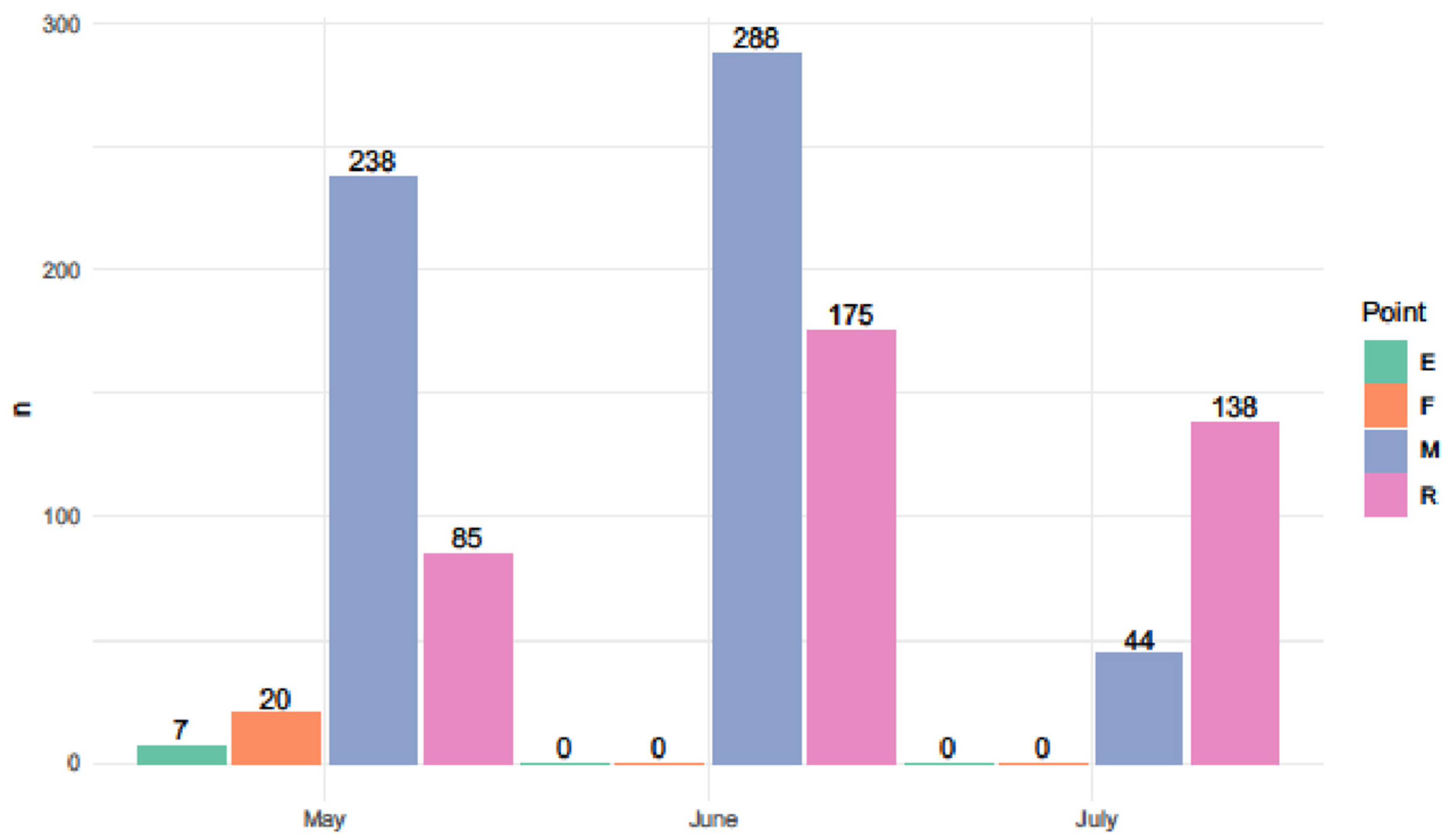
During the study, a total of 995 blood-fed female Culicoides biting midges belonging to 12 species were collected and processed: C. (Avaritia) obsoletus, C. (Beltranmyia) sphagnumensis, C. (Culicoides) grisescens, C. (Culicoides) impunctatus, C. (Culicoides) punctatus, C. (Oecata) albicans, C. (Oecata) kibunensis, C. (Oecacta) pictipennis, C. (Wirthomyia) reconditus, C. segnis (Wirthomyia), and C. (subgenus unplaced) pallidicornis (Table 1). The majority of individuals were captured in two wet habitats: 570 (57.2%) midges near Müller Dune (M) and 398 (40%) in the vicinity of the Rybachy field station (R) (Figure 2). In dry habitats, Culicoides were scarcely captured: only 20 (2.01%) individuals were caught at the Fringilla field station (F) and 7 (0.79%) near the village of Khvoinoye (K). No Culicoides were captured at the other collection locations.
The first midges were recorded on 15 May, and the highest number was collected in June at both locations (288 out of 570 in M and 175 out of 398 in R). After 13 July, no midges were found in our collections at any of the locations.
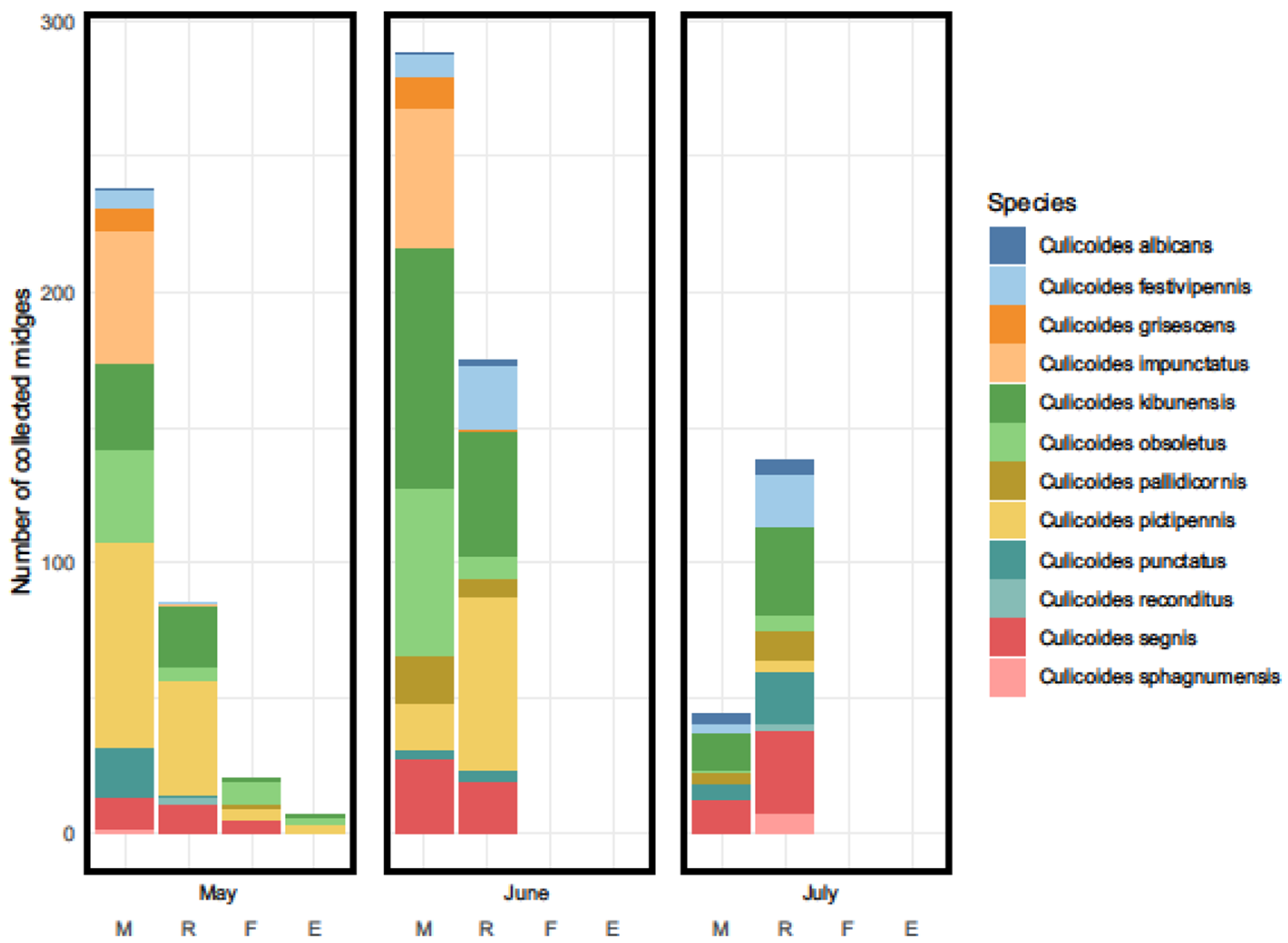
At the forest site M, the highest abundance was observed for four species: C. kibunensis (23.51%), C. impunctatus (17.54%), C. obsoletus (17.01%), and C. pictipennis (16.32%). The remaining species accounted for about 25% of the total. Nearly all C. impunctatus (100 out of 101) and C. grisescens (20 out of 21) were collected in the forest. Culicoides pictipennis was the most numerous species in May, while in June and July, C. kibunensis dominated (Figure 3).
In the reed habitat (R), two species dominated, C. pictipennis (27.64%) and C. kibunensis (25.38%), in May and June for both species and in July for C. kibunensis. At this location, C. segnis (14.82%) and C. festivipennis (10.8%) also made up a substantial proportion of the captures (Figure 3).
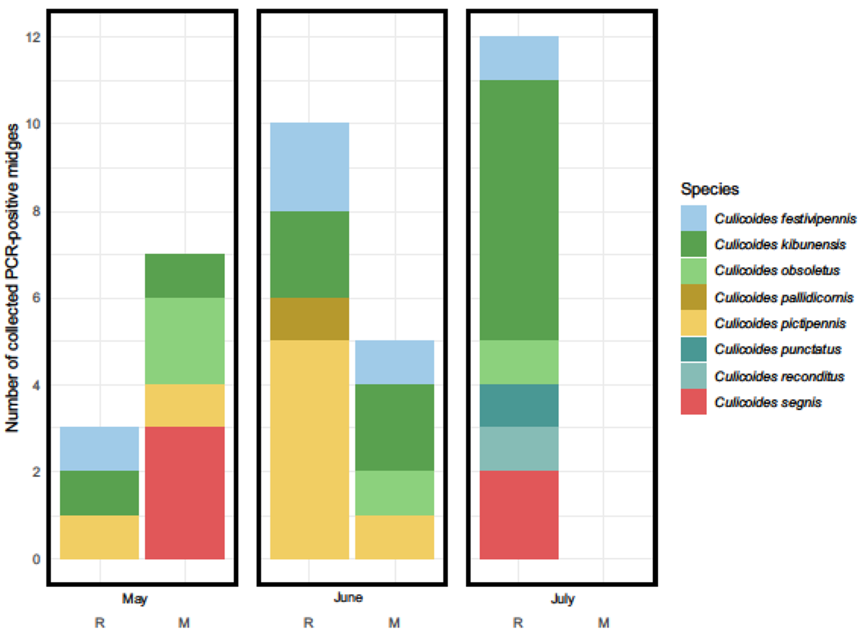
Unfortunately, we were unable to amplify haemosporidian DNA from the midges using the standard nested PCR primers described by Hellgren et al. [30], despite repeating the analysis twice. However, we obtained positive results using the primers from Drovetski et al. [31]. In total, we detected 37 individuals with haemosporidian parasites (overall infection rate of 3.71%) among females of eight species: C. kibunensis (12 positive), C. pictipennis (8), C. festivipennis (5), C. segnis (5), C. obsoletus (4), C. pallidicornis (1), C. punctatus (1), and C. reconditus (1). In the forest location (M), 12 midges with infection were found and 25 in the reeds (R). The first insects with parasites were registered on 15 May (Figure 4), and the highest haemosporidian infection rate was observed from 24 June to 6 July (19 out of 37 individuals).
Nine genetic lineages of haemosporidians were detected in this study (Table 1). Six belonged to the genus Haemoproteus (Parahaemoproteus): H. asymmetricus (hTUPHI01), H. belopolskyi (hHIICT1), H. parabelopolskyi (hSYAT02), H. minutus (hTURDUS2), and H. spp. (hBRAM1 and hHAWF6) and three to the genus Leucocytozoon: L. spp. (lBT5, lROFL6, and lSISKIN2). The most common lineage was H. parabelopolskyi (hSYAT02) at 27.02%, found in 8 C. pictipennis and 2 C. kibunensis. The lineages hBRAM1 (18.91%) and hHAWF6 (2.7%) were recorded for the first time on the Curonian Spit, as well as in midges. In this study, they were found in C. segnis (3), C. kibunensis (2), C. punctatus (1), and C. reconditus (1) for hBRAM1 and in C. kibunensis (1) for hHAWF6. One female C. pallidicornis had a mixed co-infection of several haemosporidian lineages.
Numerous sporozoites (>100 sporozoites) of H. parabelopolskyi (hSYAT02) distributed evenly throughout the smear were found in two thoraxes samples belonging to female C. pictipennis and in one C. kibunensis (Figure 5); all three were captured on 8 June.

4. Discussion

The aim of this study was to identify both competent and potential vectors of Haemoproteus parasites, as well as to evaluate midge infection rates and parasite diversity in the vectors. We identified 12 species of Culicoides on the Curonian Spit. The species composition we observed differs somewhat from the data obtained in previous years. For instance, Valkiūnas and Glukhova [34], based on collections from 1956–1957 and 1991, described only seven species, of which we found only C. obsoletus, C. punctatus, and C. reconditus, while species such as C. delta and C. stigma were completely absent in our samples. Additionally, Liutkevičius [35] noted that around 95% of midges on the Curonian Spit belonged to the species C. impunctatus, but also reported the presence of species such as C. pictipennis and C. segnis for the first time, albeit as single findings. These differences in species composition can be explained by two factors. Firstly, over the past 50–70 years, the Curonian Spit has undergone significant changes in forest cover, as well as in the maturity of pine forests, which were extensively planted in the 1950s and 1960s [36]. As the forest matured, suitable habitats for different species of Culicoides also changed. Secondly, previous studies used different methods for collecting blood-sucking insects, such as catches of feeding midges on hands, collected from bird nests, and illuminated house windows, rather than light traps. For example, the prevalence of C. impunctatus in past collections [34] can be explained by hand captures, as this species may prefer to feed on human blood.
The species composition on the Curonian Spit is most similar to data collected from various areas of the southeastern Baltic region [22,23,24,36], where species such as C. kibunensis [22,23,37], C. pictipennis [24,25,37], and the Obsoletus complex [24,38,39] dominate in different proportions. Additionally, we recorded C. albicans on the Curonian Spit for the first time, although in small numbers. This is consistent with the literature data indicating that this species is rare and occurs in low numbers [40,41].
Most midges were collected in May (32.46%) and June (46.53%), which also agrees with findings from other studies. A significant proportion of midges showed the presence of parasite’s DNA (27.02%), which were captured in May, suggesting an early start to transmission in the year of the study. The remaining PCR-positive individuals were found in June (35.13%) and July (37.85%). We did not capture any blood-fed or unfed midges from mid-July until the end of August, indicating a low circulation of Haemoproteus between birds and midges at this time. Different studies in the southeastern Baltic Coast region indicate various months for the most active transmission of bird infections by vectors: May [35], June [24], and July [25]. It appears that these three months are the most favorable for parasite transmission between the vertebrate host and vector. During this time, there is active nesting of both short- and long-distance migrants on the Curonian Spit [25]. Of course, this depends on specific weather conditions each year and in particular habitats.
It is noted that spring temperature regimes strongly influence the start of midge emergence [42], so high or low air temperatures during this period can shift the start and peak of transmission to earlier or later periods. Spring temperatures also affect the timing of bird migration, as they return from nearby and distant wintering areas [43]. Thus, there should not be gaps in time and place of presence between the parasites, birds, and vectors.
We identified 37 individuals with parasite DNA out of 995 midges across eight species, with nine genetic lineages of haemosporidians determined. It is important to note that we could not amplify the DNA of the parasites (cyt b fragment) using the standard and widely-used nested PCR method with two pairs of primers [30]. Culicoides midges are very small, and the DNA yield after extraction can be minimal [44]. Moreover, most of the extracted genetic material may belong to the host, not the parasite [45]. The nested PCR primers may have shown limited sensitivity in our study. Similar cases were described in other studies where parasites were found in preparations from PCR-negative insects [22,23].
Using the PCR protocol described by Drovetski et al. [31] (2014), we identified six Haemoproteus lineages: Haemoproteus (Parahaemoproteus): H. asymmetricus (hTUPHI01), H. belopolskyi (hHIICT1), H. parabelopolskyi (hSYAT02), H. minutus (hTURDUS2), and H. spp. (hBRAM1 and hHAWF6) and three from the genus Leucocytozoon: L. spp. (lBT5, lROFL6, and lSISKIN2).
Haemoproteus asymmetricus (hTUPHI01) and H. minutus (hTURDUS2) are two phylogenetically related species that predominantly parasitize birds of the Turdidae family [46]. These parasites are fairly widespread in Europe and can cause severe pathologies in the internal organs of some bird species [9]. Both parasite species occur on the Curonian Spit [23] and their competent vectors were identified as folows: for H. asymmetricus (hTUPHI01)—C. kibunensis, C. segnis, C. festivipennis, and C. pictipennis; for H. minutus (hTURDUS2)—C. kibunensis, C. segnis, and C. pictipennis [22,23,24,25]. In our study, we found the DNA of hTUPHI01 in C. kibunensis and C. festivipennis and hTURDUS2 in C. festivipennis, although the other listed species were also present in our study area. This suggests that these four species likely play a role in transmitting these parasites in the southeastern Baltic coast region.
Haemoproteus belopolskyi, associated with the genetic lineage hHIICT1, predominantly infects the Icterine Warbler (Hippolais icterina, Acrocephalidae) [3]. This bird species is also widespread in Europe, particularly on the Curonian Spit. The only identified natural vector for hHIICT1 in the wild is C. festivipennis [24], but potential vectors have also been identified as C. impunctatus, C. kibunensis, C. obsoletus, and C. pictipennis. We add C. segnis to this list.
Haemoproteus parabelopolskyi (hSYAT02) was the most common parasite in our study, found in 10 midge females (27.02%) of two species: C. kibunensis (3 individuals) and C. pictipennis (7 individuals). Sporozoites of hSYAT02 were also found only in these two insect species (Figure 5). This parasite species is actively transmitted in the study area among birds of the family Sylviidae [3].
The genetic lineages Haemoproteus spp. hBRAM1 and hHAWF6 have not yet been assigned to any morphospecies and are rarely found in birds. We recorded these two lineages for the first time in insects and on the Curonian Spit. Previously, hBRAM1 was found only in Bramblings (Fringilla montifringilla) in Sweden [47] and the Yamalo-Nenets Autonomous District (northern Russia) [48]. We identified hBRAM1 DNA in C. kibunensis, C. punctatus, C. reconditus, and C. segnis but did not detect any sporozoites in the salivary gland preparations of these individuals, indicating that they are likely only potential vectors. Another genetic lineage, hHAWF6, was found in Hawfinches (Coccothraustes coccothraustes) and Blackbirds (Turdus merula) in southern [49,50], central [51], and eastern [52] Europe, as well as in North Africa [49]. On the Curonian Spit, this parasite had not been previously identified in blood samples from captured birds but was detected for the first time by us in C. kibunensis. Another such finding of hHAWF6 had already been reported in C. kibunensis in Lithuania [24], and at present, this remains the only known potential vector for Haemoproteus of this genetic lineage.
Additionally, we found a single female C. pallidicornis with mixinfection of avian haemosporidians. Although this species was scarcely represented in our collections, it may still play some role in the transmission of Haemoproteus, considering that it has previously been identified as a potential vector for H. belopolskyi (hHIICT1) in Lithuania, northeast of our study area [39].
The presence of Leucocytozoon infections in midges likely indicates abortive development in these dipterans. In most cases, parasites of this genus develop in blackflies (Simuliidae). Only one species, L. caullery, from the subgenus Akiba, is known to develop in biting midges (Ceratopogonidae) [1,53], but this parasite is distributed in south and southeast Asia and has not been recorded at our study site. Non-specific avian haemosporidians are frequently found in insects feeding on birds. For example, Plasmodium is found in midges, and Haemoproteus is found in mosquitoes. There are also cases of detecting Leucocytozoon [54], although these are rare because different primer sets are used for amplifying Plasmodium/Haemoproteus and Leucocytozoon. We used primers specific to all three genera, which allowed us to detect Leucocytozoon in the midges.

5. Conclusions

This study confirms that the Culicoides species C. kibunensis and C. pictipennis play an important role in the transmission of avian Haemoproteus parasites in the southeastern Baltic region. Twelve species of midges were identified, eight of which are, apparently, involved in the transmission of Haemoproteus species on the Curonian spit. The genetic lineages H. spp. hBRAM1 and hHAWF6 were recorded for the first time on the Curonian Spit and in vectors.

Author Contributions

Experimental conception and design: E.P.; collection of fieldwork: E.P., M.E. and A.D.; biting midge identification: E.P.; dissection of biting midges: E.P. and M.E., molecular analysis: E.P. and A.M. (Alexandra Mukhina); microscopy of preparations: E.P.; manuscript writing: E.P. and A.M. (Andrey Mukhin). All authors have read and agreed to the published version of the manuscript.

Funding

This research was funded by a scientific grant (RSF № 23-24-00522 for Elena Platonova).

Institutional Review Board Statement

Not applicable.

Data Availability Statement

The data of this study are available by email inquiry.

Acknowledgments

The Authors are grateful to Diana Polikarpova for her help in the collection of biting midges in the field.

Conflicts of Interest

The authors declare no conflicts of interest.

References

  1. Valkiūnas, G. Avian Malaria Parasites and Other Haemosporidia; CRC Press: Boca Raton, FL, USA, 2005; ISBN 978-0415300971. [Google Scholar]
  2. Muriel, J.; González-Blázquez, M.; Matta Cahacho, N.E.; Vargas-León, C.M.; Marzal, A. Parasitas Sanguíneos de Anfíbios; Editora da Universidade Federal do Piauí: Teresina, Brazil, 2021. [Google Scholar]
  3. Bensch, S.; Hellgren, O.; Pérez-Tris, J. MalAvi: A public database of malaria parasites and related haemosporidians in avian hosts based on mitochondrial cytochrome b lineages. Mol. Ecol. Resour. 2009, 9, 1353–1358. [Google Scholar] [CrossRef] [PubMed]
  4. Valkiūnas, G.; Iezhova, T.A. Keys to the avian Haemoproteus parasites (Haemosporida, Haemoproteidae). Malar. J. 2022, 21, 269. [Google Scholar] [CrossRef] [PubMed]
  5. Clark, N.J. Phylogenetic uniqueness, not latitude, explains the diversity of avian blood parasite communities worldwide. Glob. Ecol. Biogeogr. 2018, 27, 744–755. [Google Scholar] [CrossRef]
  6. Bennett, G.F.; Peirce, M.A.; Ashford, R.W. Avian haematozoa: Mortality and pathogenicity. J. Nat. Hist. 1993, 27, 993–1001. [Google Scholar] [CrossRef]
  7. Palinauskas, V.; Iezhova, T.A.; Križanauskienė, A.; Markovets, M.Y.; Bensch, S.; Valkiūnas, G. Molecular characterization and distribution of Haemoproteus minutus (Haemosporida, Haemoproteidae): A pathogenic avian parasite. Parasitol. Int. 2013, 62, 358–363. [Google Scholar] [CrossRef]
  8. Duc, M.; Ilgūnas, M.; Kubiliūnaitė, M.; Valkiūnas, G. First report of Haemoproteus (Haemosporida, Haemoproteidae) megalomeronts in the brain of an avian host, with description of megalomerogony of Haemoproteus pastoris, the blood parasite of the common starling. Animals 2021, 11, 2824. [Google Scholar] [CrossRef]
  9. Ortiz-Catedral, L.; Brunton, D.; Stidworth, M.F.; Elsheikha, H.M.; Pennycott, T.; Schulze, C.; Braun, M.; Wink, M.; Gerlach, H.; Pendl, H.; et al. Haemoproteus minutus is highly virulent for Australasian and South American parrots. Parasites Vectors 2019, 12, 40. [Google Scholar] [CrossRef]
  10. Fallis, A.M.; Wood, D.M. Biting midges (Diptera: Ceratopogonidae) as intermediate hosts for Haemoproteus of ducks. Ibidem 1957, 35, 425–435. [Google Scholar] [CrossRef]
  11. Žiegytė, R.; Valkiūnas, G. Recent advances in vector studies of avian haemosporidian parasites. Ekologija 2014, 60, 73–83. [Google Scholar] [CrossRef]
  12. Bukauskaitė, D.; Iezhova, T.A.; Ilgūnas, M.; Valkiūnas, G. High susceptibility of the laboratory-reared biting midges Culicoides nubeculosus to Haemoproteus infections, with review on Culicoides species that transmit avian haemoproteids. Parasitology 2019, 146, 333–341. [Google Scholar] [CrossRef]
  13. Köchling, K.; Schaub, G.A.; Werner, D.; Kampen, H. Avian Plasmodium spp. and Haemoproteus spp. parasites in mosquitoes in Germany. Parasites Vectors 2023, 16, 369. [Google Scholar] [CrossRef] [PubMed]
  14. Bobeva, A.; Zehtindjiev, P.; Bensch, S.; Radrova, J. A survey of biting midges of the genus Culicoides Latreille, 1809 (Diptera: Ceratopogonidae) in NE Bulgaria, with respect to transmission of avian haemosporidians. Acta Parasitol. 2013, 58, 585–591. [Google Scholar] [CrossRef] [PubMed]
  15. Bobeva, A.; Ilieva, M.; Dimitrov, D.; Zehtindjiev, P. Degree of associations among vectors of the genus Culicoides (Diptera: Ceratopogonidae) and host bird species with respect to haemosporidian parasites in NE Bulgaria. Parasitol. Res. 2014, 113, 4505–4511. [Google Scholar] [CrossRef] [PubMed]
  16. Ferraguti, M.; Martínez-de la Puente, J.; Ruiz, S.; Soriguer, R.; Figuerola, J. On the study of the transmission networks of blood parasites from SW Spain: Diversity of avian haemosporidians in the biting midge Culicoides circumscriptus and wild birds. Parasites Vectors 2013, 6, 208. [Google Scholar] [CrossRef]
  17. Veiga, J.; Martinez-de la Pueante, J.; Vaclav, R.; Figuerola, J.; Valera, F. Culicoides paolae and C. circumscriptus as potential vectors of avian haemosporidians in an arid ecosystem. Parasites Vectors 2018, 11, 524. [Google Scholar] [CrossRef]
  18. Inumaru, M.; Nakamura, K.; Odagawa, T.; Suzuki, M.; Murata, K.; Sato, Y. The first detection of avian haemosporidia from Culicoides biting midges in Japan, with notes on potential vector species and the transmission cycle. Vet. Parasitol. 2023, 39, 100840. [Google Scholar] [CrossRef]
  19. Bernotienė, R.; Žiegytė, R.; Vaitkutė, G.; Valkiūnas, G. Identification of a new vector species of avian haemoproteids, with a description of methodology for the determination of natural vectors of haemosporidian parasites. Parasites Vectors 2019, 12, 307. [Google Scholar] [CrossRef]
  20. Njabo, K.Y.; Cornel, A.J.; Sehgal, R.N.; Loiseau, C.; Buermann, W.; Harrigan, R.J.; Pollinger, J.; Valkiūnas, G.; Smith, T.B. Coquillettidia (Culicidae, Diptera) mosquitoes are natural vectors of avian malaria in Africa. Malar. J. 2009, 8, 193. [Google Scholar] [CrossRef]
  21. Žiegytė, R.; Bernotiene, R. Contribution to the knowledge on black flies (Diptera: Simuliidae) as vectors of Leucocytozoon (Haemosporida) parasites in Lithuania. Parasitol. Int. 2022, 87, 102515. [Google Scholar] [CrossRef]
  22. Žiegyte, R.; Bernotienė, R.; Palinauskas, V. Culicoides segnis and Culicoides pictipennis Biting Midges (Diptera, Ceratopogonidae), new reported vectors of Haemoproteus parasites. Microorganisms 2022, 10, 898. [Google Scholar] [CrossRef]
  23. Žiegytė, R.; Palinauskas, V.; Bernotienė, R. Natural vector of avian Haemoproteus asymmetricus parasite and factors altering the spread of infection. Insects 2023, 14, 926. [Google Scholar] [CrossRef] [PubMed]
  24. Chagas, C.R.F.; Duc, M.; Kazak, M.; Valavičiūtė-Pocienė, K.; Bukauskaitė, D.; Hernández-Lara, C.; Bernotienė, R. High Abundance of Haemoproteus parasites in Culicoides (Diptera, Ceratopogonidae), with a confirmation of Culicoides reconditus as a new vector of these avian blood parasites. Insects 2024, 15, 157. [Google Scholar] [CrossRef] [PubMed]
  25. Žiegytė, R.; Platonova, E.; Kinderis, E.; Mukhin, A.; Palinauskas, V.; Bernotienė, R. Culicoides biting midges involved in transmission of haemoproteids. Parasites Vectors 2021, 14, 27. [Google Scholar] [CrossRef]
  26. Gutsevich, A.V. Blood-sucking midges (Ceratopogonidae). In Fauna of the USSR, 1st ed.; Nauka Press: Leningrad, Russia, 1973; Volume 3. [Google Scholar]
  27. Glukhova, V.M. Blood-sucking midges of the genera Culicoides and Forcipomyia (Ceratopogonidae). In Fauna of the USSR. Dipteran Insects; Nauka: Leningradskoe Otdelenie: Leningrad, Russia, 1989; Volume 3. [Google Scholar]
  28. Mathieu, B.; Ceêtre-Sossah, C.; Garros, C.; Chavernac, D.; Balenghien, T.; Carpenter, S.; Setier-Rio, M.L.; Vignes-Lebbe, R.; Ung, V.; Candolfi, E.; et al. Development and validation of IIKC: An interactive identification key for Culicoides (Diptera: Ceratopogonidae) females from the Western Palaearctic region. Parasites Vectors 2012, 5, 137. [Google Scholar] [CrossRef]
  29. Sambrook, J.; Fritsch, E.F.; Maniatis, T. Molecular Cloning: A Laboratory Manual, 2nd ed.; Cold Spring Harbor Laboratory Press: Cold Spring Harbor, NY, USA, 1989. [Google Scholar]
  30. Hellgren, O.; Waldenstrom, J.; Bensch, S. A new PCR assay for simultaneous studies of Leucocytozoon, Plasmodium, and Haemoproteus from avian blood. J. Parasitol. 2004, 90, 797–802. [Google Scholar] [CrossRef]
  31. Drovetski, S.V.; Aghayan, S.A.; Mata, V.A.; Lopes, R.J.; Mode, N.A.; Harvey, J.A.; Voelker, G. Does the niche breadth or trade–off hypothesis explain the abundance–occupancy relationship in avian Haemosporidia? Mol. Ecol. 2014, 23, 3322–3329. [Google Scholar] [CrossRef]
  32. Hall, T.A. A user-friendly biological sequence alignment editor and analysis program for Windows 98/98/NT. Nucleic. Acid. Symp. Ser. 1999, 41, 95–98. [Google Scholar]
  33. Folmer, O.; Black, M.; Hoeh, W.; Lutz, R.; Vrijenhoek, R. DNA primers for amplification of mitochondrial cytochrome c oxidase subunit I from diverse metazoan invertebrates. Mol. Mar. Biol. Biotechnol. 1994, 3, 294–299. [Google Scholar]
  34. Glukhova, V.M.; Valkiūnas, G. On the fauna and ecology of biting midges (Ceratopogonidae: Culicoides) in the Curonian spit, the methods of their collection from the birds and experimental infection with haemoproteids (Haemosporidia: Haemoproteidae). Ekologija 1993, 2, 68–73. [Google Scholar]
  35. Liutkevičius, G. The new data on the epidemiology of bird haemoproteids (Haemosporida: Haemoproteidae) on the Curonian Spit. Acta Zool. Lithuan. 2000, 2, 72–77. [Google Scholar] [CrossRef]
  36. Bernotienė, R. New data on the fauna of biting midges (Diptera, Ceratopogonidae) from Lithuania. Acta Zool. Lithuan. 2002, 12, 288–293. [Google Scholar] [CrossRef]
  37. Kazak, M.; Valavičiūtė-Pocienė, K.; Kondrotaitė, S.; Duc, M.; Bukauskaitė, D.; Hernández-Lara, C.; Bernotienė, R.; Chagas, C.R.F. Culicoides biting midges feeding behaviour as a key for understanding avian Haemoproteus transmission in Lithuania. J. Med. Entomol. 2024, 38, 530–541. [Google Scholar] [CrossRef] [PubMed]
  38. Chagas, C.R.F.; Hernández-Lara, C.; Duc, M.; Valavičiūtė-Pocienė, K.; Bernotienė, R. What can haemosporidian lineages found in Culicoides biting midges tell us about their feeding preferences? Diversity 2022, 14, 957. [Google Scholar] [CrossRef]
  39. Bernotienė, R. Peculiarities of biting midges (Ceratopogonidae) distribution and biodiversity in forest habitats. Miškininkystė 2006, 1, 35–42. [Google Scholar]
  40. Mands, V.; Kline, D.L.; Blackwell, A. Culicoides midge trap enhancement with animal odour baits in Scotland. Med. Vet. Entomol. 2004, 18, 336–342. [Google Scholar] [CrossRef]
  41. Ander, M.; Meiswinkel, R.; Chirico, J. Seasonal dynamics of biting midges (Diptera: Ceratopogonidae: Culicoides), the potential vectors of bluetongue virus, in Sweden. Vet. Parasitol. 2012, 184, 59–67. [Google Scholar] [CrossRef]
  42. Bernotienė, R.; Bartkevičienė, G.; Bukauskaitė, D. The flying activity of biting midges (Ceratopogonidae: Culicoides) in Verkiai Regional Park, southeastern Lithuania. Parasites Res. 2021, 120, 2323–2332. [Google Scholar] [CrossRef]
  43. Sokolov, L.V.; Shapoval, A.P. The long-term dynamics of the ratio of adult and young birds in the near and distant migrants during autumn migration on the Curonian Spit, the Baltic Sea. Acta Biol. Sib. 2018, 4, 71–80. [Google Scholar] [CrossRef]
  44. Milián-García, Y.; Hempel, C.A.; Janke, L.A.A.; Young, R.G.; Furukawa-Stoffer, T.; Ambagala, A.; Steinke, D.; Hanner, R.H. Mitochondrial genome sequencing, mapping, and assembly benchmarking for Culicoides species (Diptera: Ceratopogonidae). BMC Genom. 2022, 23, 584. [Google Scholar] [CrossRef]
  45. Pacheco, M.A.; Cepeda, A.S.; Bernotienė, R.; Lotta, I.A.; Matta, N.E.; Valkiūnas, G.; Escalante, A.A. Primers targeting mitochondrial genes of avian haemosporidians: PCR detection and differential DNA amplification of parasites belonging to different genera. Int. J. Parasitol. 2018, 48, 657–670. [Google Scholar] [CrossRef]
  46. Valkiūnas, G.; Ilgūnas, M.; Bukauskaitė, D.; Duc, M.; Iezhova, T.A. Description of Haemoproteus asymmetricus n. sp. (Haemoproteidae), with remarks on predictability of the DNA haplotype networks in haemosporidian parasite taxonomy research. Acta Trop. 2021, 218, 105905. [Google Scholar] [CrossRef] [PubMed]
  47. Hellgren, O.; Waldenström, J.; Peréz-Tris, J.; Szöll, E.; Si, O.; Hasselquist, D.; Križanauskienė, A.; Ottosson, U.; Bensch, S. Detecting shifts of transmission areas in avian blood parasites: A phylogenetic approach. Mol. Ecol. 2007, 16, 1281–1290. [Google Scholar] [CrossRef] [PubMed]
  48. Yusupova, D.A.; Schumm, Y.R.; Sokolov, A.A.; Quillfeldt, P. Haemosporidian blood parasites of passerine birds in north-western Siberia. Polar Biol. 2023, 46, 497–511. [Google Scholar] [CrossRef]
  49. Mata, V.A.; da Silva, L.P.; Lopes, R.J.; Drovetski, S.V. The Strait of Gibraltar poses an effective barrier to host-specialised but not to host-generalised lineages of avian Haemosporidia. Int. J. Parasitol. 2015, 45, 711–719. [Google Scholar] [CrossRef]
  50. Šujanová, A.; Špitalská, E.; Václav, R. Seasonal dynamics and diversity of haemosporidians in a natural woodland bird community in Slovakia. Diversity 2021, 13, 439. [Google Scholar] [CrossRef]
  51. Himmel, T.; Harl, J.; Matt, J.; Weissenböck, H. A citizen science-based survey of avian mortality focusing on haemosporidian infections in wild passerine birds. Malar. J. 2021, 20, 417. [Google Scholar] [CrossRef]
  52. Strehmann, F.; Becker, M.; Lindner, K.; Masello, J.F.; Quillfeldt, P.; Schumm, Y.R.; Farwig, N.; Schabo, D.G.; Rösner, S. Half of a forest bird community infected with haemosporidian parasites. Front. Ecol. Evol. 2023, 11, 1107736. [Google Scholar] [CrossRef]
  53. Santiago-Alarcon, D.; Havelka, P.; Schaefer, H.M.; Segelbacher, G. Bloodmeal analysis reveals avian Plasmodium infections and broad host preferences of Culicoides (Diptera: Ceratopogonidae) vectors. PLoS ONE 2012, 7, e31098. [Google Scholar] [CrossRef]
  54. Pramual, P.; Jomkumsing, P.; Jumpato, W.; Bunauea, S. Molecular detection of avian haemosporidian parasites in biting midges (Diptera: Ceratopogonidae) from Thailand. Acta Trop. 2021, 224, 106118. [Google Scholar] [CrossRef]
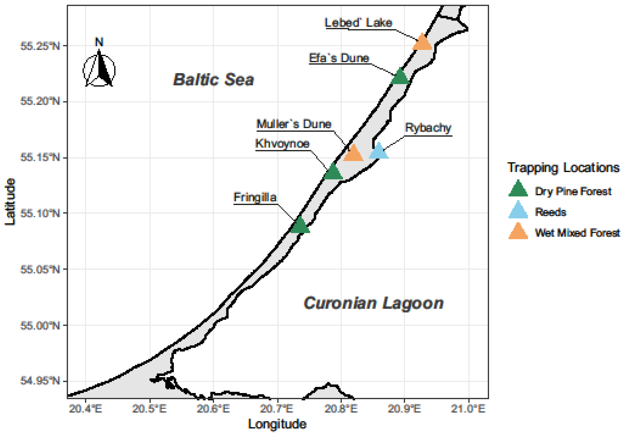
Figure 1. Map of the Curonian Spit and locations of collection points. Dry pine forests: “Fringilla” field station (F), village of Khvoinoye (K), and Dune Efa (E). Wet habitats: mixed forest near Dune Müller (M), Lake Lebed (L), and reed beds along the shore of the Curonian Lagoon on the territory of the “Rybachy” Station (R).
Diversity 16 00723 g001
Figure 2. Number of collected biting midges during the study.
Diversity 16 00723 g002
Figure 3. Number of different Culicoides species collected during the study on the Curonian Spit.
Diversity 16 00723 g003
Figure 4. Number of PCR-positive Culicoides collected during the study on the Curonian Spit.
Diversity 16 00723 g004
Figure 5. Sporozoites of Haemoproteus parabeloposkyi (hSYAT02) found in the salivary glands Culicoides pictipennis (a) and Culicoides kibunensis (b). Arrow indicates sporozoite nucleus. Scale bar 10 µm.
Diversity 16 00723 g005
Table 1. List of species of Culicoides spp. identified on Curonian spit and genetic lineages of avian haemosporidian parasites found in these biting midges. The number of midges in which these parasites were found is in brackets. The lineages of parasites whose sporozoites were found in the salivary glands are highlighted in bold.
Species of Biting MidgesReeds (Rybachy Point, R)Mixed Wet Forest (Dune Müller Point, M)
% (N)Found Genetic Lineages of Parasites% (N)Found Genetic Lineages of Parasites
Culicoides albicans1.05% (6) 2.26% (9)
C. grisescens3.51% (20) 0.25% (1)
C. festivipennis3.33% (19)hTURDUS2 (1)10.8% (43)H. asymmetricus hTUPHI01 (2), hTURDUS2 (1), lSISKIN2
C. impunctatus17.54% (100) 0.25% (1)
C. kibunensis23.51% (134)lROFL6 (2), hBRAM1 (1)25.38% (101)hHAWF6 (1), H. parabelopolskyi hSYAT02 (3), H. asymmetricus hTUPHI01 (4), hBRAM1 (1)
Obsoletus Complex17.01% (97)lBT5 (1), lSISKIN2 (2)5.03% (20)lBT5 (1)
C. pallidicornis3.86% (22) 4.27% (17)
C. pictipennis16.32% (93)H. belopolskyi hHIICT1 (1), H. parabelopolskyi hSYAT02 (1)27.64% (110)H. parabelopolskyi hSYAT02 (5)
C. punctatus27 (4.74) 6.03% (24)hBRAM1 (1)
C. reconditus0 1.51% (6)hBRAM1 (1)
C. segnis8.95% (51)hBRAM1 (3)14.8 newH. belopolskyi hHIICT1 (1), lSISKIN2 (1)
2 (59)
C. sphagnumensis0.18% (1) 1.76% (7)
Total100% (570) 100% (398)
Disclaimer/Publisher’s Note: The statements, opinions and data contained in all publications are solely those of the individual author(s) and contributor(s) and not of MDPI and/or the editor(s). MDPI and/or the editor(s) disclaim responsibility for any injury to people or property resulting from any ideas, methods, instructions or products referred to in the content.