Article

Reassessing the Diversity of the Arthropod-Pathogenic Genus Pandora Batko (Entomophthoromycotina; Erynioideae)

1
Department of Entomology, Cornell University, Ithaca, NY 14850, USA
2
UES, A Blue Halo Company, Dayton, OH 45432, USA
3
Entomology Research Laboratory, University of Vermont, Burlington, VT 05405, USA
*
Author to whom correspondence should be addressed.
Diversity 2024, 16(10), 603; https://doi.org/10.3390/d16100603
Submission received: 25 July 2024 / Revised: 16 September 2024 / Accepted: 24 September 2024 / Published: 1 October 2024
(This article belongs to the Section Phylogeny and Evolution)

Abstract

:
The fungal order Entomophthorales occurs worldwide, with most species infecting arthropods as pathogens. Species in this order can cause epizootics and change the behavior of infected hosts. Molecular data are available only for 20% of the known species, and distributions of species are seldom summarized. Significant diversity of hosts, poor molecular data availability, and poor resolution of the phylogenetic relationships within this fungal order suggest that the diversity of these fungi is not sufficiently described. The subfamily Erynioideae includes 111 arthropod pathogens, divided among six genera, with the genus Pandora being one of the most diverse genera. Sequences of 18S, 28S, and ITS for two species are used to place these Pandora species in a phylogenic tree of the subfamily; this tree also supports our synonymy of the genus Furia with Pandora. Among the two species specifically covered in this paper, Pandora gloeospora was observed during epizootics occurring in mushroom flies (Diptera: Sciaridae) on Agaricus bisporus cultures in Pennsylvania, Delaware, and Maryland (US) mushroom farms and also in Florida on Pleurotus sp. Outside the US, P. gloeospora was found infecting several Nematocera (Diptera) in Europe (France) and Asia (China). Pandora sylvestris n. sp. was collected during epizootics occurring in larvae of hickory tussock moths, Lophocampa caryae (Lepidoptera: Erebidae), in hardwood forests in Michigan and Vermont.

1. Introduction

Species in the order Entomophthorales, subphylum Entomophthoromycotina, are well known as acute pathogens infecting arthropod hosts, at times causing epizootics and changing the behavior of infected hosts before their death [1,2]. Within the order Entomophthorales, family Entomophthoraceae, the subfamily Erynioideae was named in 2005 [3]. This subfamily includes six genera: Erynia, Furia, Orthomyces, Pandora, Strongwellsea, and Zoophthora. Prior to 2005, authors proposed several different structures for classification of Erynia, Furia, Pandora, and Zoophthora [4,5,6,7]; the smaller genera Orthomyces and Strongwellsea were not included in the following re-organizational discussions. In 1964, the genus Zoophthora was described [4], and in 1966 [5], Erynia, Furia, Pandora, and Zoophthora were all named as subgenera within the genus Zoophthora. In 1989 [8], these subgenera were all raised to the generic level. However, when the subfamily Erynioideae was subsequently named, Keller and Petrini [3] stated that ‘the genera Erynia, Furia, and Pandora cannot be clearly separated’ and that ‘Furia and Pandora…are very closely related and a clear separation is almost impossible’. Keller and Petrini [3] suggested that molecular methods could help to clarify relationships. In 2013, Gryganskyi et al. [9] presented phylogenetic analyses of this order based on both molecular and non-molecular data. They demonstrated that among these four genera, Zoophthora was indeed a distinct genus, although the other three genera were not clearly separated. Unfortunately, many species in these three genera have not been isolated, and molecular data for most continue to not be available.
Recent phylogenetic analyses of the Erynioideae now suggest the existence of a cohesive clade among the sequenced species of Erynia [10]. However, species in Pandora and Furia continue to appear to be mixed on the phylogenetic tree presented in this paper. We address this problem and provide a solution by synonymizing Furia with Pandora. The genus Pandora includes thirty-eight species [10], of which fourteen are hosted by Hemiptera, twelve are hosted by Diptera, and two are hosted by Lepidoptera. In contrast, in the closely related genus Furia among the fourteen species, five infect Diptera, one infects Hemiptera, and six infect Lepidoptera. Species in both genera are found around the world.
Included in the phylogeny are new sequence data for two Pandora species, for which biological and distributional data are also presented. The well-known dipteran pathogen Pandora gloeospora, commonly seen infecting pestiferous flies in Agaricus bisporus production facilities in the mid-Atlantic area of the US, is also known from France and China to infect Diptera in the suborder Nematocera. We consider that this species is cosmopolitan, and its distribution might be impacted by human activities, namely spreading mushroom-growing technologies.
In addition, we describe a new species of Pandora that was discovered causing epizootics in larvae of outbreaking (i.e., high density) Lophocampa caryae populations in Vermont (2011) and Michigan (2016). Lophocampa caryae is in the tiger moth tribe (Arctiini) within the family Erebidae (previously Noctuidae). Larvae primarily feed on the foliage of hickory, pecan, butternut, or walnut trees, but when populations are high, they may be found feeding on other trees as well [11]. This host is distributed throughout eastern North America, but this pathogen has never been reported in the literature.
We conducted this study of the genus Pandora because, based on prior phylogenetic studies and increased knowledge about two species (one of which is new), the re-evaluation of this genus was necessary. We found that indeed the genus Furia needed to be synonymized with Pandora.

2. Material and Methods

2.1. Fungal Collection

2.1.1. Pandora gloeospora from Mushroom Flies

Mushroom flies killed by P. gloeospora were collected from a mushroom farm near Kennett Square, PA (39.43833, −75.711058), the mushroom capital of the world. Large numbers of dead adults were found attached to the walls of the growing rooms, mushroom fruit bodies of Agaricus bisporus (Basidiomycota: Agaricales), and vertical parts of the shelves. This fungus was also observed in the Flamingo Gardens and Wildlife Sanctuary, Davie, Florida (26.07452, −80.31332), where dead mushroom flies were attached to Pleurotus sp. (Basidiomycota: Agaricales) fruit bodies.
For one sample from a mushroom production facility near Kennett Square, sporulating cadavers of Lycoriella mali (Diptera: Sciaridae) were collected and sent to the USDA-ARSEF. Conidia from cadavers were showered onto EYSMA (Egg Yolk Sabouraud Maltose Agar) media for isolation [12], resulting in isolate ARSEF #13406. This isolate was subsequently cultured in liquid Grace’s insect cell culture medium as hyphal bodies for DNA extraction.

2.1.2. Fungus from Lophocampa caryae Larvae

Hickory tussock moth larvae killed by an entomophthoralean species were collected from two localized epizootics. One epizootic was discovered in Richmond, Vermont (44.448333, −73.002500), occurring over at least one week during August 2011. The second epizootic was found in Delton, Michigan (42.521049, −85.347413) on 16 September 2015. In both instances, large numbers of dead larvae were attached to tree trunks. In Vermont, most cadavers were collected from a felled butternut tree (Juglans cinerea). Cadavers were collected from tree trunks, where they had been attached by rhizoids, and conidia were showered onto lids of petri dishes where they were measured [13,14]. Conidia and hyphal bodies were stained with lactophenol cotton blue and photographed. Hyphal bodies that were observed were in the Malpighian tubules and the fat body. Resting spores were found within cadavers that did not produce conidia, and these were measured. Cadavers from Vermont that had been collected were deposited in the Entomology Research Laboratory at the University of Vermont (ERL 1970 and 1971). The fungus was isolated, and the culture was deposited in ARSEF (#11764) on EYSMA [12]. In Michigan, cadavers were cut from trunks of red oaks (Quercus rubra) using a knife. It was not possible to establish cultures from Michigan samples, but cadavers were collected individually and maintained in ethanol at 4 °C until use.

2.2. DNA Extraction, PCR Amplification and Sequencing

2.2.1. Kennett Square, Pennsylvania Sample from Lycoriella mali

A L. mali cadaver was ground in liquid nitrogen, and DNA was extracted in 2x CTAB buffer following the chloroform:isoamyl alcohol 24:1 technique as described in Gryganskyi et al. [15]. SSU (18S), LSU (28S), and ITS were sequenced. SSU primers were nssu1088R and NS24 and LSU primers were LR5 and LROR [16]. ITS primers were ITS1F [17] and ITS4 [18]. For all amplified regions, PCR conditions were as described in Hajek et al. [19]. Shrimp Alkaline Phosphatase (GE Healthcare, Piscataway, NJ, USA) and Exonuclease I (New England Biolabs, Ipswich, MA, USA) were used for PCR cleanup at 0.1 units of SAP and 0.23 units of EXO following the manufacturer’s recommendations. Purified products were sequenced via the Sanger method with both forward and reverse primers using an ABI 3730xl DNA Analyzer at Genewiz Azenta Life Sciences (South Plainfield, NJ, USA). Raw sequence data were analyzed and edited using Sequencher v. 4.1.4 software (Gene Codes Corporation, Ann Arbor, MI, USA).

2.2.2. Michigan Sample from Lophocampa caryae

Hyphae were removed from a cadaver, and DNA was extracted using a Qiagen DNeasy Plant Mini Kit (Hilden, Germany) following bead beating methods described in Hajek et al. [19]. SSU, LSU, and ITS were sequenced. SSU primers were nssu1088R and NS24 [16]. LSU primers were nu-LSU-0018-5’ [20] and nu-LSU0805-3’ [21]. For both SSU and LSU, PCR conditions were as described in Hajek et al. [19]. ITS primers were ITS1F [17] and ITS4 [18]. PCR conditions were 95 °C for 5 min, followed by 10 cycles of 95 °C for 30 s, 70 °C for 30 s (decreasing 1 °C per cycle), and 72 °C for 2 min, followed by 25 cycles of 95 °C for 30 s, 60 °C for 30 s, and 72 °C for 2 min, followed by 72 °C for 10 min. PCR amplicons were cleaned before sequencing as described above. The cleaned products were sequenced via the Sanger method at the Cornell University Biotechnology Resource Center on an ABI3730xl using the primers LROR [22], nssu1088R, or ITS5. Sequences were edited, assembled, aligned, and searched using Geneious software v. 8.1.8 (Biomatters Ltd., Auckland, New Zealand).

2.2.3. Vermont Sample from Lophocampa caryae

DNA was extracted from fungal mycelium from culture plates using the Qiagen DNeasy Power Soil kit. ITS was evaluated using primers ITS5 [18] and ITS4. The PCR program included 94 °C for 2 min, 40 cycles of 95 °C for 30 s, 52 °C for 30 s, 72 °C for 30 s, and a 10 min extension at 72 °C. PCR products were purified with the Qiaquick PCR Purification Kit before sequencing. The forward and reverse sequences were edited with Bioedit (Informer Technologies Inc., Asheville, NC, USA) to create a consensus sequence.

2.3. Phylogenetic Analyses

The first goal of this study was to construct a well-resolved, two-loci phylogeny (18S and 28S) for the subfamily Erynioideae to test whether the group is monophyletic and to resolve the major lineages within the subfamily. In order to build a phylogenetic reconstruction of this fungal group, we created a dataset including sequences from two non-protein-coding rDNA loci: SSU and LSU, 20% of which were generated by the authors of this study (Table 1). ITS sequences were excluded from the analysis because of the large amount of data and significant variability of the sequences. The sequences used for analyses belong to 44 fungal taxa, representing 10 genera: 30 Erynioideae isolates (ingroup: genera Erynia, Furia, Pandora, Strongwellsea, Zoophthora and missing molecular data for Orthomyces) and 14 isolates from other fungal lineages (outgroups). The outgroup taxa include 12 species of the closely related Entomophthoroideae subfamily and two distantly related Neoconidiobolus species; in total, the outgroup taxa represented six genera: Arthrophaga, Entomophaga, Entomophthora, Eryniopsis, Massospora, and Neoconidiobolus.
The data were combined into a single matrix with two partitions: one partition for each locus of the loci SSU and LSU. The SSU rDNA and LSU rDNA partitions included 1559 and 762 characters, respectively, for a combined data matrix of 2321 characters. The 18S sequence dataset had a missing record for one taxon, and the 28S sequence dataset had two missing records.
Sequences were aligned individually for each locus using MUSCLE [23]. Alignments were visually inspected, and ambiguous regions were excluded using Mesquite [24]. The optimized nucleotide substitution model (GTR + Γ + I) was selected using ModelTest 3.06 [25]. Maximum likelihood (ML) phylogenetic trees for each locus were estimated using Garli-2.0 [26]. To assess conflicting phylogenetic signals from the two loci, we searched for strong incongruence of the nodes by 1000 ML bootstrap replicates. Statistical support was recognized as significant with bootstrap values ≥70%.

3. Results

3.1. Phylogenetic Reconstruction

The phylogenetic tree revealed several groups within the overall Erynioideae clade. Only two of them, Erynia and Zoophthora, had strong bootstrap support. The genus Erynia, represented by three species, appeared to be basal to the whole Erynioideae clade. In contrast, species of the genus Zoophthora were placed on the most distant branch. There are two other well-defined subclades by bootstrap values, and both contain a mixture of Furia and Pandora species. However, when the molecular data of two or more strains are included in the phylogenetic reconstruction (for example, P. delphacis, P. kondoiensis, and P. neoaphidis), such branches of a single species with multiple strains have significant bootstrap support. Strongwellsea castrans, with only 18S data available, was located closer to the larger Pandora subclade (Figure 1).
Since the molecular data of the representatives of Furia and Pandora are not separate from each other on the phylogenetic tree, we propose to synonymize Furia with Pandora.

Taxonomy

Pandora Humber, Mycotaxon 34 (2): 451 (1989), emend. Mycobank #25327.
Type species: Pandora neoaphidis (Remaud. and Hennebert) Humber.
Description: Hyphal bodies irregularly subspherical to short, hyphae are unbranched or with few branches. Conidiophores digitally or dichotomously branched. Uninucleate primary conidia are ovoid, obpyriform, subcylindrical, or obclavate, straight or slightly bent. Secondary conidia of two types: 1a resembling primary, or 1b spherical conidial body with distinct papilla, often with an indistinct apical point; both types of primary conidia forcibly discharged. Capilliconidia absent. Resting spores spherical, hyaline, or colored; episporium smooth or ornamented; resting spores unknown for some species. Cystidia present, ranging from the same thickness as conidiophores to 2–3 times thicker, tapering apically. Rhizoids monohyphal, ranging from the same thickness as condiophores to 2–3 times thicker, with terminal holdfast varying from discoid to having few irregular short branches. Obligate pathogens of insects or Phalangiidae (Arachnida).
NOTE: Generic description based on Keller and Petrini [3].
Pandora virescens (Thaxt.) Hajek and Gryganskyi, comb. nov. Mycobank #854747, Empusa virescens Thaxt., Memoirs of the Boston Society of Natural History 4 (6): 133–201 (1888). Basionym. [TYPE for Furia].
Pandora americana (Thaxt.) S. Keller Sydowia 59(1): 99 (2007) Mycobank #529930, Empusa americana Thaxt., Mem. Boston Soc. Nat. Hist. 4 (6): 133–201 (1888). Basionym.
Pandora creatonoti (D.F. Yen ex Humber) Hajek and Gryganskyi, comb. nov. Mycobank #854749, Erynia creatonoti D.F. Yen, Mycotaxon 13 (3): 474 (1981). Basionym.
Pandora ellisiana (Ben Ze’ev) Hajek and Gryganskyi, comb. nov. Mycobank #854750, Erynia ellisiana Ben Ze’ev, Mycotaxon 27: 266 (1986). Basionym.
Pandora fujiana (Y.J. Huang and Z.Z. Li) Hajek and Gryganskyi, comb. nov. Mycobank #854751, Furia fujiana Y.J. Huang and Z.Z. Li, Acta Mycologica Sinica 12 (1): 1 (1993). Basionym.
Pandora fumimontana (Bałazy) Hajek and Gryganskyi, comb. nov. Mycobank #854752, Zoophthora fumimontana Balazy, Flora of Poland. Fungi (Mycota), Vol. 24: Entomophthorales: 166 (1993). Basionym.
Pandora gastropachae (Racib.) Hajek and Gryganskyi, comb. nov. Mycobank #854753, Empusa gastropachae Racib., Kosmos (Lvov) 35: 775 (1910). Basionym.
Pandora ithacensis (J.P. Kramer) Hajek and Gryganskyi, comb. nov. Mycobank #854754, Erynia ithacensis J.P. Kramer, Mycopathologia 75 (3): 160 (1981). Basionym.
Pandora montana (Thaxt.) Hajek and Gryganskyi, comb. nov. Mycobank #854755, Empusa montana Thaxt., Mem. Boston Soc. Nat. Hist. 4 (6): 133–201 (1888). Basionym.
Pandora neopyralidarum (Ben Ze’ev) Hajek and Gryganskyi, comb. nov. Mycobank #854756, Erynia neopyralidarum Ben Ze’ev, Mycotaxon 16 (1): 276 (1982). Basionym.
Pandora pieris (Ben Ze’ev) Hajek and Gryganskyi, comb. nov. Mycobank #854757, Erynia pieris Z.Z. Li and Humber, Canad. J. Bot. 62 (4): 656 (1984). Basionym.
Pandora sciarae (L.S. Olive) S. Keller, Sydowia 59(1): 104 (2007). Mycobank #529932, Empusa sciarae Olive, Botanical Gazette Crawfordsville 41 (3): 196 (1906). Basionym.
Pandora shandongensis (W.M. Wang, W.H. Lu, and Z.Z. Li) Hajek and Gryganskyi, comb nov. Mycobank #854758, Furia shandongensis W.M. Wang, W.H. Lu, and Z.Z. Li, Mycotaxon 50: 302 (1994). Basionym.
Pandora triangularis (Villac. and Wilding) Hajek and Gryganskyi, comb nov. Mycobank #854759, Erynia triangularis Villac. and Wilding, Mycological Research 98 (2): 157 (1994). Basionym.
Pandora vomitariae (Rozsypal) Hajek and Gryganskyi, comb nov. Mycobank #854760, Zoophthora vomitoriae Rozsypal, Acta Mycologica Warszawa 2: 24 (1966). Basionym.
Pandora zabri (Rozsypal ex Ben Ze’ev and R.G. Kenneth) Hajek and Gryganskyi, comb nov. Mycobank #854761, Erynia zabri Rozsypal ex Ben Ze’ev and R.G. Kenneth, Mycotaxon 14 (2): 465 (1982). Basionym.

3.2. Pandora gloeospora Biology and Distribution

In the past, P. gloeospora has been placed in the genera Entomophthora [27], Erynia [28], Zoophthora [4], Pandora [8], and Furia [29]. Our phylogenetic reconstruction shows that it belongs in the genus Pandora.
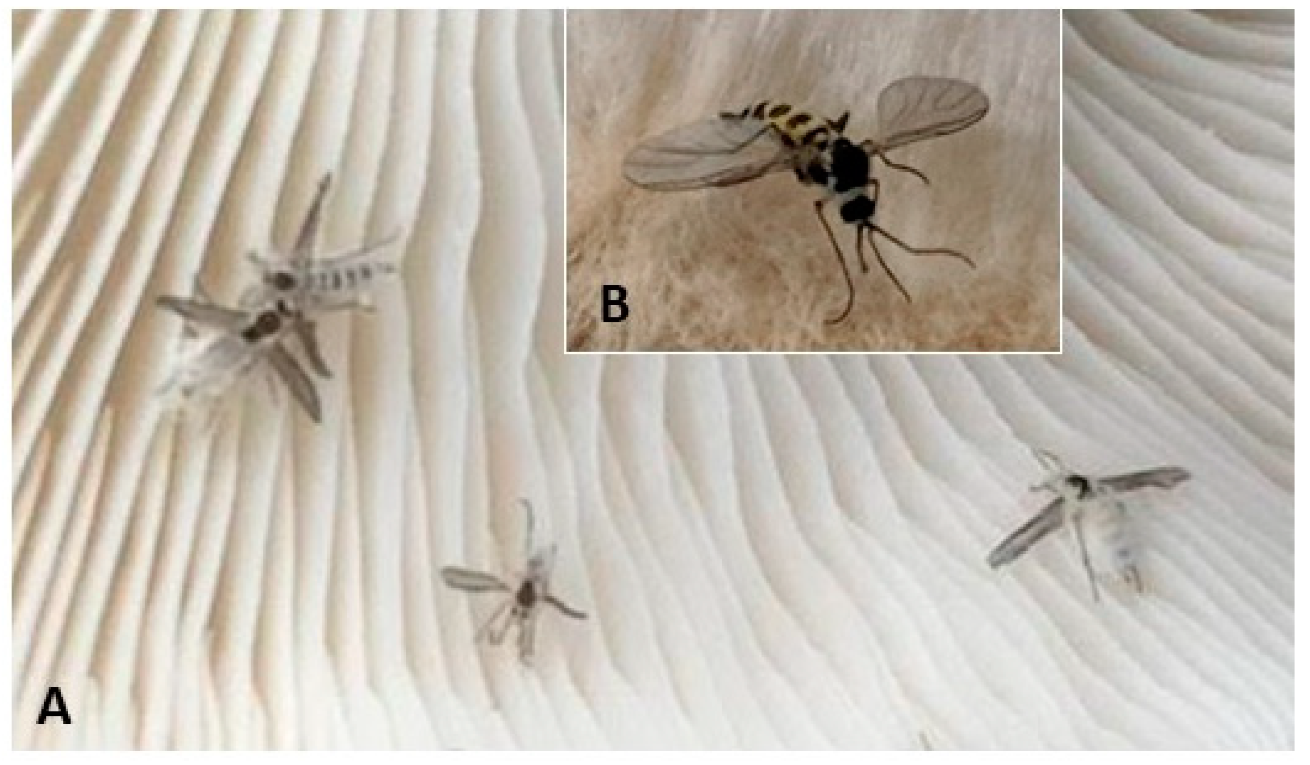
BIOLOGY. At death, wings of flies dying from P. gloeospora infections were at 90° to the length of the body (Figure 2).
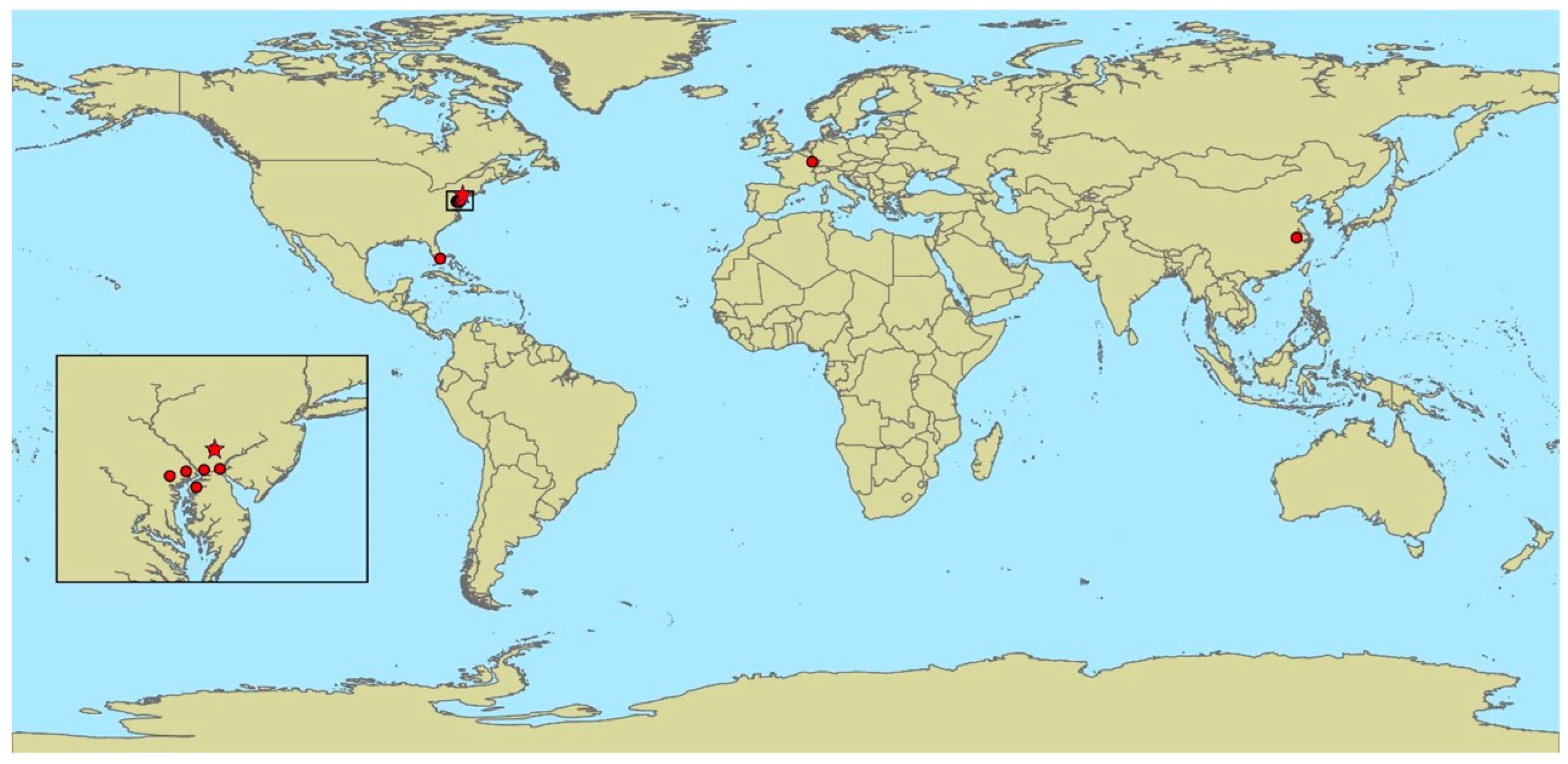
COLLECTION LOCATIONS: This species is reported from France [27], Chester Co., PA, USA [30], and Xuancheng, Anhui province, China [31]. We report now that this species also occurs in mushroom production facilities in New Castle County, Delaware, and in four counties in Maryland: Baltimore, Cecil, Harford, and Kent. The initial collection location used for the samples for the description of P. gloeospora is not clearly stated in the 1886 description [27] (Figure 3). Thus, we use the location Nancy, France, as this is the address of the author, and he describes this fungus causing epizootics in “moucherons” (= gnats and midges) living in his cultures of primitive fungi (and we assume these cultures would have been in his laboratory in Nancy).
Sciarid flies infected by P. gloeospora were detected in mushroom farms on the walls in growing rooms from 20 cm above the floor and higher, but mostly up to 2 m high. Infected flies were also found on the shelves and on mushroom fruitbodies. In the botanical garden in Florida, infected flies were detected on Pleurotus sp. fruitbodies (Figure 2A).
HOSTS: All records are from Diptera, and perhaps all are Nematocera. In the species description, Vuillemin [27] reported that this species infected ‘moucherons’, which translates to the general terms for gnat or midge. In Chester Co., Pennsylvania, USA, P. gloeospora infected Lycoriella mali (Sciaridae) [30], while in China, Psychoda sp. (Psychodidae) was infected [29].

3.3. New Species Description

Pandora sylvestris Hajek and Gryganskyi sp. nov.
HOLOTYPE: Cornell University, Plant Pathology Herbarium CUP-067659.
PARATYPES: Cornell University, Plant Pathology Herbarium CUP-067660, 067661
Holotypes and paratypes were collected in Vermont, USA: Richmond (Figure 4).
MYCOBANK: #854746.
HOST IDENTIFICATION: Lophocampa caryae Harris (late instar larvae).
COLLECTION LOCATIONS: Richmond, Chittenden County, Vermont (44.448333, −73.002500) and Delton, Barry County, Michigan (42.521049, −85.347413) (Figure 4).
DESCRIPTION: When collections were made, host populations were abundant, and larvae were later instars that were frequently oriented vertically on trees with their heads downward after dying from infections (Figure 5A). Hyphal bodies within recently dead larvae were thin, elongate, and multinucleate (Figure 5F,G). Conidiophores branched, emerging through intersegmental membranes to produce primary conidia. Primary conidia were 12.5 ± 0.3 (mean ± SE) µm long and 7.4 ± 0.1 µm wide (n = 20), and ovoid, with an evenly rounded apex tapering to a rounded basal papilla (Figure 5B–E). Cystidia were present, similar in width to conidiophores and often with tapered ends. Resting spores occurred within cadavers not producing conidia and were 37.9 ± 0.7 µm (32.6–45.6 µm) in diameter. Resting spores were black en masse, but under a compound microscope, were light brown. Episporia averaged 2.0 ± 0.9 µm thick (0–3.3 µm), consisting of convex plates of irregular shapes, averaging 6.4 + 0.4 µm (3.3–9.8 µm) in width (Figure 5H–J). Rhizoids occurred, but none were intact, as these had broken or been cut when cadavers were removed from tree bark.
GenBank numbers for the P. sylvestris samples collected in 2016 in Michigan are PQ038062 for 18S, PQ062134 for 28S, and PQ038419 for ITS. The GenBank number for the ITS for the P. sylvestris sample collected in 2011 in Vermont is PQ038420. Comparison of the ITS sequences demonstrated that the fungus sequences from the two sites were the same.
Etymology. The specific epithet describes the habitat where this species was collected.
DISTINGUISHING CHARACTERS. Resting spores appear black en masse and have patterned, bumpy epispores (Figure 4). Hosts are larval Lophocampa caryae collected in hardwood forests in Vermont and Michigan.
REMARKS. When collections were made, host populations were abundant. In Vermont, many cadavers could be seen on tree trunks and branches higher in tree canopies, and cadavers could be easily collected because one butternut tree had recently been felled. Surprisingly, this fungal species had never been described previously, although this host species occurs throughout northeastern North America, where it is perhaps best known for its urticating setae. Larvae of this native defoliator occasionally become abundant on nut trees, but at these times it will be common on several other broad-leaved trees as well. However, high populations do not persist, and little damage results [11].

4. Discussion

In this paper, we have synonymized the genus Furia with Pandora. Fungi in the genera Erynia and Zoophthora (including the species with no molecular data available) have distinct morphological and ecological features, which separate them from the rest of the subfamily Erynioideae [10]. Erynia species have characteristic conical conidia with wide cystidia and are often found in aquatic and semi-aquatic habitats. Zoophthora species have sticky secondary capilliconidia, absent in other species of this subfamily. The main distinction between the other two larger genera in this subfamily, Furia and Pandora, lies in the rhizoids that attach the infected arthropods to substrates and sometimes the cystidia that grow from the hymenium. Rhizoids of Pandora are described as 2–3× thicker than the rhizoids of Furia [8], which are similar in width to conidiophores. Attachment points of rhizoids are variable, although Pandora creates a discoid or irregularly branched and spreading holdfast [8]. Cystidia of Pandora, when present, are also reported to be thicker than conidiophores. However, information on rhizoids and cystidia is not available in all descriptions of species in Furia and Pandora. Rhizoids are often not mentioned or are included without any details, or these structures can be lost or damaged when cadavers are collected. Miller and Keil [30] stated that for P. gloeospora, patterns of rhizoids and cystidia are variable depending on the environment, time of infection, host sex, and larval diet, although the morphology of structures remained constant when expressed. In part for these reasons, Keller and Petrini [3] considered the genera Pandora and Furia almost impossible to separate based on morphology. In addition, across the Entomophthorales, frequently species have not been cultured or sequenced, and thus molecular information is not available, so morphology, based on missing information on structures such as rhizoids, must be used for identification.
Based on available sequences, our phylogenetic reconstruction supports the fact that there is no clear distinction between Pandora and Furia, as these two genera are admixed in the phylogenetic tree. Descriptions of many of the species in these genera have been based on morphology, yet the necessary rhizoids and cystidia that differ between these genera [8] are often ‘unknown or incompletely known’ [3]. Keller and Petrini [3] stated that perhaps molecular work would help to solve the question of whether these genera are justified. However, based on molecular data, admixtures between these two genera have been reported repeatedly in numerous publications: Scorsetti et al. [32], Gryganskyi et al. [9], Hajek et al. [19], Hodge et al. [33], Nie et al. [34], Keller et al. [35], and Gryganskyi et al. [10]. This is the basis for synonymizing Furia with Pandora. Both genera were initially proposed as subgenera in the same publication [5], but Pandora was described before Furia in that paper, and therefore we synonymize Furia with Pandora.
Our paper also provides distributional data and the first sequences for P. gloeospora. Vuillemin [27] originally reported that P. gloeospora-infected ‘moucherons’ were associated with his cultures of ‘lower fungi’. Miller and Keil [30] reported that this fungus infected sciarids that are pests in mushroom production facilities in Chester County, PA, and that this fungal pathogen was rare. We now report the occurrence of this fungus associated with mushroom farms in five more counties in two more mid-Atlantic US states. In Florida, this fungus was also observed infecting sciarids. In contrast, in China, Li et al. [29] found this fungus infecting Psychoda sp. (Psychodidae) at an exposed sewer. Therefore, it seems that P. gloeospora occurs in several types of habitats that can host these types of Nematocera. Curiously, in 1993, Bałazy [7] reported that Mycetophila sp. (Mycetophilidae) were infected by P. gloeospora in France, citing Vuillemin [27]. The common name for mycetophilids is ‘fungus gnat’ so we assume that is why Bałazy used this dipteran family name. However, the original publication does not provide the names Mycetophilidae or Mycetophila sp., so we have no proof that species in this fly family are infected. However, it is quite possible that P. gloeospora is a generalist that is able to infect hosts from different insect nematoceran families since it is known to infect both Sciaridae and Psychodidae.
Agaric monoculture with a daily harvest of tons of fresh mushrooms of the same age and some dozen tons of compost and casing soil, penetrated by fungal mycelium, creates favorable conditions that especially attract mushroom flies of two families (phorids in the genus Megaselia and sciarids in the genus Lycoriella), with larvae feeding on mushroom mycelium and fruit bodies. At times fly populations outbreak and the abundance of mushroom flies in growing rooms under the stable humid and warm microclimate conditions create favorable conditions for the epizootics caused by P. gloeospora. Miller and Keil [30] described P. gloeospora as being ‘rare’. We do not know the abundance of this fungus in France and China, but in mushroom production facilities in the mid-Atlantic areas of the US, infection is not rare, and levels of infection can be significant when fly populations are abundant.
Finally, we describe a new species of Pandora infecting the native hickory tussock moth in North America. At present, 80 species of Entomophthoromycotina, including P. sylvestris, are known from North America (AEH unpublished data). It is surprising that P. sylvestris has only now been described from North America. We suspect that the irregularity of outbreaks by this native hardwood defoliator specializing on forest trees that are often not dominant has thwarted the discovery of this fungal pathogen until recent years. At present, we can only report infection in one host species, and whether other hosts are infected is unknown. However, many species in the Erynioideae have narrow host ranges or are known from only one host species, so a narrow hosst range would be consistent with the trend for this subfamily [10].
In the genus Pandora (now also including Furia), DNA sequences are available for relatively few species. We look forward to the addition of more sequences as well as genomes to add molecular data for differentiation and to improve our understanding of the genera in the Erynioideae. In addition, we hope that in the future methods for collection of specimens without damage to rhizoids and cystidia will be developed so that species descriptions will include improved information about these structures when describing morphology.

5. Conclusions

This paper includes a description of a new species in the genus Pandora and new biological and distributional data for P. gloeospora. Phylogenetic analysis demonstrated that both species belong in the genus Pandora. The phylogenetic reconstruction demonstrated that based on the available sequences, the genus Pandora is admixed with the genus Furia. Morphological characters that have previously been used to separate these genera have been seriously questioned in the past [3]. Therefore, we have synonymized the genus Furia with Pandora.

Author Contributions

Conceptualization, A.E.H. and A.P.G.; investigation, S.Y.G., A.E.H., and A.P.G.; resources, C.F.S.; data curation, S.Y.G., A.E.H., and C.F.S.; writing—original draft preparation, A.E.H. and A.P.G.; writing—review and editing, A.E.H., A.P.G., T.D.B., S.Y.G., and C.F.S.; visualization, A.E.H. and A.P.G.; supervision, A.E.H. and B.L.P.; funding acquisition, B.L.P. All authors have read and agreed to the published version of the manuscript.

Funding

This research received no external funding.

Data Availability Statement

Data is contained within the article.

Acknowledgments

The description of P. sylvestris would not be possible without Jeff Boettner and Greg Dwyer (collections in Michigan) and Don Tobi (collections in Vermont) finding the epizootics and collecting specimens that they shared with us. We thank Mark W. Miller for the records and photos of P. gloeospora from Florida and Sara Cahan and Yaianna Hall (Vermont) for their assistance with the molecular work. We also thank Louela Castrillo, Kathie Hodge, Micheal Wheeler, and Kathryn Bushley of the USDA ARS ARSEF culture collection for their assistance, Jim Liebherr for assistance with the photography, Eugene Luzader and Andrew Liebhold for preparing the maps, and Bo Huang for assistance with the Chinese literature.

Conflicts of Interest

Author Andrii P. Gryganskyi was employed by the company UES. The remaining authors declare that the research was conducted in the absence of any commercial or financial relationships that could be construed as a potential conflict of interest.

References

  1. Pell, J.; Eilenberg, J.; Hajek, A.E.; Steinkraus, D.C. Biology, ecology and pest management potential of Entomophthorales. In Fungi as Biocontrol Agents: Progress, Problems and Potential; Butt, T.M., Jackson, C.W., Magan, N., Eds.; CABI Publishing: Wallingford, UK, 2001; pp. 71–153. [Google Scholar]
  2. Roy, H.E.; Steinkraus, D.C.; Eilenberg, J.; Hajek, A.E.; Pell, J.K. Bizarre interactions and endgames: Entomopathogenic fungi and their arthropod hosts. Annu. Rev. Entomol. 2006, 51, 331–357. [Google Scholar] [CrossRef] [PubMed]
  3. Keller, S.; Petrini, O. Keys to the identification of the arthropod pathogenic genera of the families Entomophthoraceae and Neozygitaceae (Zygomycetes), with descriptions of three new subfamilies and a new genus. Sydowia 2005, 57, 23–53. [Google Scholar]
  4. Batko, A. On the new genera: Zoophthora gen. nov., Triplosporium (Thaxter) gen. nov., and Entomophaga gen. nov. (Phycomycetes: Entomophthoraceae). Bull. Acad. Pol. Sci. Cl II 1964, 12, 323–326. [Google Scholar]
  5. Batko, A. On the subgenera of the fungus genus Zoophthora Batko 1964 (Entomophthoraceae). Acta Mycol. 1966, 2, 15–21. [Google Scholar] [CrossRef]
  6. Ben Ze’ev, I.; Kenneth, R.G. Features criteria of taxonomic value in the Entomophthorales. 1. A revision of the Batkoan classification. 2. A revision of the genus Erynia Nowakowski 1881 (Zoophthora Batko 1965). Mycotaxon 1982, 14, 393–475. [Google Scholar]
  7. Bałazy, S. Flora Polska, Grzyby (Mycota). Vol. 24. Owadomorkowe, Entomophthorales; Polish Academy of Sciences: Kraków, Poland, 1993; 356p. [Google Scholar]
  8. Humber, R.A. Synopsis of a revised classification for the Entomophthorales (Zygomycotina). Mycotaxon 1989, 34, 441–460. [Google Scholar]
  9. Gryganskyi, A.P.; Humber, R.A.; Smith, M.E.; Hodge, K.; Huang, B.; Voigt, K.; Vilgalys, R. Phylogenetic lineages in Entomophthoromycota. Persoonia 2013, 30, 94–105. [Google Scholar] [CrossRef]
  10. Gryganskyi, A.P.; Hajek, A.E.; Voloshchuk, N.; Idnurm, A.; Eilenberg, J.; Manfrino, R.G.; Bushley, K.E.; Kava, L.; Kutovenko, B.; Anike, F.; et al. Potential for use of species in the subfamily Erynioideae for biological control and biotechnology. Microorganisms 2024, 12, 168. [Google Scholar] [CrossRef]
  11. Rose, A.H.; Lindquist, O.H. Insects of eastern hardwood trees. Canad. For. Serv., For. Tech. Rpt. 1982, 29, 212. [Google Scholar]
  12. Hajek, A.E.; Papierok, B.; Eilenberg, J. Methods for study of the Entomophthorales, In Manual of Techniques in Invertebrate Pathology, 2nd ed.; Lacey, L., Ed.; Academic Press: Amsterdam, The Netherlands, 2012; pp. 285–316. [Google Scholar]
  13. Bosso, L.; Senatore, M.; Varlese, R.; Ruocco, M.; Garonna, A.P.; Bonanomi, G.; Mazzoleni, S.; Cristinzio, G. Severe outbreak of Fusarium solani on Quercus ilex vectored by Xylosandrus compactus. J. Plant Pathol. 2012, 94 4 Supplement, S4.99. [Google Scholar]
  14. Acosta, R.I.T.; Humber, R.A.; Sánchez-Peña, S.R. Zoophthora radicans (Entomophthorales), a fungal pathogen of Bagrada hilaris and Bactericera cockerelli (Hemiptera: Pentatomidae and Triozidae): Prevalence, pathogenicity, and interplay of environmental influence, morphology, and sequence data on fungal identification. J. Invertebr. Pathol. 2016, 139, 82–91. [Google Scholar] [CrossRef]
  15. Gryganskyi, A.P.; Golan, J.; Dolatabadi, S.; Mondo, S.; Robb, S.; Idnurm, A.; Muszewska, A.; Steczkiewicz, K.; Masonjones, S.; Liao, H.L.; et al. Phylogenetic and phylogenomic definition of Rhizopus species. G3 (Bethesda) 2018, 8, 2007–2018, Erratum in G3 (Bethesda) 2019, 9, 2789. [Google Scholar] [CrossRef] [PubMed]
  16. Kauff, F.; Lutzoni, F. Phylogeny of the Gyalectales and Ostropales (Ascomycota, Fungi): Among and within order relationships based on nuclear ribosomal RNA small and large subunits. Molec. Phylogen. Evol. 2002, 25, 138–156. [Google Scholar] [CrossRef] [PubMed]
  17. Gardes, M.; Bruns, T.D. ITS primers with enhanced specificity for basidiomycetes—Application to the identification of mycorrhizae and rusts. Mol. Ecol. 1993, 2, 113–118. [Google Scholar] [CrossRef]
  18. White, T.J.; Bruns, T.; Lee, S.; Taylor, J. Amplification and direct sequencing of fungal ribosomal RNA genes for phylogenetics. In PCR Protocols: A Guide to Methods and Applications (I); Innis, M.A., Gelfand, D.H., Sninsky, J.J., White, T.J., Eds.; Academic Press: San Diego, CA, USA, 1990; pp. 315–322. [Google Scholar]
  19. Hajek, A.E.; Gryganskyi, A.; Bittner, T.; Liebherr, J.K.; Liebherr, J.H.; Moulton, J.K.; Jensen, A.B.; Humber, R.A. Phylogenetic placement of two species known only from resting spores: Zoophthora independentia sp. nov. and Z. porteri comb nov. (Entomophthorales: Entomophthoraceae). J. Invertebr. Pathol. 2016, 140, 68–74. [Google Scholar] [CrossRef]
  20. Jensen, A.B.; Eilenberg, J. Genetic variation within the insect-pathogenic genus Entomophthora, focusing on the E. muscae complex, using PCR-RFLP of the ITS II and the LSU rDNA. Mycol. Res. 2001, 105, 307–312. [Google Scholar] [CrossRef]
  21. Kjøller, R.; Rosendahl, S. Detection of arbuscular mycorrhizal fungi (Glomales) in roots by nested PCR and SSCP (Single Stranded Confirmation Polymorphism). Plant Soil 2000, 226, 189–196. [Google Scholar] [CrossRef]
  22. Vilgalys, R.; Hester, M. Rapid genetic identification and mapping of enzymatically amplified ribosomal DNA from several Cryptococcus species. J. Bacteriol. 1990, 172, 4238–4246. [Google Scholar] [CrossRef] [PubMed]
  23. Edgar, R.C. MUSCLE: Multiple sequence alignment with high accuracy and high throughput. Nucleic Acids Res. 2004, 32, 1792–1797. [Google Scholar] [CrossRef]
  24. Maddison, W.P.; Maddison, D.R. Mesquite: A modular system for evolutionary analysis, Version 3.81. 2023. Available online: https://www.mesquiteproject.org/ (accessed on 23 July 2024).
  25. Posada, D.; Crandall, K.A. MODELTEST: Testing the model of DNA substitution. Bioinformatics 1988, 14, 817–818. [Google Scholar] [CrossRef]
  26. Zwickl, D.J. Genetic Algorithm Approaches for the Phylogenetic Analysis of Large Biological Sequence Datasets under the Maximum Likelihood Criterion. Ph.D. Thesis, The University of Texas at Austin, Austin, TX, USA, 2006; 125 p. [Google Scholar]
  27. Vuillemin, P. Études biologiques sur les champignons. Soc. Sci. (Nancy) Bull. Soc. Sci. Nancy 1886, 8 (année 19), 33–46. [Google Scholar]
  28. Remaudière, G.; Hennebert, G.L. Révision systématique de Entomophthora aphidis Hoffm. in Fres. Description de deux nouveaux pathogènes d’Aphides. Mycotaxon 1980, 11, 269–321. [Google Scholar]
  29. Li, Z.; Huang, B.; Fan, M. New species, new record, new combinations and emendation of entomophthoralean fungi pathogenic to dipteran insects. Mycosystema 1997, 16, 91–96. [Google Scholar]
  30. Miller, M.W.; Keil, C.B. Redescription of Pandora gloeospora (Zygomycetes: Entomophthorales) from Lycoriella mali (Diptera: Sciaridae). Mycotaxon 1990, 38, 227–231. [Google Scholar]
  31. Zha, L.-S.; Wen, T.-C.; Hyde, K.D.; Kang, J.-C. An updated checklist of fungal species in Entomophthorales and their host insects from China. Mycosystema 2016, 35, 666–683. [Google Scholar] [CrossRef]
  32. Scorsetti, A.C.; Jensen, A.B.; López Lastra, C.; Humber, R.A. First report of Pandora neoaphidis resting spore formation in vivo in aphid hosts. Fung. Biol. 2012, 116, 196–203. [Google Scholar] [CrossRef] [PubMed]
  33. Hodge, K.; Hajek, A.E.; Gryganskyi, A. The first entomophthoralean killing millipedes, Arthrophaga myriapodina n. gen. n. sp., causes climbing before host death. J. Invertebr. Pathol. 2017, 149, 135–140. [Google Scholar] [CrossRef]
  34. Nie, Y.; Yu, D.-S.; Wang, C.-F.; Liu, X.-Y.; Huang, B. A taxonomic revision of the genus Conidiobolus (Ancylistaceae, Entomophthorales): Four clades including three new genera. MycoKeys 2020, 66, 55–81. [Google Scholar] [CrossRef]
  35. Keller, S.; Hülsewig, T.; Jensen, A.B. Fungi attacking springtails (Sminthuridae, Collembola) with a description of Pandora batallata, sp. nov. (Entomophthoraceae). Sydowia 2022, 75, 37–45. [Google Scholar] [CrossRef]
Figure 1. Maximum likelihood phylogenetic tree of the Erynioideae subfamily. Outgroups–Neoconidiobolus thromboides and species in the Entomophthoroideae subfamily. Dashed lines for the branches with partial data.
Diversity 16 00603 g001
Figure 2. Pandora gloeospora. (A) Sciarid flies infected with P. gloeospora, on the fruit bodies of Pleurotus sp. (B) A sciarid killed by P. gloeospora on the surface of an Agaricus bisporus fruit body.
Diversity 16 00603 g002
Figure 3. Worldwide geographic distribution of Pandora gloeospora (red dots and star). Pandora gloeospora that was used for sequencing was collected at the location marked with a red star in the inserted map.
Diversity 16 00603 g003
Figure 4. Distribution of Pandora sylvestris, found only in Vermont and Michigan. Pandora sylvestris was collected at both locations (red dots) on the map.
Diversity 16 00603 g004
Figure 5. Pandora sylvestris. (A) Cadaver of late instar larva oriented vertically on bark of a tree where it is attached by rhizoids. Cadaver approx. 4.0 cm long. (BE) P. sylvestris conidia. Primary conidium of P. sylvestris being produced at the end of a conidiophore (B), producing a secondary conidium (C,D), and the cytoplasm from a primary conidium has moved into a secondary conidium (E). (F,G) Hyphal bodies of P. sylvestris with stained nuclei, in Malpighian tubules. (HJ) Resting spores of P. sylvestris. (H) Oil droplets visible inside of resting spores. (I) Epispore of entire resting spore showing bumpy surface and (J) Epispore broken, showing the bumpy surface. Scale bars: (BE) = 5 µm; (F,G) = 2; (HJ) = 20 µm.
Diversity 16 00603 g005
Table 1. GenBank accession numbers of the sequences used for phylogenetic reconstruction (sequences, generated by the authors, in bold).
Species, # of Specimens18S Accession #28S Accession #
Arthrophaga myriapodina (2)MF544092, MF544093MF544094, NG058604
Entomophaga aulicaeU35394EF392372
Ent. maimaigaEF392556EF392395
Entomophthora muscae (2)AY635820, D29948DQ273772, DQ481224
En. planchonianaAF353723MH366738
En. schizophoraeAF052402DQ481228
Erynia conicaAF368513EF392396
E. ovisporaJX242620JX242601
E. rhizosporaAF368514EF392397
Eryniopsis carolinianaAF368517EF392387
Furia americanaEF392554EF392389
F. gastropachaeEF392562EF392407
F. ithacensisAF351134EF392388
F. pierisAF368519EF392390
F. sciaraeAF368515EF392399
F. virescensEF392555EF392393
Massospora levispora (2)MH483020, MN706559 MH483016, MN706591
M. tettigatisMN706562MN706593
Neoconidiobolus thromboides (2)AF052401, JX242616JF816214, JX242597
Pandora batallata-ON176196
P. blunckiiJX242621JX242602
P. bullataHQ677592-
P. delphacisAF368521, EF392551EF392384, EF392386
P. dipterigenaAF368522EF392380
P. gammaeOM732268OM732269
P. gloeosporaPQ038061PQ062133
P. kondoiensis (2)AF351133, JX242622EF392391, JX242603
P. neoaphidis (3)EU267188, EU267192, EU267193EF392405, MH366634, MH366734
P. neopyralidarumAF368518EF392394
P. sylvestrisPQ038062PQ062134
Strongwellsea castransAF052406-
Zoophthora anglicaAF368524EF392379
Z. lanceolataEF392550EF392385
Z. occidentalisAF368525EF392402
Z. phalloidesEF392558EF392400
Z. radicans (2)D61381, JX242624MK970712, MK970714
Disclaimer/Publisher’s Note: The statements, opinions and data contained in all publications are solely those of the individual author(s) and contributor(s) and not of MDPI and/or the editor(s). MDPI and/or the editor(s) disclaim responsibility for any injury to people or property resulting from any ideas, methods, instructions or products referred to in the content.