Article

Morphological and Molecular Characterization of Discolaimus haridwarensis sp. n. (Nematoda: Dorylaimida: Qudsianematidae) from India

by
Himani Sharma
1,*,
Ashok Kumar Chaubey
1 and
Sergio Álvarez-Ortega
2
1
Nematology Laboratory, Department of Zoology, Chaudhary Charan Singh University, Meerut 250004, India
2
Departamento de Biología y Geología, Física y Química Inorgánica, Universidad Rey Juan Carlos, Campus de Móstoles, 28933 Madrid, Spain
*
Author to whom correspondence should be addressed.
Diversity 2024, 16(10), 598; https://doi.org/10.3390/d16100598
Submission received: 31 July 2024 / Revised: 2 September 2024 / Accepted: 13 September 2024 / Published: 27 September 2024

Abstract

:
Discolaimus haridwarensis sp. n. is described from agricultural fields of sugarcane in the Haridwar district, India. It is characterized by its 2.11–2.32 mm long body, lip region offset by deep constriction and 27–30 μm wide, odontostyle 20–23 μm long with aperture occupying 50–54% of its length, 436–487 μm long neck, pharyngeal expansion 57–59% of total neck length, uterus a simple tube-like structure 22–31 μm long or 0.3–0.5 times the corresponding body diameter, pars refringens vaginae absent, transverse vulva (V = 48–55), female tail conoid (34–40 µm, c = 53–68, c′ = 1.0–1.3) with rounded terminus, and males absent. The phylogenetic analysis inferred from the D2-D3 expansion segments of 28S rRNA gene and 18S rRNA gene sequences showed that Discolaimus haridwarensis sp. n. clustered with other dorylaimid species from the genus Discolaimus and the subfamily Discolaiminae.

1. Introduction

The genus Discolaimus was proposed by Cobb [1] and includes nematodes characterized by a large disc-shaped lip region, a strong odontostyle, and a rounded conoid tail [2]. D. texanus was designated as the type species for this genus. Since its establishment, the known diversity of this genus has increased significantly. As of now, it comprises 43 valid species plus one species inquirenda identified from different geographical regions [3]. Most of the known Discolaimus species were described in the late 20th century or before 2010, with only two new species described in the last decade: D. anemaqen Wu et al., 2016 [4] from China and D. miniodontii Erum et al., 2020 [5] from Pakistan. Wu et al. [4] also provided a list of valid species and a key to the species of the genus Discolaimus.
The known diversity of Discolaimus in India is relatively extensive, with fourteen species reported from this geographical region to date. These species include the following: D. acuticapitatus [6], D. affinis [6], D. agricolus [7], D. dhanachandi [8,9,10], D. laksi [11], D. major [6,7,12,13,14,15,16,17,18,19], D. papillatus [20], D. pizai [6], D. rotundicaudatus [11], D. silvicolus [7,21,22], D. similis [7,12], D. tenax [6,10,12,14,16,18,21,23] and D. texanus [13,16,18,24,25].
Discolaimus, commonly known as predatory nematodes, preys on other nematodes. These natural predators are typically found in undisturbed or stable climates and play a crucial role in organic matter recycling. In this study, a Discolaimus population was recovered during a survey in the sugarcane fields of Haridwar district, India. Detailed analyses, including morphological, molecular, and phylogenetic studies, revealed it to be a new species.

2. Materials and Methods

2.1. Sampling, Extraction, and Morphological Identification

This study was conducted across different habitats in Haridwar, Uttarakhand (29°54′40.2″ N 78°01′01.5″ E, 265 m a.s.l.), in the Indian subcontinent in the winter season (December and January, 2021). A total of ten soil samples from sugarcane fields (Saccharum officinarum) were collected at a minimum depth of 5–10 cm and brought to the Nematology Laboratory, Department of Zoology, Chaudhary Charan Singh University, Meerut, India, for further processing.
The nematodes were extracted from the soil using modified Cobb’s [26] sieving method and Baermann’s funnel techniques [27,28]. After 20 h, about 25 mL of water containing the extracted nematodes from the funnel was taken for examination under a stereo microscope at ×25 or ×50 magnification.
Nematodes were heat-killed in Ringer’s solution and then fixed in 50% TAF for 24 h. They were then fixed in 100% TAF for 5–7 days [29] and dehydrated in Seinhorst I and II [30]. Further processing was carried out by following the Sharma and Chaubey [31] description. To prevent nematodes from flattening, they were mounted in a small drop of glycerine on permanent clean glass slides with an extra amount of paraffin wax. The phase contrast microscope inbuilt software was used to perform observations and measurements. De Manian indices [32] and other ratios were calculated. A drawing tube connected to a Nikon microscope with differential interference contrast optics was used to create line drawings. One of the best-preserved specimens was photographed with a Nikon Eclipse NI-U microscope (Nikon, Tokyo, Japan) equipped with DIC and an Olympus DP23 digital camera (Olympus, Tokyo, Japan). Digital images were edited using Adobe® Photoshop® CS v.19 (Adobe Systems, San Jose, CA, USA).

2.2. DNA Extraction, PCR and Sequencing

The DNA was extracted from an individual female. The female was first washed with Ringer’s solution and then in phosphate buffer saline (pH 7.2). The female was transferred into a sterile Eppendorf tube (500 µL) containing 20 µL extraction buffer (ddH20 17.7 µL, 10X Taq buffer with (NH4)2SO4 2 µL, 1% tween 2 µL, and proteinase K 0.1 µL). This buffer was frozen at −80 °C for several days before being incubated at 65 °C for 1 h and subsequently at 95 °C for 10 min. The lysates were chilled on ice and then centrifuged at 6000× g for 2 min, and 3 μL of supernatant was used for PCR. The 18S rDNA was amplified using primers NEM18SF: 5″-CGCGAATRGCTCATTACAACAGC-3″ (forward) and NEM18SR: 5″-GGGCGGTATCTGATCGCC-3″ (reverse) [33] and partial sequence of 28S rDNA gene, D2-D3 region was amplified using primers, D2A (5″-ACAAGTACCGTGAGGGAAAGTTG-3″) (forward) and D3B (5″-TCGGAAGGAACCAGCTACTA-3″) (reverse) [34]. The 30 µL PCR product consisted of 10 µL nuclease-free distilled water, 1 µL each forward and reverse primer, 3 µL DNA template, and 15 µL Dream Taq Green PCR master mix 2X (Thermo Scientific, Waltham, MA, USA). The PCR amplifications were carried out using Verti 96 well fast thermocycler (AP Scientific, Mount Warren Park, Australia) with heated lid pre-set at 95 °C and subjected to the following cycling profile: For the 18S rDNA region, PCR conditions included 1 cycle of initial denaturation at 94 °C for 3 min, followed by 40 cycles of 94 °C (denaturation) for 30 s, 54 °C (annealing) for 30 s, and 72 °C (primer extension) for 60 s, followed by a final extension at 72 °C for 10 min. For the D2-D3 rDNA region, PCR conditions included 1 cycle of initial denaturation at 95 °C for 3 min, followed by 35 cycles of 95 °C (denaturation) for 45 s, 55 °C (annealing) for 45 s and 72 °C (primer extension) for 1 min, followed by a final extension at 72 °C for 10 min. The amplified products were electrophoresed in 1% agarose (w/v) gel using 1× TAE buffer, and stained with ethidium bromide (45 min, 100 V). The amplified PCR products were purified and sequenced by Bioserve Pvt. Ltd. Hyderabad (Hyderabad, India). BioEdit was used to edit the newly discovered sequences [35]. Sequences were deposited under the GenBank accession numbers: OM262195 (28S) and OM262197 (18S).

2.3. Phylogenetic Analyses

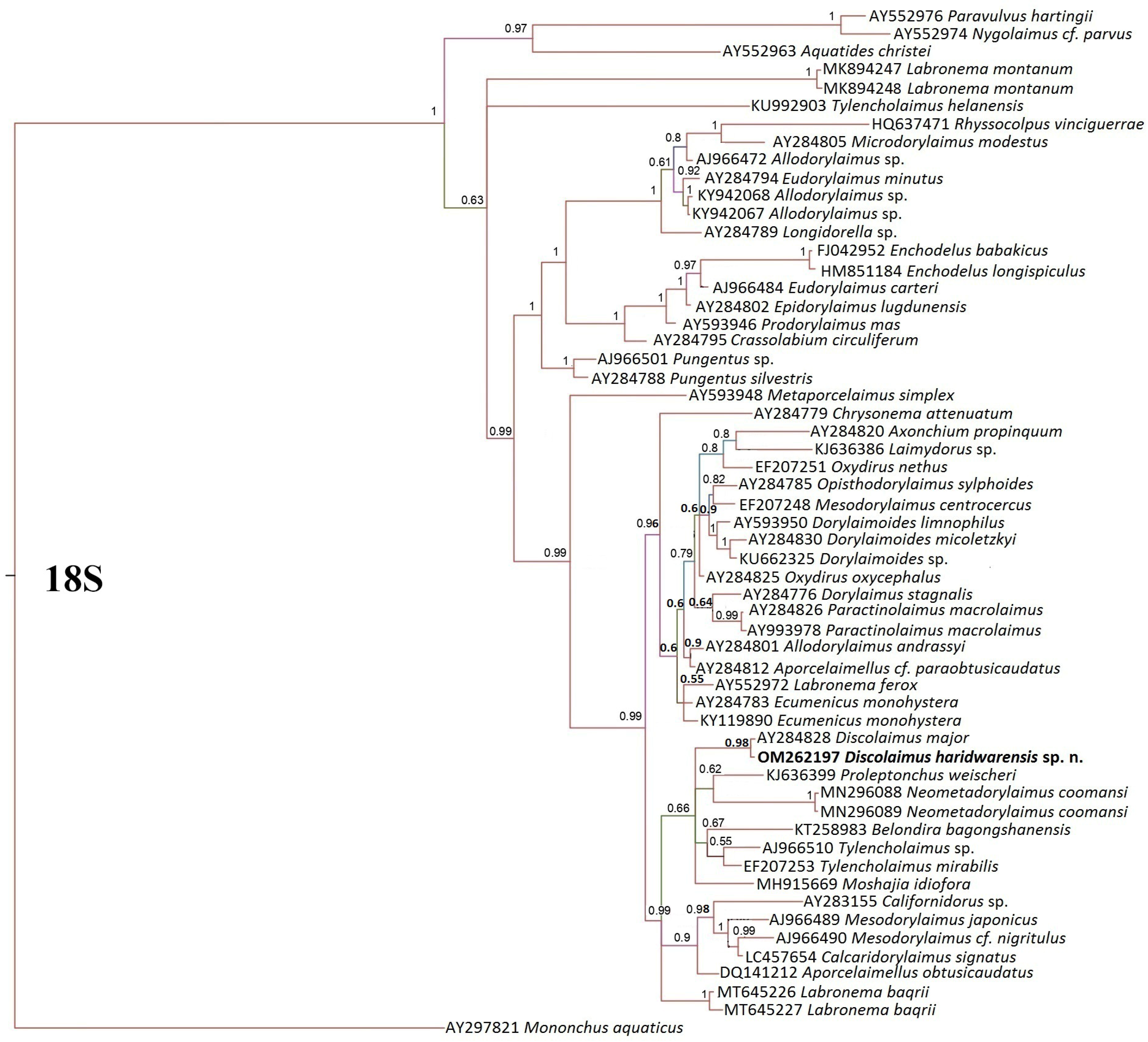
The newly obtained sequences of the D2-D3 expansion segments of the 28S rRNA and partial 18S rRNA genes were BLAST (Basic Local Alignment Search Tool) for similarity matches with the previously submitted sequences in the NCBI (National Centre for Biotechnology Information) [36]. The conserved regions, i.e., D2-D3 and 18S rDNA have been exploited for establishing the relationship among the Discolaimus species. For each amplified rDNA region (SSU and LSU), an alignment of our data with sequences from other closely related species was generated using default ClustalW parameters in MEGA 7.0 [37] and manually optimized in BioEdit [35]. Bayesian inference (BI) was used to infer the phylogenetic trees from the datasets. Mononchus truncatus (AY593064) and M. tunbridgensis (AY593063) for 28S were utilized as outgroup taxa, with all characteristics being equally weighted and gaps being treated as missing data. MrBayes 3.2.7 was used to produce Bayesian phylogenetic reconstructions [38]. The General Time Reversible substitution model with gamma-distributed rate variation across sites and a proportion of invariable sites (GTR + G + I) were used as the optimal nucleotide substitution model for the analyses. The tree was preserved per 1000 generations after running Metropolis-coupled Markov chain Monte Carlo generations (MCMC) for 1 × 10⁷ cycles.

3. Results

3.1. Systematics

  • Phylum: Nematoda Potts, 1932
  • Class: Enoplea Inglis, 1983
  • Order: Dorylaimida Pearse, 1942
  • Family: Qudsianematidae Jairajpuri, 1965
  • Genus: Discolaimus Cobb, 1913
  • Species: haridwarensis sp. n.
  • Isolates/strains: HN13

3.1.1. Discolaimus haridwarensis sp. n.

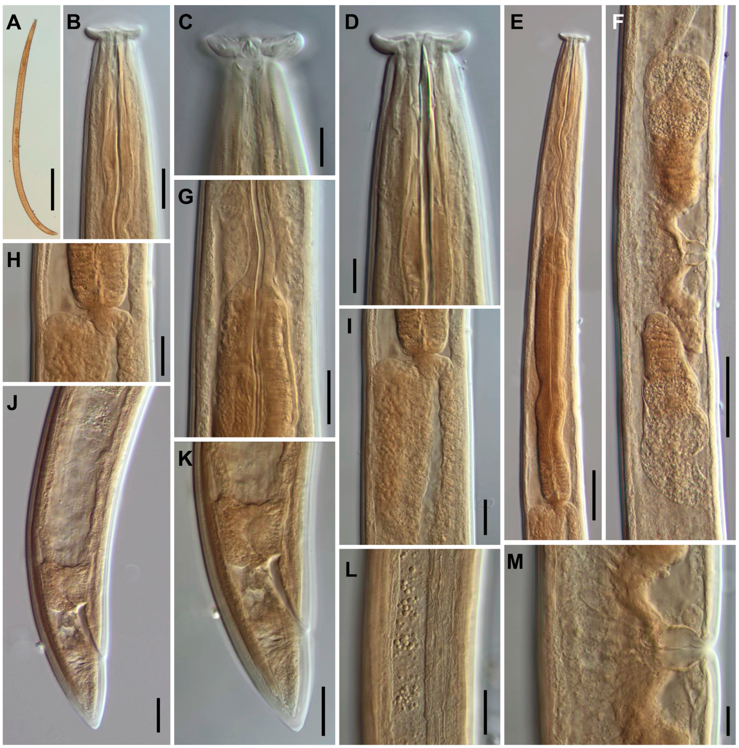
  • (Figure 1A–G and Figure 2A–M)
  • urn:lsid:zoobank.org:pub:679768CE-BC71-4CE1-B561-39B8D645C52F

3.1.2. Material Examined

Ten females in good state of preservation were examined.

3.1.3. Description

The female is described as follows. It is slender to very slender (a = 33–42) with nematodes of medium size, 2.11–2.32 mm long. The habitus is a slightly curved ventrad upon fixation. The cuticle is two-layered, 1.5–2.0 µm thick at the anterior region, 2.0–3.0 µm in mid-body, and 3.5–5.5 µm on tail; the outer layer is thin and bearing fine transverse striation throughout the body, and the inner layer thicker than the outer layer. The lateral chord is 16–18 µm wide or 28–32% of the midbody diameter. Dorsal and ventral body pores are present through the entire body, but especially visible at anterior and posterior body regions. The lip region is offset by deep constriction, 5.5–6.5 µm high and 27–30 µm wide, 0.5–0.6 times as wide as the diameter at neck base. The amphid fovea is funnel-shaped, its aperture 8.0–10.5 µm or 29–35% of lip region diameter. The odontostyle is 0.7–0.8 times as long as lip region diameter, 7.4–8.5 times longer than the width, 0.94–1.11% of total body length, with aperture 50–54% of the total length. The guiding ring simple is located at 8.0–11.0 µm from the anterior end. The odontophore is rod-like, 1.4–1.7 times longer than the odontostyle. The nerve ring is located at 35–39% of the pharynx length. The pharynx consists of a slender but muscular anterior section expanded in the posterior, with the expansion 7.5–8.3 times as long as wide, 4.2–5.4 times of the corresponding body diameter, and occupying 57–59% of the total pharynx length. The pharyngeal gland nuclei were observed, and their location [39] was as follows: DN = 45–48, S1N1 = 54–63, S1N2 = 79–82, S2N = 85–88. Cardia are conical to rounded, 12–16 μm long, with a ring-like structure surrounding its junction with pharyngeal base; a dorsal cell mass is present behind the pharyngeal base. The genital system is didelphic-amphidelphic, with both branches equally and well developed, anterior 112–144 µm long or 5–7% of total body length, and posterior 117–145 µm long or 5–7% of total body length. Ovaries are 89–118 µm long, reaching and surpassing the oviduct-uterus junction, and oocytes arranged first in two or more rows, then in a single row. The oviduct is 39.0–83.5 µm long or 0.7–1.0 times of the body diameter, consisting of a slender portion and a small pars dilatata with visible lumen and a narrowing separates oviduct and uterus. The uterus is a short simple tube-like structure, 22–31 µm long or 0.3–0.5 times the corresponding body diameter. No egg was observed in uterus. The vagina is extending inwards 16–19 µm or occupying 28–32% of body diameter at the vulva, with pars proximalis 11–14 × 12–15 µm and convergent walls surrounded by weak musculature, pars refringens absent, and pars distalis well developed, 5–7 µm long. Two cells, one anterior, one posterior, are present on both sides of vagina. The vulva, a transverse slit, is distinct. The prerectum is 0.8–1.1 and rectum is 1.0–1.1 times the anal body diameter long. The tail is conoid with rounded terminus. Two pairs of caudal pores, one sublateral, one lateral (Figure 1 and Figure 2; Table 1) are present.
  • Male. Not found.

3.1.4. Measurements

Table 1. Morphometric data of Discolaimus haridwarensis sp. n. from sugarcane fields in Haridwar, Uttarakhand, India.
HolotypeParatype
Character a9 ♀♀
L2.282.17 ± 0.89 (2.11–2.32)
a35.938.6 ± 3.8 (32.8–42.5)
b5.24.8 ± 0.3 (4.3–5.2)
c59.359.7 ± 5.8 (53.2–68.3)
c′1.01.1 ± 0.1 (1.0–1.3)
V48.351.4 ± 2.2 (48.1–55.2)
G16.25.9 ± 0.7 (5.2–6.8)
G26.45.9 ± 0.6 (5.1–6.5)
Lip region diameter29.528.4 ± 1.1 (27.0–30.0)
Odontostyle length23.021.8 ± 1.2 (20.0–23.0)
Odontophore length36.536.1 ± 2.5 (34.0–38.0)
Neck length437460.3 ± 20.9 (436–487)
Pharyngeal expansion279271.4 ± 5.9 (266–280)
Diameter at neck base62.554.3 ± 5.4 (48.5–62.5)
       at midbody63.058.5 ± 7.5 (49.0–66.0)
       at anus35.031.7 ± 2.2 (28.0–33.5)
Prerectum26.532.7 ± 5.4 (26.5–38.5)
Rectum33.532.2 ± 1.2 (30.5–33.5)
Tail38.537.2 ± 2.4 (34.0–40.0)
a All measurements are in µm (except L, in mm, ratios and percentages) and in the form of mean ± standard deviation (range). Note: n = number of measured specimens; L = overall body length; a = body length/diameter at midbody; b = body length/neck length; c = body length/tail length; c′ = tail length/body diameter at anus; G = Genital branch length/body length in %; and V = distance of anterior body end from the vulva/body length in %.

3.1.5. Type Habitat and Locality

The present population was recovered from soils around the rhizosphere of sugarcane (Saccharum officinarum) in Haridwar, Uttarakhand, India (29°54′40.2″ N 78°01′01.5″ E, 265 m a.s.l.).

3.1.6. Type Material

Female holotype and seven female paratypes were deposited at the museum of the Department of Zoology, Chaudhary Charan Singh University, Meerut, India. One female paratype was deposited in the nematode collection of the National Museum of Natural Sciences, Madrid, Spain.

3.1.7. Etymology

The species name is derived from the location Haridwar, district of Uttarakhand state, India, where the nematode specimens were recovered.

3.1.8. Diagnosis

The new species is characterized by its 2.11–2.32 mm long body, lip region 27–30 µm wide and offset by deep constriction, odontostyle 20–23 µm long, odontostyle aperture occupying 50–54%, neck 436–487 µm long, dorsal cell mass behind the pharyngeal base, pharyngeal expansion occupying 57–59% of the total neck length, uterus a simple tube-like structure and 22–31 µm long or 0.3–0.5 times the corresponding body diameter, vulva transverse (V = 48–55), female tail conoid (34–40 µm, c = 53–68, c′ = 1.0–1.3) with rounded terminus, and male absent.

3.1.9. Relationships

Morphologically and morphometrically, the new species is similar to D. major Thorne, 1939 [40] (= D. perplexans Siddiqi, 1964 [12] by Peña-Santiago et al. [41]) and D. trapezoides Siddiqi, 2005 [42]. D. haridwarensis sp. n. can be distinguished from D. major by its larger body size (2.11–2.32 vs. almost always 1.5–2.0 mm long, see description by Peña-Santiago et al. [41]), shorter odontostyle (20–23 vs. 24–29 µm), absence of large cell bodies around the cardia (vs. usually present), longer tail (34–40 vs. 22–30 µm), and absence of blind prerectal sac (vs. bearing a short blind prerectal sac). And from D. trapezoides, it can be differentiated in its larger body size (2.11–2.32 vs. 1.9–2.0 mm long), narrower lip region (27–30 vs. 30–33 µm wide), shorter odontostyle (20–23 vs. 28–30 µm long and 0.7–0.8 vs. 0.9–1.0 times as long as lip region diameter), shorter neck (436–487 vs. 510–520 µm long and b = 4.3–5.2 vs. 3.7–4.0), shorter female genital branch (G = 5.1–6.8 vs. 8.3–9.5), and longer tail (34–40 vs. 30–35 µm). The new species also resembles D. anemaqen, but it can be differentiated by its wider lip region (27–30 vs. 25–27 µm), longer odontostyle (20–23 vs. 19–20 µm), lateral chord lacking (vs. bearing) gland bodies, and presence (vs. absence) of a dorsal cell mass behind the pharyngeal base.
In having a body size larger than 0.7 and smaller than 2.4 mm, the basal part of pharynx not offset from anterior part by a constriction and without two sheaths, prerectum less than 4 times the anal body diameter long, odontostyle more than 16 μm in average (following the key to the species of the genus Discolaimus by Wu et al. [4]), the new species resembles D. agricolus Sauer & Annels, 1985 [43], D. auritus Lordello, 1955 [44], D. discocephalus Tulaganov, 1949 [45], D. neomajor Siddiqi, 2005 [42], D. papillatus Khan, Ahmad & Jairajpuri, 1994 [20], D. silvicolus Sauer & Annels, 1985 [43] and D. zicsii Andrássy, 1968 [46]. It can be distinguished from D. agricolus by its larger body size (2.11–2.32 vs. 1.23–1.98, after Peña-Santiago et al. [41]), absence of cell bodies close to the cardia area (vs. medium sized cell bodies just close to cardia area, sometimes forming a longitudinal series, after Peña-Santiago et al. [41]), prerectum without a blind sac (vs. bearing a distinct blind sac), and longer tail (34–40 vs. 19–28 µm). It can be distinguished from D. auritus by its larger body size (2.11–2.32 vs. 1.64 mm long), shorter odontostyle (20–23 vs. 25 µm), more posterior vulva position (V = 48–55 vs. 44), and comparatively longer tail (c′ = 1.0–1.3 vs. c′ < 1.0) and lacking (vs. bearing) a digitate terminus. It can be distinguished from D. discocephalus in its larger body size (2.11–2.32 vs. 1.6 mm long) and from D. neomajor in its wider lip region (27–30 vs. 24–27 µm), shorter odontostyle (20–23 vs. 24–27 µm), shorter female genital branch (G = 5.1–6.8 vs. 8.0–9.5), more posterior vulva position (V = 48–55 vs. 41–42), and longer tail (34–40 vs. 24 µm). It can be distinguished from D. papillatus by its larger body size (2.11–2.32 vs. 1.70–1.90 mm long), longer odontostyle (20–23 vs. 18–19 µm), shorter prerectum (30–34 vs. 45–60 µm long or 0.8–1.1 vs. 1.6–2.3 times the anal body diameter long), and male absent (vs. present). It can be distinguished from D. silvicolus by its larger body size (2.11–2.32 vs. 1.61–1.89 mm long), pharyngeal expansion without sheath (vs. with a thick irregular sheath), and absence of cell bodies close to the cardias area (vs. large cells with usually prominent nucleoli occurs posterior to the cardiac region) and from D. zicsii in its less slender body (a = 33–42 vs. 52), larger body size (2.11–2.32 vs. 1.7 mm long), prerectum without a blind sac (vs. bearing a distinct blind sac), and comparatively longer tail (c = 53–68 vs. 78).

3.2. Molecular Characterization

One partial 18S rRNA gene sequence (884 bp) and one D2-D3 of 28S rRNA gene sequence (734 bp) were studied for this species in this study. Both sequences were deposited in the NCBI GenBank with accession numbers OM262197 and OM262195, respectively.

3.3. Phylogenetic Analysis

The D2-D3 of 28S rRNA gene alignment was 1269 bp in length and contained 65 sequences. Phylogenetic relationships of the new species within selected dorylaimid nematodes are given in Figure 3. The phylogenetic analysis showed that the new species clustered together with other representatives of the family Qudsianematidae (Carcharodiscus Andrássy, 1991 [47] + Discolaimoides Heyns, 1963 [48] + Discolaimus Cobb, 1913 [1]). Only two named species of Discolaimus genus have been molecularly characterized based on 28S rDNA namely, D. bicorticus and D. major. The sequences of D. haridwarensis sp. n. (OM262195) formed a well-supported clade with D. bicorticus (OK142270) and an unidentified Discolaimus species (KY750793) with Bayesian posterior probability (BPP) of 1.00, confirming its identity within the Discolaimus genus. Together, D. haridwarensis sp. n., D. bicorticus and Discolaimus sp. clustered inside of a well-supported clade that included Discolaimoides symmetricus (MT776559 and EF207238), Carcharodiscus banaticus (AY593023 and AY593024) and D. major (AY593025 and AY593026), having in common the absence of the pars refringens vaginae. However, the relationships between the Discolaimus species were not resolved, most probably due to the limited molecular data available. Therefore, additional molecular data of other Discolaimus species are needed to clarify the monophyly of the genus.
The 18S rRNA gene alignment was 2628 bp in length and contained 57 sequences. Phylogenetic relationships of the new species within selected dorylaimid nematodes are given in Figure 4. The phylogenetic analysis showed that the new species clustered with D. major (AY593028) within a poorly supported clade that included some representatives of Belondiridae (Belondira Thorne, 1939 [40]), Leptonchidae (Proleptonchus Lordello, 1955 [44]) and Tylencholaimidae (Neometadorylaimus Jairajpuri & Ahmad, 1992 [49] + Tylencholaimus de Man, 1876 [50]). These groups share the common characteristic of lacking the pars refringens vaginae, except in Neometadorylaimus. The internal relationship of the Discolaimus species remains unresolved, likely due to the limited molecular data available.

4. Discussion

The newly identified taxon, Discolaimus haridwarensis sp. n., exhibits similarities with the most common and widely distributed species of the genus Discolaimus, D. major. However, both species can be distinguished by some important morphological features, including body size and odontostyle length. Additionally, the current molecular data for both species support a separate status for D. haridwarensis sp. n.
Molecular data for Discolaimus species are currently insufficient, comprising 21 sequences, including one whole genome (JAQIWJ000000000) and eight rDNA partial sequences for D. major (MT776558, MH216059, MH206070, AY284828, AY593026, AY593025, EF207239, EF207252). There are also three rDNA partial sequences for D. texanus (AY911958, AY911957, AY146485) and one partial rDNA sequence each for D. auritus (AY146484), D. bicorticus (OK142270), and D. similis (AY146501). Additionally, six sequences belong to unidentified species of the Discolaimus genus (MT796643, MG421342, KY750844, KY750793, KY750790, and KY750789). These latter six sequences belong to COI, 18S, and 28S molecular markers and require careful attention in their usage. It is noteworthy that none of the described Discolaimus species to date have included molecular data in their descriptions, including the most recent species described by [4,5]. Consequently, no reference sequences (i.e., molecular data from type populations or type localities) for species of this genus are currently available. Only limited molecular data, without associated published morphological characterizations, have been reported. This lack of comprehensive data makes the taxonomic confirmation of the available Discolaimus molecular data challenging.
The phylogenetic tree based on the D2-D3 region of 28S rRNA showed that the new species clustered with D. bicorticus (OK142270) and an unidentified Discolaimus species (KY750793) (1.00 BPP). Furthermore, it forms a clade with its closest relative, i.e., Carcharodiscus and Discolaimoides genera, as all three genera belong to the family Qudsianematidae. The phylogenetic tree based on the 18S rRNA showed that the new species clustered with Discolaimus major (AY593028) (0.98 BPP). The Qudsianematidae clade forms a well-supported clade with Belondiridae, Leptonchidae, and Tylencholaimidae families (0.99 BPP), as the species in these families lack pars refringens vaginae. The monophyly of Discolaimus species remains unresolved due to limited molecular data.

Author Contributions

Conceptualization, H.S., A.K.C. and S.Á.-O.; data curation, H.S. and S.Á.-O.; investigation, H.S. and S.Á.-O.; methodology, H.S.; visualization, H.S. and S.Á.-O.; writing—original draft, H.S.; writing—review and editing, S.Á.-O. and A.K.C. All authors have read and agreed to the published version of the manuscript.

Funding

This research received no external funding.

Institutional Review Board Statement

Not applicable.

Data Availability Statement

The data and material of this manuscript are available from the corresponding author on reasonable request.

Acknowledgments

The authors thank the Department of Zoology, Chaudhary Charan Singh University, Meerut, for providing the necessary laboratory facilities for conducting the experiments. The last author is especially grateful for the financial support received by “Acción financiada por la Comunidad de Madrid en el marco del Convenio Plurianual con la Universidad Rey Juan Carlos en la línea de actuación 1, Programa de “Estímulo a la investigación de jóvenes doctores”. Ref. proyecto M2730 Acrónimo NEMAVID”.

Conflicts of Interest

The authors declare no conflicts of interest.

References

  1. Cobb, N.A. New nematode genera found inhabiting freshwater and non-brackish soils. J. Wash. Acad. Sci. 1913, 3, 432–444. [Google Scholar]
  2. Andrássy, I. Free-Living Nematodes of Hungary (Nematoda errantia). III. Pedozoologica Hungarica 5; Hungarian Natural History Museum and Systematic Research Group of the Hungarian Academy of Sciences: Budapest, Hungary, 2009; 608p. [Google Scholar]
  3. Peña-Santiago, R. Dorylaimida Mundi (Nematoda): Checklist of Genera and Species, with Their Records; Monographic Papers on Nematology 7; University of Jaén: Jaén, Spain, 2021. [Google Scholar]
  4. Wu, W.; Yan, L.; Xu, C.L.; Wang, K.; Jin, S.Y.; Xie, H. A new species of the genus Discolaimus Cobb, 1913 (Nematoda: Dorylaimida: Qudsianematidae) from Qinghai, China. Zootaxa 2016, 4088, 129–138. [Google Scholar] [CrossRef] [PubMed]
  5. Erum, I.; Nasira, K.; Sagir, H.; Raza, S.; Firoza, K. Description of Discolaimus miniodontii n. sp. and Laevides hunderansis n. sp. with notes on Discolaimoides spatilabium (Nematoda: Dorylaimida) from District Ghizer, Gilgit-Baltistan, Pakistan. Pakistan J Nematol. 2020, 38, 1–13. [Google Scholar]
  6. Sharma, V. Diversity of Terrestrial nematodes in Uttarakhand. J. Exp. Zool. India 2011, 14, 575–582. [Google Scholar]
  7. Sharma, V. A preliminary study of nematodes in Govind Wildlife Sanctuary, Uttarkashi, Uttarakhand, India. Ann. For. Res. 2013, 21, 92–94. [Google Scholar]
  8. Gantait, V.V.; Bhattacharya, T.; Chatterjee, A. A new species of the genus Discolaimus Cobb, 1913 (Qudsianematidae: Dorylaimida) from West Bengal, India with revised key to the genus. Proc. Zool. Soc. 2009, 62, 67–73. [Google Scholar] [CrossRef]
  9. Gantait, V.V.; Chatterjee, A.; Bhattacharya, T. Trophic groups of nematodes associated with banana plantation in Paschim Medinipur district of West Bengal, India. Trop. Ecol. 2011, 52, 331–335. [Google Scholar]
  10. Sen, D.; Chatterjee, A.; Manna, B. Two New and Two Known Species of Dorylaimoidea (Nematoda) from West Bengal, India with a Key to the Species of the Genus Indodorylaimus Ali and Prabha, 1974. Rec. Zool. Surv. India 2012, 112, 11–20. [Google Scholar] [CrossRef]
  11. Khan, E.; Laha, S.K. Seven new dorylaimid nematodes from Indian Agricultural Research Institute Farm, Delhi, India. Indian J. Nematol. 1982, 12, 232–249. [Google Scholar]
  12. Siddiqi, M.R. Studies on Discolaimus spp. (Nematoda: Dorylaimidae) from India. Z. Zool. Syst. Evol. 1964, 2, 174–184. [Google Scholar] [CrossRef]
  13. Lal, A.; Khan, E. Nematodes associated with forest trees in nurseries and reserved natural forests of northern India. Indian J. For. 1988, 11, 276–282. [Google Scholar]
  14. Bilgrami, A.L.; Kulshreshtha, R.; Pervez, R. Community analysis of predaceous and freeliving nematodes of Aligarh district. Indian J. Nematol. 1998, 27, 104–110. [Google Scholar]
  15. Pervez, R. Prevalence of plant parasitic and predatory nematodes associated with different crops of Badaun districts (Uttar Pradesh). Curr. Nematol. 2004, 15, 43–46. [Google Scholar]
  16. Baqri, Q.H.; Bohra, P. Studies on Plant and Soil Nematodes Associated with Crops of Economic Importance in Gujarat; Records of the Zoological Survey of India, Occasional Paper; Zoological Survey of India: Kolkata, Indian, 2005; No. 240; 48p. [Google Scholar]
  17. Bohra, P.; Baqri, Q.H. Biodiversity of plant and soil nematodes from Andaman and Car Nicobar Islands. Rec. Zool. Surv. India 2005, 105, 43–49. [Google Scholar] [CrossRef]
  18. Bohra, P.; Baqri, Q.H.; Dwivedi, A.K. Diversity of phytophagous nematodes associated with economically important crops in Gujarat, India. Indian J. Nematol. 2005, 35, 130–133. [Google Scholar]
  19. Pervez, R.; Ali, S.S. Biodiversity of the predatory nematodes associated with pulse crops from Jhansi district of Uttar Pradesh. Curr. Nematol. 2008, 19, 57–61. [Google Scholar]
  20. Khan, Z.A.; Ahmad, W.A.; Jairajpuri, M.S. Description of two new species of Dorylaimoidea from India. Fund. Appl. Nematol. 1994, 17, 147–151. [Google Scholar]
  21. Rahaman, P.F.; Ahmad, I. Community analysis of predatory nematode species from Aligarh soils, India. Nematol. Mediterr. 1995, 23, 57–60. [Google Scholar]
  22. Sharma, V. Ten new records of free-living nematodes from Uttarakhand, India. Indian J. For. 2018, 41, 39–42. [Google Scholar]
  23. Ahmad, W.; Jairajpuri, M.S. Some new and known species of Dorylaimoidea. Nematologica 1982, 28, 39–61. [Google Scholar] [CrossRef]
  24. Mohilal, N.; Dhanachand, C. Investigations of soil nematodes from Manipur, species of the family Qudsianematidae. Uttar Pradesh J. Zool. 2001, 21, 41–45. [Google Scholar]
  25. Baniyamuddin, M.; Ahmad, W. Some new and known species of Dorylaimoidea (Dorylaimida: Nematoda) from natural forest of Arunachal Pradesh, India. J. Nematode Morphol. Syst. 2007, 10, 75–95. [Google Scholar]
  26. Cobb, N.A. Estimating the nematode population of the soil Bureau of Plant Industry. U. S. Dep. Agric. Agric. Technol. Circ. 1918, 1, 19–24. [Google Scholar]
  27. Goodey, J.B. Laboratory Methods for Work with Plant and Soil Nematodes; Technical Bulletin No. 2; Ministry of Agriculture, Fisheries and Food: London, UK, 1957; 47p. [Google Scholar]
  28. Southey, J.F. Laboratory Methods for Work with Plant and Soil Nematodes; Reference Book 402; Ministery of Agriculture, Fisheries and Food: London, UK, 1985. [Google Scholar]
  29. Courtney, W.D.; Polley, D.; Miller, V.L. TAF, an improved fixative in nematode technique. Plant Dis. Rep. 1955, 39, 570–571. [Google Scholar]
  30. Seinhorst, J.W. A rapid method for the transfer of nematodes from fixative to anhydrous glycerin. Nematologica 1959, 4, 67–69. [Google Scholar] [CrossRef]
  31. Sharma, H.; Chaubey, A.K. Molecular and phenotypic characterization of Hemicriconemoides rosae (Rathour et al., 2003) from mustard rhizosphere in India. J. Basic Appl. Zool. 2023, 84, 16. [Google Scholar] [CrossRef]
  32. De Man, J.G. Die einheimischen, frei in der reinen Erde und imsüssen Wasser lebenden Nematoden. Tijdschr. Ned. Dierkd. Ver. 1880, 5, 1–104. [Google Scholar]
  33. Floyd, R.M.; Rogers, A.D.; Lambshead, P.J.D.; Smith, C.R. Nematode-specific PCR primers for the 18S small subunit rRNA gene. Mol. Ecol. Notes 2005, 5, 611–612. [Google Scholar] [CrossRef]
  34. De Ley, P.; Félix, M.A.; Frisse, L.M.; Nadler, S.A.; Sternberg, P.W.; Thomas, W.K. Molecular and morphological characterisation of two reproductively isolated species with mirror-image anatomy (Nematoda: Cephalobidae). Nematology 1999, 1, 591–612. [Google Scholar] [CrossRef]
  35. Hall, T.A. BioEdit: A user-friendly biological sequence alignment and analysis program for Windows 95/98.NT. Nucleic Acids Symp. Ser. 1999, 41, 95–98. [Google Scholar]
  36. Altschul, S.F.; Gish, W.; Miller, W.; Myers, E.W.; Lipman, D.J. Basic local alignment search tool. J. Mol. Biol. 1990, 215, 403–410. [Google Scholar] [CrossRef]
  37. Kumar, S.; Stecher, G.; Tamura, K. MEGA7: Molecular evolutionary genetics analysis version 7.0 for bigger datasets. Mol. Biol. Evol. 2016, 33, 1870–1874. [Google Scholar] [CrossRef] [PubMed]
  38. Ronquist, F.; Teslenko, M.; van der Mark, P.; Ayres, D.L.; Darling, A.; Höhna, S.; Larget, B.; Liu, L.; Suchard, M.A.; Huelsenbeck, J.P. MrBayes 3.2: Efficient Bayesian phylogenetic inference and model choice across a large model space. Syst. Biol. 2012, 61, 539–542. [Google Scholar] [CrossRef] [PubMed]
  39. Loof, P.A.A.; Coomans, A. On the development and location of the oesophageal gland nuclei in the Dorylaimina. Zesz. Probl. Postepow. Nauk Roln. 1970, 92, 79–161. [Google Scholar]
  40. Thorne, G. A monograph of the nematodes of the superfamily Dorylaimoidea. Capita Zool. 1939, 8, 1–261. [Google Scholar]
  41. Peña-Santiago, R.; Torres, B.; Liébanas, G.; Abolafia, J. Nematodes of the order Dorylaimida from Andalucía Oriental, Spain. The genus Discolaimus Cobb, 1913. II. Two previously known species, with comments on their taxonomy. Russ. J. Nematol. 2002, 10, 79–88. [Google Scholar]
  42. Siddiqi, M.R. Ten new species of Discolaimus Cobb, 1913 (Nematoda: Dorylaimida). Int. J. Nematol. 2005, 15, 215–229. [Google Scholar]
  43. Sauer, M.R.; Annells, C.M. Species of Discolaimus (Nematoda: Dorylaimoidea) from Australia. Nematologica 1985, 31, 121–133. [Google Scholar] [CrossRef]
  44. Lordello, L.G.E. On the morphology of Proleptonchus aestivus n. gen., n. sp. and Dorylaimus lourdesae n. sp., two new soil nematodes from Brazil. Proc. Helminthol. Soc. Wash. 1955, 22, 71–75.
  45. Tulaganov, A.T. Plant parasitic and soil nematodes in Uzbekistan. Izdat. Akad. Nauk. Uzbeksk. SSR Tashkent. 1949; 3–227. (In Russian) [Google Scholar]
  46. Andrássy, I. The scientific results of the Hungarian Soil Zoological Expedition to the Brazzaville-Congo, 31. Nematodes from Groundwater. Ann. Univ. Sci. Budapestinensis Rolando Eötvös Nomin. (Sect. Biol.) 1968, 9–10, 3–26. [Google Scholar]
  47. Andrássy, I. The superfamily Dorylaimoidea (Nematoda)—A review. Family Qudsianematidae, II. Opusc. Zool. 1991, 24, 3–55. [Google Scholar]
  48. Heyns, J. A report on South African nematodes of the genera Labronema Thorne, Discolaimus Cobb, Discolaimoides n. gen., and Discolaimium Thorne (Nemata: Dorylaimoidea). Proc. Helminthol. Soc. Wash. 1963, 30, 1–6. [Google Scholar]
  49. Jairajpuri, M.S.; Ahmad, W. Dorylaimida: Free-Living, Predacious and Plant-Parasitic Nematodes; Brill: Leiden, The Netherlands, 1992. [Google Scholar]
  50. De Man, J.G. Onderzoekingen over vrij in de aarde levende Nematoden. Tijdschr. Ned. Dierkd. Ver. 1876, 2, 78–196. [Google Scholar]
Figure 1. Discolaimus haridwarensis sp. n. (female, line drawings). (A,B) anterior region; (C) neck region; (D) anterior genital branch; (E) vagina; (F) cardia and pharyngo-intestinal junction; (G) posterior body region.
Diversity 16 00598 g001
Figure 2. Discolaimus haridwarensis sp. n. (female, light micrographs). (A) entire body; (B,D) anterior region in lateral median view, B showing in detail the dorsal body pores; (C) anterior region in lateral surface view; (E) neck region; (F) genital branch; (G) pharyngeal expansion; (H,I) cardia and pharyngo-intestinal junction, I showing in detail the dorsal cell mass present behind the pharyngeal base; (J,K) posterior body region; (L) lateral chord; (M) vagina. (Scale bars: (A) = 400 µm; (B,GL) = 20 µm; (C,D,M) = 10 µm; (E,F) = 50 µm.).
Diversity 16 00598 g002
Figure 3. Phylogenetic relationships within populations and species of Discolaimus as inferred from Bayesian analysis using the 28S rRNA gene sequence dataset under General Time Reversible substitution model with gamma-distributed rate variation across sites and a proportion of invariable sites (GTR + G + I). Newly obtained sequence is indicated in bold letters.
Diversity 16 00598 g003
Figure 4. Phylogenetic relationships within populations and species of Discolaimus as inferred from Bayesian analysis using the 18S rRNA gene sequence dataset under General Time Reversible substitution model with gamma distributed rate variation across sites and a proportion of invariable sites (GTR + G + I). Newly obtained sequence is indicated in bold letters.
Diversity 16 00598 g004
Disclaimer/Publisher’s Note: The statements, opinions and data contained in all publications are solely those of the individual author(s) and contributor(s) and not of MDPI and/or the editor(s). MDPI and/or the editor(s) disclaim responsibility for any injury to people or property resulting from any ideas, methods, instructions or products referred to in the content.