3.3. Taxonomy
Lampyrinae Rafinesque, 1815
Photinini LeConte, 1881
Scissicauda McDermott, 1964
Figure 3,
Figure 4,
Figure 5,
Figure 6,
Figure 7,
Figure 8,
Figure 9,
Figure 10,
Figure 11,
Figure 12,
Figure 13,
Figure 14,
Figure 15,
Figure 16 Scissicauda McDermott, 1964: 10, 39; 1966: 87.
Schistura Olivier, 1911:51 (nec Schistura McClevelland, 1838 Actinopterygii).
Aethra Laporte, 1833 (partim). Olivier in Wytsman 1907: 16; Blackwelder 1944: 353. Lychnuris Motschulsky, 1853 (partim). McDermott 1966 (quid pro quo).
Schistura Olivier, 1911: 51; McDermott 1964: 10, 39.
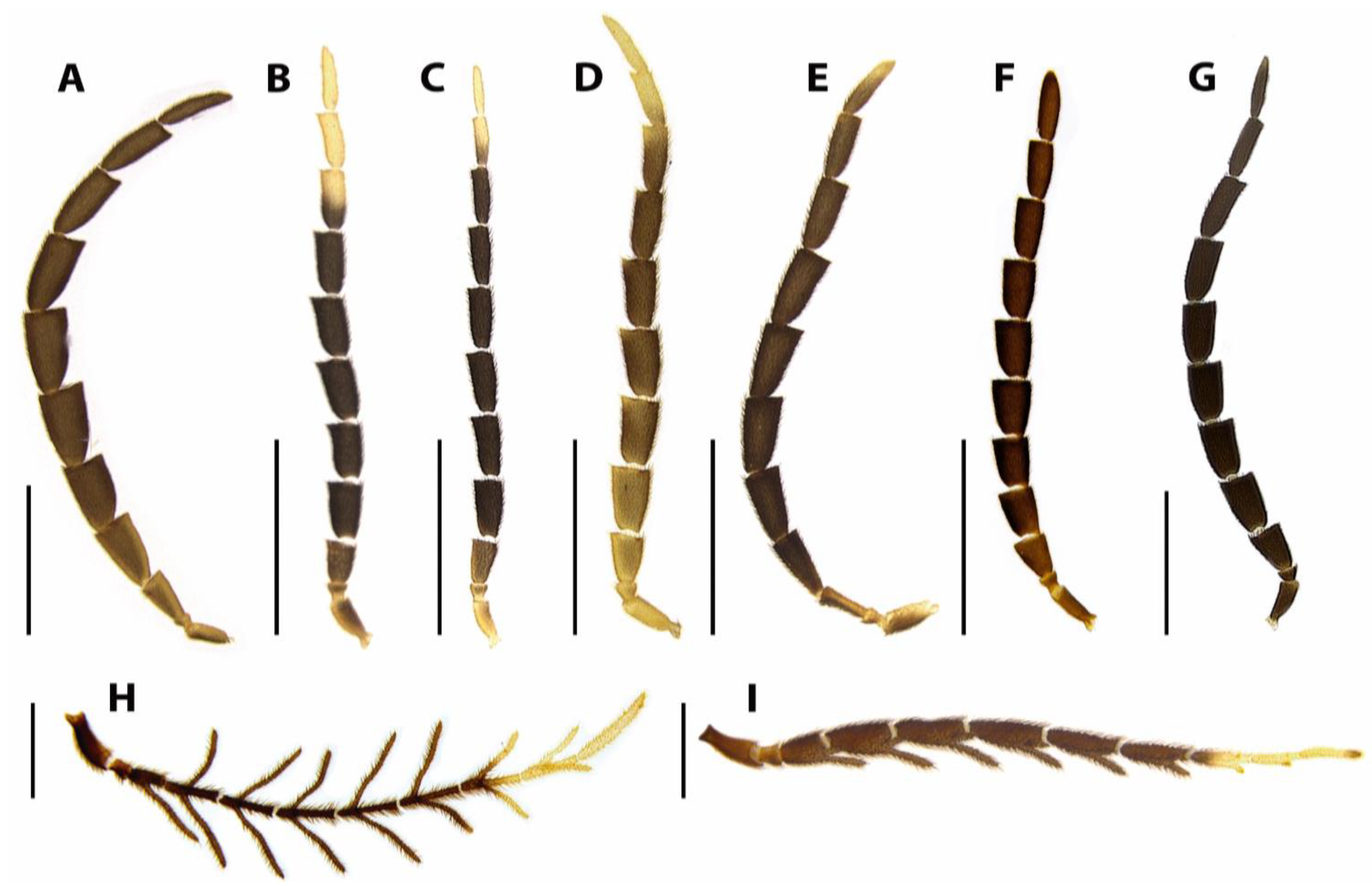
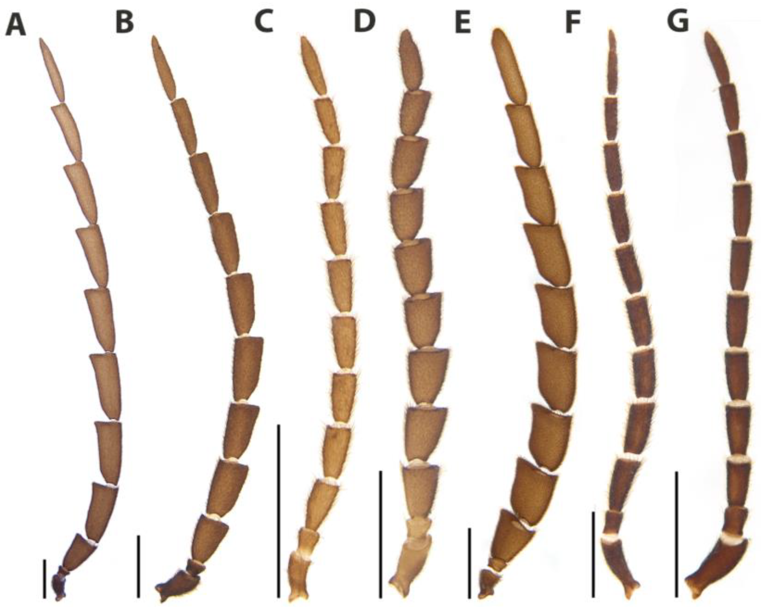
Diagnosis. Antenna 11-segmented (
Figure 6), antenommeres III–X compressed and serrate or cylindrical and flabellate, uniramose or biramose, with dense, upright bristles, rami at most twice longer than antennomere body (smaller than antennomere in
S. asymmetrica), attached basally (distally in
S. asymmetrica). Antennal sockets large (
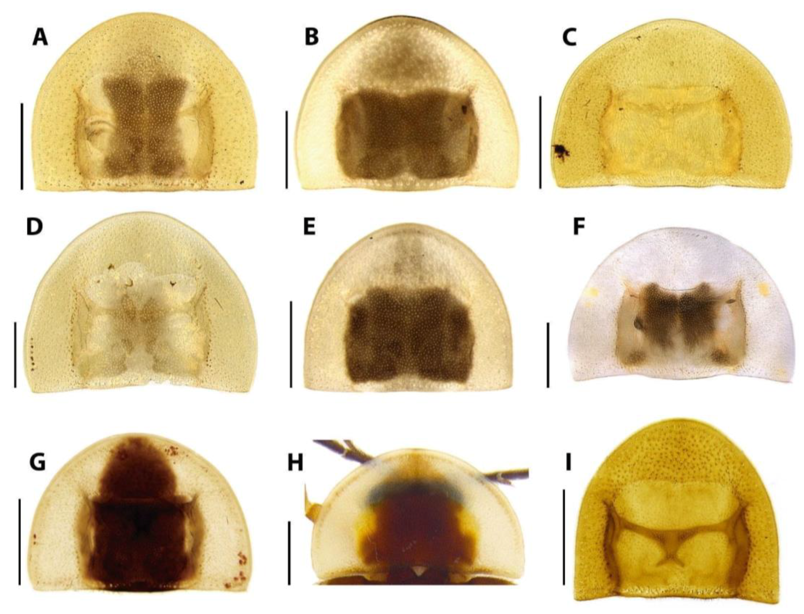
Figure 5A–D,I–L), two-thirds of frontal width, close-set, reniform, antennifer process distinct. Occiput as wide as one-third head width. Apical maxillary palpomere lanceolate. Apical labial palpomere securiform. Pronotum roughly semilunate (except in
S. neyi), with a marginal row of gross, deep punctures in dorsal view (
Figure 7A–H). Abdominal terga with posterior angles progressively produced and acute (except in
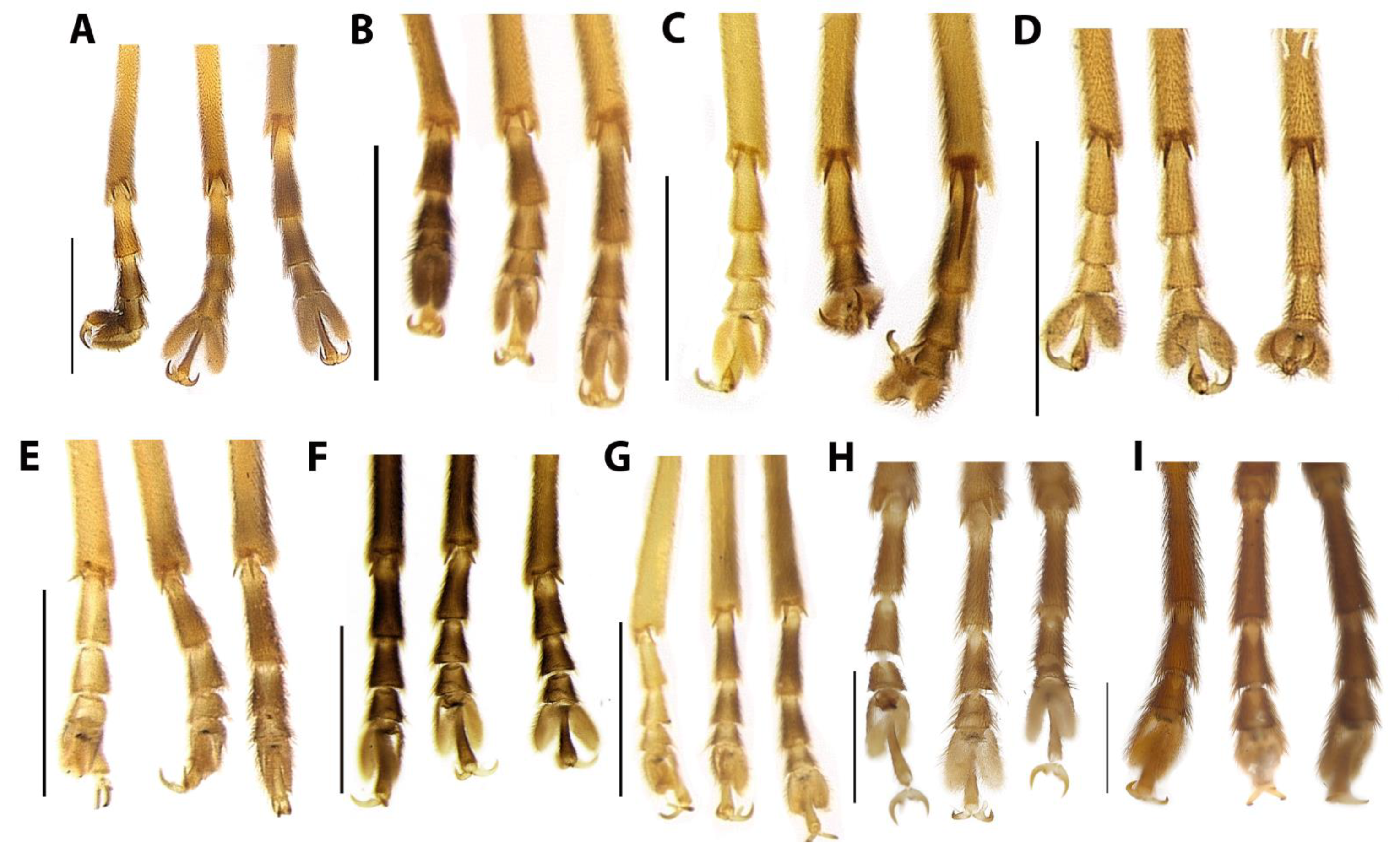
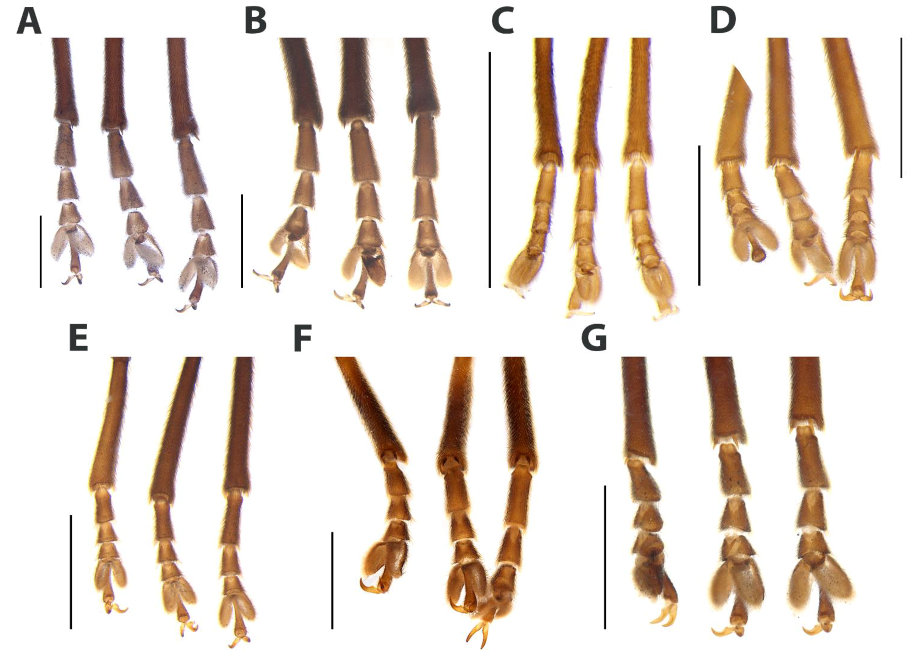
S. aurata). Tibial spurs present. Tarsomere I 2× longer than II, II 2× longer than III, III of subequal length as IV. Tarsomere IV bilobed, lobes reaching two-thirds of length of tarsomere V (
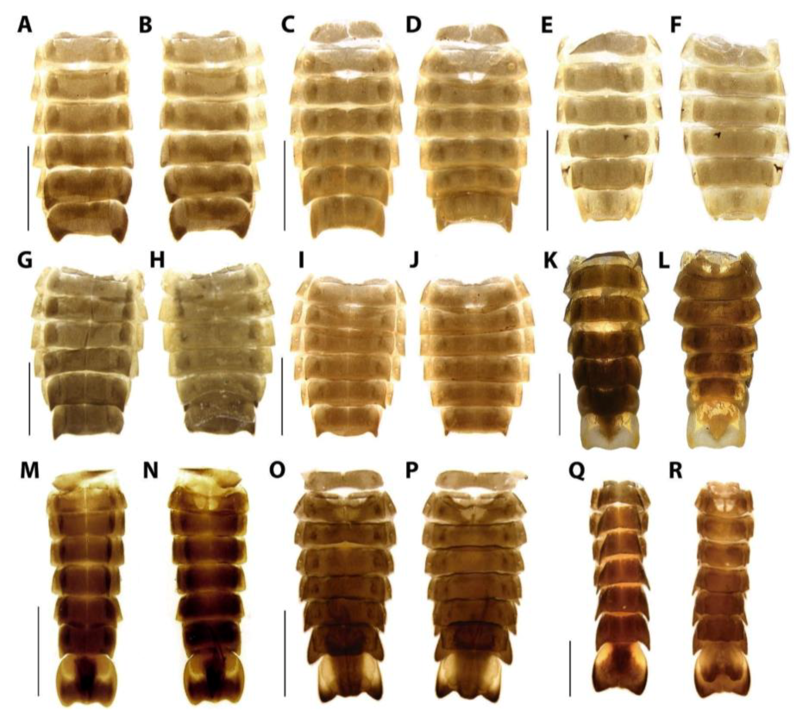
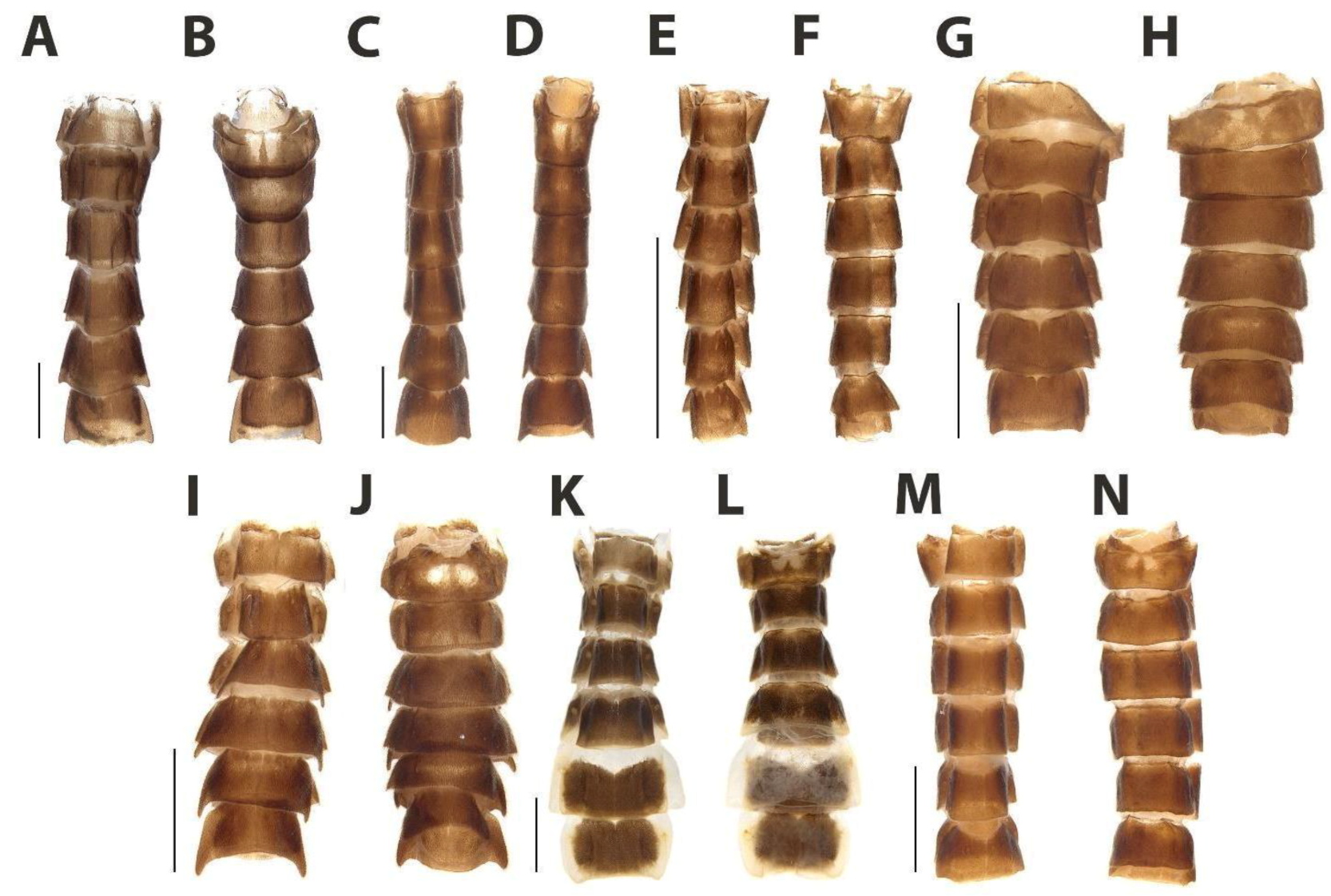
Figure 8). Abdominal sterna VI and VII without lanterns, VII with posterior angles projected, embracing anterior angles of pygidium, or poorly developed (
Figure 10). Males with sternum IX retracted under VIII or partially exposed (
Figure 3B,D,F,H,J,L and
Figure 4B,D,F) 2× or 3× as long as syntergite (
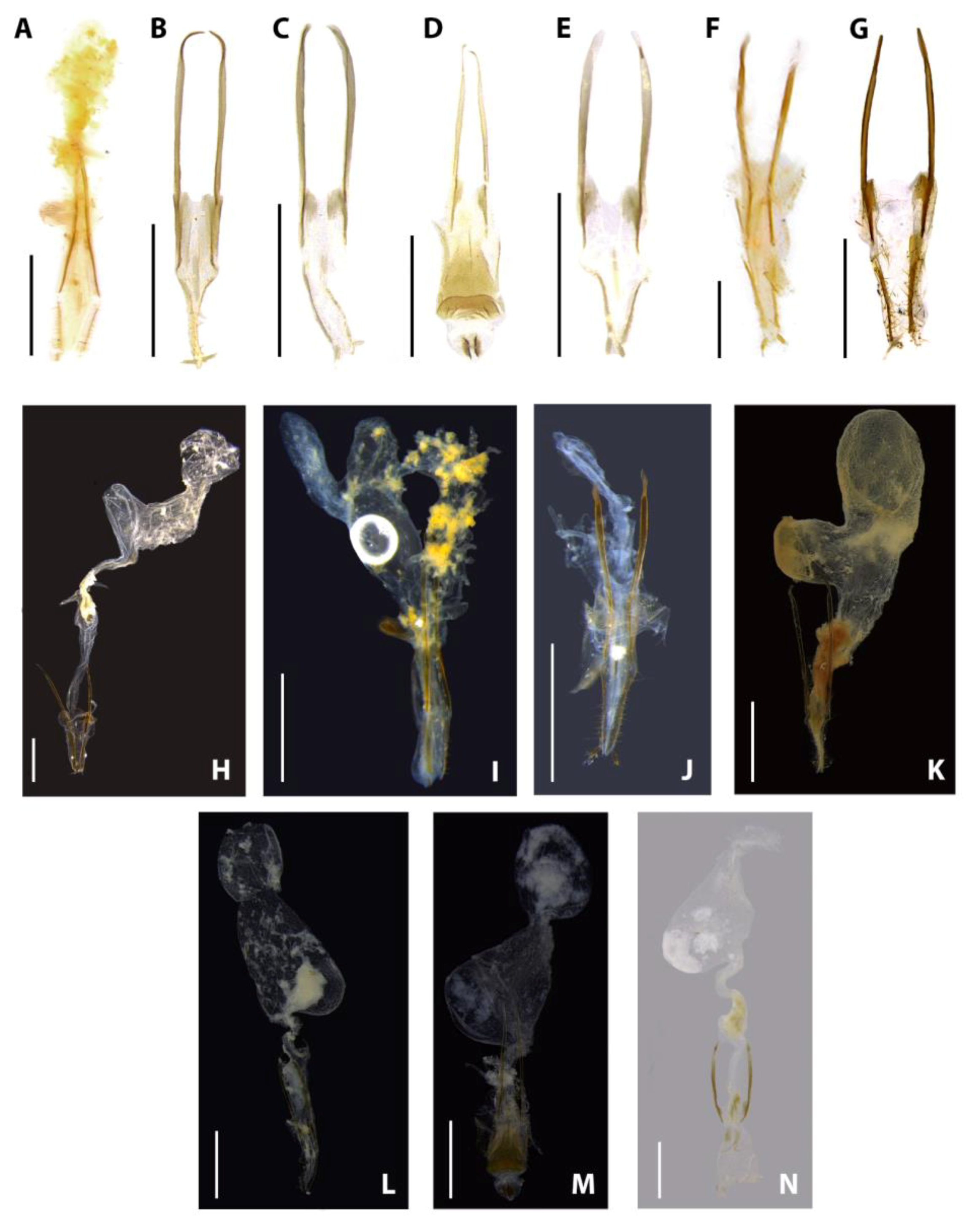
Figure 12A–Q). Phallus with dorsal and ventral plates, with dorsal plate basally fused to parameres, symmetric; ventral plate with lateral margins sinuose, weakly sclerotized; parameres symmetric, apically rounded, with a ventrobasal process rudimentary or extended beyond phallus (
Figure 13).
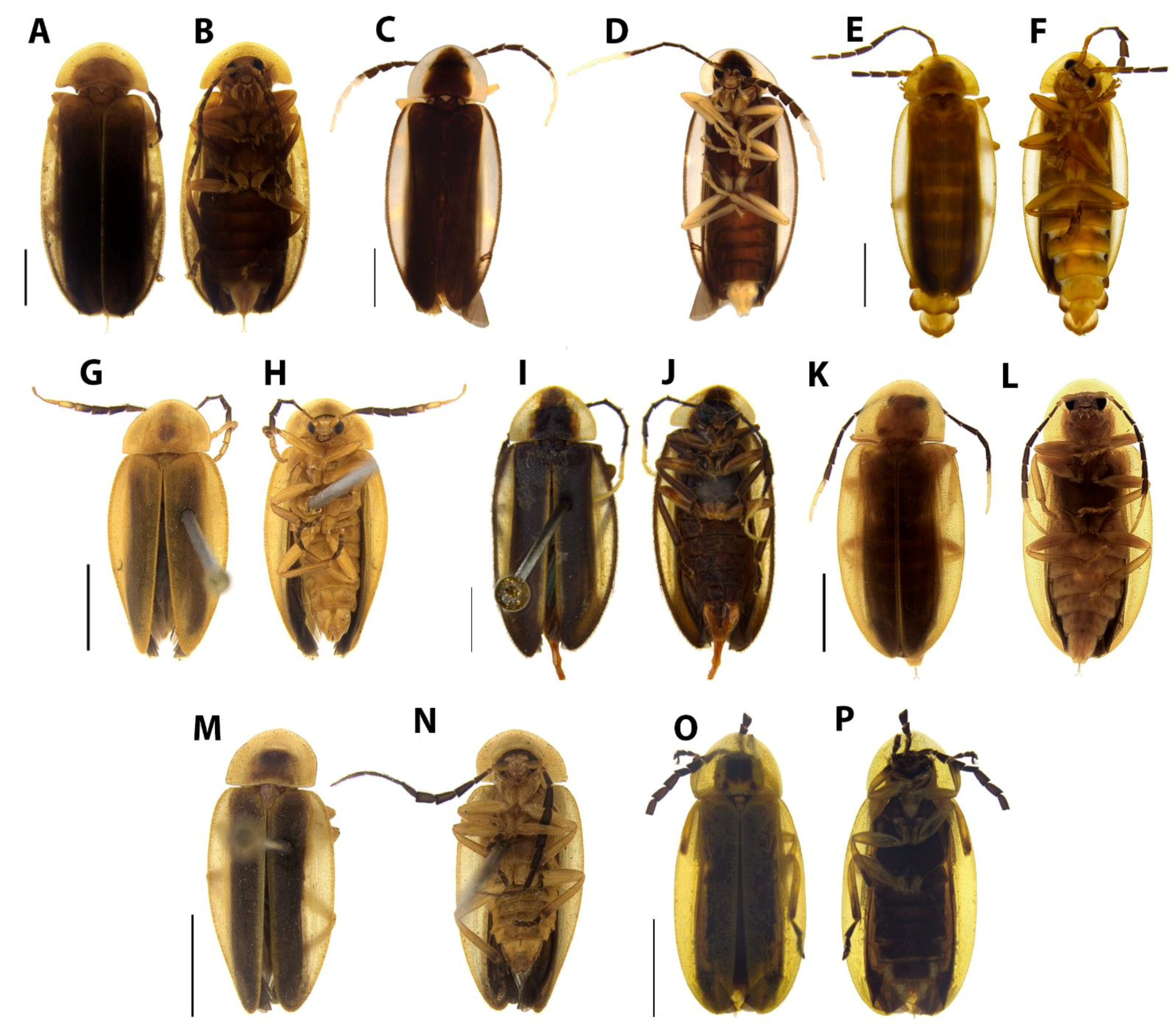
Figure 3.
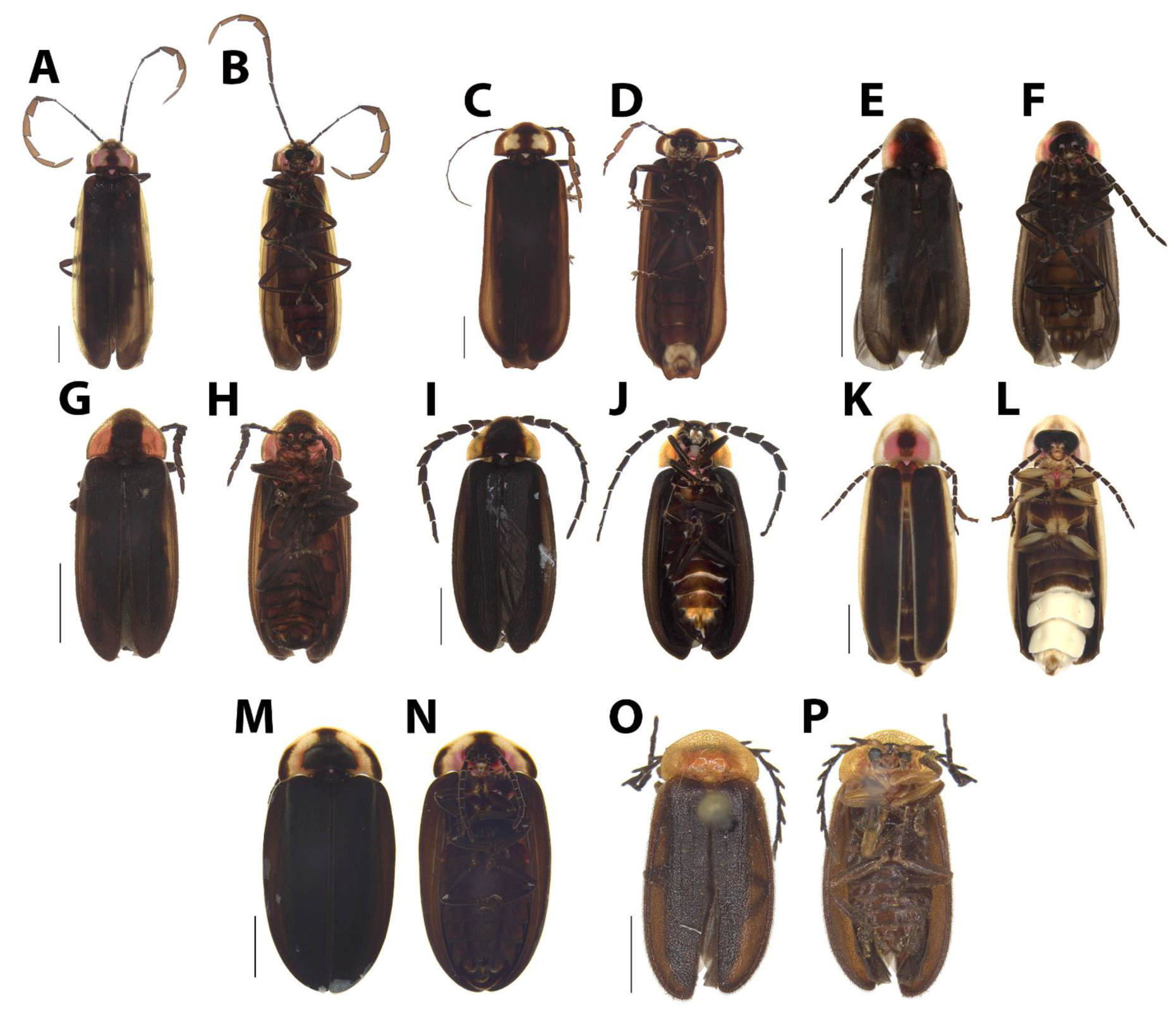
Male habitus, dorsal and ventral views. (A,B), Scissicauda truncata sp. nov. (C,D), Scissicauda antennata sp. nov. (E,F), Scissicauda malleri (Pic, 1935) comb. nov. (G,H), Scissicauda jamari sp. nov. (I,J), Scissicauda gomesi sp. nov. (K,L), and Scissicauda aurata sp. nov. Scale bar: 2 mm (A–L).
Figure 4.
Male habitus, dorsal and ventral views. (A,B) Scissicauda biflabellata sp. nov. (C,D), Scissicauda asymmetrica sp. nov. (E,F), and Scissicauda neyi sp. nov. Scale bar: 2 mm (A–F).
Figure 4.
Male habitus, dorsal and ventral views. (A,B) Scissicauda biflabellata sp. nov. (C,D), Scissicauda asymmetrica sp. nov. (E,F), and Scissicauda neyi sp. nov. Scale bar: 2 mm (A–F).
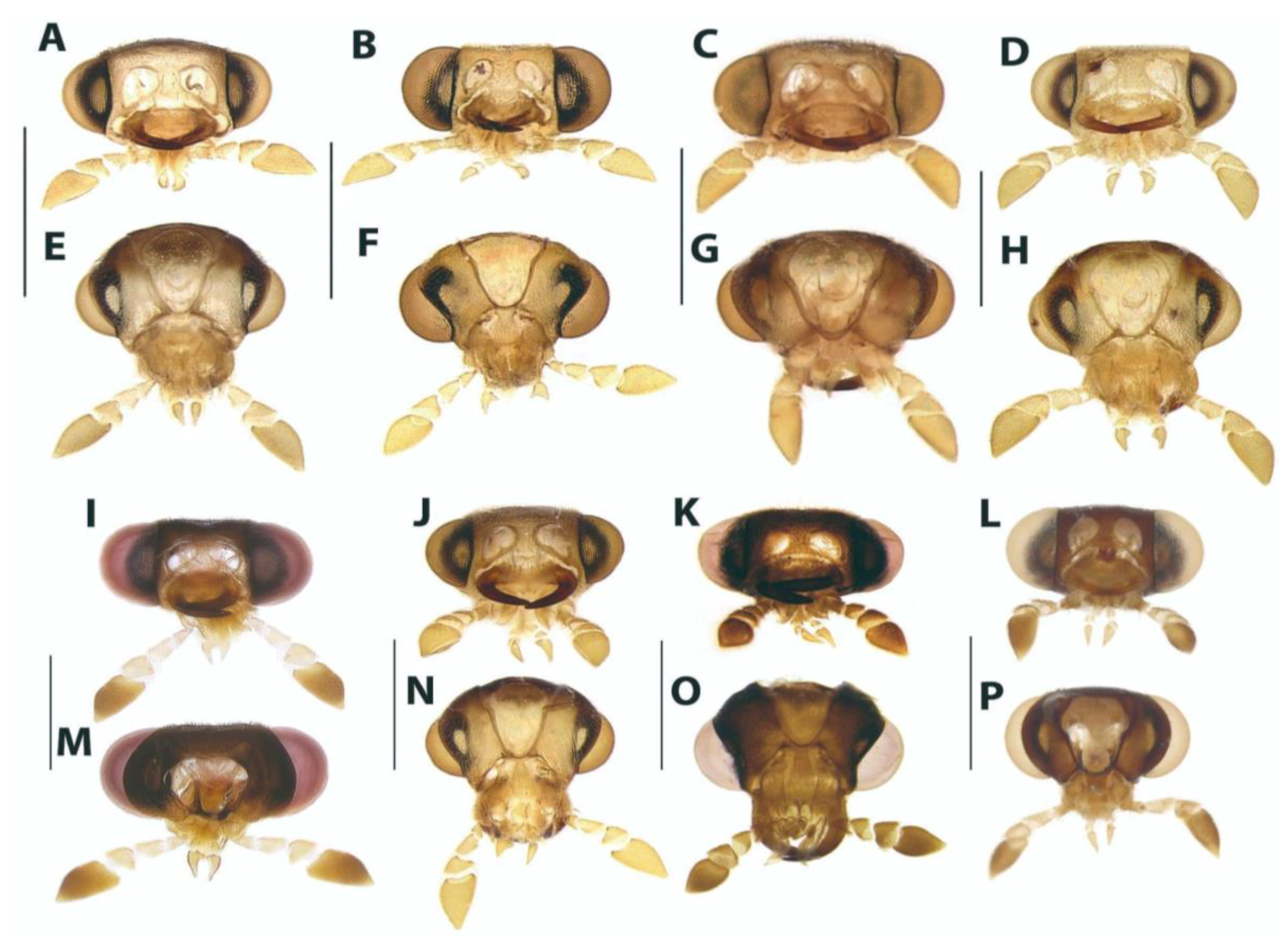
Figure 5.
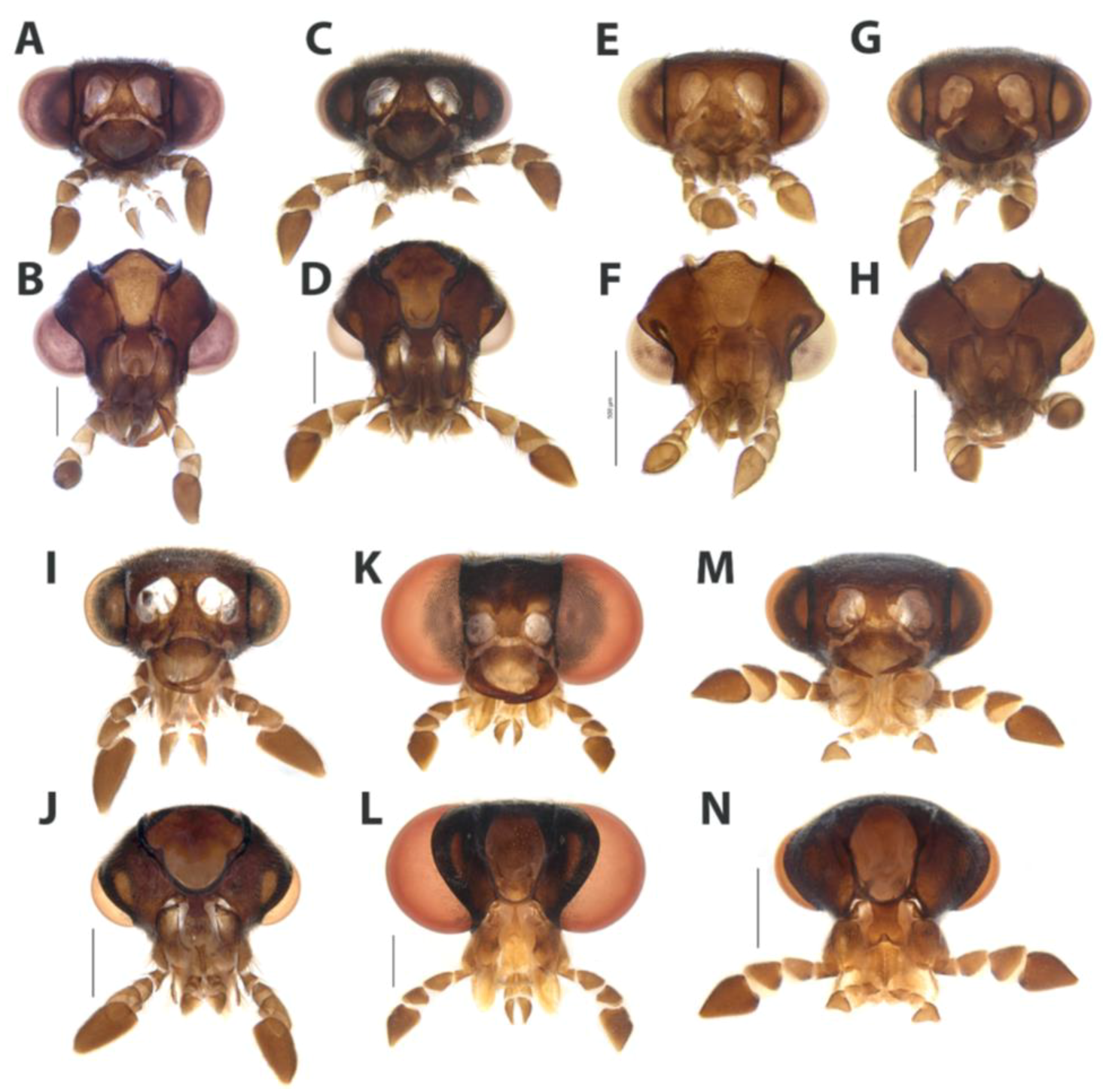
Head capsule, frontal and occipital views. (A,E) Scissicauda gomesi sp. nov. (B,F) Scissicauda aurata sp. nov. (C,G) Scissicauda antennata sp. nov. (D,H) Scissicauda jamari sp. nov. (I,M) Scissicauda malleri (Pic, 1935) comb. nov. (J,N) Scissicauda truncata sp. nov. (K,O) Scissicauda neyi sp. nov. (L,P) Scissicauda biflabellatasp. nov. Scale bar: 1 mm (A–P).
Figure 6.
Antenna, lateral view. (A) Scissicauda antennata sp. nov. (B) Scissicauda truncata sp. nov. (C) Scissicauda aurata sp. nov. (D) Scissicauda jamari sp. nov. (E) Scissicauda gomesi sp. nov. (F) Scissicauda neyi sp. nov. (G) Scissicauda malleri (Pic, 1935) comb. nov. (H) Scissicauda biflabellata sp. nov. (I) Scissicauda asymmetrica sp. nov. Scale bar: 1 mm (H,I); 2 mm (A–G).
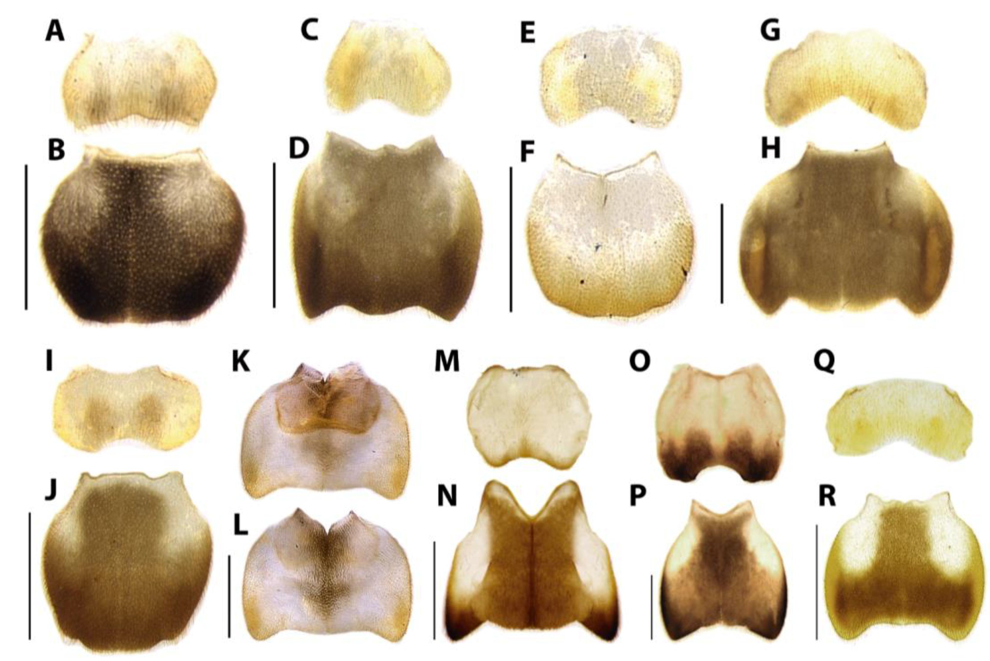
Figure 7.
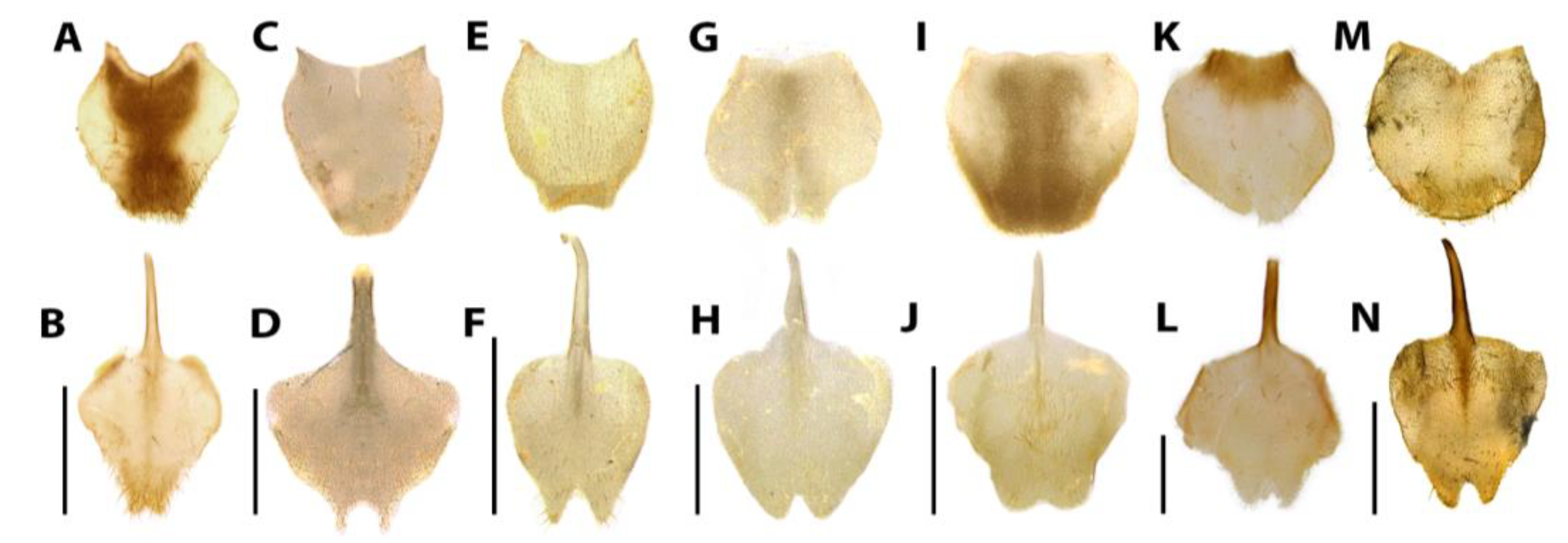
Male pronotum, dorsal view. (A) Scissicauda truncata sp. nov. (B) Scissicauda antennata sp. nov. (C) Scissicauda aurata sp. nov. (D) Scissicauda jamari sp. nov. (E) Scissicauda gomesi sp. nov. (F) Scissicauda malleri (Pic, 1935) comb. nov. (G) Scissicauda biflabellata sp. nov. (H) Scissicauda asymmetrica sp. nov. (I) Scissicauda neyi sp. nov. Scale bar: 1 mm (A–I).
Figure 8.
Detail of tarsus and claws: proleg, mesoleg, and metaleg, respectively. (A) Scissicauda malleri (Pic, 1935) comb. nov. (B) Scissicauda antennata sp. nov. (C) Scissicauda jamari sp. nov. (D) Scissicauda aurata sp. nov. (E) Scissicauda gomesi sp. nov. (F) Scissicauda neyi sp. nov. (G) Scissicauda truncata sp. nov. (H) Scissicauda biflabellata sp. nov. (I) Scissicauda asymmetrica sp. nov. Scale bar: 1 mm (A–I).
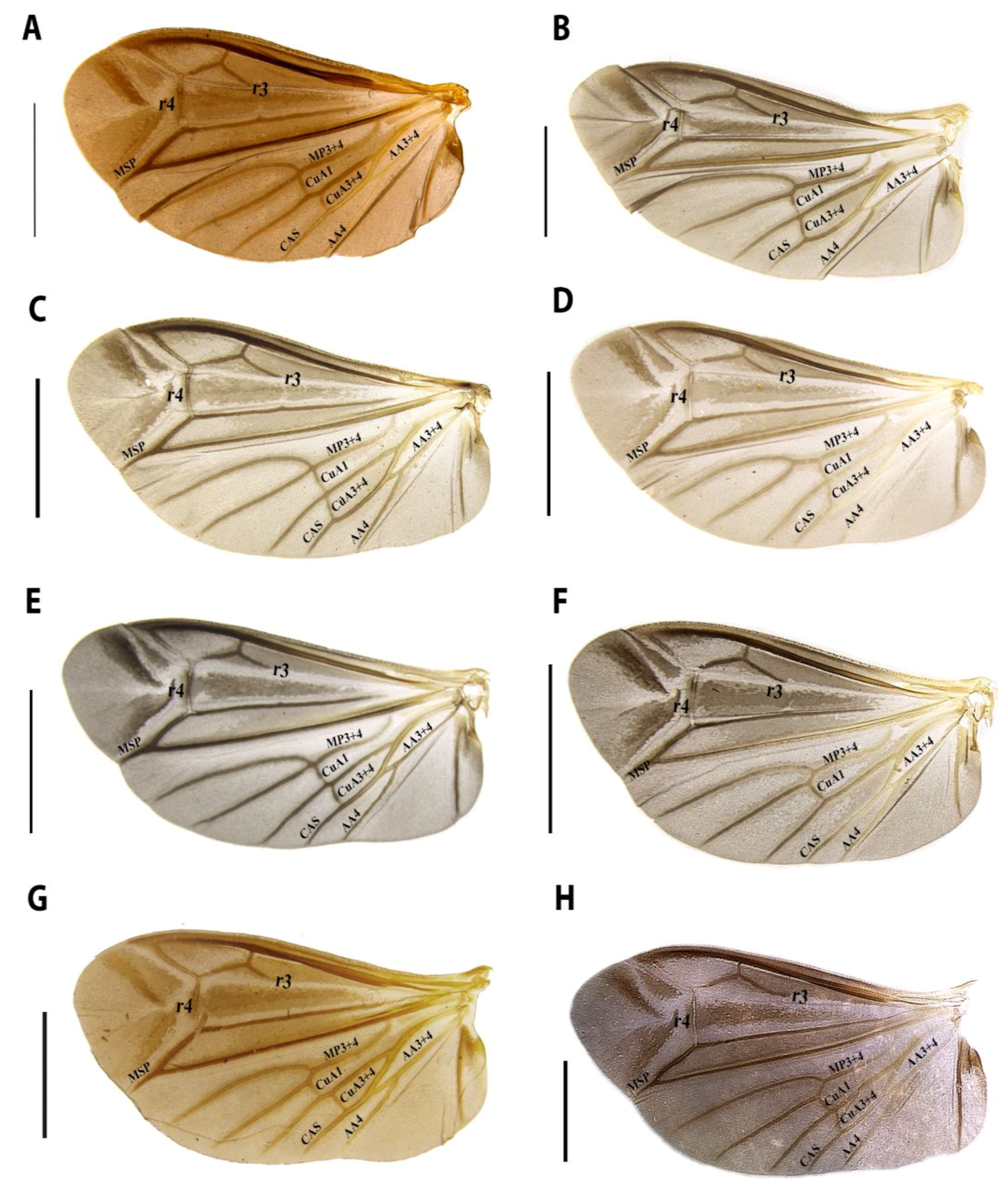
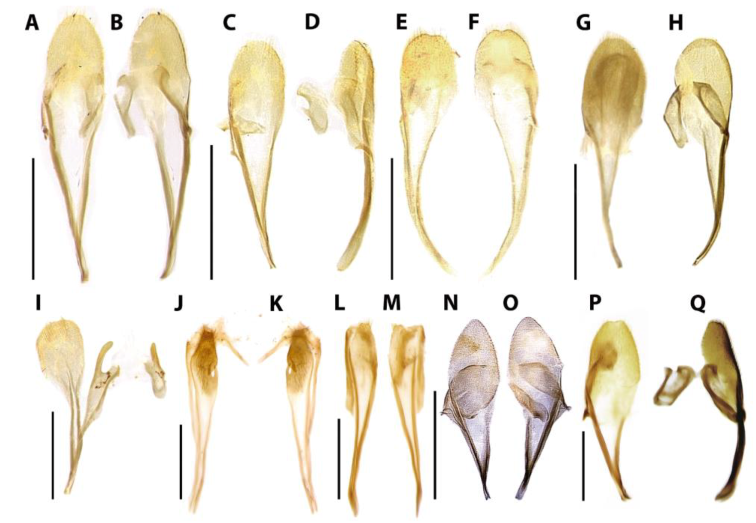
Figure 9.
Hind wing, dorsal. (
A)
Scissicauda biflabellata sp. nov. (
B)
Scissicauda jamari sp. nov. (
C)
Scissicauda gomesi sp. nov. (
D)
Scissicauda antennata sp. nov. (
E)
Scissicauda truncata sp. nov. (
F)
Scissicauda aurata sp. nov. (
G)
Scissicauda neyi sp. nov. (
H)
Scissicauda malleri (Pic, 1935) comb. nov. CAS = Cubitoanal Strut (Cu + CuP + AA3), MSP = Medial spur, r = radial cross-vein, AA = Anal Anterior, CuA = Cubitus Anterior, MP = Media Posterior (Lawrence et al. 2021 [
18]). Scale bar: 2 mm (
A–
H).
Figure 10.
Abdominal sclerites, dorsal and ventral views. (A,B) Scissicauda truncata sp. nov. (C,D) Scissicauda antennata sp. nov. (E,F) Scissicauda aurata sp. nov. (G,H) Scissicauda jamari sp. nov. (I,J) Scissicauda gomesi sp. nov. (K,L) Scissicauda malleri (Pic, 1935) comb. nov. (M,N) Scissicauda neyi sp. nov. (O,P) Scissicauda biflabellata sp. nov. (Q,R) Scissicauda asymmetrica sp. nov. Scale bar: 2 mm (A–R).
Figure 11.
Sternum VIII, ventral view and pygidium, dorsal view. (A,B) Scissicauda truncata sp. nov. (C,D) Scissicauda antennata sp. nov. (E,F) Scissicauda aurata sp. nov. (G,H) Scissicauda jamari sp. nov. (I,J) Scissicauda gomesi sp. nov. (K,L) Scissicauda malleri (Pic, 1935) comb. nov. (M,N) Scissicauda biflabellata sp. nov. (O,P) Scissicauda asymmetrica sp. nov. (Q,R) Scissicauda neyi sp. nov. Scale bar: 1 mm (A–R).
Figure 12.
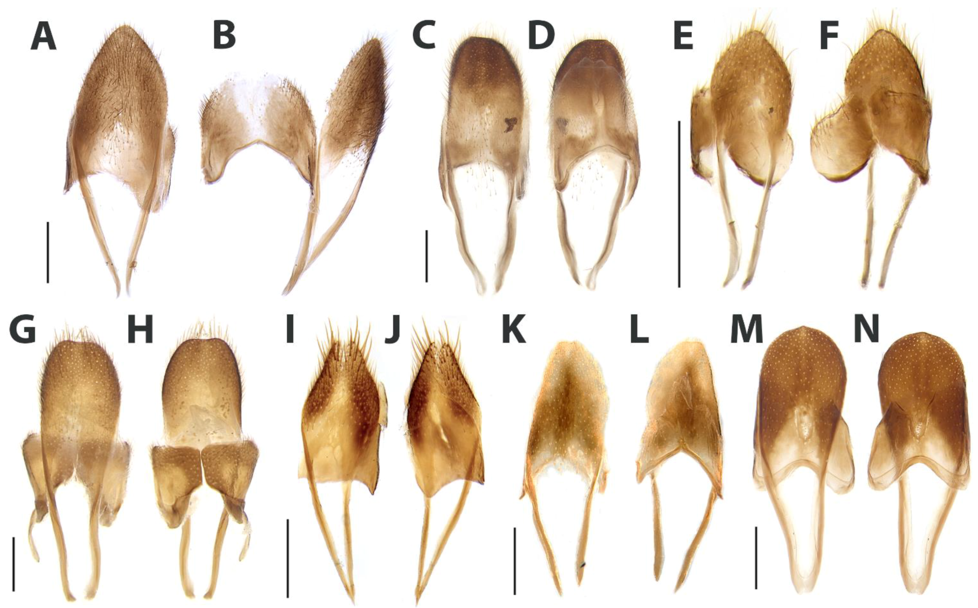
Syntergite, dorsal view and sternite IX, ventral view. (A,B) Scissicauda truncata sp. nov. (C,D) Scissicauda antennata sp. nov. (E,F) Scissicauda aurata sp. nov. (G,H) Scissicauda jamari sp. nov. (I) Scissicauda gomesi sp. nov. (J,K) Scissicauda asymmetrica sp. nov. (L,M) Scissicauda biflabellata sp. nov. (N,O) Scissicauda malleri (Pic, 1935) comb. nov. (P,Q) Scissicauda neyi sp. nov. Scale bar: 1 mm (A–Q).
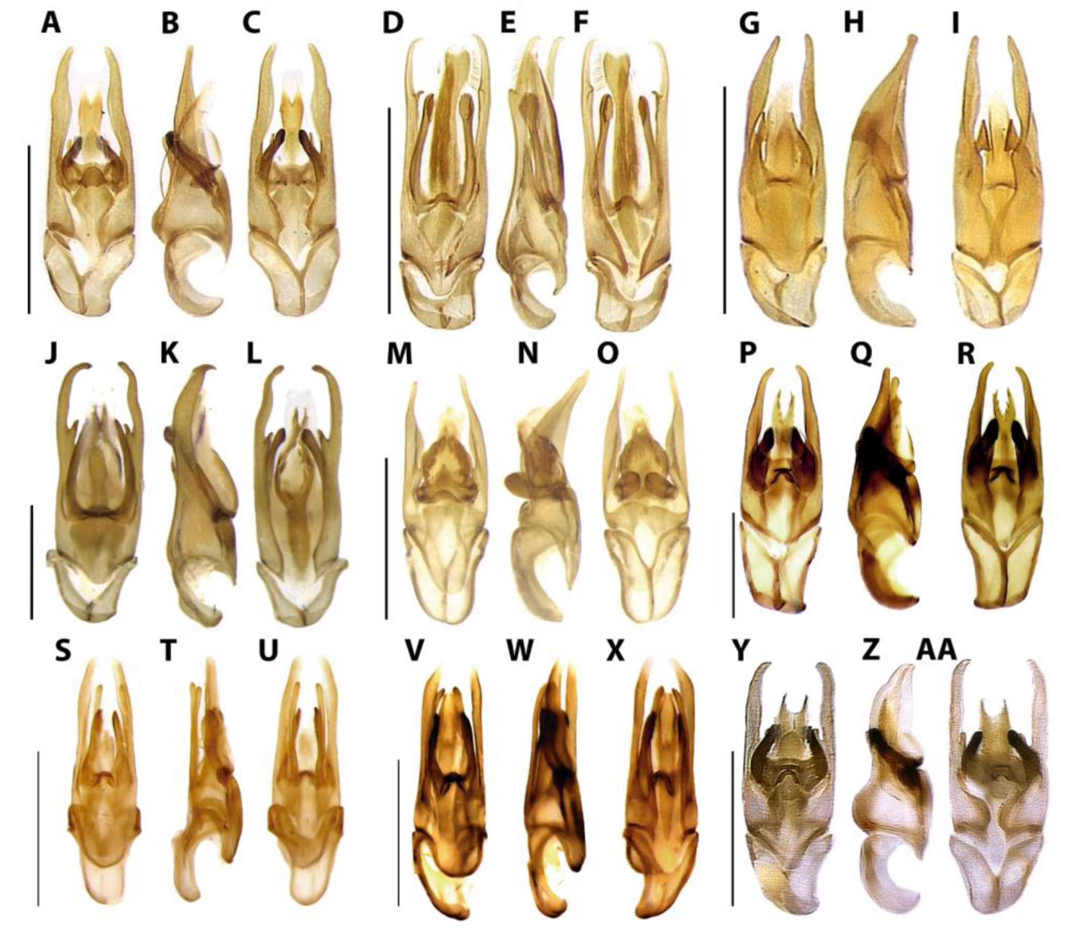
Figure 13.
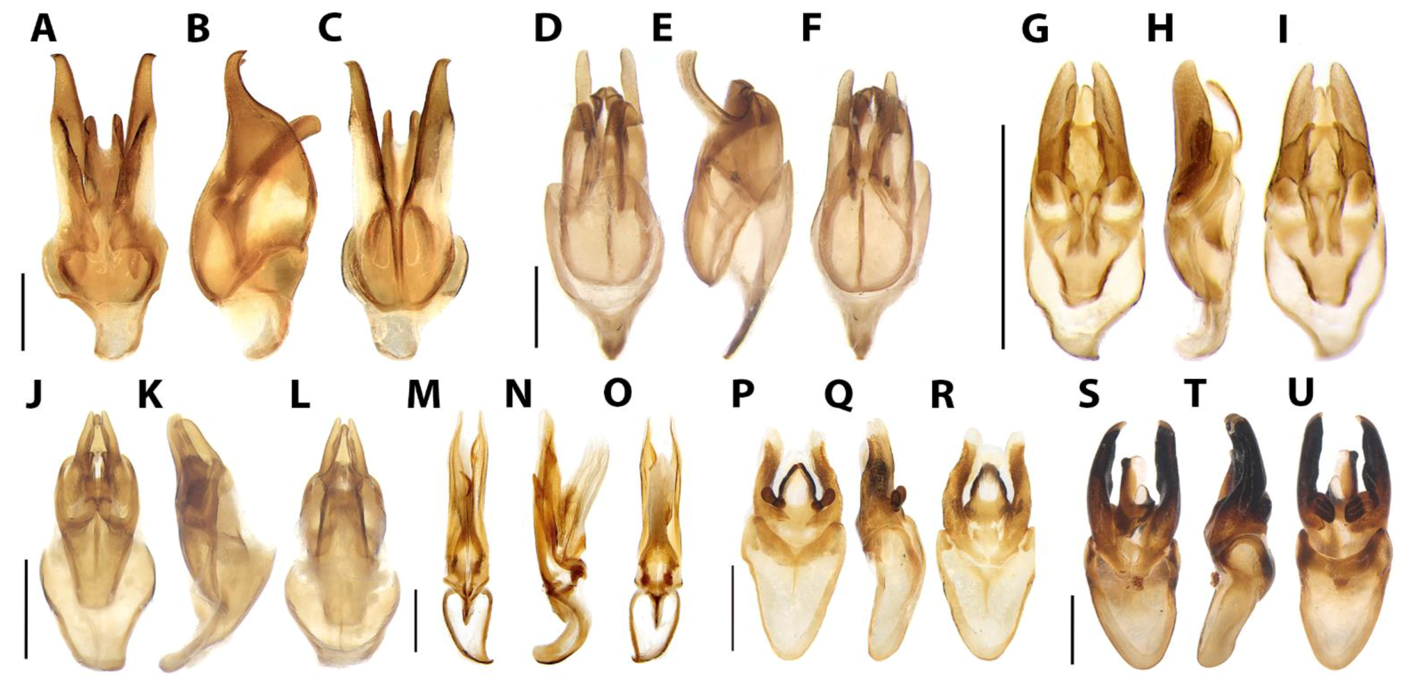
Aedeagus, dorsal and ventral views. (A–C) Scissicauda truncata sp. nov. (D–F) Scissicauda aurata sp. nov. (G–I) Scissicauda antennata sp. nov. (J–L) Scissicauda jamari sp. nov. (M–O) Scissicauda gomesi sp. nov. (P–R) Scissicauda neyi sp. nov. (S–U) Scissicauda biflabellata sp. nov. (V–X) Scissicauda asymmetrica sp. nov. (Y–AA) Scissicauda malleri (Pic, 1935) comb. nov. Scale bar: 1 mm (A–AA).
Redescription. Male. Head (
Figure 5) entirely covered by pronotum in dorsal view; capsule almost 2× wider than long, slightly longer than high. Frons slightly prominent dorsally, swollen. Antennal sockets reniform, about two-thirds of frons width; antennifer process conspicuous. Vertex somewhat convex. Antenna (
Figure 6) 11-segmented, scape constricted basally, pedicel almost as long as wide and constricted medially or basally, antennomeres III–X serrate to flabellate, compressed, subequal in length (III slightly smaller than IV in
S. biflabellata), with double or single lamellae present (with dense upright bristles) or absent, lamellae slender, apical antennomere slightly longer than the subapical one. Frontoclypeus slightly curved. Labrum connected to frontoclypeus by a membranous suture, 2× as wide as long, anterior margin poorly defined. Mandibles long and slender, arcuate, apex acute, internal tooth absent, external margin sparsely setose in basal ½. Maxilla with cardo well-sclerotized, stipe oblong in ventral view, posterior margins truncate, weakly or well-sclerotized, palpi 4-segmented; palpomere III subtriangular; IV lanceolate, with internal margin covered with minute, dense bristles, almost 3× longer than III. Labium with mentum weakly or well-sclerotized and bristled, completely divided sagittally; submentum weakly or well-sclerotized and bristled, subcordiform, elongate; palpi 3-segmented, palpomere III securiform. Gular sutures almost indistinct; gular bar transverse, 2× as wide as the submentum minimal width. Occiput piriform, as wide as one-third posterior width (
Figure 5E–H,M–P). Tentorium long and slender, almost as high as half head high, projected internally almost on the half of its length, strongly curved backwards. Thorax. Pronotum (
Figure 7) semilunar, posterior angles acute; disc subquadrate in dorsal view, notably convex, regularly punctured, punctures small and bristled; with a line of distinct deep marginal punctures; pronotal expansions well-developed, anterior expansion maximal length almost half as long as the disc, posterior expansions straight; slightly wider than humeral distance. Hypomeron 3× longer than tall. Prosternum 4× as wide as its major length; slightly constricted parasagittally (in
S.balena and
S.disjuncta). Proendosternite elongated or clavate, slightly longer than prosternal process minimal width. Mesoscutellum with posterior margin rounded. Elytra rounded (subparallel in
S. disjuncta), 3–5× as long as wide, pubescent, secondary pubescence absent, with a line of conspicuous punctures all over sutural and lateral margins. Hind wing (
Figure 9) well-developed, posterior margin sinuose, 2× as long as wide, r3 almost as long as r4, radial cell 2–3× wider than long, almost reaching anterior margin, costal row of setae inconspicuous; CuA1, CuA2, CuA3+4 (absent in
S. aurata), CAS, MP3+4, AA3+4, and AA4 present; MSP of three fourths r4 length, reaching or almost reaching distal margin, J indistinct. Allinotum slightly wider than long, lateral margins slightly convergent posteriad, posterior margin straight; prescutum extending slightly less than half metascutum length; rounded area of scutum weakly sclerotized, scutum–prescutal plates sclerotized, extending ridges almost up to posterior margin; metascutellum glabrous. Mesosternum weakly sclerotized, acute medially, attached to metasternum by a suture almost as wide as mesosternum. Mesoepimeron attached to metasternum by membrane. Mesosternum/mesanepisternum suture inconspicuous. Mesanepisternum/mesepimeron suture conspicuous. Metasternum oblique and strongly depressed by mesocoxae, anterior medial keel prominent up to anterior one-third, discrimen indistinct, lateral margins divergent posteriad up to lateral-most part of metacoxa, then convergent posteriad, posterior margin bisinuose. Femur slightly shorter or same length as tibia. Anterior pro- and mesoclaws with posterior branch toothed. Tibial spurs formula 2-2-2, symmetric or asymmetric (
Figure 8). Tarsomere I 2–2.6× longer than II, II 2× longer than III, III subequal in length to IV, IV bilobed, lobes reaching two-thirds V length. Mesendosternum with two parasagittal projections directed outwards, irregularly alate. Metendosternum spatulate, 1.5–2× longer than wide, median projection acute anteriad, with two lateral laminae. Abdomen (
Figure 10). Tergum I with anterior margin membranous, laterotergite membranous; spiracle obliquely attached to thorax, more vertically. Terga II–VII with posterior angles progressively produced and acute posteriad (except in
S. aurata, not progressively produced), posterior margins progressively bisinuose or emarginate. Sterna II–VIII or II–IX visible, sternum II with two median close-set vitreous spots. Spiracles dorsal, at almost half sterna lengths. Sternum VIII with larval lanterns elongate or absent. Sternum VIII (
Figure 11A,C,E,G,I,K,M,O,Q) clearly longer than VII or slightly longer; Pygidium (
Figure 11B,D,F,H,J,L,N,P) almost 2× longer than VIII, anterior margin emarginated or indented, posterior margin sub-rounded, truncated, indented, emarginated or bisinuose; Sternum IX (
Figure 12) asymmetric, posterior margin acute, truncate or subrounded. Syntergite (
Figure 12B,D,F,H,I,K,M,O,Q) consisting of paired lateral plates convergent posteriad (putatively tergite IX or paraproct), median transversal suture absent or medially divided, bearing bristles posteriorly, membrane-connected at sternum IX, 1/3 or 2/3 shorter than sternum IX, membrane-connected at sternum IX along its length. Aedeagus (
Figure 13) with phallus consisting of a dorsal plate basally fused to parameres, symmetric, medially grooved, projected dorsolaterally toward apex; ventral plate with lateral margins sinuose, weakly sclerotized; parameres symmetric, apically rounded, with a ventrobasal process rudimentary or projected and extended beyond the phallus.
Female. Pygidium (
Figure 15A,C,E,G,I,K,M) with sides rounded, posterior margin subrounded, truncate, or slightly emarginate. Sternum VIII as long as wide, spiculum ventrale long and slender, as long as 3/4 or 2/4 as the core sternum length (
Figure 15B,D,F,H,J,L,N). Internal genitalia (
Figure 16H–N) with a large and somewhat rounded spermatophore-digesting gland anterior to the common oviduct, and a more anterior and a slightly larger spermatophore-digesting gland. Valvifers free, twisted basally, 3× longer than coxite; coxites medially fused, coxital baculi well developed, sclerotized, divergent basally; styli minute, sclerotized; proctiger indistinct (
Figure 16). In [
9] interpretation of the internal parts of
S. disjuncta suggests that it lacks a spermatophore. We suggest that this interpretation be revisited upon the availability of new specimens to be dissected. An alternative interpretation is that what [
9] regards as the spermatophore-digesting gland and common oviduct are instead the spermatheca and the spermatophore-digesting gland, respectively.
Remarks.
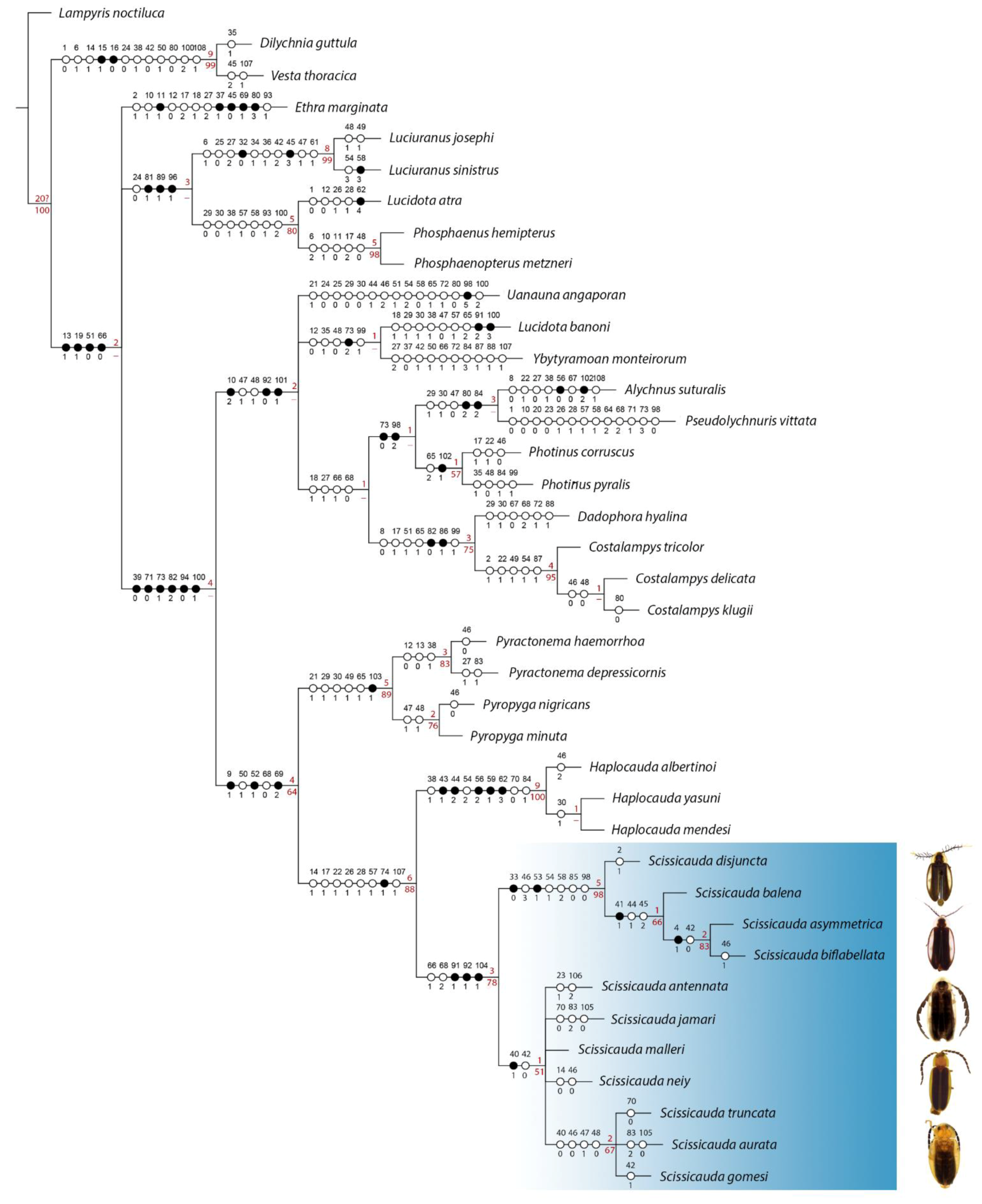
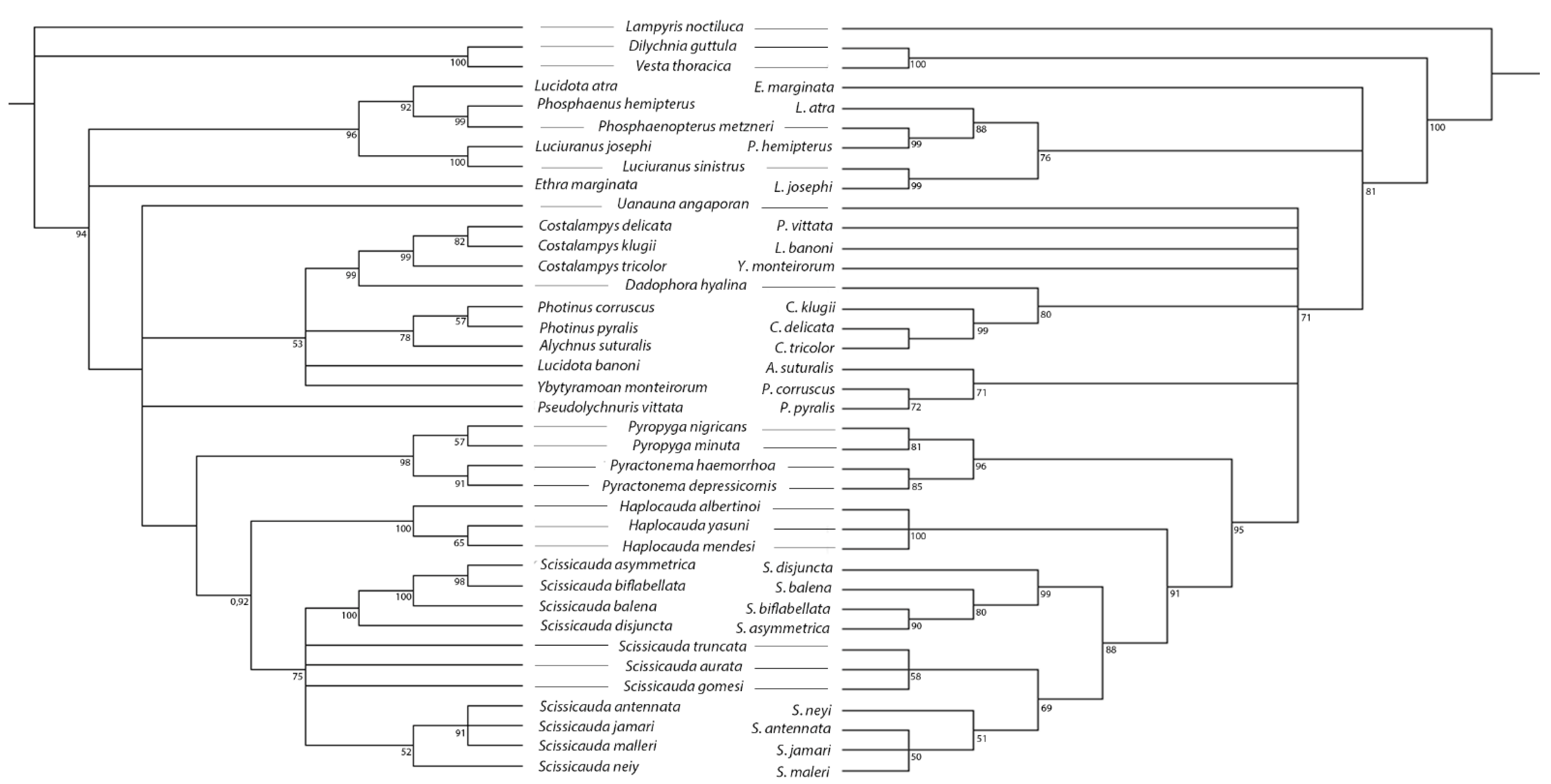
Scissicauda is recovered here as closest to
Haplocauda, then
Pyropyga +
Pyractonema. While
Pyropyga +
Pyractonema have the central third of the frons fused to labrum and lack claw teeth,
Haplocauda and
Scissicauda have the labrum entirely connected to frons by membrane and have toothed pro- and mesoclaws.
Scissicauda can be distinguished from these three aforementioned genera by male genitalic traits. The most distinctive feature is the dorsal plate with a deep transversal groove (
Figure 13). “Typical
Scissicauda” (i.e., with modified terminalia; see below) are somewhat similar to
Haplocauda, from which it can be distinguished by the lack of a median pointed projection on the sternum VIII (
Figure 11A,C,E,G,I,K,M,O,Q). “Atypical
Scissicauda” (i.e., with regular/unmodified terminalia; see below) are externally similar to many
Lucidota and the smaller
Pyractonema species, from which it can be distinguished by the aedeagal morphology, especially the dorsal plate bearing a deep transversal groove. A more thorough comparison with the rather distant
Costalampys was provided in [
4].
Scissicauda was placed in Lampyrinae:
incertae sedis in [
2].Here, based on its regular-sized arcuate mandibles, pronotum bearing anterior and lateral expansions, dorsal abdominal spiracles, and asymmetrical aedeagal sheath, in addition to the phylogenetic results in [
2,
5], and this paper (see above),
Scissicauda is herein placed in Photinini.
Distribution. The distribution of
Scissicauda and its species may be underestimated due to lack of targeted sampling. However, a few interesting patterns emerge from their distribution (
Figure 26). “Typical”
Scissicauda” seems to be restricted to the Atlantic Forest biome (in the Parana dominion sensu 36), and it is likely to be endemic to it, since they were never found elsewhere. On the other hand, “atypical”
Scissicauda are scattered across South America eastern to the Andes, mainly in the Amazon Basin (Boreal and Southern Brazilian dominions sensu 36). Notable exceptions are
S. malleri comb. nov., the only “atypical”
Scissicauda in the Atlantic Forest, and
S. neyi in the Cerrado biome (in the Chacoan dominion).
Checklist of Scissicauda species
Scissicauda antennata sp. nov.
Scissicauda asymmetrica sp. nov.
Scissicauda aurata sp. nov.
Scissicauda balena (Silveira, Mermudes & Bocakova, 2016)
Scissicauda biflabellata sp. nov.
Scissicauda disjuncta (E. Olivier, 1896)
Scissicauda gomesi sp. nov.
Scissicauda jamari sp. nov.
Scissicauda malleri (Pic, 1935) comb. nov.
Scissicauda neyi sp. nov.
Scissicauda truncata sp. nov.
Key to Scissicauda
species based on males | 1 | Antennae uniflabellate or biflabellate (Figure 6H,I)........................................................................................................2 |
| 1′ | Antennae serrate (Figure 6A–G).........................................................................................................................................4 |
| 2 | Antennae with lamellae as long as core antennomere (Figure 6I); pygidium emarginated on anterior margins |
| | (Figure 11P).......................................................................................................................Scissicauda asymmetrica sp. nov. |
| 2′ | Antennae with lamellae longer than core antennomere (Figure 6H); pygidium strongly indented or slightly |
| | emarginated on anterior margins (Figure 11N)………………………………………………...........................................3 |
| 3 | Antennae biflabellate (Figure 6H); sternum VIII with posterior margin emarginate (Figure 11M); pygidium with |
| | anterior margin strongly indented (Figure 11N); paramere with a long ventral rod (Figure 13S–U).......................... |
| | ................................................................................................................................................Scissicauda biflabellata sp. nov. |
| 3′ | Antennae uniflabellate; sternum VIII with posterior margin trisinuose; pygidium with anterior margin slightly |
| | emarginate; paramere with a rudimentary ventral rod (see [9])......................Scissicauda disjuncta (E. Olivier, 1896). |
| 4 | Sternum IX retracted under VIII; paramere with a long ventral rod (see [9]) ............................................................. |
| | …………………………………………………………….…..Scissicauda balena Silveira, Mermudes & Bocakova, 2016. |
| 4′ | Sternum IX partially exposed (Figure 3B); parameres with a rudimentary ventral rod (Figure 13J) .........................5 |
| 5 | Tibial spurs of metaleg distinctly asymmetrical in length (Figure 8A–C); Sternum VIII with anterior margin |
| | distinctly narrower than anterior 1/3 of the pygidium (Figure 11G–H) ........................................................................6 |
| 5′ | Tibial spurs of metaleg symmetrical in length (Figure 8D–I); Sternum VIII with anterior margin as wide as |
| | anterior 1/3 of pygidium (Figure 11B,F,J) .........................................................................................................................8 |
| 6 | Antennae almost as long as whole body (Figure 3C); pygidium 1.8× wider than anterior margin (Figure 11D)….. |
| | ...................................................................................................................................................Scissicauda antennata sp. nov. |
| 6′ | Antennae shorter than body length (Figure 3A); pygidium 2.5× wider than anterior margin (Figure 11H).............7 |
| 7 | Antennomeres X–XI yellowish (Figure 6D); pygidium with anterior margin emarginated (Figure 11H); sternum |
| | IX with posterior margin rounded (Figure 12G)...................................................................Scissicauda jamari sp. nov. |
| 7′ | Antennae entirely brown (Figure 6G); pygidium with anterior margin medially indented (Figure 11L); sternum |
| | IX with posterior margin acuminate (Figure 12N)……................................Scissicauda malleri (Pic, 1935) comb. nov. |
| 8 | Pygidium with posterior margin subrounded (Figure 11F,J); parameres curved dorsally in lateral view |
| | (Figure 13E)...........................................................................................................................................................................9 |
| 8′ | Pygidium with posterior margin truncate or posterolateral angles longer than the midline, rounded (Figure 11B,R); |
| | parameres almost straight in lateral view (Figure 13B,Q)……………………………………................................……10 |
| 9 | Pygidium with anterior margin almost straight (Figure 11J); phallic ventral plate at least 2× longer than phallobase |
| | (Figure 13O), parameres with ventral rod at the basal half of paramere (Figure 13O) …Scissicauda gomesi sp. nov. |
| 9′ | Pygidium with anterior margin emarginated (Figure 11F); phallic ventral plate at least 3× longer than phallobase; |
| | parameres with ventral rod at the apical half of paramere (Figure 13F).............................Scissicauda aurata sp. nov. |
| 10 | Pronotum with anterior expansion 1.4× longer than lateral expansion (Figure 7A); pygidium with posterior |
| | margin truncate (Figure 11B)..................................................................................................Scissicauda truncata sp. nov. |
| 10′ | Pronotum with anterior expansion almost 2× longer than lateral expansion (Figure 7I); pygidium with |
| | posterolateral angles longer than the midline (Figure 11R)......................................................Scissicauda neyi sp. nov. |
3.3.1. Scissicauda antennata sp. nov. Zeballos, Roza, and Silveira
Figure 3C,D,
Figure 5C–G,
Figure 6A,
Figure 7B,
Figure 8B,
Figure 9D,
Figure 10C,D,
Figure 11C,D,
Figure 12C,D,
Figure 13G–I and
Figure 14E,F.
urn:lsid:zoobank.org:act:B97D4A19-E3EE-48A9-801E-F5367B66B7E9
Type material. HOLOTYPE (♂, INPA, pinned), label data: “BRAZIL, Rondônia: Nova, Mamoré, Parque Estadual de, Guajará-Mirim. Rio Formoso//101926 S–643388 W, 20-, 27.x.1995, J. Vidal & L.S., Aquino. Arm. de Malaise//Scissicauda antennata HOLOTYPE [red label]”. PARATYPES: (4 ♂, INPA, pinned, 1 completely dissected and stored in microvial with glycerin) “BRAZIL, Rondônia: Nova, Mamoré, Parque Estadual de, Guajará-Mirim. Rio Formoso//101926 S–643388 W, 20-, 27.x.1995, J. Vidal & L.S., Aquino. Arm. de Malaise [yellow label]”. (2 ♂, INPA, pinned) “BRAZIL, Rondônia, Guajará-, Rio Ouro Preto, Bananal, 105823 S–650539 W//20–27.x.1995, J. A., Rafael & A.L.Henriques, Arm. Malaise [yellow label]”. (1 ♂, INPA, pinned) “BRAZIL: AM, QUERARI, São Gabriel da Cachoeira, 2° Pel. Esp. de Fronteira, 01°05″ N/69°51″ W//05.iv-27.v.1993, Vidal, J; Ferreira, R.L.M. col.//Malaise [yellow label]”.
Type locality. Brazil, Rondônia: Nova Mamoré, Parque Estadual de Guajará-Mirim.
Material examined. (5 ♂, INPA, pinned) “BRAZIL, Rondônia: Nova, Mamoré, Parque Estadual de, Guajará-Mirim. Rio Formoso//101926 S–643388 W, 20-, 27.x.1995, J. Vidal & L.S., Aquino. Arm. de Malaise”. (5 ♂, INPA, pinned) “BRAZIL, Rondônia, Guajará-, Rio Ouro Preto, Bananal, 105823S-650539W//20–27.x.1995, J. A., Rafael & A.L.Henriques, Arm. Malaise”. (1 ♂, INPA, pinned) “BRAZIL: AM, QUERARI, São Gabriel da Cachoeira, 2° Pel. Esp. de Fronteira, 01°05”N/69°51”W//05.iv-27.v.1993, Vidal, J; Ferreira, R.L.M. col.//Malaise”.
Distribution: Brazil: Rondônia, Nova Mamoré (see South Brazilian dominion in
Figure 26) e Amazonas, São Gabriel da Cachoeira (see Boreal Brazilian dominion in
Figure 26).
Etymology: Antennata is a Latin adjective that means “which bears an antennae”. This epithet is given because of the long male antennae.
Diagnosis. Males with antennomere compressed, serrate, as long as body length, (
Figure 6A). Metatibia with two spurs asymmetrical not reaching half the length of first tarsomere (
Figure 8B). Pygidium with posterior margin bisinuose (
Figure 11D). Sternum IX (
Figure 12C) with posterior margin rounded. Syntergite completely divided by a membranous line (
Figure 12D). Phallus dorsal plate strongly rounded basally not reaching half the length of the phallobase; phallic groove rudimentary; phallic ventral plate as long as paramere length; parameres curved dorsally (lateral view); ventrobasal process rudimentary (
Figure 13G–I).
Females. Sternum VIII (
Figure 14F) longer than wide. Pygidium with anterior margin emarginated, posterior margin subrounded (
Figure 14E).
Description. Color pattern. Integument overall blackish-brown to light brown. Scape and pedicel blackish-brown to light brown, rest of antennomeres dark brown (
Figure 6A). Pronotum largely yellowish at sides and slenderly anterior at the disc, pronotal disc brown (
Figure 7B); hypomeron yellowish or light brown with margins yellowish. Elytron with pale yellow lateral-longitudinal vittae on the anterior 2/3 of the elytron, sutural margin blackish-brown to pale yellow and outer lateral line light brown (
Figure 3C). Sternites blackish-brown, coxae, trochanters, and femora blackish-brown to light brown. Abdominal sternites brown to light brown (
Figure 3D). Pygidium medially light brown, posterior margin blackish-brown (
Figure 11D).
Male. Antennae (
Figure 6A) pedicel almost as long as wide and constricted medially; antennomeres III slightly smaller than IV, antennomeres IV–X subequal in length, antennomere XI slightly longer than the X, compressed. Pronotum 1.2× wider than long (
Figure 7B). Metatibia with two spurs asymmetrical not reaching half the length of first tarsomere (
Figure 8B). Elytral expansion half of pronotal disc width. Hind wing with cross-veins CuA1 (more apical than MP3+4 split) and CuA3+4 present, forming a continuous line (
Figure 9D). Sternum VIII with posterior margin emarginated (
Figure 11C). Sternum IX with posterior margin rounded, almost a quarter longer than the aedeagus. Syntergite completely divided into two asymmetric plates connected by a membrane, without posterior projection (
Figure 12C–D). Pygidium with sides rounded, posterior margin bisinuose, anterior margin emarginate (
Figure 11D). Phallus dorsal plate strongly rounded basally, phallic groove at half of its length, moderately curved; dorsal plate arms projecting ventrally towards the apex, embracing the ventral plate, with apical margin truncate and wide; ventral plate slightly longer than phallobase; parameres curved in lateral, with apical margin rounded, ventrobasal process rudimentary (
Figure 13G–I).
Female. Sternum VIII (
Figure 14F) longer than wide, convergent posteriorly, posterior margin indented medially, Pygidium with anterior margin emarginated, posterior margin subrounded (
Figure 14E).
Remarks. S. antennata sp. nov. (
Figure 3C) is similar in dorsal coloration pattern (overall blackish-brown to light brown) to
S. gomesi sp. nov. (
Figure 3I).
Scissicauda antennata is unique among its congenerics by the antenna as long as total body length, and by the wing with a cross-vein CuA1 more apical than MP3+4 split (
Figure 9D).
3.3.2. Scissicauda asymmetrica sp. nov. Zeballos, Roza, and Silveira
Figure 4C,D,
Figure 6I,
Figure 7H,
Figure 8I,
Figure 10Q,R,
Figure 11O,P,
Figure 12J,K,
Figure 13V–X,
Figure 14C,D,
Figure 15K,L and
Figure 16F,J.
urn:lsid:zoobank.org:act:BB7868F2-9DD6-47C0-A59D-0EE7C4A89E63
Type material. Holotype (♂, DZRJ, pinned dissected and stored in microvial with glycerin), label data: “Brasil. Rio de Janeiro. Angra dos Reis, Ilha grande x.2017, malaise 23°08′47″ S, 441 m 44°11′09.4″ W, L. Silveira, L. Campello, S. Vaz col. Paratypes (3 ♀, DZRJ), label data: “Brasil, RJ, Itatiaia, P. N. de, Itatiaia. Malaise, Pensario p2 1280m, XII.2014, R. Monteiro col.”; (♂, DZRJ, dissected and stored in microvial with glycerin), label data: “Brasil, RJ, Itatiaia, Parque Nacional de Itatiaia, 1 male, sendo predado por fêmea de Photuris sp., 31.I.2012, Silveira L., Araújo C. col.; (♂, DZRJ) label data: “Brasil, MG, Itajubá, R.B.M. Serra dos Toledos, Malaise, 16.II-17.III.2017, Rosa e Lopes col.”.
Type locality: Brazil, Rio de Janeiro, Parque Estadual da Ilha grande.
Distribution: Brazil: Rio de Janeiro, Angra dos Reis e Itatiaia; e Minas Gerais, Itajubá (see Parana dominion in
Figure 26).
Etymology: Asymmetrica is a Latin adjective that means “asymmetric”. This epithet is given because of the asymmetrical lamellae of the male antenna.
Diagnosis. Males with antennomere III–IX compressed, filiform with double apical lamellae asymmetrical in size, the internal much smaller, antennomere XI with single apical lamellae (
Figure 6I). Metatibial spurs symmetrical (
Figure 8I). Pygidium (
Figure 11P) strongly indented on both anterior and posterior margins, with lateral margins subparallel and posterior margin obliquely prolonged externally. Sternum IX abruptly constricted anteriad at half its length, posterior margin convergent. Syntergite consisting of paired lateral plates, joined posteriad (
Figure 12J–K). Phallus dorsal plate subtruncate basally, phallic groove at half of its length, moderately curved; ventral plate 2× phallobase length; parameres straight (lateral view), ventrobasal process digitiform, extending slightly beyond ventral plate, shorter than paramere itself (
Figure 13V–X).
Females. Sternum VIII (
Figure 15L) longer than wide. Spiculum ventrale 2/4 sternum length (
Figure 15L). Pygidium with anterior margin almost straight, posterior margin slightly emarginated (
Figure 15K).
Description. Color pattern. Integument overall blackish-brown to light brown. Scape and pedicel brown or yellowish, rest of antennomeres brown except VIII which can be yellowish, and IX–XI which are always yellowish (
Figure 6I). Pronotum yellowish at sides and anterior margin, with a paired yellow parasagittal vittae (
Figure 7H); hypomeron yellowish. Elytron with pale yellow lateral-longitudinal and thin sutural vittae (
Figure 4D). Sternites light brown, coxae, trochanters and femora yellowish to light brown, tibiae and tarsi brown. Abdominal sternites light brown, VIII paired rounded yellow parasagittal vittae near the margin or almost completely yellowish (
Figure 4C). Pygidium laterally and medially dark brownish (
Figure 11P).
Male. Antennae with pedicel almost as long as wide and constricted medially; antennomeres III slightly smaller than IV, IV–X subequal in length, IX–XI progressively thinner, and III–IX compressed filiform with double apical lamellae, which are asymmetrical in size, the internal much smaller, X with a single apical lamellae, XI one-third longer than X (
Figure 6I). Pronotum 1.7× wider than long (
Figure 7H). Metatibial spurs symmetrical (
Figure 8I). Elytral expansion as long as disc width (
Figure 4C). Hind wing with anterior cross-vein CuA1 (more basal than MP3+4 split) and CuA3+4 present. Sternum VIII with posterior margin rounded (
Figure 11O). Sternum IX abruptly constricted anteriad at half its length, one-third longer than aedeagus, posterior margin convergent (
Figure 12J). Syntergite consisting of paired lateral plates, joined posteriad (
Figure 12K). Pygidium strongly indented on both anterior and posterior margins, with lateral margins subparallel and posterior margin obliquely prolonged externally (
Figure 11P). Phallus dorsal plate subtruncate basally, phallic groove at half of its length, moderately curved; ventral plate 2× phallobase length; parameres straight (lateral view), ventrobasal process digitiform, extending slightly beyond ventral plate, shorter than paramere itself (
Figure 13V–X).
Female. Sternum VIII longer than wide, rounded, constricted at posterior third, posterior margin indented medially. Spiculum ventrale long and slender, 2/4 sternum length (
Figure 15L). Pygidium with anterior margin constricted, almost straight, constricted at posterior third (
Figure 15K).
Remarks. S. asymmetrica sp. nov. (
Figure 4C) is similar in dorsal coloration pattern (overall blackish-brown to light brown) to the other “typical
Scissicauda”.
S. asymmetrica shares with
S. biflabellata (
Figure 4A) the biflabellate antennae. In
Scissicauda asymmetrica, the lamellae are asymmetrical and inserted apically in the antennomere (versus basally inserted and symmetrical lamellae in
S. biflabellata).
3.3.3. Scissicauda aurata sp. nov. Zeballos, Roza, and Silveira
Figure 3K,L,
Figure 5B–F,
Figure 6C,
Figure 7C,
Figure 8D,
Figure 9F,
Figure 10E,F,
Figure 11E,F,
Figure 12E,F,
Figure 13D–F,
Figure 14K,L,
Figure 15E,F and
Figure 16C,H.
urn:lsid:zoobank.org:act:A7F2C55C-933B-484D-B3D9-383527742DF1
Type material. HOLOTYPE (♂, INPA, pinned), label data: “BRAZIL, AM, Novo Aripuanã, Res. Soka gakkai, Malaise-ár. aberta, 06–10.xii.1999, J. Vidal//INPA-COL, 001245. Scissicauda aurata”. PARATYPE (♂, ♀, INPA, pinned, completely dissected and stored in microvial with glycerin), label data: “BRAZIL, AM, Reserva Ducke 6–8.x.21, Pensilvânia, luz negra, Bento & Bevilaqua legs.//Scissicauda aurata”.
Type locality: Brazil: Amazonas, Novo Aripuanã.
Distribution: Brazil: Amazonas, Manaus e Novo Aripuanã (see South Brazilian dominion in
Figure 26).
Etymology: Aurata is a Latin adjective meaning “golden”. Refers to the predominant color of that species.
Diagnosis. Males with antennomere compressed, serrate, with antennal lamellae absent, slightly longer than half length of body (
Figure 6C). Anterior pro- and mesoclaws bifid. Metatibia with two spurs symmetrical (
Figure 8D). Pygidium with posterior margin sub-rounded (
Figure 11F). Sternum IX with posterior margin rounded. Syntergite divided into two asymmetrical plates connected by a membrane, without posterior projection (
Figure 12E–F). Phallus dorsal plate strongly rounded basally, with phallic groove phallic groove in the basal half, moderately curved; phallic ventral plate at least 3× phallobase length; parameres curved dorsally, ventrobasal process rudimentary in the apical half of paramere (
Figure 13D–F). Females (
Figure 14K–L) with Sternum VIII longer than wide. Spiculum ventrale 3/4 sternum length (
Figure 15E). Pygidium with anterior margin emarginated, posterior slightly emarginated (
Figure 15F).
Description. Color pattern. Integument overall yellowish. Scape and pedicel light brown, antennomeres IV–IX blackish-brown, antennomeres X–XI yellowish (
Figure 6C). Pronotum, elytron, hypomeron, sternites, legs and abdominal sternites yellowish. (
Figure 7C). Pygidium anteriorly translucent and posteriorly yellowish (
Figure 11F). Female. Antennomeres IX–XI yellowish.
Male. Antennae (
Figure 6C) with the scape constricted basally, pedicel almost as long as wide and constricted medially; antennomeres IV–X subequal in length, XI slightly longer than the X. Pronotum 1.3× wider than long (
Figure 7C). Anterior, pro- and mesoclaws bifid, metatibia with two spurs symmetrical (
Figure 8D). Elytral expansion 1/3 of pronotal disc width (
Figure 3K). Hind wing with cross-vein CuA1 more basal than MP3+4 split, cross-vein CuA3+4 absent. Sternum VIII with posterior margin emarginate (
Figure 11E). Sternum IX with anterior margin rounded, slightly longer than aedeagus. Syntergite divided into two asymmetrical plates connected by a membrane, without posterior projection (
Figure 12E–F). Pygidium with posterior margin sub-rounded (
Figure 11F). Phallus dorsal plate strongly rounded basally, phallic groove at half length, moderately curved; ventral plate at least 3× phallobase length; parameres slightly curved dorsally (lateral view), ventrobasal process rudimentary in the apical half of paramere (
Figure 13D–F).
Female. Sternum VIII longer than wide, rounded, constricted at posterior third, posterior margin indented medially. Spiculum ventrale long and slender, 3/4 sternum length (
Figure 15F). Pygidium with anterior margin emarginated, posterior margin slightly emarginated, constricted at posterior quarter (
Figure 15E).
Remarks. S. aurata sp. nov. is similar to
S. truncata sp. nov. and
S. gomesi sp. nov. in having the posterolateral corners of the pygidium barely conspicuous and smaller than the median third (
Figure 11B,F,J).
Scissicauda aurata and
S. neyi sp. nov. are the only ones with a pronotum completely yellowish (
Figure 7C,I).
S. aurata is unique by its integument color (almost entirely yellowish;
Figure 3K,L), ventral plate of phallus at least 3× phallobase length (
Figure 13F), parameres with ventrobasal process rudimentary in the apical half of paramere (
Figure 13F).
3.3.4. Scissicauda biflabellata sp. nov. Zeballos, Roza, and Silveira
Figure 4A,B,
Figure 5L–P,
Figure 6H,
Figure 7G,
Figure 8H,
Figure 9A,
Figure 10O,P,
Figure 11M,N,
Figure 12L,M,
Figure 13S–U,
Figure 15A,B and
Figure 16A,I.
urna:lsid:zoobank.org:act:E4CAD15D-2D72-47F8-ABE1-EA39D538FBF9
Type material. HOLOTYPE: (♂, DZUP, pinned) label data: “Dept° Zool, UF-Parana//Brazil. Espirito Santo. Conceição da Barra, 11.9.1969, C & C.T. Elias col.//DZUP 429998//Scissicauda biflabellata, HOLOTYPE, det. Roza 2023.” PARATYPES: (♂, DZUP, pinned) label data: “Dept° Zool, UF-Parana//Brazil. Espirito Santo. Conceição da Barra, 8–14.X.1968, C & C.T. Elias col.//DZUP 430019//Scissicauda biflabellata, PARATYPE, det. Roza 2023.” (♂, DZUP, pinned) label data: “Dept° Zool, UF-Parana//Brazil. Espirito Santo. Conceição da Barra, 11.VIII.1969, C & C.T. Elias col.//DZUP 430015//Scissicauda biflabellata, PARATYPE, det. Roza 2023.” (♂, DZUP, pinned) label data: “Dept° Zool, UF-Parana//Brazil. Espirito Santo. Conceição da Barra, 11.IX.1969, C & C.T. Elias col.//DZUP 429999//Scissicauda biflabellata, PARATYPE, det. Roza 2023.” (♂, DZUP, pinned) label data: “Dept° Zool, UF-Parana//Brazil. Espirito Santo. Conceição da Barra, 17.IX.1969, C & C.T. Elias col.//DZUP 430017//Scissicauda biflabellata, PARATYPE, det. Roza 2023.” (♀, DZUP, pinned, abdomen dissected and stored in microvial with glycerin) label data: “Dept° Zool, UF-Parana//Brazil. Espirito Santo. Conceição da Barra, 17.IX.1969, C & C.T. Elias col.//DZUP 430016//Scissicauda biflabellata, PARATYPE, det. Roza 2023.” (♂, DZUP, pinned, entire specimen dissected and stored in microvial with glycerin) label data: “Dept° Zool, UF-Parana//Brazil. Espirito Santo. Conceição da Barra, 5.I.1970, C & C.T. Elias col.//DZUP 430018//Scissicauda biflabellata, PARATYPE, det. Roza 2023.”.
Type locality: Brazil. Espírito Santo, Conceição da Barra.
Distribution: Brazil. Espírito Santo, Conceição da Barra (see Parana dominion in
Figure 26).
Etymology: Biflabellata is a Latin adjective meaning “with double lamellae”.
Diagnosis. Males with antennomere compressed, filiform, with basal double lamellae symmetrical in size (
Figure 6H). Anterior pro- and mesoclaws bifid. Metatibial spurs symmetrical (
Figure 8H). Pygidium strongly indented on both anterior and posterior margins, with lateral margins subparallel and posterior margin obliquely prolonged externally (
Figure 11M,N). Sternum IX abruptly constricted anteriad at half its length, one-third longer than aedeagus, posterior margin convergent. Syntergite consisting of paired lateral plates joined posteriad (
Figure 12L–M). Phallus dorsal plate rounded basally, phallic groove at half of its length, moderately curved; ventral plate subequal to phallobase length; parameres straight (lateral view), ventrobasal process digitiform, extending slightly beyond ventral plate, shorter than paramere itself (
Figure 13S–U). Females with Sternum VIII longer than wide. Spiculum ventrale 3/4 the sternum length (
Figure 15A). Pygidium with anterior margin emarginated, posterior margin truncated (
Figure 15B).
Description. Color pattern. Integument overall blackish-brown (
Figure 4B). Scape and pedicel brown, antennomeres IX–XI yellowish (
Figure 6H). Pronotum largely yellowish at sides and with anterior portion of the expansion medially brown and laterally yellowish, with paired yellow parasagittal vittae at disc; hypomeron yellowish with lateral and ventral margins darkened (
Figure 7G). Elytron with pale yellow lateral-longitudinal and sutural vittae (
Figure 4A). Sternites blackish-brown, trochanters and femorae yellowish, tibiae and tarsi dark brown (
Figure 4B). Abdominal sternites light brown (
Figure 10O), except sternite VIII which is yellowish. Pygidium laterally and medially dark brownish (
Figure 11N).
Male. Antennae with pedicel almost as long as wide and constricted medially; antennomeres III slightly smaller than IV, IV–X subequal in length, compressed, filiform, with basal double lamellae, lamellae almost 2× as long as antennomere IV, progressively smaller until antennomere X, which is one-third longer than antennomere; antennomere XI 1.5× longer than the X (
Figure 6H). Pronotum 1.4× wider than long (
Figure 7G). Anterior pro- and mesoclaws bifid. Metatibial spurs symmetrical (
Figure 8H). Elytral expansion two-thirds of elytral disc width (
Figure 4A). Hind wing with anterior cross-vein CuA1 (more basal than MP3+4 split) and CuA3+4 present (
Figure 9A). Sternum VIII with posterior margin emarginated (
Figure 11M). Sternum IX abruptly constricted anteriad at half its length, one-third longer than aedeagus, posterior margin convergent (
Figure 12L). Syntergite consisting of paired lateral plates joined posteriad (
Figure 12M). Pygidium strongly indented on both anterior and posterior margins, with lateral margins subparallel and posterior margin obliquely prolonged externally (
Figure 11N). Phallus dorsal plate rounded basally, phallic groove at half of its length, moderately curved; ventral plate subequal to phallobase length; parameres straight (lateral view), ventrobasal process digitiform, extending slightly beyond ventral plate, shorter than paramere itself (
Figure 13S–U).
Female. Sternum VIII longer than wide, rounded, convergent posteriorly, posterior margin indented medially. Spiculum ventrale long and slender, 3/4 sternum length (
Figure 15B). Pygidium with anterior margin emarginated, posterior margin truncated, convergent posteriorly (
Figure 15A).
Remarks. S. biflabellata sp. nov. (
Figure 4A) is similar in dorsal color pattern (in general dark brown to light brown) to other “typical
Scissicauda”.
S. biflabellata shares biflabellate antennae with
S. asymmetrica but with longer lamellae (
Figure 6H) (versus short in
S. asymmetrica).
3.3.5. Scissicauda gomesi sp. nov. Zeballos, Roza, and Silveira
Figure 3I,J,
Figure 5A–E,
Figure 6E,
Figure 7E,
Figure 8E,
Figure 9C,
Figure 10I,J,
Figure 11I,J,
Figure 12I,
Figure 13M–O,
Figure 14M,N,
Figure 15I,J and
Figure 16E,N.
urn:lsid:zoobank.org:act:7A3DF14C-C12C-4D9B-AF0B-482E12B8F462
Type material. HOLOTYPE (♂, INPA, pinned), label data: “BRAZIL, PA, Repartimento, Vicinal 08, 042642S-, 495425W, 28.xi.2001, J.A., Rafael & J. Vidal, Malaise.//Scissicauda gomesi HOLOTYPE [red label]”. PARATYPES: (8 ♂, 1 ♀, INPA, pinned), label data: “BRAZIL, PA, Repartimento, Vicinal 08, 042642S-, 495425W, 28.xi.2001, J.A., Rafael & J. Vidal, Malaise.//Scissicauda gomesi PARATYPES [yellow label]”. (3 ♂, INPA, pinned), label data: “BRAZIL, PA, Tucuruí, Morro do Senador, 035923 S; 494445 W, 01.xii.2001, Malaise, J.A. Rafael & J. Vidal.//Scissicauda gomesi PARATYPES [yellow label]”. (1 ♂, INPA, pinned), label data: “BRAZIL, Pará, Tucuruí, Ig. Água Fria, 035052S-, 494704W, 02.xii.2001.//Scissicauda gomesi PARATYPES [yellow label]”. (3 ♂, INPA, pinned), label data: “BRAZIL, Pará, Tucuruí, Faz. Senador, 035948 S-, 494603 W, 01.xii.2001, J.A., Rafael & J. Vidal, Malaise.//Scissicauda gomesi PARATYPES [yellow label]”. (1 ♂, INPA, pinned), label data: “BRAZIL, Pará, Tucuruí, Próx. Barragem, 02.xii., 2001, J.A.Rafael & J. Vidal, Malaise.//Scissicauda gomesi PARATYPES [yellow label]”.(1 ♂, MZUSP, pinned), label data: “BRAZIL, PA, Abel Figueiredo, 4.850128° S 48.531963° W, 27.XI.2013, T. Camijo col.//Scissicauda gomesi PARATYPES [yellow label]”. (4 ♂, 1 ♀, INPA, pinned), label data: “BRAZIL, MA, Vila Nova, dos Martirios, F. Sta Rosa//050707S-481519W, 06.xii., 2001, F.L. Oliveira &, J. Vidal, arm. Malaise. Scissicauda gomesi PARATYPES [yellow label]”. (7 ♂, INPA, pinned), label data: “BRAZIL, MA, S. Pedro da, Água Branca, F. Esplanada, 045905S-480803W, 05.xii., 2001, J.A. Rafael; F.L., Oliveira & J. Vidal, Malaise. Scissicauda gomesi PARATYPES [yellow label]”. (2 ♂, 3 ♀, INPA, pinned), label data: “BRAZIL, MA, S. Pedro da, Água Branca, F. Esplanada, 045905S-480803W, 07.xii., 2001, J.A. Rafael; F.L., Oliveira & J. Vidal, Malaise. Scissicauda gomesi PARATYPES [yellow label]”.
Type locality: Novo Repartimento, Pará, Brazil.
Distribution: Brazil, Pará, Novo Repartimento (see South-eastern Amazonian dominion in
Figure 26) and Maranhão, Vila Nova (see Boreal Brazilian dominion in
Figure 26).
Etymology: This species was named in honor of the lampyrid researcher William Gomes, of the National Institute for Research in the Amazon, who helped locate and sort the specimens used in this paper.
Diagnosis. Males with antennomere compressed, serrate, with antennal lamellae absent, slightly longer than half the length of the body (
Figure 6E). Anterior pro- and mesoclaws bifid. Metatibia with two spurs symmetrical (
Figure 8E). Pygidium with the length of median region longer than posterior angles (
Figure 11J). Sternum IX with posterior margin rounded. Syntergite divided into two asymmetrical plates, connected by membrane, with a long projection on the posterior margin of each plate (
Figure 12I). Phallus dorsal plate subtruncate basally, reaching half the length of the phallobase, phallic groove moderately curved, phallic ventral plate as long as the paramere length, base of paramere nearby in the ventral region, paramere ventrobasal process short, in basal third of paramere, paramere slightly curved dorsally in lateral view (
Figure 13M–O). Females (
Figure 14M–N) with Sternum VIII longer than wide. Spiculum ventrale 2/4 sternum length (
Figure 15I). Pygidium with anterior margin almost straight, posterior margin truncated (
Figure 15J).
Description. Color pattern. Integument overall blackish-brown to light brown. Scape and pedicel blackish-brown to light brown, antennomeres X–XI blackish-brown, and apex of XI light brown (
Figure 6E). Pronotum largely yellowish at sides and slenderly anterior at the disc; disc blackish-brown (
Figure 7E) and hypomeron yellowish. Elytron with pale yellow lateral-longitudinal, sutural margin blackish-brown and outer lateral line light brown (
Figure 3I). Sternites, coxae, trochanters, femora and abdominal sternites blackish-brown to light brown (
Figure 3J). Pygidium overall blackish-brown, with anterior corners yellowish pale (
Figure 11J).
Male. Antennae (
Figure 6E) with scape constricted basally, pedicel almost as long as wide and constricted medially; antennomeres IV–XI subequal in length, XI slightly longer than the X. Pronotum 1.2× wider than long (
Figure 7E). Anterior pro- and mesoclaws bifid, metatibia with two spurs symmetrical (
Figure 8E). Elytral expansion almost 1/3 of pronotal disc width (
Figure 3I). Hind wing with cross-vein CuA1 (more basal than MP3+4 split) and CuA3+4 present, forming a continuous line. Sternum VIII with posterior margin emarginate, pygidium with the length of median region longer than the posterior angles (
Figure 11I). Sternum IX with anterior margin rounded, slightly longer than aedeagus. Syntergite divided into two asymmetrical plates, connected by a membrane, with a long projection on the posterior margin of each plate (
Figure 12I). Phallus dorsal plate strongly rounded basally, dorsal plate arms projecting ventrally, embracing the ventral plate, with apical margin rounded, phallic grooves at half length, moderately curved; ventral plate almost as long than phallobase, with a row of bristles on the side margins; parameres slightly curved dorsally (lateral view), base of paramere nearby in the ventral region, parameres ventrobasal process rudimentary (
Figure 13M–O).
Female. Sternum VIII longer than wide, rounded, constricted at posterior third, posterior margin slightly emarginated. Spiculum ventrale long and slender, 2/4 sternum length (
Figure 15N). Pygidium with anterior margin almost straight, posterior margin truncated, convergent posteriorly (
Figure 15M).
Remarks. S. gomesi sp. nov. is similar to
S. antennata sp. nov. by the overall coloration of integument (blackish-brown to light brown;
Figure 3C,I).
S. gomesi is unique among the aforementioned species in having a posterior projection in the syntergite (absent in the other species) and by the length of the ventral phallus plate as long as the phallobase (
Figure 13O).
3.3.6. Scissicauda jamari sp. nov. Zeballos, Roza, and Silveira
Figure 3G,H,
Figure 5D–H,
Figure 6D,
Figure 7D,
Figure 8C,
Figure 9B,
Figure 10G,H,
Figure 11G,H,
Figure 12G,H,
Figure 13J–L,
Figure 14G,H,
Figure 15G,H and
Figure 16D,M.
urn:lsid:zoobank.org:act:A7D01EBD-28F7-4321-814A-F8817ED78811
Type material. HOLOTYPE (♂, INPA, pinned), label data: “BRAZIL, RO, Itapuã do Oeste, Flona Jamari, Igarapé Preto, 09°11′16.0″ S–62°56′57.0″ W//07.x.2014, Varredura, J.A., Rafael, F.F. Xavier F°, R.M., Vieira & R.H. Aquino.//Scissicauda jamari HOLOTYPE [red label]”. PARATYPES: (1 ♂, 2 ♀, INPA, pinned), label data: “BRAZIL, RO, Itapuã do Oeste, Flona Jamari, Igarapé Preto, 09°11′16.0″ S–62°56′57.0″ W//07.x.2014, Varredura, J.A., Rafael, F.F. Xavier F°, R.M., Vieira & R.H. Aquino.//Scissicauda jamari PARATYPE [yellow label]”. (♂, INPA, pinned), label data: “BRAZIL, AM, Reserva Ducke 6–8.x.21, Lençol, luz mista, Bento & Bevilaqua legs. Scissicauda jamari PARATYPE [yellow label]”. (3 ♂, INPA), label data: “BRAZIL, AM, Manicoré, cachoeira. 05°29′44″ S; 60°, 49′21″ W. Malaise de solo, Floresta úmida. ix.2004, Silva & Pena leg. Scissicauda jamari PARATYPE [yellow label]”. (3 ♂, INPA), label data: “BRAZIL, Amazonas, Reserva F. Ducke, Malaise. 04.ix.1990., J. Vidal. Scissicauda jamari PARATYPE [yellow label]”.
Type locality: Brazil, Rondônia, Itapuã do Oeste, Flona Jamari.
Distribution: Brazil, Rondônia, Itapuã do Oeste (see South Brazilian dominion in
Figure 26) e Amazonas, Manaus e Manicoré (see Boreal Brazilian dominion in
Figure 26).
Etymology: The specific epithet is based on the Jamari National Forest, a Sustainable Use Conservation Unit located in the municipality of Itapuã do Oeste, in Rondônia, where the type specimen was collected. Noun in apposition.
Diagnosis. Males with antennomere compressed, serrate, with antennal lamellae absent, slightly longer than half length of body (
Figure 6D). Anterior pro- and mesoclaws bifid. Metatibia with two spurs asymmetrical, exceeding half the length of the first tarsomere (
Figure 8C). Pygidium with the length of posterior angles longer than the median region, with sides rounded, anterior margin emarginate, 3× smaller than the greatest width of pygidium (
Figure 11H). Sternum IX with posterior margin rounded. Syntergite divided into two plates connected by a membrane, with the posterior region with a long projection (
Figure 12G–H). Phallus dorsal plate strongly rounded basally, slightly projected but not reaching half the length of the phallobase, phallic groove moderately curved, phallic ventral plate symmetrically or asymmetrically, paramere ventrobasal process short, at apical 2/3, parameres slightly curved dorsally in the lateral view (
Figure 13J–L). Females (
Figure 14G–H) with Sternum VIII longer than wide. Spiculum ventrale 2/4 sternum length (
Figure 15G). Pygidium with anterior margin almost straight, posterior margin emarginated (
Figure 15H).
Description. Color pattern. Integument overall yellowish to blackish-brown. Scape, pedicel and antennomeres XI yellowish to light brown, III–X and apex of IX yellowish, antennomeres V–VIII and ¾ basal of IX blackish-brown (
Figure 6D). Pronotum largely yellowish at sides and anterior margin, disc with central and posterior regions blackish-brown (
Figure 7D), hypomeron yellowish. Elytron completely blackish-brown or with lateral-longitudinal vittae, sutural margin, and outer lateral line yellowish (
Figure 3G). Sternites yellowish to blackish-brown, coxae, trochanters, and femora yellowish to blackish-brown. Abdominal sternites yellowish to light brown (
Figure 3H). Pygidium overall brown, anterior corner pale yellow (
Figure 11H).
Male. Antennae (
Figure 6D) with scape constricted basally, pedicel almost as long as wide and constricted medially; antennomeres IV–X subequal in length, XI slightly longer than the X. Pronotum 1.3× wider than long (
Figure 7D). Anterior pro- and mesoclaws bifid. Metatibia with two spurs asymmetrical, exceeding half the length of the first tarsomere (
Figure 8C). Elytral expansion 1/3 of pronotal disc width (
Figure 3G). Hind wing with cross-veins CuA1 (more basal than MP3+4 split) and CuA3+4 present, forming a continuous line (
Figure 9B). Sternum VIII with posterior margin emarginate (
Figure 11G). Sternum IX with anterior margin rounded, 1.2× longer than aedeagus. Syntergite divided into two plates connected by a membrane, without posterior projection (
Figure 12G–H). Pygidium with the length of the posterior angles longer than the median region, with sides rounded, anterior margin emarginate, 3x smaller than the greatest width of pygidium. Phallus dorsal plate strongly rounded basally, phallic groove at half-length, moderately curved; ventral plate 2.5× longer than phallobase; parameres curved dorsally in lateral view, ventrobasal process rudimentary (
Figure 13J–L).
Female. Sternum VIII longer than wide, rounded, convergent posteriorly, posterior margin indented medially. Spiculum ventrale long and slender, 2/4 sternum length (
Figure 15H). Pygidium with anterior margin almost straight, posterior margin slightly emarginated, constricted at the posterior quarter (
Figure 15G).
Remarks. S. jamari sp. nov. is similar to
S. antennata sp. nov. and
S. malleri comb. nov. in having metatibia with two spurs asymmetrical (
Figure 8A–C). The general yellowish coloration of the integument of
S. jamari sp. nov. is superficially similar to
S. aurata sp. nov., except in variations with blackish-brown integument (
Figure 3K,L).
Scissicauda jamari can be distinguished from other species by having the length of the ventral phallus plate twice longer than the phallobase (
Figure 13L).
3.3.7. Scissicauda malleri (Pic, 1935) comb. nov.
Figure 3E,F,
Figure 5I–M,
Figure 6G,
Figure 7F,
Figure 8A,
Figure 9H,
Figure 10K,L,
Figure 11K,L
Figure 12N–O,
Figure 13Y–AA,
Figure 14O,P,
Figure 15M,N and
Figure 16G,H,
Supplementary Material S3.
Type material. HOLOTYPE (
Supplementary Material S3) (♂, MNHN, pinned), label data: voi
Lucidota imcopta//
Lucidota malleri//Ma 248, h, 12//TYPE”.
Examined material. (4 ♂, DZRJ, pinned), label data: “MG, RPPN Caraça, 19–20/XI/22, Trilha do Tanque, Pensilvania, M. L. Monné//Scissicauda malleri”. (1 ♀, DZRJ, pinned), label data: “BRAZIL, RIO DE JANEIRO, Parque Nacional de Itatiaia, 2018, A. Soares col.//Scissicauda malleri”. (1 ♂, DZRJ, pinned), label data: “Serra do Caraça, MG BRAZIL, 27.XI a 5.xii.1972, Exp. Mus. Zool.//Scissicauda malleri”. (2♂, UFRJ, pinned), label data: “BRAZIL, MG, Passa Quatro, Roza, Leocádio col.//Scissicauda malleri”. (1♂, DZRJ, pinned), label data: “BRAZIL, MG, Passa Quatro, 26.xi.2016 Mermudes, Roza, Leocádio e Lopez col.//Scissicauda malleri”. (4 ♂, MNRJ, pinned), label data: “MG. RPPN Caraça, 19.20/XI/22, Trilha do Tanque, Pensilvania, M.L.Monné//Scissicauda malleri”. (1 ♀, MNRJ, pinned), label data: “Brasil, Rio de Janeiro, Parque Nacional do Itatiaia, 2018, A. Soares col.//MNRJ- ENT7-43147//Scissicauda malleri”.
Type locality: Corupá, Santa Catarina, Brazil.
Distribution: Brazil, Santa Catarina, Corupá; Rio de Janeiro, Parque Nacional de Itatiaia; Minas Gerais, Passa Quatro e Serra do Caraça (see Parana dominion in
Figure 26).
Diagnosis. Males with antennomere compressed, serrate, with antennal lamellae absent, slightly longer than half the length of the body (
Figure 6G). Anterior pro- and mesoclaws bifid. Metatibia with two spurs asymmetrical, less than half the length of the first tarsomere (
Figure 8A). Pygidium with the length of posterior angles longer than the median region, with sides rounded, anterior margin indented, 3× smaller than the greatest width of pygidium (
Figure 11L). Sternum IX with posterior margin lanceolate. Syntergite divided into two plates connected by a membrane (
Figure 12N–O). Phallus dorsal plate strongly rounded basally, slightly projected but not reaching half the length of the phallobase, phallic groove moderately curved, phallic ventral plate with apical margin strongly emarginate, parameres slightly curved dorsally (lateral view), ventrobasal process rudimentary, almost in the middle of paramere (
Figure 13Y–AA).
Description. Color pattern. Integument overall yellowish to blackish-brown. Scape, pedicel and antennomeres XI blackish-brown (
Figure 6G). Pronotum yellowish, disc blackish-brown (
Figure 7F); hypomeron yellowish. Elytron entirely yellowish (
Figure 3E). Sternites yellowish to blackish-brown, coxae, trochanters, and femora yellowish to blackish-brown. Abdominal sternites yellowish to light brown (
Figure 3F). Pygidium overall yellowish (
Figure 11L).
Male. Antennae (
Figure 6G) with scape constricted basally, pedicel with lateral margins divergent apically; antennomeres IV–XI subequal in length, XI slightly longer than the X. Pronotum 1.4× wider than long (
Figure 7F). Anterior pro- and mesoclaws bifid. Metatibia with two spurs asymmetrical, less than half length of first tarsomere (
Figure 8A). Elytral expansion 1/3 of the elytra width (
Figure 3E). Hind wing with cross-vein CuA1 (more apical than MP3+4 split) and CuA3+4 present. Sternum VIII with posterior margin emarginate (
Figure 11K). Sternum IX with anterior margin lanceolate, almost longer than aedeagus. Syntergite divided into two plates connected by a membrane, without posterior projection (
Figure 12N–O). Pygidium with the length of posterior angles longer than the median region, with sides rounded, anterior margin indented, 3× smaller than the greatest width of pygidium (
Figure 12L). Phallus dorsal plate strongly rounded basally, slightly projected but not reaching half the length of the phallobase, phallic groove at half-length, moderately curved; ventral plate 1.7× longer than phallobase, with apical margin strongly emarginate; parameres slightly curved dorsally (lateral view), ventrobasal process rudimentary (
Figure 13Y–AA).
Female. Sternum VIII longer than wide, rounded, constricted at posterior third, posterior margin indented medially. Spiculum ventrale long and slender, 3/4 sternum length (
Figure 15N). Pygidium with anterior margin indented medially, posterior margin rounded (
Figure 15M).
Remarks. S. malleri sp. nov. is superficially similar to
S. antennata sp. nov.,
S. neyi sp. nov. and
S. gomesi sp. nov. in having the antennae entirely blackish-brown (
Figure 3A,E–G).
S. malleri is unique among the aforementioned species in having sternum IX with posterior margin acuminate (
Figure 12N). Additionally, we noted that the reference cited in McDermott (1966) for the original description of
Lucidota malleri should be corrected to: Pic, M. 1935. Mélanges Exotico Entomologiques, 66 (i.e., instead of Êchange LI, 1935, p. 9).
3.3.8. Scissicauda neyi sp. nov. Zeballos, Roza, and Silveira
Figure 4E,F,
Figure 5K–O,
Figure 6F,
Figure 7I,
Figure 8F,
Figure 9G,
Figure 10M,N,
Figure 11Q,R,
Figure 12P,Q and
Figure 13P–R.
urn:lsid:zoobank.org:act:3C9370C5-3145-45AE-839B-38DA940D8165.
Type material. Holotype. (♂, DZUP, stored in alcohol), label data: “Brasil, MT, Chapada dos Guimarães, P1.1, 12–18.XI.2013, Malaise, G. Melo col.” Paratypes. (22 ♂, DZUP, pinned, 1 dissected and stored in microvial with glycerin). (8 ♂, MZSP, stored in alcohol) label data: “Brasil, MS, Aquidauana, Malaise, 08, 20°25′59″ S, 55°39′20.8″ W, 11–26.XI.2011, Lamas, Nihei e Eq. col. Sisbiota CNPq Fapesp”
Type locality: Brazil, Mato Grosso, Chapada dos Guimarães.
Distribution: Brazil, Mato Grosso, Chapada dos Guimarães; Mato Grosso do Sul, Aquidauana (see Chacoan dominion in
Figure 26).
Etymology: This species was named in honor of Ney de Souza Pereira, known by the stage name of Ney Matogrosso. Ney is a shining artist originally from the Mato Grosso state, where part of the type series, including the holotype, was collected.
Diagnosis. Males with antennomere compressed, serrate, with antennal lamellae absent, slightly longer than half length of body (
Figure 6F). Anterior pro- and mesoclaws bifid. Metatibia with two symmetrical spurs (
Figure 8F). Pygidium with the length of posterior angles longer than the median region, with sides rounded, anterior margin emarginate, 2× smaller than the greatest width of pygidium (
Figure 11R). Sternum IX with posterior margin truncate and slightly emarginate. Syntergite divided into two asymmetrical plates connected by a membrane, without posterior projection (
Figure 12P–Q). Phallus dorsal plate strongly rounded basally, fleebly projected, barely reaching the phallobase, phallic groove moderately curved, phallic ventral plate symmetrically Y-shaped, paramere ventrobasal process short, slightly past the height of phallic groove, parameres almost straight in the lateral view (
Figure 13P–R).
Description. Color pattern. Integument overall yellowish to blackish-brown. Scape, pedicel and antennomeres III yellowish to light brown, antennomeres IV–XI blackish-brown (
Figure 6F). Pronotum largely yellowish at sides and anterior margin, disc orange-yellowish, sometimes with central paired longitudinal brownish macula (
Figure 7I); hypomeron yellowish. Elytron with lateral-longitudinal vittae, sutural margin and outer lateral line yellowish (
Figure 4E). Prosternum yellowish brown, meso- and metasternum blackish-brown, coxae, trochanters, and femora yellowish to blackish-brown. Abdominal sternites light brown to blackish-brown, with yellowish margins (
Figure 4F). Pygidium medially light brown, anterior corners whiteish, lateral and posterior margins yellowish brown (
Figure 11R).
Male. Antennae with scape constricted basally, pedicel as long as wide and constricted medially; III–X antennomeres compressed, serrate, III–IX slightly crescent in length, IX and X subequal in length, XI slightly longer than the X (
Figure 6F). Pronotum 1.1–1.2× wider than long, anterior margin rounded, lateral margins subparallel (
Figure 7I). Anterior pro- and mesoclaws bifid. Metatibia with two symmetrical spurs (
Figure 8F). Elytral expansion 1/3 of pronotal disc width (
Figure 4E). Hind wing with cross-vein CuA1 (more basal than MP3+4 split) and CuA3+4 forming a continuous line (
Figure 9G). Sternum VIII with posterior margin emarginate (
Figure 11Q). Sternum IX with anterior margin truncate and slightly emarginate, 1.2× longer than aedeagus. Syntergite divided into two asymmetrical plates connected by a membrane, without posterior projection (
Figure 11P–Q). Pygidium with the length of posterior angles longer than the median region, with sides rounded, anterior margin emarginate, 2× smaller than the greatest width of pygidium (
Figure 11R). Phallus dorsal plate strongly rounded basally, feebly projected, barely reaching the phallobase, phallic groove slightly past the half-length, moderately curved, phallus ventral plate symmetrically Y-shaped, 1.5× longer than phallobase, paramere almost straight (lateral view), ventrobasal process short, slightly past the height of phallic groove (
Figure 13P–R).
Female. Unknown.
Remarks.
Scissicauda neyi sp. nov. is the only
Scissicauda species with a pronotum with an anterior expansion almost 2× longer than lateral expansion (
Figure 7I). All the other species have anterior expansion up to 1.7× greater than lateral expansion.
S. neyi sp. nov. is also the first species of this genus known from the Cerrado biome (
Figure 26).
3.3.9. Scissicauda truncata sp. nov. Zeballos, Roza, and Silveira
Figure 3A,B,
Figure 5J–N,
Figure 6B,
Figure 7A,
Figure 8G,
Figure 9E,
Figure 10A,B,
Figure 11A,B,
Figure 12A,B,
Figure 13A–C,
Figure 14A,B,
Figure 15C,D and
Figure 16B,K.
urna:lsid:zoobank.org:act:529EE2A3-6702-4F85-B741-760245F861C2.
Type material. HOLOTYPE (♂, INPA, pinned), label data: “BRAZIL, AM, Reserva Ducke 6–8.x.21, Pensilvânia, luz negra, Bento & Bevilaqua legs. Scissicauda truncata HOLOTYPE”. PARATYPES: (3 ♂, INPA), label data: “BRAZIL, AM, Reserva Ducke 6–8.x.21, Pensilvânia, luz negra, Bento & Bevilaqua legs. Scissicauda truncata PARATYPES. (♂, INPA), label data: “BRAZIL, Amazonas, Manaus- INPA, 22-x-1979, Jorge Arias. Scissicauda truncata PARATYPES [yellow label]”.
Type locality: Fragment of the Ducke Reserve, in Manaus, close to the accommodations.
Etymology: “Truncata” is a Latin adjective that means “truncate”. Refers to the shape of the posterior margin of the pygidium.
Diagnosis. Males with antennomere compressed, serrate, with antennal lamellae absent, slightly longer than half length of body (
Figure 6B). Anterior pro- and mesoclaws bifid. Metatibia with two spurs symmetrical (
Figure 8G). Pygidium with posterior margin truncate, anterior margin slightly emarginate, with sides rounded (
Figure 11B). Sternum IX with posterior margin oval. Syntergite divided into two asymmetrical plates connected by a membrane, with posterior projection absent (
Figure 12A,B). Phallus dorsal plate subtruncate basally, not reaching half the length of the phallobase, phallic groove strongly curved, phallic ventral plate as long as the paramere length, paramere ventrobasal process short, at the height of phallic groove, paramere almost straight in lateral view (
Figure 13A–C). Females (
Figure 14A,B) with Sternum VIII as long as wide. Spiculum ventrale 2/4 sternum length (
Figure 15C). Pygidium with anterior margin emarginated, posterior margin truncated (
Figure 15D).
Description. Color pattern. Integument overall blackish-brown to yellowish. Scape, pedicel and antennomere III brown to yellowish, antennomere IV–VIII and base of IX blackish-brown, apex of antennomere IX and antennomeres X–XI yellowish (
Figure 6B). Pronotum yellowish at sides and anterior margin, central region of disc brown (
Figure 7A), hypomeron yellowish. Elytron with yellowish lateral-longitudinal, sutural margin blackish-brown and outer lateral line yellowish (
Figure 3A). Sternites, trochanters and coxae yellowish, femora yellowish to blackish-brown. Abdominal sternites brown to yellowish (
Figure 3B). Pygidium dark brown and anterior angles creamy white (
Figure 11B).
Male. Antennae with scape constricted basally, pedicel almost as long as wide and constricted medially; antennomeres III–X subequal in length, XI slightly longer than the X (
Figure 6B). Pronotum 1.2× wider than long (
Figure 7A). Anterior pro- and mesoclaws bifid. Metatibia with two spurs symmetrical (
Figure 8G). Elytral expansion 1/3 of pronotal disc width (
Figure 3A). Hind wing with cross-vein CuA1 (more basal than MP3+4 split) and CuA3+4 forming a continuous line (
Figure 9E). Sternum VIII with posterior margin emarginate (
Figure 11A). Sternum IX with anterior margin oval, 1.4× longer than aedeagus. Syntergite divided into two asymmetrical plates connected by a membrane, without posterior projection (
Figure 12A–B). Pygidium with posterior margin truncate, with sides rounded (
Figure 11B). Phallus dorsal plate subtruncate basally, phallic groove at half length, moderately curved; ventral plate almost as long as phallobase; parameres ventrobasal process rudimentary, parameres slightly curved dorsally in the lateral view (
Figure 13A–C).
Female. Sternum VIII as long as wide, rounded, convergent posteriorly, posterior margin indented medially. Spiculum ventrale long and slender, 2/4 sternum length (
Figure 15D). Pygidium with anterior margin emarginated, posterior margin truncated, convergent posteriorly (
Figure 15C).
Remarks. In the aedeagus, the shape of the dorsal (
Figure 13A,P,Y) and ventral phallus plate (
Figure 3C,R,AA) of
S. truncata sp. nov. (
Figure 3A) is similar to
S. malleri sp. nov. (
Figure 3E) and
S. neyi sp. nov. (
Figure 4E).
S. truncata is distinct from these other two species by its pygidium with posterior margin truncate and posterior angles not developed (versus posterior angles of the pygidium well-developed in
S. neyi and
S. malleri) (
Figure 11B).
Figure 14.
Female habitus, dorsal and ventral views. (A,B) Scissicauda truncata sp. nov. (C,D) Scissicauda asymmetrica sp. nov. (E,F) Scissicauda antennata. (G,H) Scissicauda jamari sp. nov. (I,J) Scissicauda biflabellata sp. nov. (K,L) Scissicauda aurata sp. nov. (M,N) Scissicauda gomesi sp. nov. (O,P) Scissicauda malleri comb. nov. Scale bar: 2 mm (A–F,I–L, O–P); 3 mm (G,H,M,N).
Figure 15.
Pygidium, dorsal view (A,C,E,G,I,K,M) and Sternum VIII, ventral view (B,D,F,H,J,L,N). (A,B) Scissicauda biflabellata sp. nov. (C,D) Scissicauda truncata sp. nov. (E,F) Scissicauda aurata sp. nov. (G,H) Scissicauda jamari sp. nov. (I,J) Scissicauda gomesi sp. nov. (K,L) Scissicauda asymmetrica sp. nov. (M,N) Scissicauda malleri. Scale bar: 1 mm (A–N).
Figure 16.
Ovipositor, dorsal view (A–G) and Internal anatomy of the reproductive tract (H–N). (A,I) Scissicauda biflabellata sp. nov. (B,K) Scissicauda truncata sp. nov. (C,H) Scissicauda aurata sp. nov. (D,M) Scissicauda jamari sp. nov. (E,N) Scissicauda gomesi sp. nov. (F,J) Scissicauda asymmetrica sp. nov. (G,H) Scissicauda malleri. Scale bar: 1 mm (A–N).
