Article

Genome-Based Species Diversity Assessment in the Pseudomonas chlororaphis Phylogenetic Subgroup and Proposal of Pseudomonas danubii sp. nov. Isolated from Freshwaters, Soil, and Rhizosphere

by
Magdalena Mulet
1,
María José Martínez
1,
Margarita Gomila
1,
Johanna Dabernig-Heinz
2,
Gabriel E. Wagner
2,
Clemens Kittinger
2,
Gernot Zarfel
2,
Jorge Lalucat
1,3 and
Elena García-Valdés
1,3,*
1
Microbiologia, Departament de Biologia, Edifici Guillem Colom, Universitat de les Illes Balears, 07122 Palma de Mallorca, Spain
2
Diagnostic and Research Institute of Hygiene, Microbiology and Environmental Medicine, Medical University of Graz, 8010 Graz, Austria
3
Institut Mediterrani d’Estudis Avançats (IMEDEA, CSIC-UIB), 07122 Palma de Mallorca, Spain
*
Author to whom correspondence should be addressed.
Diversity 2023, 15(5), 617; https://doi.org/10.3390/d15050617
Submission received: 28 March 2023 / Revised: 24 April 2023 / Accepted: 28 April 2023 / Published: 2 May 2023
(This article belongs to the Special Issue Pseudomonas Biology and Biodiversity)

Abstract

:
The Pseudomonas chlororaphis phylogenetic subgroup of species, within the Pseudomonas fluorescens group, currently includes seven bacterial species, all of which have environmental relevance. Phylogenomic analyses help clarify the taxonomy of strains in the group and allow for precise identification. Thirteen antibiotic-resistant strains isolated in a previous study from nine different sampling sites in the Danube River were suspected to represent a novel species and are investigated taxonomically in the present study, together with four other strains isolated from the Woluwe River (Belgium) that were phylogenetically closely related in their rpoD gene sequences. The strains were characterized phenotypically, chemotaxonomically (fatty acid composition and main protein profiles), and phylogenetically. They could not be assigned to any known Pseudomonas species. Three genomes of representative strains were sequenced and analyzed in the context of the genome sequences of closely related strains available in public databases. The phylogenomic analysis demonstrates the need to differentiate new genomic species within the P. chlororaphis subgroup and that Pseudomonas piscis and Pseudomonas aestus are synonyms. This taxonomic study demonstrates that 14 of the characterized isolates are members of the Pseudomonas_E protegens_A species in the GTDB taxonomy and that they represent a novel species in the genus Pseudomonas, for which we propose the name Pseudomonas danubii sp. nov. with strain JDS02PS016T (=CECT 30214T = CCUG 74756T) as the type strain. The other three strains (JDS08PS003, rDWA16, and rDWA64) are members of the species Pseudomonas_E protegens_B in the GTDB taxonomy and need further investigation for proposal as a new bacterial species.

1. Introduction

Pseudomonas is a widely distributed bacterial genus with exceptional metabolic versatility and species diversity, occupying many ecological niches [1]. It is the genus of gram-negative bacteria with the highest number of species. In the current taxonomy, at least 282 validly described Pseudomonas species are considered in the List of Prokaryotic Names with Standing in Nomenclature (visited in February 2023) [2] The correct species affiliation of new isolates is a prerequisite for accurate ecological studies, and species delineation within the genus has been revised recently by using phylogenomic approaches [3,4,5]. The genus has been phylogenetically divided into 14 groups [6] and some of them have been proposed recently as new genera [4,5,7]. The Pseudomonas genus has also been studied in a comparative analysis of the core proteome to find the major evolutionary groups [8]. The Pseudomonas fluorescens group contains the highest number of species and is subdivided into several subgroups. One of them, the Pseudomonas chlororaphis subgroup, is widely distributed and includes strains of species isolated from rhizosphere samples (Pseudomonas sessilinigenes) [9], others are known to be plant beneficial (“Pseudomonas aestus”, P. chlororaphis, Pseudomonas protegens, Pseudomonas sesami), another is a xenobiotic degrader (Pseudomonas saponiphila), and others have been isolated from diseased freshwater fish (Pseudomonas piscis) or from the intestines of freshwater fish (P. chlororaphis subsp. piscium). P. chlororaphis subsp. piscium was suggested as a potential probiotic to control bacterial disease in freshwater fish. Recently, strains classified in Pseudomonas fluorescens subclades I and II have been isolated from human respiratory samples from a cystic fibrosis patient [10,11]. Clade II of Scales and collaborators coincides with the P. chlororaphis phylogenetic subgroup.
In a previous study screening for antibiotic-resistant environmental Pseudomonas, 611 strains were isolated from water samples along the course of the Danube River [12]. Thirteen isolates affiliated phylogenetically by their rpoD gene sequence were noted for belonging to the same phylospecies within the P. chlororaphis subgroup of species but could not be ascribed to any known Pseudomonas species [13]. These strains were provisionally classified as new phylospecies 6. In a previous study [14], we isolated two isolates closely related to phylospecies 6 from water samples of the Woluwe River (Belgium). The main objectives of the present study are the clarification of the taxonomy of strains in the P. chlororaphis subgroup of species from a phylogenomic perspective, the taxonomical characterization of the newly isolated strains, and to propose them as representatives of a new Pseudomonas species, for which the name Pseudomonas danubii is proposed. The phylogenomic approach also demonstrates that at least 12 other Pseudomonas strains, isolated mainly from freshwaters and whose genomes have been sequenced by other authors, also belong to P. danubii. The genome-based taxonomy of the whole P. chlororaphis subgroup is also discussed, and it is concluded that “P. aestus” [15] and P. piscis [16] are synonyms, and in addition to P. danubii, at least two other phylogenomic species can be delineated within the group.

2. Materials and Methods

2.1. Bacterial Strains and Culture Conditions

The 13 Pseudomonas isolates characterized taxonomically in the present study were isolated from freshwater samples taken in 9 different locations on the course of the Danube River, and 4 strains were isolated from one sampling point located near the source in the Woluwe River (Belgium) [12,13,14]. Details on the sampling sites, geographical origin, and references are summarized in Table 1 and Supplemental Table S1. Bacterial isolations were performed following standard procedures: 0.5 mL of the Danube River samples were plated on complex media (Endo Agar, Xylose Lysine Deoxycholate Agar and Chromocult Coliform Agar; all Merck, Rahway, NJ, USA) and incubated at 37 °C for 18–24 h. The Woluwe River sample was plated on Pseudomonas-selective CFC medium (Merck) and incubated overnight at 30 °C. The fact that different temperatures were used for isolation is due to the different initial objectives of the 2 studies: the search for antibiotic-resistant bacteria or the specific search for Pseudomonas. Isolated colonies were checked for purity on complex media. Species type strains are indicated by T. Species names that were published but not validated are marked within brackets.
The isolates were cultured routinely in LB medium (Lysogeny Broth, Difco, Forn El Chebbak, Lebanon) at 30 °C. For long-term storage, strains were kept in 20% glycerol at −70 °C.

2.2. Phylogenetic Analyses of the 16S rRNA, gyrB, rpoB, and rpoD Gene Sequences and Multilocus Sequence Analysis (MLSA)

The template DNA for the PCRs was obtained with a Wizard Genomic DNA Purification kit (Promega, Madison, WI, USA) from an overnight culture of a fresh colony inoculated in 4 mL Lysogeny broth. The PCR amplification for the 16S rRNA, gyrB, rpoB, and rpoD genes, the primers used, the purification of the amplified products, the DNA sequencing conditions, and the sequence analysis procedures have been previously described [23]. Alternatively, the 16S rRNA gene sequences were retrieved from the corresponding whole genome sequences or from public databases. The GenBank/EMBL/DDBJ accession numbers of the gene sequences analyzed were obtained from public databases or were determined in the present study as indicated in Table 1 and Table S2 and in the phylogenetic trees.
The partial nucleotide gene sequences of the 16S rRNA (1350 nt), gyrB (RNA gyrase subunit B, 802 nt), rpoD (RNA polymerase subunit D, 689 nt), and rpoB (RNA polymerase beta subunit, 916 nt) genes were used to generate individual trees. The concatenation of the sequences of 4 genes, 3757 nucleotides, permitted the generation of a multilocus sequence analysis (MLSA) tree to locate the putative new Pseudomonas species in the described Pseudomonas phylogenetic groups of species. Distance matrices were generated by the Jukes and Cantor method (JC) [24], and the tree was reconstructed with neighbor-joining (NJ) [25]. Trees were also constructed by maximum likelihood (ML) and maximum parsimony (MP) methods as implemented in the MEGA package [26].
Additionally, a more robust MLSA was performed with the genome sequences obtained as described below by reconstructing a phylogenetic tree with the autoMLST web server (https://automlst.ziemertlab.com accessed on 21 November 2022) following the standard procedure described by Alanjary et al. [27]. Briefly, 100 housekeeping monocopy gene sequences with the lowest dN/dS ratio were automatically selected, and the sequences were concatenated and aligned using MAFFT. IQ-TREE implemented in the web server was used to infer the final species tree.

2.3. Genome Sequencing

Genomic DNA from the strains JDS02PS016T, JDS10PS014, and JDS08PS003 was isolated using the Nucleospin Microbial DNA Kit (Macherey-Nagel, Nordrhein-Westfalen, Germany) as described previously [28]. DNA quality and concentration were assessed by Nanodrop and Qubit 4 Fluorometer (Thermo Fisher Scientific, Waltham, MA, USA) measurements. The sequencing library was prepared using the “Native Barcoding Kit 24 (Q20+)” (SQK-NBD112.24) (Oxford Nanopore Technologies, Ltd., Oxford, UK) according to the manufacturer’s instructions. The library was sequenced on a MinION sequencer (Oxford Nanopore Technologies, Ltd., Oxford, UK) (www.nanoporetech.com) using an R10.4 flow cell and default parameters in MinKNOW, version 21.11.8. The FAST5 files containing the raw data were base-called with super accurate mode and demultiplexed with Guppy version 6.0.6.
Additionally, for strain JDS02PS016T, paired-end library reads were generated using the Illumina HiSeq 2000 platform. The quality of the Illumina paired-end reads was analyzed with FastQC, version 0.11.3 (www.bioinformatics.babraham.ac.uk/projects/fastqc/). Nanopore reads were split with Duplex Tools 0.2.9 (ONT) de novo assembled with Flye 2.9 software (“--nano-hq” parameter) [29], and polished with racon 1.3.1 (“-m 8 -x -6 -g -8 -w 500”) and medaka 1.5.0 (“-m r104_e81_sup_g5015” parameter). For strain JDS02PS016T, hybrid polishing of the Flye assembly using the Illumina reads was performed with Polypolish version 0.5.0, after trimming Illumina reads with Trimmomatic, v.0.39 (“PE HEADCROP:10 SLIDINGWINDOW:5:20”).
The draft genomes obtained were annotated using the NCBI Prokaryotic Genome Annotation Pipeline (PGAP). These whole-genome shotgun sequences have been deposited at DDBJ/ENA/GenBank under accession numbers CP116502 (JDS02PS016T), CP120725 (JDS10PS014), and CP120724 (JDS08PS003). The version described in this paper is the first version. The three genomes were also annotated with PROKKA on the KBase website (https://docs.kbase.us/ accessed on 21 November 2022) and with RAST (Rapid Annotation using Subsystem Technology; https://rast.nmpdr.org/rast.cgi, accessed on 1 March 2023) [30].

2.4. Phylogenomic Analyses

The genome relatedness of the 3 isolates, JDS02PS016T, JDS10PS014, and JDS08PS003, to the whole-genome shotgun sequences of all species type strains and closely related strains in the P. chlororaphis phylogenetic group of species available in public databases was determined based on the average nucleotide identity determined with the BLASTN algorithm (ANIb). It was calculated using the JSpecies software tool available at http://jspecies.ribohost.com/jspeciesws/ accessed on 21 November 2022 [31]. Additionally, digital DNA–DNA hybridization between the selected strains was performed by the genome-to-genome distance (GGDC) method. GGDC was calculated using a web service (http://ggdc.dsmz.de accessed on 21 November 2022) [32] and the recommended BLAST method. The presented GGDC results are based on the recommended Formula 2. The similarity to the genomes of closely related species type strains was also calculated using the Type Strain Genome Server (TYGS), a free bioinformatics platform (https://tygs.dsmz.de accessed on 21 November 2022) [33].
Orthologous genes in the genomes of the three sequenced isolates were analyzed using the MICROBIALIZER web server (https://microbializer.tau.ac.il/index.html accessed on 21 November 2022) [34] together with the genome sequences of the species type strains in the P. chlororaphis group and closely related strains. Briefly, the server extracts the orthologous sets of genes in each genome and analyzes the gene presence–absence patterns. The default settings were used: maximal e-value cutoff: 0.01; identity minimal percent cutoff: 70.0%; minimal percentage for core: 100.0%. The Jaccard similarity index implemented in the PAST package of programs was used as a measure of similarity in pairwise comparisons. The percentage of shared genes was calculated pairwise with the Jaccard index and represented in a dendrogram with the PAST program [35]. The Jaccard index of similarity was calculated as SJ = a/(a + b + c + d), in which a is the number of genes that were present in both genomes of each pair, b and c are the number of genes present in one strain but absent in the other, and d is the number of orthologs absent in both strains. The final matrix was represented in a UPGMA dendrogram with PAST [35]. The orthologous genes shared by the pairs of strains were also represented in a split tree decomposition as discussed by Huson and Bryant [36] as well as in a heatplot using the same program. SplitsTree (version 5) software was used for computing unrooted phylogenetic networks from molecular sequence data. A Venn diagram was constructed on the web page https://bioinfogp.cnb.csic.es/tools/venny/ accessed on 21 November 2022 [37] to differentiate the three genome-sequenced strains.

2.5. Genome Insights

The presence of antibiotic resistance genes was screened on the CARD website [38] (https://card.mcmaster.ca; accessed on February 2023). The presence of prophages in the genomes was screened with the PHASTER web server (PHAge Search Tool Enhanced Release; http://phaster.ca/ accessed on 21 November 2022) [39].
Analysis and comparison of the functional annotations were performed using the Kyoto Encyclopedia of Genes and Genomes (KEGG) Automatic Annotation Server [40].

2.6. Morphological, Biochemical, and Physiological Tests

The bacteria were cultured routinely on LB medium at 30 °C. Production of fluorescent pigments was tested on King B medium (Pseudomonas agar F, Difco), and pyocyanin production was tested on King A medium (Pseudomonas agar P, Difco). The strains were phenotypically characterized using API 20 NE strips (bioMérieux, Marcy l’Etoile, France) and Biolog GEN III MicroPlates (Biolog, Hayward, CA, USA) following the manufacturer’s recommendations. Different growth temperatures (4, 10, 25, 30, 37, and 42 °C) and the pH range that allowed for bacterial growth (4–11) were tested in LB medium. Growth in the presence of NaCl (0–10% w/v) was tested in NB medium (nutrient broth, Difco).
Cell size, morphology, and flagellum insertion were determined by transmission electron microscopy of cells from the exponential growth phase in LB medium. A Hitachi Model H600 electron microscope was used at 75 kV. The samples were negatively stained with phosphotungstic acid (1%, pH 7.0) as previously described [41].
Antibiotic resistance was tested in duplicate following standard procedures that were described previously [12].

2.7. Chemotaxonomic Analysis

The main protein profiles of the isolates, together with their closely related species type strains, were obtained by WC-MALDI-TOF MS. Data were obtained by the Scientific-Technical Services of the University of Balearic Islands (Spain) with a Bruker instrument. Sample treatment and analysis with the Biotyper program were previously described in detail [42].
Whole-cell fatty acid methyl ester (FAME) analyses were performed at the Spanish Type Culture Collection (CECT) in Valencia, Spain (http://cect.org/identificaciones accessed on 21 November 2022). Fatty acids were extracted and prepared according to highly standardized protocols as described for the MIDI Microbial Identification System. The cellular fatty acid content was analyzed by gas chromatography with an Agilent 6850 with the MIDI Microbial Identification System using the RTSBA6 method and the Microbial Identification Sherlock software package version 6.1 [43].

3. Results

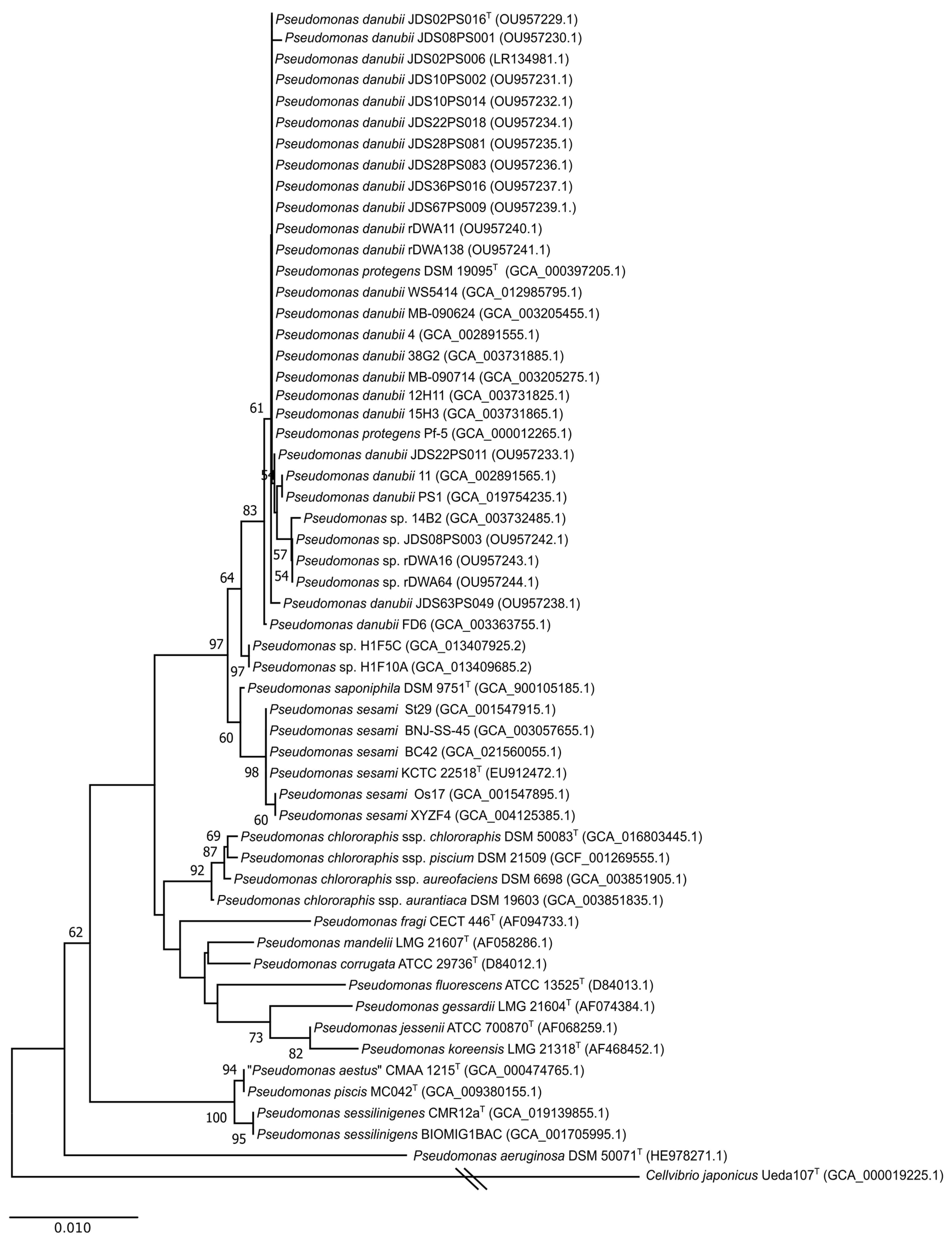
3.1. 16S rRNA and rpoD Gene Phylogenies

Figure 1 shows the phylogenetic relationships of the 16S rRNA gene of the 17 isolates, together with their closely related strains available in public databases. All 17 sequences were grouped in the same branch, together with the P. protegens type strain, at similarities ranging from 99.9 to 100%. P. sesami was the next closest type strain (similarity 99.1–99.2%), followed by P. saponiphila (98.7–98.8%), although they appeared in distant branches of the tree. The 16S rDNA sequence clearly included the 17 isolates in the P. fluorescens group within the P. chlororaphis phylogenetic subgroup of species. Another 10 nontype strains assigned in the Genome Taxonomy Data Base (GTDB; [44]) to the putative species Pseudomonas_E protegens_A and Pseudomonas_E protegens_B clustered in the same branch and were included in the phylogenetic analyses.
The analysis of the rpoD gene sequence is a powerful tool for species differentiation in the genus Pseudomonas, with a higher discrimination power than the 16S RNA gene sequence [6,35,45,46,47]. The corresponding phylogenetic tree is depicted in Figure S1. All strains were located in two separated branches of the P. chlororaphis subgroup of species, close to P. saponiphila and P. protegens type strains, but, as shown in Table S3, at distances higher than the 96% species threshold established for the rpoD gene partial sequence in the genus Pseudomonas [6]. Other type strains affiliated in the P. protegens-P. saponiphila phylogenetic branch in the 16S rRNA analysis, such as P. sesami, were separated in the rpoD tree.

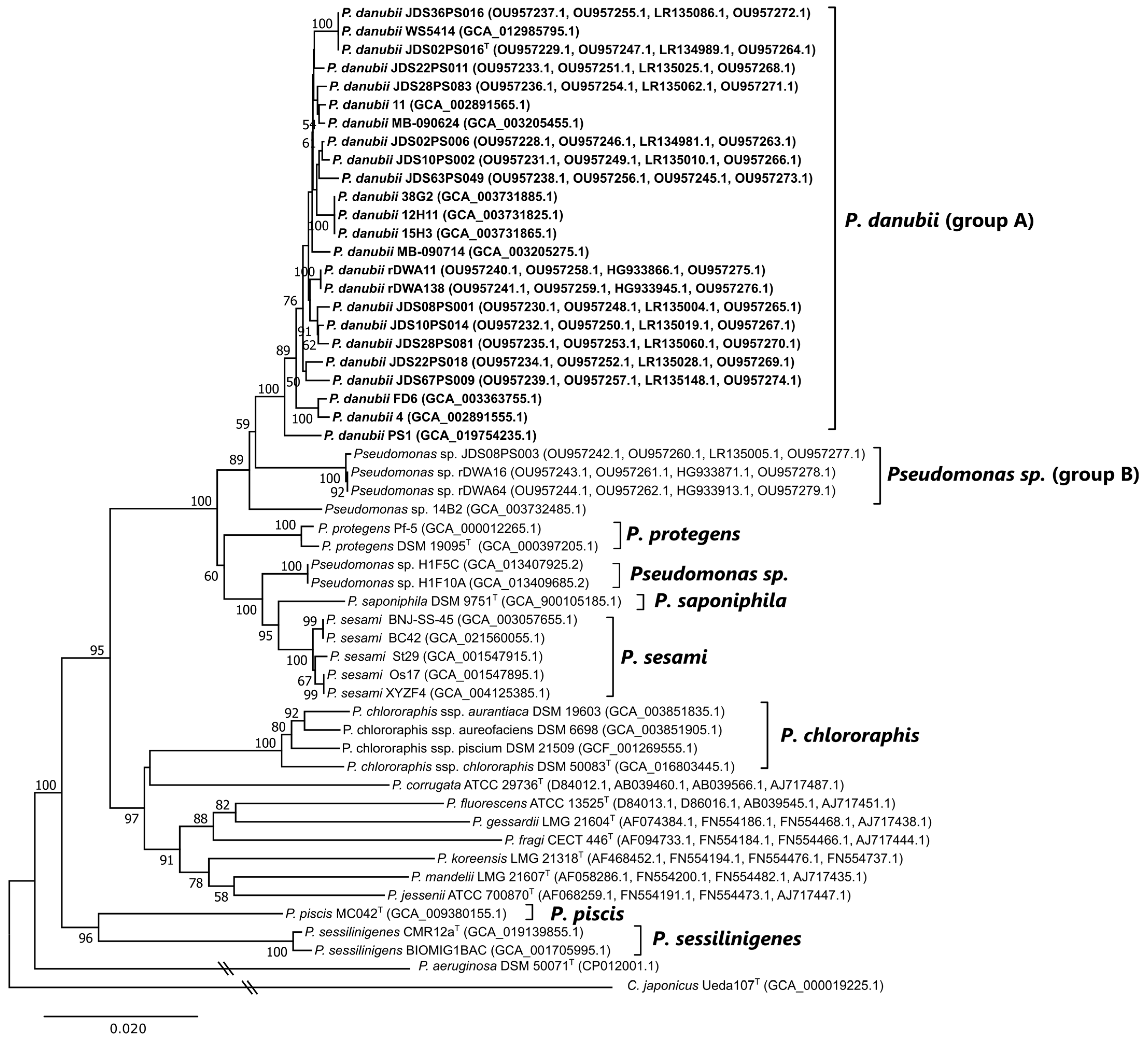
3.2. Multilocus Sequence Analysis (MLSA)

The concatenated partial sequences of the four housekeeping genes were analysed, and the results confirmed the groupings based on the rpoD gene sequences previously observed (Figure 2). The closest related type strains were P. saponiphila and P. protegens. The phylogenetic distances of the 17 isolates under study to P. saponiphila were below the 97% species threshold established for the four-gene concatenated multilocus sequence analysis [6]. The distances to P. protegens range from 96.6 to 97.1%, the borderline for species differentiation in the four-gene MLSA (Figure 2 and Table S3). The distances among the 14 isolates of the major group (A) range from 99.2 to 100%. Two isolates from the Woluwe River (rDWA16, rDWA64) and one isolate from the Danube River (JDS08PS003) clustered separately from the other 14 isolates at phylogenetic distances at the borderline for species differentiation in the four-gene analysis. These three isolates formed a second homogeneous Group B (distances of 99.9–100% among them, 97.6–97.9% with the other 14 strains studied, and 96.6–96.7% with the P. protegens type strain). P. protegens and P. saponiphila type strains are at a distance of 96.8%. The distances among the four P. chlororaphis subspecies range from 98.2–98.6% in the four-gene MLSA. These results indicate that the 17 isolates cannot be ascribed to their closest related species P. protegens or to P. saponiphila in the P. chlororaphis subgroup of species, and this assumption was corroborated in this investigation by genomic comparisons. The strains XY2F4, Os17, St29, BC42, and BNJ-SS-45, whose identification was considered inconclusive in the NCBI species taxonomy check, clustered together with the P. sesami type strain (Figure 2).

3.3. Genome Characteristics

Two isolates from the main group A and one strain of group B were selected for genome sequencing. The genome characteristics and assembly metrics of the three sequenced isolates of the present study are given in Table 2. The genome sizes range from 6.7 Mbp to 7.1 Mbp (6.6–7.6 in the group), the GC content ranges from 62.1% to 62.2% (61.8–63.8% in the group), and the numbers of coding sequences are in the range of the other strains in the group.

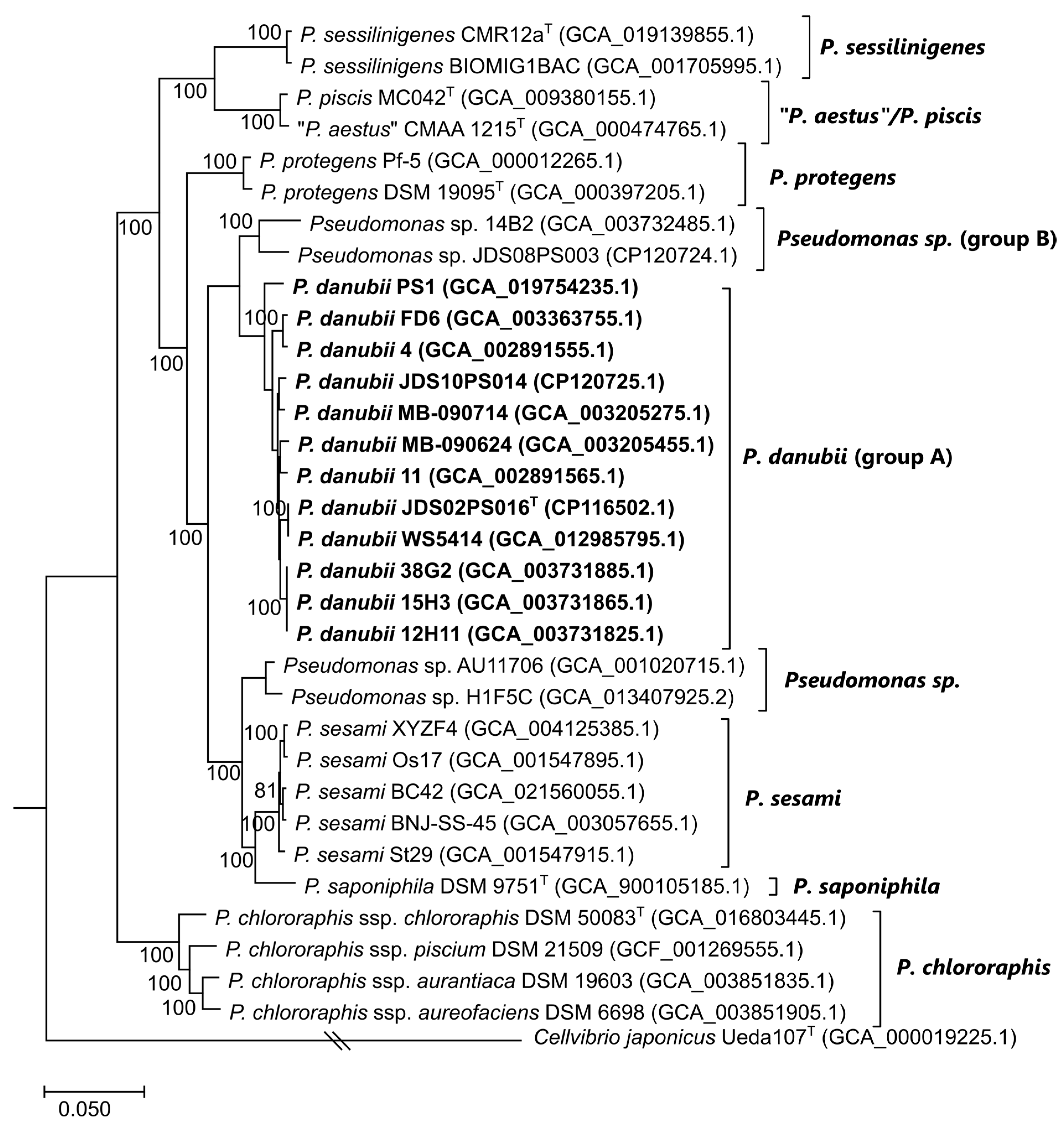
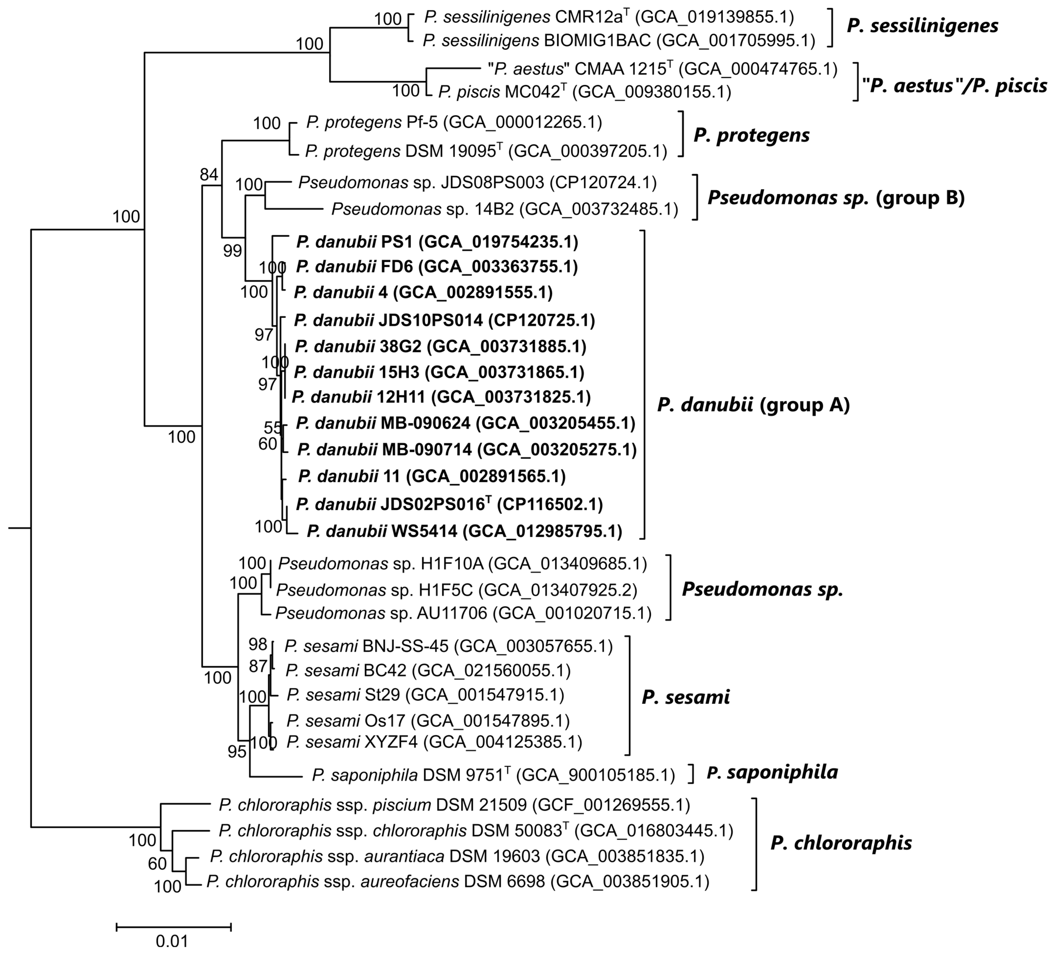
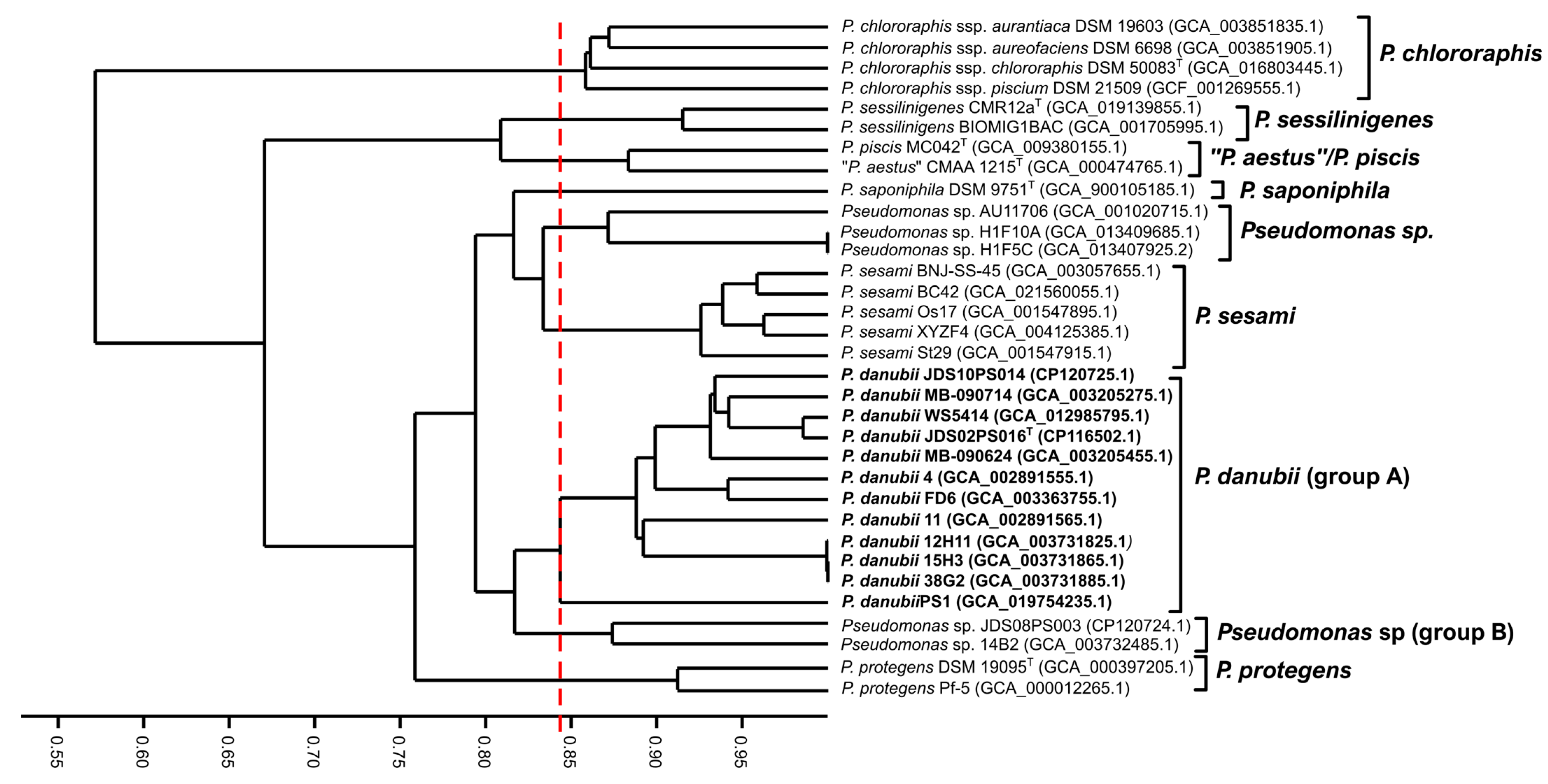
3.4. Genome-Based Phylogeny and Species Circumscription

The genome sequences of the three isolates under study were compared among themselves and to publicly available genome sequences of strains in the P. chlororaphis subgroup of species, including the type strains of 10 species or subspecies in the subgroup. Nine strains classified as Pseudomonas protegens_A and one strain classified as Pseudomonas protegens_B in the GTDB taxonomy were included in two types of phylogenomic analyses, autoMLST and core-gene phylogeny. Genome sequences were retrieved from the NCBI database, and their accession numbers are given in Table 1.
Figure 3 represents the analysis based on the concatenated sequences of 100 housekeeping genes (Table S4) selected by the autoMLST program (96,596 nt in total). The topology of the tree was supported by high bootstrap values, and it was highly coincident with the four-gene phylogenetic tree in the P. chlororaphis phylogenetic subgroup. Eight clusters of strains can be distinguished. The three isolates under study are monophyletic and clearly separated from the known species. Isolates JDS02PS016 and JDS10PS014 are located in the same branch, together with the strain classified as Pseudomonas_E protegens_A. Isolate JDS08PS003 is close to the strain classified as Pseudomonas_E protegens_B. “P. aestus” and P. piscis type strains are closely related in the analysis, suggesting that they conform to a single species.
The MICROBIALIZER web server (https://microbializer.tau.ac.il/index.html accessed on 21 November 2022) [34] was utilized for a deeper phylogenomic analysis of the set of genomes selected in the P. chlororaphis subgroup of species. Orthologous genes were analyzed in the sequenced genomes of the three isolates under study and in twenty additional strains selected. A total of 11,081 orthologous genes were found among the 34 genomes analyzed, and 3471 of them conformed to the core proteome (i.e., genes shared among all strains) of the P. chlororaphis phylogenetic subgroup of species. The final species tree is a maximum-likelihood phylogenetic tree reconstructed based on the analysis of the aligned sequences of the core proteome with a length of 1,230,882 nucleotides (Figure 4). The tree was highly concordant with the four-gene and one-hundred-gene phylogenetic trees previously obtained, confirming that the three strains under study are monophyletic. Their phylogeny was also assessed, including the Cellvibrio japonicum type strain as an outgroup with the same settings in the program. The number of core genes was then limited to 249, but the groupings were identical, and the topology of the tree was almost identical, with one exception: the branch of the P. protegens strains separated earlier from the P. danubi strains (Figure S2). All trees were supported with high bootstrap values.
For the species circumscription, two indices were calculated, ANIb and GGDC. The results are shown in Table S5 and in Figure S3. Both similarity values of the three strains under study in comparison with the type strains of the species in the P. chlororaphis phylogenetic group are below the species cutoff established for each method (95–96% for ANIb and 70% for GGDC), confirming that isolates JDS02PS016T, JDS10PS014, and JDS08PS003 do not belong to any known species. Isolates JDS02PS016T and JDS10PS014, together with the nine isolates classified as Pseudomonas_E protegens_A, belong to the same genomic species. Isolate JDS08PS003 and the strain classified as Pseudomonas_E protegens_B belong to a different genomic species related to the other group at ANIb values of 93.35–93.84% and GGDC values of 54–56%. However, the calculated indices between isolates JDS08PS003 and 14B2 are 94.56–94.52% (for ANIb) and 61.90% (for GGDC), values considered on the borderline for genomic species differentiation. A similar situation could be observed in the P. chlororaphis species: three subspecies are recognized, and the lowest ANI values among them range between 93.95 and 94.42%. The results confirm that “P. aestus” and P. piscis are members of the same phylogenomic species and have to be considered synonyms, with an ANI of 98%. The cluster of five strains identified as P. sesami in the MLSA analysis also conforms to a homogenous genomic group, with ANI values higher than 98.64%, separated from P. saponiphila but with identities of 94.03–94.42% in ANI. Strains AU11706, H1F10A, and H1F5C constitute a potential new species, yet to be described, which is close to P. sesami and P. saponiphila. The phylogram obtained in the TYGS web server was the same as the tree constructed with the GGDC values and proposes the same genomic species in the group.

3.5. Gene Content Comparisons

In an attempt to clarify the possible species status of those strains on the borderline of species circumscription, we analyzed the genes shared by all strains in the group. The 11,081 orthologous genes detected in the MICROBIALIZER analysis were studied by three different approaches. The Jaccard index matrix among the pairwise comparisons of the strains is depicted in the dendrogram represented in Figure 5. The clustering of strains in the same species is maintained. Strains from each of the nine phylogenomic species share at least 85% of the orthologous genes, but the Jaccard index for strains of the abovementioned closely related groups is higher than 80%, and a clear threshold cannot be delineated. Similar results were observed in the split tree decomposition (Figure S4a) obtained with SplitsTree. The presence–absence of the orthologous genes was also represented in a heatmap (Figure S4b) that clearly shows the combinations of orthologous genes that differentiate the species in the group.
A more detailed comparison was performed among two genomes of strains in the group of P. danubii (isolates JDS02PS016T and JDS10PS014) and the closely related isolates JDS08PS003 and 14B2 assigned to Pseudomonas_E protegens_B in the GTDB taxonomy. A Venn diagram was constructed on the web page https://bioinfogp.cnb.csic.es/tools/venny/ accessed on 21 November 2022 [37], as shown in Figure S5. Only 321 genes (4%) were shared by strains JDS08PS003 and 14B2 and were not present in the other two genomes. However, most of these 321 genes were also present in the genomes of other members of the P. chlororaphis phylogenetic group. Only 22 genes were exclusive to this potential species, Pseudomonas_E protegens_B, and no characteristic differential trait was encoded by these genes.

3.6. Genome Insights

The KEGG analysis of the proteome of the sequenced three isolates of the proposed new species reveals the presence of genes of metabolic pathways that are relevant for their ecological potential. Some traits that have been considered in the taxonomy of Pseudomonas were found. Starch can be hydrolyzed by means of alpha-amylase and iso-amylase, and the degradative products can then be metabolized through glycolysis or the pentose phosphate cycle; benzoate can be metabolized through catechol, which is cleaved in the ortho position and followed by the ortho pathway enzymes. All the necessary enzymes for the assimilatory reduction of nitrate to ammonium were detected, as well as those for the synthesis of type II and type VI secretion systems. The type VI secretion system might be related to the alginate biosynthesis potential and secretion for biofilm formation. The strains can be beta-lactam resistant due to the presence of RND efflux pumps and the synthesis of a class C beta-lactamase.
The presence of antibiotic resistance genes was screened on the CARD website. Table 3 summarizes the genes and the potential resistance against antibiotics found. Seven strict hits were detected by protein homology in the three genomes. Following the Antibiotic Resistance Ontology (“ARO”) terminology, three to four adeF determinants were identified, as well as fosA and fosA8 and vanG, vanW, and yajC. The potential resistances determined were related to antibiotic efflux pumps and antibiotic target inactivation.
Prophages are important modulators of the characteristics of their host and are species- or strain-specific [48]. As specified in Table S6, three, four, or five sequence regions in each genome contained phage genes. A region containing 40 to 56 protein-coding sequences in the four genomes was considered intact and was most similar to the reference Salmonella phage 118970_sal3 selected by the server. A nucleotide BLAST search in the NCBI database revealed that at least some of these sequences are also present in at least 50 isolates of the P. chlororaphis phylogenetic subgroup of strains comprising most species in the group, suggesting the presence of this phage in their common ancestor. The significance of this phage needs further investigation but was not the main subject of this study. Another intact phage (48 protein-coding sequences) was found in strain 14B2, and other incomplete regions differentiated the isolates studied.

3.7. Phenotypic Traits: Morphology, Physiology, Biochemical Traits, and Chemotaxonomy

The colonies were round or oval, flat, opaque, and beige colored with regular margins, and measured 2–3 mm in diameter after incubation on LB agar plates (Luria-Bertani, Difco) at 30 °C for 48 h. The cells were short rods (2.5–1.9 µm long and 1.3–0.5 µm wide), motile by a single or two polar-inserted flagella (Figure S6) and stained gram-negative. P. protegens, P. saponiphila, P. chlororaphis, P. koreensis, and P. fluorescens type strains were included in the analysis as controls and for comparative purposes. The results are detailed in Table 4 and Table S7. The metabolism was strictly aerobic and non-fermentative, and the strains were not able to reduce nitrate to nitrite. Catalase and oxidase tests were positive. Growth was observed in liquid LB medium at temperatures between 4 and 37 °C, and the strains were able to grow at pH values between 5 and 9 or 10. All isolates tolerated NaCl concentrations up to 6% (w/v). Only four of the fourteen strains in the P. danubii proposed species were able to grow in the presence of 8% NaCl in the GENIII galleries. Fluorescent pigments were produced on King B (Difco) but not on King A (Difco) media. Many of the sugars and organic acids tested were assimilated. The 17 strains were not efficient in the use of Tween 40, a characteristic that differentiates them from their closest relatives. The 17 isolates under study were phenotypically homogeneous, and few biochemical tests could differentiate them from their closest related P. protegens, P. saponiphila, and P. chlororaphis type strains. Table 4 shows the differences found in the biochemical tests. The use of adipate, arabitol, sucrose, and aspartic acid differentiates P. danubii from P. protegens, P. saponiphila, and P. chlororaphis type strains. Substrates used by these type strains, such as serine, saccharic acid, and formic acid, are not used by P. danubii strains.
Five of the seventeen Danube isolates were considered multidrug resistant in a previous study [12] because they revealed resistance to three (MDR3) or four (MDR4) antibiotic classes. The antibiotic resistance patterns were analyzed for the Woluwe strains and are reported in Table S8 together with the previously obtained results. The five MDR isolates from the Danube River were JDS02PS016T (TZP (piperacillin/tazobactam), CAZ (ceftazidime), GM (gentamicin), MEM (meropenem), and CIP (ciprofloxacin); JDS02PS020 to CAZ, MEM, and CIP; JDS22PS018 to CAZ, IPM (imipenem), and MEM; JDS28PS083 to CAZ, FEP (cefepime), IPM, and MEM; and JDS28PS113 to CAZ, IPM, and MEM. Isolate JDS10PS014 was only resistant to MEM, and isolate JDS08PS003 was sensitive to all antibiotics tested. The four Woluwe River isolates showed the same antibiotic sensitivity profile, being resistant to carbapenems only (MEM and IPM). The three strains classified in Pseudomonas_E protegens_B were more sensitive to antibiotics than the P. danubii strains.
The total fatty acid methyl ester composition (FAME) and the main protein profiles were determined for phenotypic chemotaxonomic characterization. The FAME results were compared with the profiles of the three closest related type strains and are shown in Table S9. As in other Pseudomonas, the major fatty acid components were C16:0 (30.93%) and summed feature three (27.84%). The major difference from P. saponiphila, P. protegens, and P. chlororaphis is the absence of dodecanoic acid (C12:0).
The mass to charge (m/z) data dendrogram of the whole cells is shown in Figure S7. As reported in a previous study, the 17 investigated strains clustered together in the dendrogram close to P. saponiphila, P. protegenes, and P. chlororaphis type strains at a distance level of 100 arbitrary units (Figure S7a), confirming our previous results indicating the difficulty in differentiating the species in this phylogenetic group by their major protein profile in the context of more distant strains [13]. However, a detailed analysis of the strains in the P. chlororaphis phylogenetic group alone demonstrated clear differences (Figure S7b) and the consistency of the newly proposed species.

4. Discussion

The P. chlororaphis subgroup in the Pseudomonas fluorescens phylogenetic group of species is a well-defined phylogenomic branch within the proposed Pseudomonas_E genus in the GTDB taxonomy. It is worth highlighting that all studied genomic indices are concordant and that the rpoD alone was predictive of the new species. The species included in the P. chlororaphis subgroup are “P. aestus” (not validly published) [15], P. chlororaphis (with four subspecies) [17,18], P. piscis [16], P. protegens [19], P. saponiphila [20], P. sesami [21], and P. sessilinigenes [9]. The group as a whole is widely distributed, with relevant ecological properties, including interactions with plants and fish. Only isolate AU11706 was isolated from a clinical sample [11]. The phylogenetic analysis and genomic indices obtained in the present study indicate that P. piscis and “P. aestus” are synonyms, as suggested in previous studies, and that the 17 isolates taxonomically characterized in this study conform to a monophyletic branch that represents one or two new species. Fifteen out of seventeen strains belong to the proposed species Pseudomonas_E protegens_A in the GTDB taxonomy, which contains nine isolates isolated from diverse habitats and geographical locations (Table 1). Ten strains classified as P. protegens and considered inconclusive in the NCBI taxonomy check are reclassified in the species P. danubii in the present study.
The other three taxonomically studied strains (JDS08PS003, rDWA11, and rDWA64) belong to the proposed species Pseudomonas_E protegens_B represented thus far by a single strain (14B2) isolated from the Missouri River. These last four strains are on the borderline for species differentiation with the main group represented by Pseudomonas_E protegens_A strains. In our opinion, more isolates and/or more genomes are needed to decide if Pseudomonas_E protegens_B constitute one or two different species or if both have to be considered subspecies of P. danubii because the phylogenomic indices are at the same level as those among strains of the four P. chlororaphis subspecies. The difficulty in differentiating species or subspecies in P. chlororaphis has been discussed previously [3].
The phylogenomic analysis also demonstrates that strains XY2F4, Os17, St29, BC42, and BNJ-SS-45 are members of P. sesami, a species on the borderline for genomic differentiation with P. saponiphila. These strains were not classified previously at the species level, or were identified as P. protegens strains, but their species assignation was considered inconclusive in the NCBI taxonomy check or were considered different species in the GTDB taxonomy. The five strains are monophyletic, and the genomic indices between strains are higher than the species thresholds. Their ANIb values range from 94.18 to 94.35 with the P. saponiphila type strain, and the corresponding GGDC values range from 58.3 to 58.6. The genome sequence of the P. sesami type strain is needed to clarify the species status. Experimental DNA–DNA hybridization of P. sesami with P. saponiphila was reported to be lower than 70%, which justified the separation into two species [21].
The strains P. fluorescens AU11706 and P. protegens H1F10A and H1F5C are members of a different phylogenomic species, classified as P. fluorescens_AP in the GTDB taxonomy. These strains were isolated from two quite different habitats: strains thus far classified as P. protegens (H1F10A and H1F5C) were isolated from samples taken in Yellowstone National Park (USA) together with other strains, and strain AU11706 was isolated from cystic fibrosis sputum in Michigan (USA) and initially classified as Pseudomonas fluorescens [10]. The ecological and potential pathogenicity characteristics of these strains raise interesting questions that merit specific investigations.
Genome analysis also revealed the metabolic and ecological potential of the new species. However, it must be kept in mind that the presence of genes for a specific trait may not always indicate expression. Two examples are assimilatory nitrate reduction or resistance to antibiotics. Genes for the reduction of nitrate to nitrite and nitrite to ammonium are present in the genomes, but the phenotypic tests rendered negative results. Likewise, genes for the synthesis of efflux pumps might confer resistance to many antibiotics, but strains with this gene content were experimentally found to be antibiotic sensitive. All isolates investigated for their antibiotic resistance were of environmental, aquatic origins. Those isolated from a sample taken in the Woluwe River near the source were only resistant to carbapenems and sensitive to the other antibiotics tested. These waters are not considered contaminated [14], and thus, carbapenem resistance must be intrinsic to the strains. However, the MDR strains of the new species have most likely acquired resistance and are in habitats in which resistance can provide a selective advantage.
On the basis of the phylogenetic, genomic, phenotypic, and chemotaxonomic properties described in the present investigation, we propose that fourteen strains of our culture collection isolated from nine freshwater samples of two rivers (the Danube and Woluwe), and nine strains classified Pseudomonas_E protegens_A in the GTDB taxonomy, belong to a new species for which the name Pseudomonas danubii is proposed, with strain JDS02PS01T (=CECT 30214 T = CCUG 74756 T) as the type strain. A list of the 23 P. danubii strains is given in Table 1. Genetic and phenotypic analyses indicate that a simple distinction between species can be obtained on the basis of the rpoD gene sequence and their main protein profiles obtained by MALDI TOF mass spectrometry. Adipate assimilation, a weak utilization of tween 40 and serine, and a lack of assimilation of formic acid differentiates P. danubii isolates from the other closely related species.

5. Protologue

Description of Pseudomonas danubii sp. nov.

P. danubii sp. nov. (da.nu’bi.i. L. gen. n. danubii, of the Danube, referring to the sampling site of the type strain).
Rod-shaped cells, 2.5–1.9 µm long and 1.3–0.5 µm wide, forming colonies after 24 h incubation at 30 °C on LB. The cells are gram-negative, motile by means of one or two polar-inserted flagella, and exhibit growth at pH of 5 to 9 and in the range of 4 to 37 °C (optimum 30 °C); no growth detected at 42 °C. NaCl concentrations up to 6% (w/v) are tolerated. Fluorescent pigments were produced on King B but not on King A medium. Strictly aerobic, not fermentative. Catalase and oxidase positive. Nitrate is not reduced. The API 20NE test was positive for arginine dihydrolase and hydrolysis of gelatin but not esculin and was negative for urease and beta-galactosidase; it was positive for the assimilation of glucose, mannose, mannitol, N-acetyl-D-glucosamine, gluconate, caprate, adipate, malate, citrate, and phenylacetate but negative for arabinose and maltose. In Biolog GENIII testing, plates were positive for assimilation of alfa-D-glucose, p-hydroxy-phenylacetic acid, D-mannose, D-mannitol, glycyl-L-proline, gamma-amino-butyric acid, D-fructose, D-arabitol, L-alanine, D-trehalose, myo-inositol, L-arginine, D-gluconic acid, L-lactic acid, beta-hydroxy-D,L butyric acid, D-cellobiose, and D-salicin. Adipate assimilation, weak utilization of tween 40 and serine, and lack of assimilation of formic acid differentiate it from the other closely related species. The major fatty acids are C16:0 and summed feature 3 (C15:0 iso 2OH and C16:1 w7c). The absence of C12:0 differentiates it from closely related species. Simple distinction between the species can be obtained on the basis of the rpoD gene sequence.
The type strain is JDS02PS016T (=CECT 30214T = CCUG 74756T). The GenBank accession number for the rRNA gene sequence is OU957229.1, and that for the genome is CP116502. The G+C content of the type strain is 62.3 mol% based on the draft genome sequence, and it ranges from 62.0 to 62.5 mol% in 11 strains of the species.

Supplementary Materials

The following supporting information can be downloaded at https://www.mdpi.com/article/10.3390/d15050617/s1. Figure S1. Phylogenetic tree based on the rpoD gene sequence. Figure S2. Maximum likelihood phylogenetic tree of strains studied in the P. chlororaphis subgroup of species based on the concatenated sequences of 249 core genes. Figure S3. (a) Dendrograms of the aggregated ANIb values. (b) Dendrogram of the GGDC similarities among the studied strains. Figure S4. Graphical representation of the distribution of the 11,081 orthologous genes among the strains: (a) The phyletic pattern is represented in an unrooted network by the split decomposition method. (b) Heat plot of the phyletic pattern representing the presence (red) or absence (blue) of the orthologous genes. Figure S5. Venn diagram of the shared orthologous genes of two sequenced strains of P. danubii (JDS02PS016T and JDS10PS014) and two closely related strains of Pseudomonas_E protegens_B in the GTDB taxonomy (JDS08PS003 and 14B2). Figure S6. Electron microscopy of negatively stained flagellated cells of strain JDS02PS016T in the exponential growth phase (left). Colony morphology of strain JDS02PS016T after incubation at 30 °C for 48 h on LB agar (right). Figure S7. Dendrogram of the main proteins obtained by MALDI-TOF mass spectrometry. (A) with outgroups; (B) only species in the P. chlororaphis subgroup. Table S1. Sampling sites, geographical origins, and references of the strains included in the present study. Table S2. GenBank accession numbers of sequences used in this study. Table S3. Pairwise sequence similarities of the 16S rRNA, concatenated genes (16S rRNA, gyrB, rpoB, and rpoD) and rpoD genes between Pseudomonas danubii and the strains of the Pseudomonas fluorescens group included in this study. Table S4. One hundred housekeeping genes (filtered from 450 total entries) were selected for the autoMLST analysis. Table S5: Genomic indices among the studied strains. Table S6. Prophage regions detected by the PHASTER server in the genome sequences of the four strains sequenced in the present study. Table S7. Physiological and biochemical characteristics of Pseudomonas danubii and related Pseudomonas strains and species type strains. Table S8. Antibiotic susceptibility tests of the studied strains. (S: sensitive, R: resistant). Table S9. Cellular fatty composition (%) derived from FAME analysis of Pseudomonas danubii JDS02PS016T and the type strains of closely related Pseudomonas species [49].

Author Contributions

Conceptualization, C.K., G.Z., J.L. and E.G.-V.; bioinformatic analysis M.M., M.J.M., M.G. and G.E.W.; investigation, M.M., M.J.M., M.G., J.D.-H. and G.E.W.; resources, M.G., E.G.-V. and G.Z.; data curation, J.L. and C.K.; writing, J.L., E.G.-V., C.K. and G.Z.; project administration and funding acquisition, M.G., E.G.-V. and G.Z. All authors have read and agreed to the published version of the manuscript.

Funding

This study was funded by the Spanish Ministry of Science and Innovation MICIN/AEI, 10.13039/501100011033, project PID2020-119449RB-I00 (to MG and EGV).

Institutional Review Board Statement

Not applicable.

Informed Consent Statement

Not applicable.

Data Availability Statement

All sequence data obtained in the present study have been deposited in publicly available databases (NCBI/GenBank) under the accession numbers given in the text.

Acknowledgments

The authors would like to thank B. Schink for advice on taxonomic nomenclature and etymology and M. Avram for help with the MICROBIALIZER analyses. The FAME analysis was performed at the Spanish Collection of Type Strains (CECT, Valencia, Spain). MALDI-TOF m/s spectra were obtained by R. Gomila at the Scientific-Technical services of UIB. The International Commission for the Protection of the Danube River (ICPDR) is gratefully acknowledged for the sampling and administrative organization.

Conflicts of Interest

The authors declare no conflict of interest.

Abbreviations

ANIgenome-aggregated average nucleotide identity
autoMLSTautomated multilocus species tree
FAMEfatty acid methyl ester analysis
GGDCgenome to genome distance calculator
MALDI-TOF MSmatrix-assisted laser desorption/ionization (MALDI) time-of-flight (TOF) mass spectrometry
MLSAmultilocus sequence analysis

References

  1. Palleroni, N.J.; Genus, I. Pseudomonas. In Bergey’s Manual of Systematic Bacteriology. Part B. the Gammaproteobacteria; Brenner, D.J., Krieg, N.R., Staley, J.T., Eds.; Springer: New York, NY, USA, 2005; pp. 323–379. [Google Scholar]
  2. Parte, A.C.; Sardà Carbasse, J.; Meier-Kolthoff, J.P.; Reimer, L.C.; Göker, M. List of Prokaryotic names with Standing in Nomenclature (LPSN) moves to the DSMZ. Int. J. Syst. Evol. Microbiol. 2020, 70, 5607–5612. [Google Scholar] [CrossRef] [PubMed]
  3. Lalucat, J.; Mulet, M.; Gomila, M.; García-Valdés, E. Genomics in Bacterial Taxonomy: Impact on the Genus Pseudomonas. Genes 2020, 11, 139. [Google Scholar] [CrossRef]
  4. Rudra, B.; Gupta, R.S. Phylogenomic and comparative genomic analyses of species of the family Pseudomonadaceae: Proposals for the genera Halopseudomonas gen. nov. and Atopomonas gen. nov., merger of the genus Oblitimonas with the genus Thiopseudomonas, and transfer of some misclassified species of the genus Pseudomonas into other genera. Int. J. Syst. Evol. Microbiol. 2021, 71, 5011. [Google Scholar] [CrossRef]
  5. Saati-Santamaría, Z.; Peral-Aranega, E.; Velázquez, E.; Rivas, R.; García-Fraile, P. Phylogenomic Analyses of the Genus Pseudomonas Lead to the Rearrangement of Several Species and the Definition of New Genera. Biology 2021, 10, 782. [Google Scholar] [CrossRef]
  6. Mulet, M.; Lalucat, J.; García-Valdés, E. DNA sequence-based analysis of the Pseudomonas species. Environ. Microbiol. 2010, 12, 1513–1530. [Google Scholar] [CrossRef] [PubMed]
  7. Lalucat, J.; Gomila, M.; Mulet, M.; Zaruma, A.; García-Valdés, E. Past, present and future of the boundaries of the Pseudomonas genus: Proposal of Stutzerimonas gen. Nov. Syst. Appl. Microbiol. 2022, 45, 126289. [Google Scholar] [CrossRef]
  8. Nikolaidis, M.; Mossialos, D.; Oliver, S.G.; Amoutzias, G.D. Comparative Analysis of the Core Proteomes among the Pseudomonas Major Evolutionary Groups Reveals Species-Specific Adaptations for Pseudomonas aeruginosa and Pseudomonas chlororaphis. Diversity 2020, 12, 289. [Google Scholar] [CrossRef]
  9. Girard, L.; Lood, C.; Höfte, M.; Vandamme, P.; Rokni-Zadeh, H.; van Noort, V.; Lavigne, R.; De Mot, R. The Ever-Expanding Pseudomonas Genus: Description of 43 New Species and Partition of the Pseudomonas putida Group. Microorganisms 2021, 9, 1766. [Google Scholar] [CrossRef]
  10. Scales, B.S.; Dickson, R.P.; LiPuma, J.J.; Huffnagle, G.B. Microbiology, Genomics, and Clinical Significance of the Pseudomonas fluorescens Species Complex, an Unappreciated Colonizer of Humans. Clin. Microbiol. Rev. 2014, 27, 927–948. [Google Scholar] [CrossRef]
  11. Scales, B.S.; Erb-Downward, J.R.; LiPuma, J.J.; Huffnagle, G.B. Draft Genome Sequences of Five Pseudomonas fluorescens Subclade I and II Strains, Isolated from Human Respiratory Samples. Genome Announc. 2015, 3, e00837-15. [Google Scholar] [CrossRef]
  12. Kittinger, C.; Lipp, M.; Baumert, R.; Folli, B.; Koraimann, G.; Toplitsch, D.; Liebmann, A.; Grisold, A.J.; Farnleitner, A.H.; Kirschner, A.; et al. Antibiotic Resistance Patterns of Pseudomonas spp. Isolated from the River Danube. Front. Microbiol. 2016, 7, 586. [Google Scholar] [CrossRef] [PubMed]
  13. Mulet, M.; Montaner, M.; Román, D.; Gomila, M.; Kittinger, C.; Zarfel, G.; Lalucat, J.; García-Valdés, E. Pseudomonas Species Diversity along the Danube River Assessed by rpoD Gene Sequence and MALDI-TOF MS Analyses of Cultivated Strains. Front. Microbiol. 2020, 11, 2114. [Google Scholar] [CrossRef] [PubMed]
  14. Sánchez, D.; Matthijs, S.; Gomila, M.; Tricot, C.; Mulet, M.; García-Valdés, E.; Lalucat, J. rpoD Gene Pyrosequencing for the Assessment of Pseudomonas Diversity in a Water Sample from the Woluwe River. Appl. Environ. Microbiol. 2014, 80, 4738–4744. [Google Scholar] [CrossRef] [PubMed]
  15. Vasconcellos, R.L.F.; Santos, S.N.; Zucchi, T.D.; Silva, F.S.P.; Souza, D.T.; Melo, I.S. Pseudomonas aestus sp. nov., a plant growth-promoting bacterium isolated from mangrove sediments. Arch. Microbiol. 2017, 199, 1223–1229. [Google Scholar] [CrossRef] [PubMed]
  16. Liu, Y.; Rao, Q.; Blom, J.; Lin, Q.; Luo, T. Pseudomonas piscis sp. nov., isolated from the profound head ulcers of farmed Murray cod (Maccullochella peelii peelii). Int. J. Syst. Evol. Microbiol. 2020, 70, 2732–2739. [Google Scholar] [CrossRef]
  17. Peix, A.; Valverde, A.; Rivas, R.; Igual, J.M.; Bahena, M.H.R.; Mateos, P.; Santa-Regina, I.; Rodríguez-Barrueco, C.; Martínez-Molina, E.; Velázquez, E. Reclassification of Pseudomonas aurantiaca as a synonym of Pseudomonas chlororaphis and proposal of three subspecies, P. chlororaphis subsp. chlororaphis subsp. nov., P. chlororaphis subsp. aureofaciens subsp. nov., comb. nov. and P. chlororaphis subsp. aurantiaca subsp. nov., comb. nov. Int. J. Syst. Evol. Microbiol. 2007, 57, 1286–1290. [Google Scholar] [CrossRef]
  18. Burr, S.; Gobeli, S.; Kuhnert, P.; Goldschmidt-Clermont, E.; Frey, J. Pseudomonas chlororaphis subsp. piscium subsp. nov., isolated from freshwater fish. Int. J. Syst. Evol. Microbiol. 2010, 60, 2753–2757. [Google Scholar] [CrossRef]
  19. Ramette, A.; Frapolli, M.; Saux, M.F.-L.; Gruffaz, C.; Meyer, J.-M.; Défago, G.; Sutra, L.; Moënne-Loccoz, Y. Pseudomonas protegens sp. nov., widespread plant-protecting bacteria producing the biocontrol compounds 2,4-diacetylphloroglucinol and pyoluteorin. Syst. Appl. Microbiol. 2011, 34, 180–188. [Google Scholar] [CrossRef]
  20. Lang, E.; Burghartz, M.; Spring, S.; Swiderski, J.; Spröer, C. Pseudomonas benzenivorans sp. nov. and Pseudomonas saponiphila sp. nov., Represented by Xenobiotics Degrading Type Strains. Curr. Microbiol. 2010, 60, 85–91. [Google Scholar] [CrossRef]
  21. Madhaiyan, M.; Poonguzhali, S.; Saravanan, V.S.; Selvapravin, K.; Duraipandiyan, V.; Al-Dhabi, N.A. Pseudomonas sesami sp. nov., a plant growth-promoting Gammaproteobacteria isolated from the rhizosphere of Sesamum indicum L. Antonie Van Leeuwenhoek 2017, 110, 843–852. [Google Scholar] [CrossRef]
  22. Paulsen, I.T.; Press, C.; Ravel, J.; Kobayashi, D.Y.; Myers, G.; Mavrodi, D.; DeBoy, R.T.; Seshadri, R.; Ren, Q.; Madupu, R.; et al. Complete genome sequence of the plant commensal Pseudomonas fluorescens Pf-5. Nat. Biotechnol. 2005, 23, 873–878, Erratum in Nat. Biotechnol. 2006, 24, 466. [Google Scholar] [CrossRef] [PubMed]
  23. Mulet, M.; Gomila, M.; Ramírez, A.; Cardew, S.; Moore, E.R.B.; Lalucat, J.; García-Valdés, E. Uncommonly isolated clinical Pseudomonas: Identification and phylogenetic assignation. Eur. J. Clin. Microbiol. Infect. Dis. 2017, 36, 351–359. [Google Scholar] [CrossRef] [PubMed]
  24. Jukes, T.; Cantor, C. Evolution of protein molecules. In Mammalian Protein Metabolism; Munro, H.N., Ed.; Academic Press: New York, NY, USA, 1969; pp. 21–132. [Google Scholar]
  25. Saitou, N.; Nei, M. The neighbor-joining method: A new method for reconstructing phylogenetic trees. Mol. Biol. Evol. 1987, 4, 406–425. [Google Scholar] [CrossRef] [PubMed]
  26. Tamura, K.; Stecher, G.; Kumar, S. MEGA11: Molecular Evolutionary Genetics Analysis Version 11. Mol. Biol. Evol. 2021, 38, 3022–3027. [Google Scholar] [CrossRef]
  27. Alanjary, M.; Steinke, K.; Ziemert, N. AutoMLST: An automated web server for generating multi-locus species trees highlighting natural product potential. Nucleic Acids Res. 2019, 47, W276–W282. [Google Scholar] [CrossRef]
  28. Wagner, G.E.; Dabernig-Heinz, J.; Lipp, M.; Cabal, A.; Simantzik, J.; Kohl, M.; Scheiber, M.; Lichtenegger, S.; Ehricht, R.; Leitner, E.; et al. Real-Time Nanopore Q20+ Sequencing Enables Extremely Fast and Accurate Core Genome MLST Typing and Democratizes Access to High-Resolution Bacterial Pathogen Surveillance. J. Clin. Microbiol. 2023, 61, e0163122. [Google Scholar] [CrossRef]
  29. Kolmogorov, M.; Bickhart, D.M.; Behsaz, B.; Gurevich, A.; Rayko, M.; Shin, S.B.; Kuhn, K.; Yuan, J.; Polevikov, E.; Smith, T.P.L.; et al. metaFlye: Scalable long-read metagenome assembly using repeat graphs. Nat. Methods 2020, 17, 1103–1110. [Google Scholar] [CrossRef]
  30. Aziz, R.K.; Bartels, D.; Best, A.A.; DeJongh, M.; Disz, T.; Edwards, R.A.; Formsma, K.; Gerdes, S.; Glass, E.M.; Kubal, M.; et al. The RAST server: Rapid annotations using subsystems technology. BMC Genom. 2008, 9, 75. [Google Scholar] [CrossRef]
  31. Richter, M.; Rosselló-Móra, R.; Oliver Glöckner, F.O.; Peplies, J. JSpeciesWS: A web server for prokaryotic species circumscription based on pairwise genome comparison. Bioinformatics 2016, 32, 929–931. [Google Scholar] [CrossRef]
  32. Meier-Kolthoff, J.P.; Auch, A.F.; Klenk, H.-P.; Göker, M. Genome sequence-based species delimitation with confidence intervals and improved distance functions. BMC Bioinform. 2013, 14, 60. [Google Scholar] [CrossRef]
  33. Meier-Kolthoff, J.P.; Carbasse, J.S.; Peinado-Olarte, R.L.; Göker, M. TYGS and LPSN: A database tandem for fast and reliable genome-based classification and nomenclature of prokaryotes. Nucleic Acids Res. 2022, 50, D801–D807. [Google Scholar] [CrossRef] [PubMed]
  34. Avram, O.; Rapoport, D.; Portugez, S.; Pupko, T. M1CR0B1AL1Z3R—A user-friendly web server for the analysis of large-scale microbial genomics data. Nucleic Acids Res. 2019, 47, W88–W92. [Google Scholar] [CrossRef]
  35. Hammer, Ø.; Harper, D.A.T.; Ryan, P.D. PAST: Paleontological statistics software package for education and data analysis. Palaeontol. Electron. 2001, 4, 9. Available online: http://palaeo-electronica.org/2001_1/past/issue1_01.htm (accessed on 1 February 2023).
  36. Huson, D.H.; Bryant, D. Application of Phylogenetic Networks in Evolutionary Studies. Mol. Biol. Evol. 2006, 23, 254–267. [Google Scholar] [CrossRef]
  37. Oliveros, J.C. Venny. An Interactive Tool for Comparing Lists with Venn Diagrams. Available online: http://bioinfogp.cnb.csic.es/tools/venny/index.html (accessed on 1 February 2023).
  38. Alcock, B.P.; Huynh, W.; Chalil, R.; Smith, K.W.; Raphenya, A.R.; A Wlodarski, M.; Edalatmand, A.; Petkau, A.; A Syed, S.; Tsang, K.K.; et al. CARD 2023: Expanded curation, support for machine learning, and resistome prediction at the comprehensive antibiotic resistance database. Nucleic Acids Res. 2023, 51, D690–D699. [Google Scholar] [CrossRef] [PubMed]
  39. Arndt, D.; Grant, J.; Marcu, A.; Sajed, T.; Pon, A.; Liang, Y.; Wishardt, D.S. PHASTER: A better faster version of the PHAST phage search tool. Nucleic Acids Res. 2016, 44, W16–W21. [Google Scholar] [CrossRef]
  40. Moriya, Y.; Itoh, M.; Okuda, S.; Yoshizawa, A.C.; Kanehisa, M. KAAS: An automatic genome annotation and pathway reconstruction server. Nucleic Acids Res. 2007, 35 (Suppl. S2), W182–W185. [Google Scholar] [CrossRef] [PubMed]
  41. Lalucat, J. Analysis of Refractile (R) Bodies. In Methods in Microbiology; Academic Press: Cambridge, MA, USA, 1988; Volume 20, pp. 79–90. [Google Scholar]
  42. Sánchez, D.; Mulet, M.; Rodríguez, A.C.; David, Z.; Lalucat, J.; García-Valdés, E. Pseudomonas aestusnigri sp. nov., isolated from crude oil-contaminated intertidal sand samples after the Prestige oil spill. Syst. Appl. Microbiol. 2014, 37, 89–94. [Google Scholar] [CrossRef]
  43. Sasser, M. Identification of Bacteria by Gas Chromatography of Cellular Fatty Acids; MIDI Technical Note 101; MIDI Inc.: Newark, NJ, USA, 1990. [Google Scholar]
  44. Parks, D.H.; Chuvochina, M.; Waite, D.W.; Rinke, C.; Skarshewski, A.; Chaumeil, P.-A.; Hugenholtz, P. A standardized bacterial taxonomy based on genome phylogeny substantially revises the tree of life. Nat. Biotechnol. 2018, 36, 996–1004. [Google Scholar] [CrossRef]
  45. Mulet, M.; Bennasar, A.; Lalucat, J.; García-Valdés, E. An rpoD-based PCR procedure for the identification of Pseudomonas species and for their detection in environmental samples. Mol. Cell. Probes 2009, 23, 140–147. [Google Scholar] [CrossRef]
  46. Ghyselinck, J.; Coorevits, A.; Van Landschoot, A.; Samyn, E.; Heylen, K.; De Vos, P. An rpoD gene sequence based evaluation of cultured Pseudomonas diversity on different growth media. Microbiology 2013, 159, 2097–2108. [Google Scholar] [CrossRef]
  47. Girard, L.; Lood, C.; Rokni-Zadeh, H.; Van Noort, V.; Lavigne, R.; De Mot, R. Reliable Identification of Environmental Pseudomonas Isolates Using the rpoD Gene. Microorganisms 2020, 8, 1166. [Google Scholar] [CrossRef] [PubMed]
  48. Fortier, L.-C.; Sekulovic, O. Importance of prophages to evolution and virulence of bacterial pathogens. Virulence 2013, 4, 354–365. [Google Scholar] [CrossRef] [PubMed]
  49. Tao, X.; Zhang, H.; Gao, M.; Li, M.; Zhao, T.; Guan, X. Pseudomonas species isolated via high-throughput screening significantly protect cotton plants against verticillium wilt. AMB Express 2020, 10, 193. [Google Scholar] [CrossRef] [PubMed]
Figure 1. Phylogenetic tree based on the 16S RNA gene sequence (Jukes–Cantor, Maximum Likelihood). Bootstrap values higher than 50 are indicated in the nodes. Bar indicates sequence divergence.
Diversity 15 00617 g001
Figure 2. Phylogenetic tree based on the 4-gene concatenated sequences (Jukes–Cantor, Maximum Likelihood). Bootstrap values higher than 50 are indicated in the nodes. Bar indicates sequence divergence. Strains assigned to the new species P. danubii are highlighted in bold.
Diversity 15 00617 g002
Figure 3. ML phylogenetic tree based on the 100 selected genes in the autoMLST analysis. Bootstrap values higher than 50 are indicated in the nodes. Bar indicates sequence divergence. Strains assigned to the new species P. danubii are highlighted in bold.
Diversity 15 00617 g003
Figure 4. ML phylogenetic tree based on the 3471 concatenated gene sequences of the core genome. Bootstrap values higher than 50 are indicated in the nodes. Bar indicates sequence divergence. Strains assigned to the new species P. danubii are highlighted in bold.
Diversity 15 00617 g004
Figure 5. UPGMA dendrogram based on the Jaccard index calculated from the phyletic pattern. Strains assigned to the new species P. danubii are highlighted in bold.
Diversity 15 00617 g005
Table 1. P. danubii strains analyzed and phylogenetically closely related strains included in this study.
StrainGenomePrevious Identification in the GTDB Taxonomy or Corresponding ReferenceNCBI Organism Name in the Assembly InformationNCBI Taxonomy: Species Check in the Assembly InformationIdentification in This StudyReference
JDS02PS006-Pseudomonas sp. PSNew6--P. danubii[12,13]
JDS02PS016TCP116502Pseudomonas sp. PSNew6--P. danubii[12,13]
JDS08PS001-Pseudomonas sp. PSNew6--P. danubii[12,13]
JDS10PS002-Pseudomonas sp. PSNew6--P. danubii[12,13]
JDS10PS014CP120725Pseudomonas sp. PSNew6--P. danubii[12,13]
JDS22PS011--Pseudomonas sp. PSNew6--P. danubii[12,13]
JDS22PS018-Pseudomonas sp. PSNew6--P. danubii[12,13]
JDS28PS081-Pseudomonas sp. PSNew6--P. danubii[12,13]
JDS28PS083-Pseudomonas sp. PSNew6--P. danubii[12,13]
JDS36PS016-Pseudomonas sp. PSNew6--P. danubii[12,13]
JDS63PS049-Pseudomonas sp. PSNew6--P. danubii[12,13]
JDS67PS009-Pseudomonas sp. PSNew6--P. danubii[12,13]
rDWA11-Pseudomonas sp.--P. danubii[14]
rDWA138-Pseudomonas sp.--P. danubii[14]
JDS08PS003CP120724Pseudomonas sp. PSNew6--Pseudomonas sp.[12,13]
rDWA16-Pseudomonas sp.--Pseudomonas sp.[14]
rDWA64-Pseudomonas sp.--Pseudomonas sp.[14]
Closely related type strains in the Pseudomonas chlororaphis phylogenetic subgroup
CMAA 1215 TGCA_000474765.1P. aestusP. aestusinconclusiveP. piscis[15]
DSM 19603 TGCA_003851835.1P. chlororaphis ssp. aurantiacaP. chlororaphis ssp. aurantiacaOK; type materialP. chlororaphis ssp. aurantiaca[17]
DSM 6698 TGCA_003851905.1P. chlororaphis ssp. aureofaciensP. chlororaphis ssp. aureofaciensOK; type materialP. chlororaphis ssp. aureofaciens[17]
DSM 50083 TGCA_016803445.1P. chlororaphis ssp. chlororaphisP. chlororaphis ssp. chlororaphisOK; type materialP. chlororaphis ssp. chlororaphis[17]
DSM 21509 TGCF_001269555.1P. chlororaphis ssp. pisciumP. chlororaphis ssp. pisciumOK; type materialP. chlororaphis ssp. piscium[18]
DSM 19095 TGCA_000397205.1P. protegensP. protegensOK; type materialP. protegens[19]
DSM 9751 TGCA_900105185.1P. saponiphilaP. saponiphilaOK; type materialP. saponiphila[20]
KCTC 22518Tnot availableP. sesamiP. sesami P. sesami[21]
CMR12a TGCA_019139855.1P. sessilinigenesP. sessilinigenesOK; type materialP. sessilinigenes[9]
MC042 TGCA_009380155.1P. piscisP. piscisinconclusiveP. piscis[16]
Closely related strains in public databases of genome sequences
BIOMIG1BACGCA_001705995.1Pseudomonas_E sp001705835Pseudomonas sp.OKP. sessilinigensNCBI web page
FD6GCA_003363755.1Pseudomonas_E protegens_AP. protegensinconclusiveP. danubiiNCBI web page
11GCA_002891565.1Pseudomonas_E protegens_AP. protegensinconclusiveP. danubiiNCBI web page
WS5414GCA_012985795.1Pseudomonas_E protegens_APseudomonas sp.inconclusiveP. danubiiNCBI web page
MB-090624GCA_003205455.1Pseudomonas_E protegens_AP. protegensinconclusiveP. danubiiNCBI web page
4GCA_002891555.1Pseudomonas_E protegens_AP. protegensinconclusiveP. danubiiNCBI web page
38G2GCA_003731885.1Pseudomonas_E protegens_AP. protegensinconclusiveP. danubiiNCBI web page
MB-090714GCA_003205275.1Pseudomonas_E protegens_AP. protegensinconclusiveP. danubiiNCBI web page
12H11GCA_003731825.1Pseudomonas_E protegens_AP. protegensinconclusiveP. danubiiNCBI web page
15H3GCA_003731865.1Pseudomonas_E protegens_AP. protegensinconclusiveP. danubiiNCBI web page
Go58GCA_017347385.1Pseudomonas_E protegens_AP. protegensinconclusiveP. danubiiNCBI web page
B2-1059GCF_026016285.1not foundPseudomonas sp.inconclusiveP. danubiiNCBI web page
PS1GCA_019754235.1not foundP. protegensinconclusiveP. danubiiNCBI web page
14B2GCA_003732485.1Pseudomonas_E protegens_BP. protegensinconclusivePseudomonas sp.NCBI web page
AU11706GCA_001020715.1Pseudomonas_E fluorescens_APP. fluorescensinconclusivePseudomonas sp.[11]
H1F5CGCA_013407925.2Pseudomonas_E fluorescens_APP. protegensinconclusivePseudomonas sp.NCBI web page
H1F10AGCA_013409685.2Pseudomonas_E fluorescens_APP. protegensinconclusivePseudomonas sp.NCBI web page
Pf-5GCA_000012265.1Pseudomonas_E protegensP. protegensOKP. protegens[22]
PF-1GCA_005887595.1Pseudomonas_E protegensP. protegensOKP. protegensNCBI web page
St29GCA_001547915.1Pseudomonas_E sp001547895Pseudomonas sp.inconclusiveP. sesamiNCBI web page
Os17GCA_001547895.1Pseudomonas_E sp001547895Pseudomonas sp.inconclusiveP. sesamiNCBI web page
XYZF4GCA_004125385.1Pseudomonas_E sp001547895P. protegensinconclusiveP. sesamiNCBI web page
BNJ-SS-45GCA_003057655.1Pseudomonas_E sp001547895P. protegensinconclusiveP. sesamiNCBI web page
BC42GCA_021560055.1not foundPseudomonas sp.inconclusiveP. sesamiNCBI web page
NFPP19GCA_900110785.1Pseudomonas_E protegensPseudomonas sp.OKP. protegensNCBI web page
NFPP12GCA_900103205.1Pseudomonas_E protegensPseudomonas sp.OKP. protegensNCBI web page
NFPP10GCA_900109535.1Pseudomonas_E protegensPseudomonas sp.OKP. protegensNCBI web page
NFPP05GCA_900114815.1Pseudomonas_E protegensPseudomonas sp.OKP. protegensNCBI web page
NFPP08GCA_900113795.1Pseudomonas_E protegensPseudomonas sp.OKP. protegensNCBI web page
NFPP09GCA_900119575.1Pseudomonas_E protegensPseudomonas sp.OKP. protegensNCBI web page
R26GCF_002112545.1Pseudomonas_E piscisPseudomonas sp.OKP. piscisNCBI web page
B6(2017)GCF_002112765.1Pseudomonas_E piscisPseudomonas sp.OKP. piscisNCBI web page
FW50712TSAGCF_017350535.1Pseudomonas_E piscisPseudomonas sp.OKP. piscisNCBI web page
FW50714TSAGCF_017350515.1Pseudomonas_E piscisPseudomonas sp.OKP. piscisNCBI web page
Table 2. Genomic characteristics and assembly metrics of Pseudomonas danubii sp. nov. JDS02PS016T, JDS10PS014, and Pseudomonas sp. JDS08PS003 strains.
P. danubii JDS02PS016TP. danubii JDS10PS014Pseudomonas sp. JDS08PS003
GeneBank IDCP116502CP120725CP120724
BioProjectPRJNA922378PRJNA922378PRJNA922378
BioSampleSAMN32652947SAMN33777683SAMN33777684
Genome size (bp)6,722,3266,746,0287,107,113
GC-content (%)62.362.362.1
Total genes602160556357
Protein-coding genes (CDS)593159656267
RNA genes (clusters)909090
tRNAs707070
Pseudogenes585665
Table 3. Genomes of the three sequenced strains were analyzed for the presence of antibiotic resistance genes in the CARD website (https://card.mcmaster.ca/analyze/rgi accessed on 21 November 2022). Seven strict hits were detected by protein homology in the three genomes.
Strain
JDS02PS016TJDS10PS014JDS08PS003
ARO
Term
AMR Gene
Family
Drug ClassResistance Mechanism% Identity of Matching Region% Length of Reference
Sequence
% Identity of
Matching Region
% Length of
Reference
Sequence
% Identity of
Matching Region
% Length of
Reference Sequence
adeFresistance-nodulation-cell division (RND)
antibiotic efflux pump
fluoroquinolone antibiotic,
tetracycline antibiotic
antibiotic efflux41.4898.8741.4898.8741.3898.87
adeF44.0297.4544.0297.4544.5997.45
adeF67.36100.0067.36100.0067.36100.00
adeF 67.79100.76
FosA8fosfomycin thiol transferasephosphonic acid antibioticantibiotic inactivation61.7697.87
FosA 73.13102.22
vanGglycopeptide
resistance gene cluster, Van
ligase
glycopeptide antibioticantibiotic target
alteration
36.91104.3036.91104.3036.91104.30
vanW gene in vanG
cluster
glycopeptide antibioticantibiotic target
alteration
30.57100.7130.57100.7128.83102.91
YajCresistance-nodulation-cell division (RND)
antibiotic efflux pump
fluoroquinolone antibiotic, cephalosporin, glycylcycline, penam, tetracycline antibiotic, oxazolidinone antibiotic,
glycopeptide antibiotic,
rifamycin antibiotic, phenicol antibiotic, disinfecting agents, and antiseptics
antibiotic efflux90.18100.0090.18100.0089.29100.00
Table 4. Characteristics differentiating Pseudomonas danubii from the type strains of the most related species. +, positive; −, negative; w, weak. Strains: Pseudomonas danubii sp. nov. (1. JDS02PS016T, 2. JDS02PS006, 3. JDS08PS001, 4. JDS10PS002, 5. JDS10PS014, 6. JDS22PS011, 7. JDS22PS018, 8. JDS28PS081, 9. JDS28PS083, 10. JDS36PS016, 11. JDS63PS049, 12. JDS67PS009, 13. rDWA11, and 14. rDWA138), Pseudomonas sp. (15. JDS08PS003, 16. rDWA16, and 17. rDWA64), 18. P. protegens DSM 19095T, 19. P. saponiphila DSM 9751T, 20. P. chlororaphis subsp. chlororaphis DSM 50083T, 21. P. koreensis LMG 21318T, and 22. P. fluorescens ATCC 13525T.
Characteristics12345678910111213141516171819202122
NaCl (%) (/v)0–6 0–6 0–6 0–6 0–6 0–6 0–6 0–6 0–6 0–6 0–6 0–6 0–50–50–6 0–6 0–6 0–6 0–50–60–60–8
pH5–105–105–105–95–95–105–105–95–105–95–95–105–95–95–95–95–95–105–105–95–95–9
API 20 NE test:
Reduction of nitrate to nitrite+
Reduction of nitrite to N2+
Hydrolysis of gelatin++++++++++++++++++++
Assimilation of:
        Arabinose++
        Adipate+++++++++++++++++
        Phenylacetate+++++++++++++++++++
BIOLOG GENIII test:
Carbon source utilization assays
        D-Sorbitol+
        p-Hydroxy-Phenylacetic Acid++++++++++++++++++++
        Tween 40wwwww+wwww+++++++
        glycyl-L-proline+++++++++++++++++++++
        D-Galacturonic Acid+
        Methyl Pyruvatewwwwwwwwww+w
        D-Arabitol+++++++++++++++++++++
        alfa-Hydroxy Butyric Acidwwwwwwwwwww+
        D-Trehalose+++++++++++++++++++++
        D-Galactose+++
        myo-Inositol+++++++++++++++++++++
        D-Glucuronic Acid+
        alfa-Keto-Butyric Acidwwwww+w+w+
        D-Fucosewwwwwwwwww++
        D-glucose-6-PO4+++++++++++++++++++
        Glucuronamide++++wwwwwwww+ww+w+++
        Sucrose++++++++++++++++++++
        D-fructose-6-PO4+++++++++++++++++++ww
        Mucic Acid+++
        D-Malic Acidw+++++
        L-Rhamnose+
        D-Aspartic Acid+
        D-Serinew++w+++
        D-Saccharic Acid+++
        Bromo-Succinic Acidww++
        Formic Acid +++++
Chemical sensitivity assays
        Sodium Butyrate+++++++++++++++++++w+
        8% NaClwww+www+w++w+++++
Disclaimer/Publisher’s Note: The statements, opinions and data contained in all publications are solely those of the individual author(s) and contributor(s) and not of MDPI and/or the editor(s). MDPI and/or the editor(s) disclaim responsibility for any injury to people or property resulting from any ideas, methods, instructions or products referred to in the content.

