Article

Introducing Cyanodorina gen. nov. and Cyanodorina ovale sp. nov. (Microcystaceae, Chroococcales), a Novel Coccoid Cyanobacterium Isolated from Caohai Lake in China Based on a Polyphasic Approach

1
Key Laboratory of Ecological Impacts of Hydraulic Projects and Restoration of Aquatic Ecosystem of Ministry of Water Resources, Institute of Hydroecology, Ministry of Water Resources & Chinese Academy of Sciences, Wuhan 430079, China
2
Yangtze River Basin Ecological Environment Monitoring and Scientific Research Center, Yangtze River Basin Ecological Environment Supervision and Administration Bureau, Ministry of Ecological Environment, Wuhan 430010, China
3
College of Life and Environmental Sciences, Wenzhou University, Wenzhou 325035, China
4
State Key Laboratory of Freshwater Ecology and Biotechnology, Institute of Hydrobiology, Chinese Academy of Sciences, Wuhan 430072, China
*
Authors to whom correspondence should be addressed.
Diversity 2023, 15(3), 329; https://doi.org/10.3390/d15030329
Submission received: 4 November 2022 / Revised: 13 February 2023 / Accepted: 15 February 2023 / Published: 23 February 2023
(This article belongs to the Special Issue Diversity and Ecology of Algae in China)

Abstract

:
The Chroococcales is one of the least studied cyanobacterial orders comprising the non-baeocyte-producing coccoids cyanobacteria with stacked and fasciculated thylakoids. During a survey of aquatic biodiversity in Caohai Lake in Guizhou Province, China, a coccoid-like cyanobacterium was isolated. It was characterized using a polyphasic approach, based on morphology, electron microscopy, and molecular phylogenetic analyses. This species’ colonies exhibited morphological similarity to those of Microcystis species but differed in their larger colony sizes and widely oval cells. The 16S rRNA gene sequence of this species had the maximum homology, corresponding to 93.10%, to that of the genus Microcystis. The results of 16S rRNA gene threshold value and 16S rRNA phylogenetic analyses confirmed that the studied species belongs to the family Microcystaceae but is phylogenetically distinct from the other species of Microcystaceae. Furthermore, The D1–D1′, Box–B helix, and V3 helix of the 16S–23S ITS region were also different from those previously described in Microcystaceae taxa. Combining the morphological, ecological, and molecular features of the coccoid-like cyanobacterium, we here propose the establishment of the Cyanodorina gen. nov. and the Cyanodorina ovale sp. nov.

1. Introduction

Cyanobacteria are the most diverse group of prokaryotes, with morphologies including simple unicellular bacteria, colonies of coccoid cells and filaments associated with special cells, such as heterocytes or akynetes, and false or true branching [1]. The systematics and taxonomy of unicellular cyanobacteria represent a major challenge due to the scarcity of distinct morphological traits and a relatively high level of evolutionary convergence [2]. Komárek et al. and Mareš classified the coccoid cyanobacteria into five major groups, i.e., the Gloeobacterales, the Synechococcales, the Pleurocapsales, the Chroococcidiopsidales, and the Chroococcales [3,4].
The non-baeocyte-producing coccoid cyanobacteria with stacked and fasciculated thylakoids mostly belong to the Chroococcales, which is probably the least studied major group of cyanobacteria [2,3]. The members of this order remain taxonomically neglected and poorly understood, with the possible exception of the genus Microcystis, because some species in this genus can form blooms and are harmful for human health [5]. Chroococcales are difficult to cultivate in the laboratory because of their uncharacteristic morphology and the loss of mucilage and colonial features in culture, which may lead their misidentification both on the species and the genus levels [2].
Polyphasic taxonomic approaches have been widely applied in modern cyanobacterial taxonomy, allowing constructing monophyletic genera, such as several important groups of heterocytous cyanobacteria and numerous nonheterocytous filamentous types [2]. The whole classification of cyanobacteria has undergone extensive restructuring and revisions at the species, genus, family, order level in recent years [3]. Despite such progress, only a few studies have dealt with the modern taxonomy of the Chroococcalean genera such as Chroococcus, Cryptococcum, Inacoccus, Halothece, Aphanothece, Cyanobacterium, Synechocystis, Sphaerocavum, Geminocystis, Chalicogloea, Gloeothece, Gloeocapsa, Cyanothece, and Euhalothece [2,6,7,8,9,10,11,12,13]. Rigonato et al. [13] combined morphological and molecular analyses and revealed that Sphaerocavum is a morphotype of Microcystis. Other studies established taxa via gathering all available informative data on morphology, cell ultrastructure, ecology, physiology, or biochemical traits [2,6,7,8,9,10,11,12].
However, a new proposal for cyanobacterial taxonomic classification above the genus level was presented based on a robust phylogenetic analysis of recently available genomic data and the broadly sampled 16S rRNA gene phylogeny [14]. In the latest cyanobacteria taxonomy system, to achieve the monophyly at order rank, Strunecký (2022) [14] divided Cyanophyceae into 20 orders: Gloeobacterales, Thermostichales, Aegeococcales, Pseudanabaenales, Gloeomargaritales, Acaryochloridales, Prochlorotrichales, Synechococcales, Nodosilineales, Oculatellales, Leptolyngbyales, Geitlerinematales, Desertifilales, Oscillatoriales, Coleofasciculales, Spirulinales, Chroococcales, Gomontiellales, Chroococcidiopsidales, and Nostocales. The Chroococcales previously underwent several fundamental taxonomic reorganizations, revealing its complexity. The family Microcystaceae is now in the order Chroococcales. Up to now, 34 genera have been deposited under the family Microcystaceae.
In the present study, several complementary methods, including morphological observation, TEM, phylogenetic analysis of the 16S rRNA gene and ITS (16S-23S ITS) secondary structure prediction, and assessment of the source habitat type were applied for in-depth analyses of isolated cyanobacteria. Above all, we propose that the examined bacteria represent a new genus within Microcystaceae.

2. Materials and Methods

2.1. Study Area and Sample Collection

The samples were collected by a Peterson grab sampler from the bottom of Caohai Lake in Guizhou Province, China (26°49′ N, 104°15′ E, 2171.7 m a.s.l.), on 18 April 2022. The grab sampler collected a 10 cm thick sediment layer. The collected samples were stored in a sample bottle and then divided into four parts: the first part was fixed with formaldehyde, the second part was fixed with Lugol’s reagent, the third part was used for field preculture, and the remaining one was kept cool in a portable fridge and brought back to the lab within 3 days. The globular population of the collected samples was inoculated into sterile BG11 medium as a preculture in 5 mL centrifuge tubes. During the field investigation period, the preculture tubes were exposed to sunlight indoors by temporarily placing them by a window, and room temperature was maintained to preserve the biological activity of the inoculum. In the laboratory, single balls were inoculated in sterile BG11 medium in screw-neck glass tubes using a lab-made Pasteur pipette under an inverted microscopy with 100× magnification (Olympus CKX31, Tokyo, Japan). The tubes were placed at 25 °C, alternating 12 h of light (35 μmol photons·m–2·s–1) and 12 h of darkness. The cultures have been maintained for five months now.

2.2. Morphological Characterization

In the laboratory, the samples were observed under a light microscope (Nikon Eclipse 80i, Japan). Morphological and morphometrical studies were carried out according to the description of Boone and Castenholz and Komárek and Anagnostidis [15,16]. The cell size was measured in more than 50 individuals using a Nikon eclipse 80i light microscope with a DS-Ri1 digital camera (Nikon, Tokyo, Japan). The images were analyzed using the NIS-Elements D 3.2.
The ultrastructure of the studied species was examined by transmission electron microscopy (TEM). The samples were fixed and dehydrated according to Geng et al. [17]. Fresh samples were fixed using 2.5% glutaraldehyde in 0.1 M phosphate buffer at a pH 7.2 and 4 °C for 3 days. Then, these samples were washed using 0.1 M phosphate buffer, after which they were post-fixed using 1% osmium tetroxide for 2 h and washed again using 0.1 M phosphate buffer to remove osmium tetroxide. Next, they were dehydrated using a sequential ethanol gradient (30, 50, 70, 90, and 100%) and embedded in Spurr’s resin. Uranyl acetate (2%) and lead citrate were used for staining. The samples were sliced using an ultramicrotome (Leica UC7, Weztlar, Germany). The slices were fixed on a copper net. Then, images of the processed samples were finally observed using a transmission electron microscope (Hitachi HT-7700, Tokyo, Japan) at an accelerating voltage of 80 kV.

2.3. DNA Extraction and PCR Amplification

A single cultured ball was picked under the stereoscope (Nikon SMZ 1500, Japan), washed in sterile water for several times, and then transferred into a 1.5 mL centrifuge tube. Total genomic DNA was extracted from the cyanobacterial cells using the Clarke’s method [18]. The 16S rRNA gene and the 16S–23S ITS region, 2000 bp fragments, were PCR-amplified in a PCR Thermal Cycler (Applied Biosystems 2720, Waltham, MA, USA), using the primer sets pA and B23S [19,20]. The PCR reaction, with a total volume of 20 µL, contained: 8 µL of sterile water, 1 µL of genomic DNA (100 ng/µL), 0.5 µL of each primer (10 µmol/L), and 10 µL of 2× PCR mix with Taq polymerase (Cat TSE001, Beijing Tsingke Biotech Co. Ltd., Beijing, China). The positive PCR products were purified with a PCR purification kit (Omega, USA) and cloned into the pMD18-T vector (Takara, Kusatsu, Japan) according to the manufacturer’s instructions. Five positive bacterial clones were randomly chosen per clone library and cultured for 12–14 h at 37 °C. Plasmids were extracted with the TIANprep Rapid Mini Plasmid Kit according to the manufacturer’s instructions. The plasmids, including the target fragment, were sequenced using the ABI 3730 automated sequencer (Applied Biosystems). Thereafter, the available nucleotide sequences were deposited in the GenBank database, with accession numbers OP382354─OP382358.

2.4. Phylogenetic Analysis

Sequences of the putative relatives were obtained from GenBank. All the sequences were aligned using MAFFT 7.037, and ambiguous gap regions were manually adjusted [21]. The final phylogenetic trees were constructed using neighbor-joining (NJ), maximum likelihood (ML), and Bayesian inference (BI). The NJ analysis using the Kimura-2 model upon default parameters with 1000 bootstrap replicates was run via the MEGA5 program package [22]. The ML analysis was performed on the IQ-TREE web server with 10,000 bootstrap replicates by using ultrafast bootstrapping [23]. The best fitting models, K2P + I + G4 and GTR + F + I + G4, were selected for the ML and BI analyses via the Akaike Information Criterion (AIC) in ModelFinder [24]. The BI analysis was conducted with MrBayes v3.2.6 in the CIPRES Science Gateway V 0.3.3 [25,26]. In the BI analyses, two runs of eight Markov chains were run for 10,000,000 generations, sampling every 100 generations, with 25% of the sampled trees discarded as burn-in. The consensus phylogenetic trees thus obtained were visualized in MEGA5, with Gloeobacter violaceus as the outgroup.
Calculation of the p-distance in the 16S rRNA was carried out by MEGA5 and used to calculate the sequence similarity (100 × (1 − p)) for the 16S rRNA data.

2.5. ITS Secondary Structure Prediction

The secondary structure folding analysis was carried out with 16S─23S ITS sequences [27]. The folding of conserved regions (D1─D1′, Box B and V3 helix) was analyzed using RNA structure (version 6.4) and compared [28].

3. Results

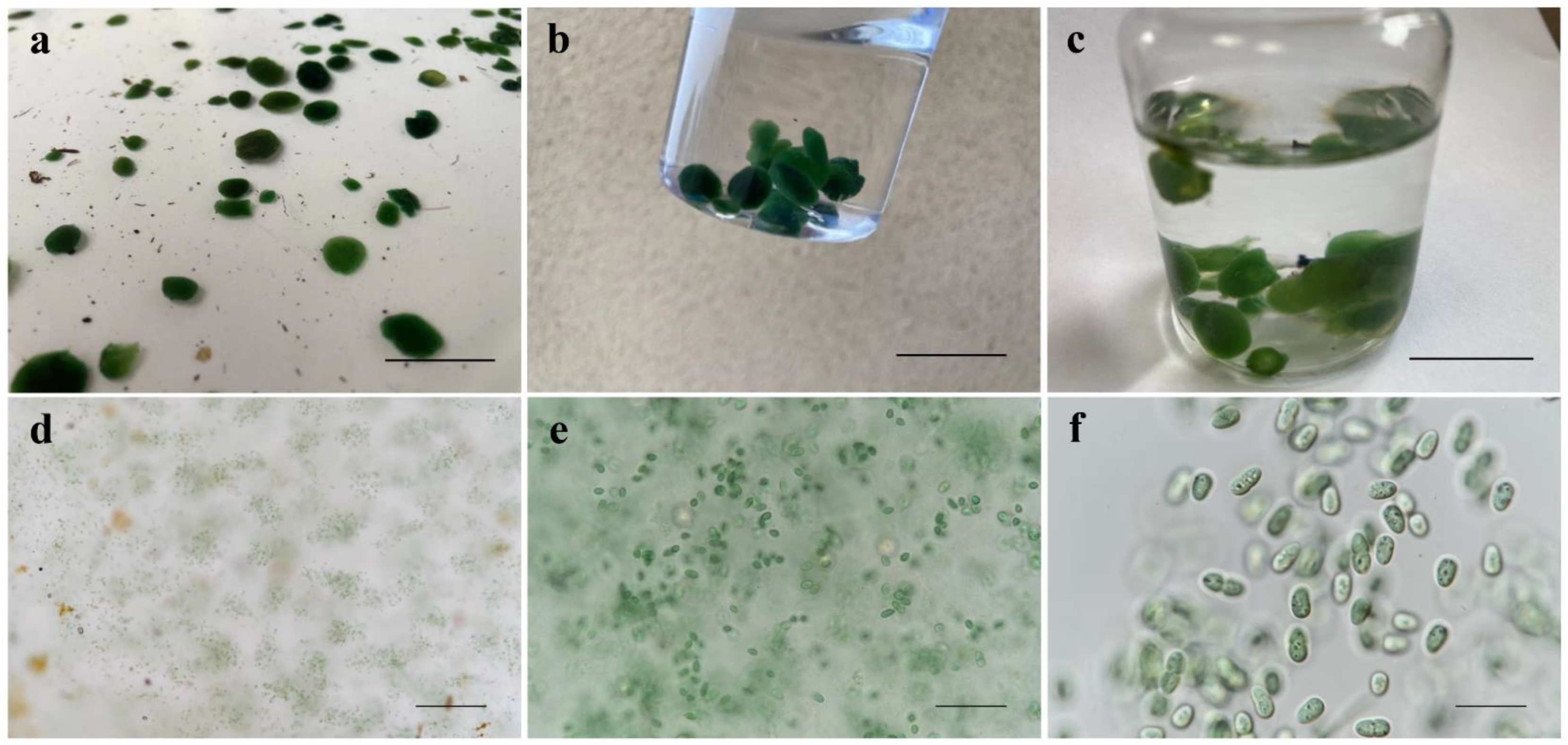
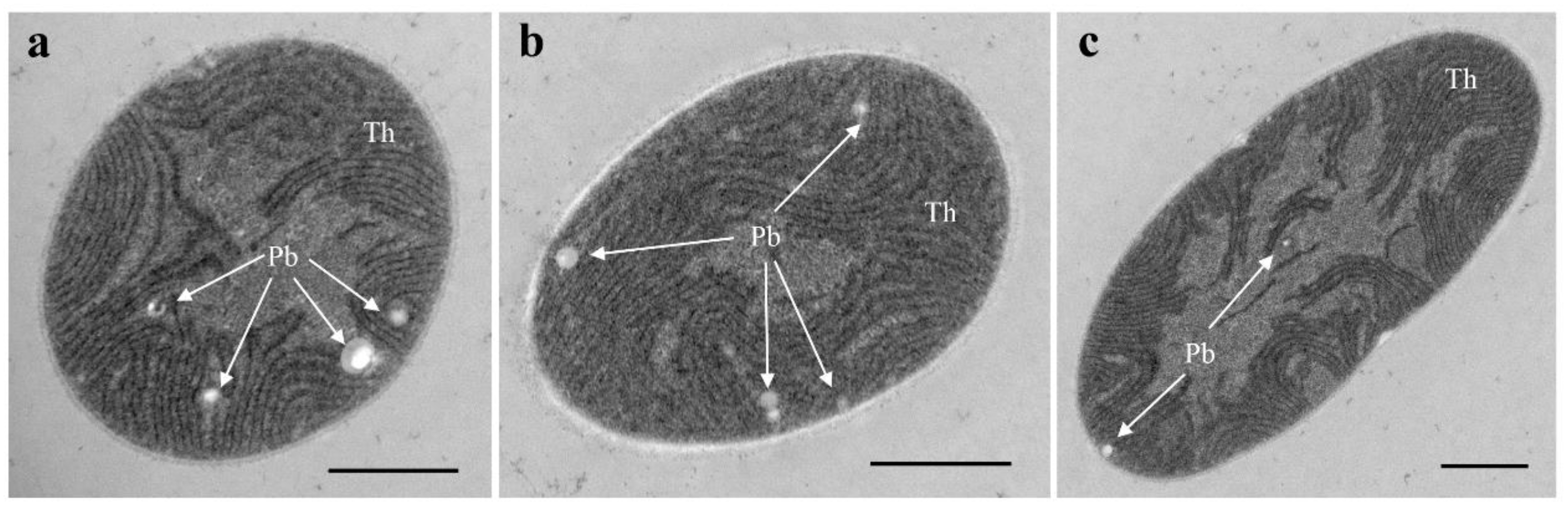
3.1. Morphological Description

Class: Cyanophyceae
Order: Chroococcales
Family: Microcystaceae
Cyanodorina W. Chen et G. Song & P. Ma gen. nov.
Description: Coccoid cyanobacteria. Colonies macroscopic, benthic epipelic cyanobacteria, with irregularly and densely arranged cells in a common sheath, dark green when fresh, yellow when old. Unicellular bacteria without envelopes, blue-green when fresh, yellow or yellow green when old. Stacked thylakoids formed fascicles of short sections in different number crossing the entire cell.
Etymology: The epithet Cyanodorina indicates that the colony forms a ball in Cyanobacteria.
Type species: Cyanodorina ovale
Cyanodorina ovale W. Chen et G. Song & P. Ma sp. nov. (Figure 1 and Figure 2).
Diagnosis: By macroscopic and microscopic observation, this species colonies exhibited morphological similarity to the Microcystis species but differed in their larger sizes and the presence of widely oval cells. However, phylogeny based on the 16S rRNA gene showed that this species had a unique position close to some coccoid cyanobacterial genera such as Microcystis, Chalicogloea, Cyanoarbor. Moreover, the lower similarity in the 16S rRNA gene sequence of this species to those of coccoid cyanobacterial genera and the significant differences between this species and those of coccoid cyanobacterial genera, as regards the length and the secondary structure of the 16S–23S ITS region, supported it as a new cyanobacterial genus.
Description: Colonies more or less irregularly spherical, macroscopic, usually composed of small groups of cells (subcolonies), embedded in a wide, fine, homogeneous, colorless sheath. Cells appeared green to blue-green, widely oval, longer than wide, 6.54–8.21–8.92 µm long, and 4.51–5.08–5.69 µm wide, with a length/width ratio of 1.26–1.60–1.80; unicells dividing by simple binary fission, absence of clearly delimited and concentrically lamellated mucilaginous envelopes around individual cells. Stacked thylakoids formed fascicles of short sections in different number crossing the entire cell, as documented by TEM analysis (Figure 2).
Etymology: The name of the species was chosen for the oval shape of the cells.
Type locality: Isolated from a water sample in Caohai Lake, Guizhou Province, China (18 April 2022, 26°49′ N, 104°15′ E, 2171.7 m a.s.l.).
Holotype designated here: Dry and fixed samples are stored at the Freshwater Algal Herbarium (HBI), Institute of Hydrobiology, Chinese Academy of Science, Wuhan, China, as specimens No. GZ202213 (dry), No. GZDX202213 (formaldehyde), No. GZDL202213 (Lugol’s).
Habitat: The surface sediment. Caohai Lake is a light eutrophic lake. In April 2022, the mean values of water temperature, pH, transparency, conductivity of electricity, dissolved oxygen, turbidity were 15.1 °C, 8.2, 53.25 cm, 410.5 µs cm−1, 6.66 mg L−1, and 10.45 NTU, respectively. The mean values of total nitrogen, total phosphorus, nitrate nitrogen, ammonia nitrogen, orthophosphate were 1.51 mg L−1, 0.08 mg L−1, 0.133 mg L−1, 0.53 mg L−1, 0.013 mg L−1, respectively. The mean concentration of chlorophyll a was 21.25 µg L−1.

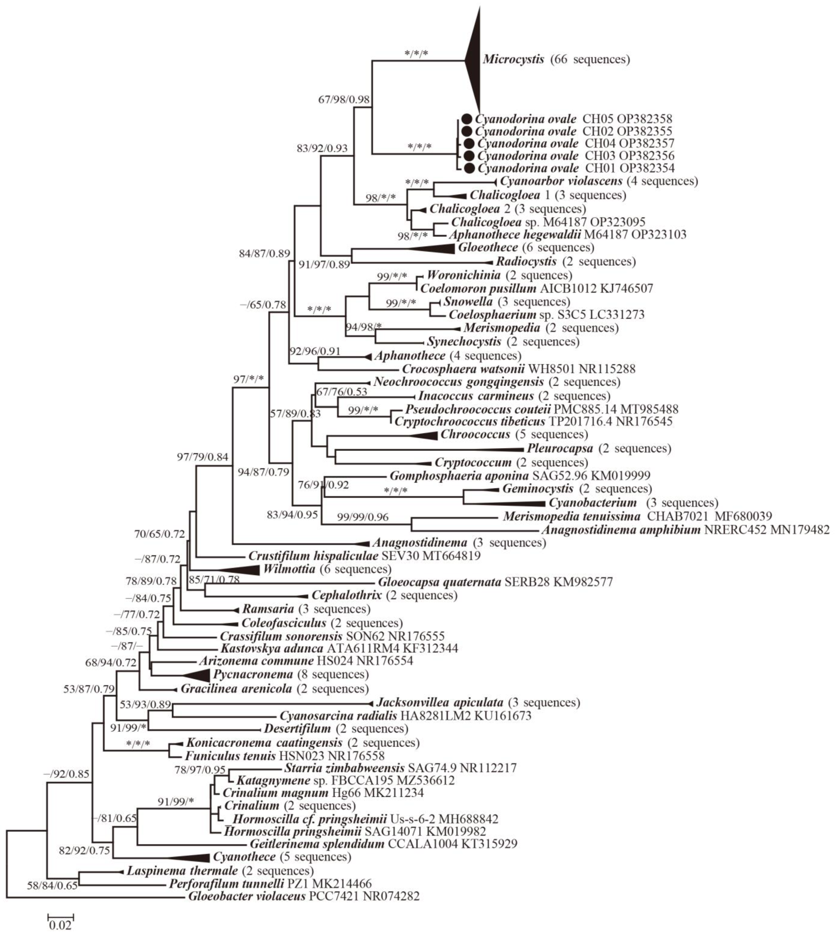
3.2. Molecular and Phylogenetic Analysis

For the calculation of the 16S rRNA p-distance matrix, we used sequences of representative taxa from families within the order Chroococcales. The sequences of Cyanodorina appeared to share 99.73–100.00% similarity to all five clones, and the five clones’ maximum similarity between Microcystis and other coccoid cyanobacterial genera was 93.10% (Table 1). Cyanodorina was found to share 93.10% sequence similarity with Microcystis aeruginosa, 93.00% with Cyanoarbor violascens, 91.30% with Aphanothece sacrum, 92.40% with Chalicogloea cavernicola.
In total, 187 representative taxa sequences were included in the phylogenetic analysis to assess the placement of the coccoid cyanobacterial (Figure 3 and Table S1). NJ, ML, and Bayesian inference analyses produced similar tree topologies in our phylogenies. The 16s rRNA phylogeny indicated that the studied species was distinct from the species of the other genus of Chroococcales (67% and 98% NJ and ML bootstrapping percentage (BP) and 0.98 posterior probability (PP)). Cyanodorina appeared as sister or parallel to other genera.

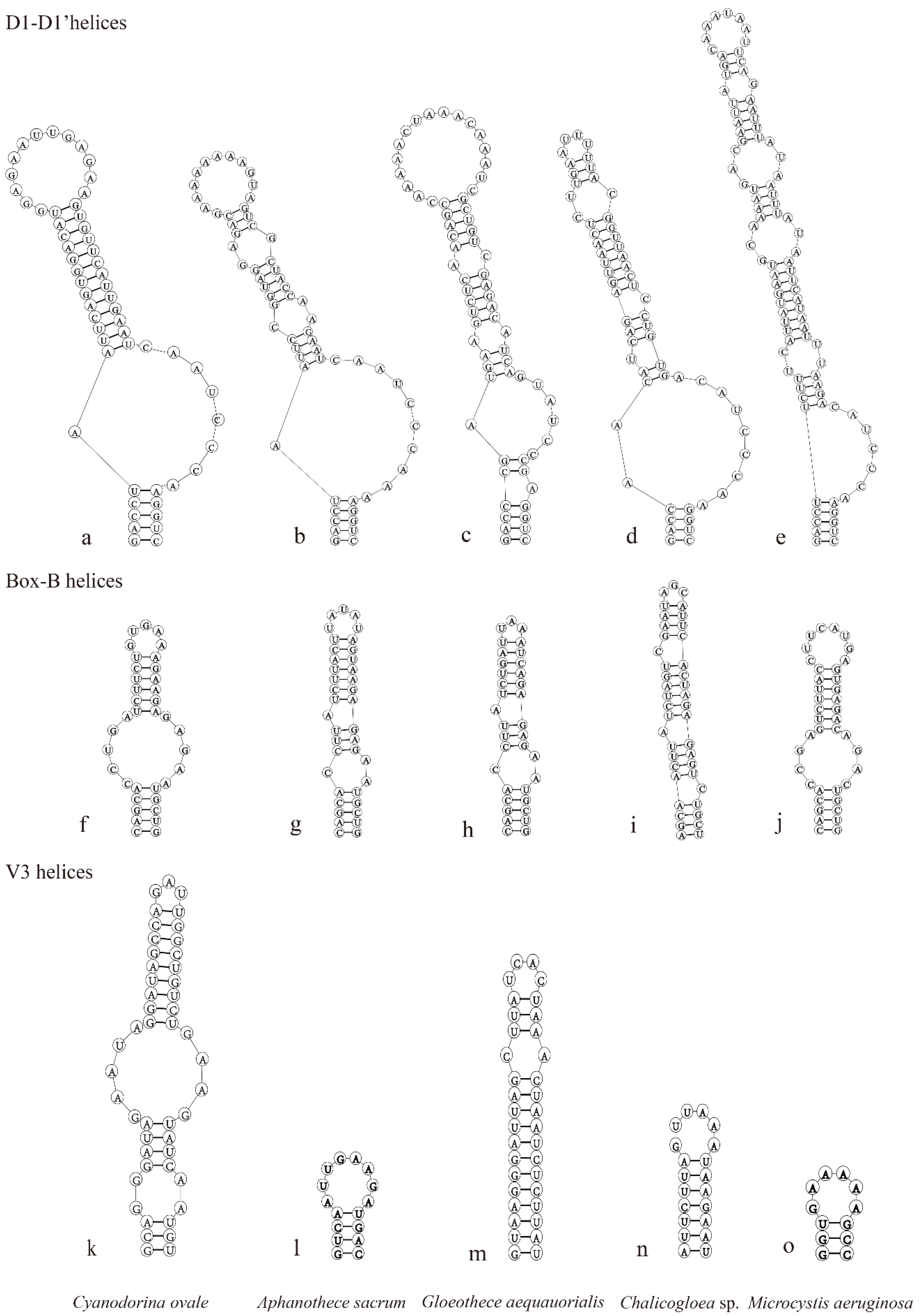
3.3. Comparison of ITS Regions between 16S and 23S rRNA Genes and Secondary Structures

The full-length of the ITS sequence of this species was 460 bp (Table 2), containing only one tRNAIle. The ITS secondary structures of the isolated Cyanodorina ovale were compared with the ITS structures of representative taxa from four genera in Microcystaceae, which were Gloeothece aequatorialis, Chalicogloea cavernicola, Microcystis aeruginosa, and Aphanothece sacrum. The ITS region of Cyanodorina ovale was different from those of the other taxa both in nucleotide sequence and in length of some regions. The D1-D1′ helix exhibited five distinct patterns within these five taxa (Figure 4a–e). All the outgroups differed from Cyanodorina ovale in the number of unilateral and bilateral bulges and base pairs. The Box-B helix was relatively conserved in Cyanodorina ovale (Figure 4f–j). This structure of Cyanodorina ovale differed from those of all other four taxa; all taxa possess highly variable helices and share no similar patterns with each other. The V3 helices of Cyanodorina ovale CH01 appeared conspicuously different from those of other taxa in the four genera in sequence length and stem–loop structures (Figure 4k–o, Table 2). These results provided strong evidence for the phylogenetic conclusion obtained above, further indicating that this species belongs to the new genus.

4. Discussion

Cyanobacteria are one of the most studied groups of aquatic microbes because of their importance in fishery, biological, azotification, environmental protection, and research on classifying systems [29]. Simultaneously, with the availability of 16S rRNA and ITS regions, α-level taxonomy and revision of extant taxa are rapidly progressing at the species and genus level [14]. In modern cyanobacterial taxonomy, the genus should be a monophyletic cluster, and the species should be well characterized using a polyphasic approach. The polyphasic approach has become the gold standard in cyanobacterial taxonomy by providing all available informative data on morphology, cell ultrastructure, ecology, physiology, and biochemical traits [30,31,32,33].
Coccoid cyanobacteria show high heterogeneity and include various developmental lines, which differ in important phenotypic and ultrastructural markers [6]. Of the five widely accepted groups in the coccoid cyanobacteria, Gloeobacterales and Synechococcales either lack thylakoids or possess parietal thylakoids, and Pleurocapsales, Chroococcales, and Chroococcidiopsidales show complex arrangements of thylakoids [34]. As regards Chroococcales with fasciculated thylakoids, only a few studies have chosen the polyphasic approach [6,7,8,9,35]. A large number of essential taxa are still lacking molecular data which would enable a comprehensive phylogenetic evaluation [36,37,38,39]. Despite such fuzzy status quo, a robust phylogenetic backbone based on multilocus analysis strongly supports the re-integration of Chroococcales and Pleurocapsales into a single taxonomic unit to sustain monophyly and define the orders on a comparable level of phylogenetic branching through all the Cyanophyceae [14].
The present study described a new coccoid cyanobacterial taxon isolated from Caohai Lake, China. We compared the characteristics of the 13 previously described genera of Chroococcales (11 of these genera are Microcystaceae) (Table 3). It was found that Gloeothece, Cyanothece, Rippkaea, Aphanothece, Cyanoaggregatum possess a similar cell shape to Cyanodorina ovale but favor different habitats. Cyanodorina ovale is morphologically similar to species of the genus Microcystis in colonial form and cellular ultrastructure. As described, Microcystis cells are often spherical or spheroidal with a diameter of 1.7–8.1 µm and colonies from 40 µm to 3 mm. the colonial forms are gelatinous, free-floating, and spherical, discoid, or irregular [40]. The ultrastructure of Microcystis cells showed that stacked thylakoids formed fascicles of short sections in different numbers, crossing the entire cell [41]. Therefore, the new coccoid cyanobacterial taxon appeared different from Microcystis in cell morphology, cell size, and colonial size. Moreover, this new coccoid cyanobacterial taxon grows on the surface of the sediment. It is different from the floating Microcystis.
Meanwhile, the highest homology between 16S rRNA gene sequences with respect to the existing cyanobacterial taxa appeared to be 93.10% (Microcystis aeruginosa NIES298), and the unique phylogenetic position indicated a high possibility of a novel genus. The observation that the secondary structures D1–D1′ helix, Box B helix, and V3 helix in the 16S–23S ITS region containing two tRNAs appeared different from those of phylogenetically close coccoid representative taxa of four genera supports the independence of the isolated species at the genus level.
Cyanodorina ovale possesses a spherical, oval, or cylindrical shape with widely rounded ends and stacked and fasciculated thylakoids, which is typical of Microcystaceae [14]. This species’ 16S rRNA appeared to share less than 93.10% genetic similarity with the other taxa that were the focus of this study, and this value was well below the genetic cut-off proposed by Yarza et al. of 94.5% [40]. Simultaneously, Yarza et al. [40] proposed a value of 86.5% to delineate families in regard to bacterial and archaeal species based on 16S rRNA gene sequences similarity [34]. In our study, the 16S rRNA gene phylogeny of this new taxon presented similarities with that of the known cyanobacterial genera in Microcystaceae, grouped in a distinct isolated clade (branch supports ≥ 98%). Following the conventional taxonomic research procedures, the observed species would belong to a new Microcystacean genus based on the obvious difference between Cyanodorina ovale and other Microcystacean species.
In conclusion, one new species of Microcystaceae was separated on the basis of a combination of different analyses, i.e., of the morphology, 16S rRNA gene dissimilarity and phylogeny, and secondary structures of the 16S-23S ITS region. On the basis of this evidence, we established a new Microcystacean genus—Cyanodorina—to correctly accommodate the new species Cyanodorina ovale within the family Microcystaceae.

Supplementary Materials

The following supporting information can be downloaded at: https://www.mdpi.com/article/10.3390/d15030329/s1, Table S1: List of all strains examined in this study, with NCBI Genbank accession numbers.

Author Contributions

Conceptualization, W.C. and G.S.; methodology, W.C.; software, S.L. and Y.X.; formal analysis, W.C. and G.S.; investigation, S.L. and Y.X.; data curation, W.C.; writing—original draft preparation, W.C. and P.M.; writing—review and editing, G.S. and P.M.; visualization, R.G.; funding acquisition, W.C. All authors have read and agreed to the published version of the manuscript.

Funding

This research was funded by the National Natural Science Foundation of China (No. 52200231) and Knowledge Innovation Program of Wuhan-Basic Research (202202080101017).

Institutional Review Board Statement

Not applicable.

Informed Consent Statement

Not applicable.

Data Availability Statement

Not applicable.

Acknowledgments

The first author thanks Zhenfei Xing for technical assistance with the TEM images.

Conflicts of Interest

The authors declare no conflict of interest.

References

  1. Waterbury, J.B. The Cyanobacteria-Isolation, Purification and Identification. In Prokaryotes, 3rd ed.; Dworkin, M., Falkow, S., Rosenberg, E., Schleifer, K.H., Stackebrandt, E., Eds.; Springer: New York, NY, USA, 2006; Volume 4, pp. 1053–1073. [Google Scholar] [CrossRef]
  2. Mareš, J.; Johansen, J.R.; Hauer, T.; Zima, J., Jr.; Ventura, S.; Cuzman, O.; Tiribilli, B.; Kaštovský, J. Taxonomic resolution of the genus Cyanothece (Chroococcales, Cyanobacteria), with a treatment on Gloeothece and three new genera, Crocosphaera, Rippkaea and Zehria. J. Phycol. 2019, 55, 578–610. [Google Scholar] [CrossRef]
  3. Komárek, J.; Kaštovský, J.; Mareš, J.; Johansen, J.R. Taxonomic classification of cyanoprokaryotes (cyanobacterial genera) 2014, using a polyphyletic approach. Preslia 2014, 86, 295–335. [Google Scholar]
  4. Mareš, J. Multilocus and SSU rRNA gene phylogenetic analyses of available cyanobacterial genomes, and their relation to the current taxonomic system. Hydrobiologia 2018, 811, 19–34. [Google Scholar] [CrossRef]
  5. Harke, M.J.; Steffen, M.M.; Gobler, C.J.; Otten, T.G.; Wilhelm, S.W.; Wood, S.A.; Paerl, H.W. A review of the global ecology, genomics, and biogeography of the toxic cyanobacterium, Microcystis spp. Harmful Algae 2016, 54, 4–20. [Google Scholar] [CrossRef] [PubMed] [Green Version]
  6. Kovačić, L.; Jezberová, J.; Komárková, J.; Kopecky., J.; Komárek, J. Ecological characteristics and polyphasic taxonomic classification of stable pigment-types of the genus Chroococcus (Cyanobacteria). Preslia 2011, 83, 145–166. [Google Scholar]
  7. Margheri, M.C.; Ventura, S.; Kaštovský, J.; Komárek, J. The taxonomic validation of the cyanobacterial genus Halothece. Phycologia 2008, 47, 477–486. [Google Scholar] [CrossRef]
  8. Komárek, J.; Kaštovský, J.; Jezberová, J. Phylogenetic and taxonomic delimitation of the cyanobacterial genus Aphanothece and description of Anathece gen. nov. Eur. J. Phycol. 2011, 46, 315–326. [Google Scholar] [CrossRef] [Green Version]
  9. Roldán, M.; Ramírez, M.; Del Campo, J.; Hernández-Mariné, M.; Komárek, J. Chalicogloea cavernicola gen. nov., sp. nov. (Chroococcales, Cyanobacteria), from low-light aerophytic environments: Combined molecular, phenotypic and ecological criteria. Int J. Syst. Evol. Microbiol. 2013, 63, 2326–2333. [Google Scholar] [CrossRef]
  10. Wang, Y.L.; Jia, N.N.; Geng, R.Z.; Yu, G.L.; Li, R.H. Phylogenetic insights intochroococcus-like taxa (Chroococcales, Cyanobacteria), describing Cryptochroococcus tibeticus gen. nov. sp. nov. and Limnococcus fonticola sp. nov. from Qinghai-Tibet plateau. J. Phycol. 2021, 57, 1739–1748. [Google Scholar] [CrossRef]
  11. Mogany, T.; Swalaha, F.M.; Allam, M.; Senzo Mtshali, P.; Ismail, A.; Kumari, S.; Bux, F. Phenotypic and genotypic characterisation of an unique indigenous hypersaline unicellular cyanobacterium, Euhalothece sp. nov. Microbiol. Res. 2018, 211, 47–56. [Google Scholar] [CrossRef]
  12. Gama, W.A.; Rigonato, J.; Fiore, M.F.; Sant’Anna, C.L. New insights into Chroococcus (Cyanobacteria) and two related genera: Cryptococcum gen. nov. and Inacoccus gen. nov. Eur. J. Phycol. 2019, 54, 315–325. [Google Scholar] [CrossRef]
  13. Rigonato, J.; Sant’Anna, C.L.; Giani, A.; Azevedo, M.T.P.; Gama, W.A.; Viana, V.F.L.; Fiore, M.F.; Werner, V.R. Sphaerocavum: A coccoid morphogenus identical to Microcystis in terms of 16S rDNA and ITS sequence phylogenies. Hydrobiologia 2018, 811, 35–48. [Google Scholar] [CrossRef]
  14. Strunecký, O.; Ivanova, A.P.; Mareš, J. An updated classification of cyanobacterial orders and families based on phylogenomic and polyphasic analysis1. J. Phycol. 2022; accepted. [Google Scholar]
  15. Boone, D.R.; Castenholz, R.W. The Archaea and the Deeply Branching and Phototrophic Bacteria. In Bergey’s Manual of Systematic Bacteriology, 2nd ed.; Garrity, G., Ed.; Springer: New York, NY, USA, 2001; Volume 1, pp. 1–721. [Google Scholar]
  16. Komárek., J.; Anagnostidis., K. Cyanoprokaryota 1. Teil: Chroococcales. In Süsswasserflora von Mitteleuropa 19/1; Ettl, H., Gärtner, G., Heynig, H., Eds.; Gustav Fischer: Jena, Germany; Stuttgart, Germany; Lübeck, Germany; Ulm, Germany, 1998; pp. 1–548. [Google Scholar]
  17. Geng, R.Z.; Li, W.K.; Chao, A.M.; Guo, X.Y.; Li, H.; Yu, G.L.; Li, R.H. Establishment of a New Filamentous Cyanobacterial Genus, Microcoleusiopsis gen. nov. (Microcoleaceae, Cyanobacteria), from Benthic Mats in Open Channel, Jiangxi Province, China. Diversity 2021, 13, 548. [Google Scholar] [CrossRef]
  18. Clarke, J.D. Cetyltrimethyl ammonium bromide (CTAB) DNA miniprep for plant DNA isolation. Cold Spring Harb. Protoc. 2009, 2009, pdb.prot5177. [Google Scholar] [CrossRef] [PubMed]
  19. Lepère, C.; Wilmotte, A.; Meyer, B. Molecular diversity of Microcystis strains (Cyanophyceae, Chroococcales) based on 16S rRNA sequences. Syst. Geogr. Plants 2000, 70, 275–283. [Google Scholar] [CrossRef]
  20. Edwards, U.; Rogall, T.; Blöcker, H.; Emde, M.; Böttger, E.C. Isolation and direct complete nucleotide determination of entire genes. Characterization of a gene coding for 16S ribosomal RNA. Nucleic Acids Res. 1989, 17, 7843–7853. [Google Scholar] [CrossRef] [PubMed] [Green Version]
  21. Katoh, K.; Standley, D.M. MAFFT multiple sequence alignment software version 7: Improvements in performance and usability. Mol. Biol. Evol. 2013, 30, 772–780. [Google Scholar] [CrossRef] [Green Version]
  22. Kumar, S.; Stecher, G.; Li, M.; Knyaz, C.; Tamura, K. MEGA X: Molecular Evolutionary Genetics Analysis across computing platforms. Mol. Biol. Evol. 2018, 35, 1547–1549. [Google Scholar] [CrossRef]
  23. Trifinopoulos, J.; Nguyen, L.T.; von Haeseler, A.; Minh, B.Q. W-IQ-TREE: A fast online phylogenetic tool for maximum likelihood analysis. Nucleic Acids Res. 2016, 44, 232–235. [Google Scholar] [CrossRef] [Green Version]
  24. Kalyaanamoorthy, S.; Minh, B.Q.; Wong, T.K.F.; Von Haeseler, A.; Jermiin, L.S. ModelFinder: Fast model selection for accurate phylogenetic estimates. Nat. Methods 2017, 14, 587–589. [Google Scholar] [CrossRef] [Green Version]
  25. Miller, M.A.; Schwartz, T.; Pickett, B.; He, S.; Klem, E.; Scheuermann, R.H.; Passarotti, M.; Kaufman, S.; O’Leary, M.A. A RESTful API for Access to Phylogenetic Tools via the CIPRES Science Gateway. Evol. Bioinform. 2015, 11, 43–48. [Google Scholar] [CrossRef] [PubMed]
  26. Ronquist, F.; Teslenko, M.; Van der Mark, P.; Ayres, D.L.; Darling, A.; Höhna, S.; Larget, B.; Liu, L.; Suchard, M.A.; Huelsenbeck, J.P. MrBayes 3.2: Efficient bayesian phylogenetic inference and model choice across a large model space. Syst. Biol. 2012, 61, 539–542. [Google Scholar] [CrossRef] [PubMed] [Green Version]
  27. Boyer, S.; Johansen, J.R.; Flechtner, V.R. Phylogeny and genetic variance in terrestrial Microcoleus (Cyanophyceae) species based on sequence analysis of the 16S rRNA gene and associated 16S-23S ITS region. J. Phycol. 2002, 38, 1222–1235. [Google Scholar] [CrossRef]
  28. Reuter, J.S.; Mathews, D.H. RNAstructure: Software for RNA secondary structure prediction and analysis. BMC Bioinform. 2010, 11, 129. [Google Scholar] [CrossRef] [PubMed] [Green Version]
  29. Sandrini-Neto, L.; Geraudie, P.; Santana, M.S.; Camus, L. Effects of dispersed oil exposure on biomarker responses and growth in juvenile wolfish Anarhichas denticulatus. Environ. Sci. Pollut. Res. Int. Res. 2016, 23, 21441–21450. [Google Scholar] [CrossRef]
  30. Comte, K.; Holland, D.P.; Walsby, A.E. Changes in cell turgor pressure related to uptake of solutes by Microcystis sp. strain8401. FEMS Microbiol. 2007, 61, 399–405. [Google Scholar] [CrossRef]
  31. Dvořák, P.; Poulíčková, A.; Hašler, P.; Belli, M.; Casamatta, D.A.; Papini, A. Species concepts and speciation factors in cyanobacteria, with connection to the problems of diversity and classification. Biodivers. Conserv. 2015, 24, 739–757. [Google Scholar] [CrossRef] [Green Version]
  32. Komárek, J. Several problems of the polyphasic approach in the modern cyanobacterial system. Hydrobiologia 2018, 811, 7–17. [Google Scholar] [CrossRef]
  33. Sciuto, K.; Rascio, N.; Andreoli, C.; Moro, I. Polyphasic characterization of ITD-01, a cyanobacterium isolated from the Ischia Thermal District (Naples, Italy). Fottea 2011, 11, 31–39. [Google Scholar] [CrossRef]
  34. Wang, Y.L.; Cai, F.F.; Jia, N.N.; Li, R.H. Description of a novel coccoid cyanobacterial genus and species Sinocapsa zengkensis gen. nov. sp. nov. (Sinocapsaceae, incertae sedis), with taxonomic notes on genera in Chroococcidiopsidales. Phytotaxa 2019, 409, 146–160. [Google Scholar] [CrossRef]
  35. Korelusová, J.; Kaštovský, J.; Komárek, J. Heterogeneity of the cyanobacterial genus Synechocystis and description of a new genus, Geminocystis. J. Phycol. 2009, 45, 928–937. [Google Scholar] [CrossRef] [PubMed]
  36. Castenholz, R.W.; Wilmotte, A.; Herdman, M.; Rippka, R.; Waterbury, J.B.; Iteman, I.; Hoffmann, L. Bergey’s Manual of Systematic Bacteriology; Springer Science+Business Media: New York, NY, USA, 2001. [Google Scholar]
  37. Nelissen, B.; Van de Peer, Y.; Wilmotte, A.; De Water, R. An early origin of plastids within the cyanobacterial divergence is suggested by evolutionary trees based on complete 16S rRNA sequences. Mol. Biol. Evol. 1995, 12, 1166–1173. [Google Scholar] [PubMed] [Green Version]
  38. Ohki, K.; Kamiya, M.; Honda, D.; Kumazawa, S.; Ho, K.K. Morphological and phylogenetic studies on unicellular dizaotrophic Cyanobacteria (Cyanophytes) isolated from the coastal waters around singapore. J. Phycol. 2008, 44, 142–151. [Google Scholar] [CrossRef]
  39. Komárek, J.; Cepák, V.; Kaštovský, J.; Sulek, J. What are the cyanobacterial genera Cyanothece and Cyanobacterium? Contribution to the combined molecular and phenotype taxonomic evaluation of cyanobacterial diversity. Algol. Stud./Arch. Für Hydrobiol. 2004, 113, 1–36. [Google Scholar] [CrossRef]
  40. Yarza, P.; Yilmaz, P.; Pruesse, E.; Glöckner, F.O.; Ludwig, W.; Schleifer, K.-H.; Whitman, W.B.; Euzéby, J.; Amann, R.; Rpsselló-Móra, R. Uniting the classification of cultured and uncultured bacteria and archaea using 16S rRNA gene sequences. Nat. Rev. Microbiol. 2014, 12, 635–645. [Google Scholar] [CrossRef] [PubMed]
  41. Sendall, B.C.; Mcgregor, G.B. Cryptic diversity within the Scytonema complex: Characterization of the paralytic shellfish toxin producer Heterosyctonema crispum, and the establishment of the family Heteroscytonemataceae (Cyanobacteria/Nostocales). Harmful Algae 2018, 80, 158–170. [Google Scholar] [CrossRef] [PubMed]
Figure 1. Field sample morphology of Cyanodorina ovale. (a) Cyanodorina ovale in the tray after sediment cleaning. (b,c) Cyanodorina ovale in liquid BG11 medium. (df) Colonies at different magnification. Scale bars: (ac) 2 cm, (d) 200 µm, (e) 50 µm, (f) 20 µm.
Diversity 15 00329 g001
Figure 2. TEM micrographs of Cyanodorina ovale. (a) Cross section of a cell. (b) Longitudinal section of a cell. (c) Dividision of a cell. Pb, polyphosphate body, Th, thylakoids. Scale bars: 1 µm.
Diversity 15 00329 g002
Figure 3. Maximum likelihood (ML) phylogenetic tree of cyanobacteria based on 16S rRNA sequences (1020 bp) representing the current GenBank data for Chroococcalean cyanobacteria. Bootstrap values greater than 50% are shown on the ML tree for the NJ/ML methods and Bayesian posterior probabilities. * indicates bootstrapping values of 100 for NJ, ML, and BI posterior probabilities of 1.00. Sequences from GenBank are indicated by accession numbers. The main clades are indicated by numbers. Sequences from this study are denoted with solid circles.
Diversity 15 00329 g003
Figure 4. Secondary structures of D1–D1′ helices (ae), Box-B helices (fj) and V3 helices of various taxa. (a,f,k) Cyanodorina ovale CH01. (b,g,l) Aphanothece sacrum FPU3. (c,h,m) Gloeothece aequauorialis SAG 36.87. (d,i,n) Chalicogloea sp. BACA0589. (e,j,o) Microcystis aeruginosa UUEX ‘B 2667.
Diversity 15 00329 g004
Table 1. Sequence similarity comparison of the 16S rRNA gene between Cyanodorina ovale CH01 and species of close genera.
1234567891011121314151617181920
1. Cyanodorina ovale CH01 clone1
2. Microcystis aeruginosa NIES29893.10
3. Neochroococcus gongqingensis CHAB4018 93.00 91.50
4. Cyanoarbor violascens C193.00 92.30 91.90
5. Chalicogloea cavernicola CCALA97592.40 91.30 91.10 94.80
6. Cryptochroococcus tibeticus TP201716.492.20 91.20 94.50 92.00 91.20
7. Pseudochroococcus couteii PMC885.1492.10 91.50 94.40 92.00 91.10 99.10
8. Gloeothece aequatorialis SAG36.87 clone791.90 91.90 92.40 91.80 91.40 91.10 91.00
9. Inacoccus carmineus CCIBt347591.60 90.80 94.20 91.20 90.00 93.90 94.00 91.30
10. Gomphosphaeria aponina SAG52.9691.50 89.90 94.00 90.70 90.50 93.20 93.20 91.20 93.80
11. Pannus brasiliensis CCIBt3594 91.30 94.90 90.20 90.50 90.70 89.90 90.10 91.10 89.50 89.30
12. Aphanothece sacrum H591.30 90.50 92.60 91.60 91.60 92.10 92.60 91.20 92.90 93.00 90.80
13. Eucapsis minor SAG 14.99 91.10 93.50 91.90 91.50 90.80 90.10 90.20 90.70 90.90 90.30 92.80 90.90
14. Radiocystis geminata MAC121490.80 91.00 91.50 91.40 91.20 90.10 90.10 91.40 92.20 91.30 89.70 91.30 91.40
15. Chroococcus subviolaceus CCIBt354990.80 91.50 92.80 91.80 90.20 92.30 92.70 91.20 93.40 91.70 89.70 91.20 89.30 90.10
16. Cryptococcum komarkovae CCALA5490.30 88.90 93.00 90.10 90.00 92.20 92.20 89.90 91.90 91.60 89.10 91.20 89.80 89.70 91.30
17. Geitlerinema splendidum CCALA100490.00 87.80 90.30 88.50 88.80 90.20 90.50 88.10 90.40 89.80 88.50 90.10 87.60 88.20 91.20 89.90
18. Anagnostidinema amphibium NRERC45288.90 89.70 89.80 88.50 87.90 89.50 89.60 89.00 89.30 90.00 88.10 89.10 88.00 89.40 90.10 87.40 87.60
19. Geminocystis herdmanii PCC630888.00 86.60 88.70 87.00 87.10 90.00 89.90 87.00 89.10 90.20 86.80 89.10 86.70 87.30 87.30 88.40 87.50 86.70
20. Alborzia kermanshahica S2 86.90 85.90 86.50 89.90 91.50 85.80 85.80 86.60 84.90 85.60 86.00 86.70 86.90 86.60 84.70 85.00 83.40 82.20 82.50
21. Cyanobacterium stanieri MAC321786.50 84.60 86.50 84.30 84.70 87.70 87.90 85.30 87.70 88.90 84.70 87.60 84.80 85.90 86.00 86.50 86.80 85.10 92.80 80.00
Table 2. Analyses of the ITS of the 16S–23S region for Cyanodorina ovale CH01 and other related strains.
OrganismsGenBankITS Total Length (nt)D1–D1′ Helix Length (nt)D2 Region tRNAIletRNAAlaBox B Helix Length (nt)Box A SpacerV3
Helix Length (nt)
Cyanodorina ovale CH01OP38235446058CTTTCAAACTCT+-37GCACCTTGAAAA48
Aphanothece sacrum FPU3AB116658.245393CTTTCAAACTAG+-40GCACCTTGAAAA16
Gloeothece aequauorialis SAG 36.87MF781064.1 44960CTTTCAAACTTA--43GAACCTTGAAAA36
Chalicogloea sp. BACA0589OM732250.148167CTTTCAAACTCT+-35GCACCTTGAAAA20
Microcystis aeruginosa UUEX ‘B 2667 HQ625424.140463CTTTCAAACTAG+-39GAACCTTGAAAA12
Table 3. Comparison of the characteristics of the 13 previously described genera of Chroococcales (“-” indicates that the feature is unknown).
GenusCell ShapeCell Size (μm)Colony FormationColony Size (μm)SheathsThylakoid ArrangementHabitat
Cyanodorinaoval 6.54–8.92 × 4.51–5.69Yes5000–20,000 YesIrregular fasciclesFreshwater, benthic
Microcystisspherical or hemispherical1.7–8.1Yes40–3000YesIrregular fasciclesFreshwater, free floating
Pannusround1–4Yes126.5–314.0YesIrregular fasciclesPlanktonic in brackish bays and reservoirs, benthic in stagnant freshwater
Eucapsissemicircle8–14Yes<50Yes-Freshwater, benthic
Chalicogloeaspherical2.6–4.3Yes<100.0YesIrregular fasciclesSalpetre Cave
Cyanoarborsubspherical1–5Yes200.0–10,000.0YesIrregular fasciclesSalty and alkaline waters, high mts lakes,
Gloeotheceoval to cylindrical5–20 × 3–12Yes, rarely absent13.0–40.0 long, 10.0–26.0 wideYesIrregular fasciclesFreshwater, terrestrial
Cyanotheceoval to cylindrical2.5–5 × 7.5–10No NoSpecial (radial with a central network)Freashwater, terrestrial
Crocosphaeraspherical to cylindrical2.8–7.1 × 1.2–1.8No NoIrregular fasciclesMarine
Zehriaspherical to cylindrical2.0–5.6 × 2.0–4.0No NoIrregular fasciclesMarine
Rippkaeaoval to cylindrical4–9 × 3–5No NoIrregular fasciclesSemi-terrestrial (soil of rice field)
Aphanotheceoval to cylindrical1–12Yesmicro to macroscopicYesIrregular fasciclesFreshwater, terrestrial
Cyanoaggregatumoval or cylindrical2.4–3.8 × 1.4–2.0Yes92.0–340.0 long, 62.0–160.0 diameter Yes-lagoon
Disclaimer/Publisher’s Note: The statements, opinions and data contained in all publications are solely those of the individual author(s) and contributor(s) and not of MDPI and/or the editor(s). MDPI and/or the editor(s) disclaim responsibility for any injury to people or property resulting from any ideas, methods, instructions or products referred to in the content.

