Article

North–South Differentiation of Black Flies in the Western Cordillera of North America: A New Species of Prosimulium (Diptera: Simuliidae) †

1
Department of Plant and Environmental Sciences, Clemson University, Clemson, SC 29634-0310, USA
2
C.P. Gillette Museum of Arthropod Diversity, Fort Collins, CO 80528, USA
*
Author to whom correspondence should be addressed.
urn:lsid:zoobank.org:pub: 7BBA84B5-BD26-4001-8E66-8FF7FE1AEBFA.
Diversity 2023, 15(2), 212; https://doi.org/10.3390/d15020212
Submission received: 17 December 2022 / Revised: 26 January 2023 / Accepted: 28 January 2023 / Published: 2 February 2023
(This article belongs to the Special Issue Diversity, Distribution and Phylogeny of Vector Insects)

Abstract

:
Glaciation has been a powerful determiner of species distributions and the genetic structure of populations. Contemporary distributions of many organisms in North America’s Western Cordillera reflect the influence of Pleistocene glaciation. We identified a pattern of north–south differentiation in the genus Prosimulium of western North America, which reflects the separation of northern and southern populations by the North American Ice Sheet during the Pleistocene Epoch. The taxonomic implication is that new species exist within nominal species, requiring formal description or revalidation of names currently in synonymy. We morphologically and cytogenetically examined populations of one nominal species of black fly, Prosimulium esselbaughi Sommerman, over its known range from Alaska south to California and Colorado. Chromosomal and morphological evidence supports the presence of two species, P. esselbaughi sensu stricto from Alaska to at least southern British Columbia, and a new species, Prosimulium supernum in the central Rocky Mountains and high Sierra Nevada range of the United States. The new species is described in all life stages above the egg, along with its polytene chromosomes. The existence of differentiated populations of other nominal species of black flies in northern and southern North America provides a system for investigating possible co-differentiation of vectors and parasites.

1. Introduction

Mountains provide rich opportunities for population isolation and, therefore, are often hotspots for speciation, biodiversity, and endemism. North America’s vast Western Cordillera, running from the Brook’s Range of Alaska southward through the Rockies, Cascades, Coast Ranges, and Sierra Nevada, has an ancient history of tectonic influences [1,2]. Superimposed on the orogenic consequences have been cyclical glacial and interglacial periods, most recently the Last Glacial Period (ca. 115,000–12,000 BP) with its Late Glacial Maximum about 26,500 to 19–20,000 BP [3]. Species distributions and genetic structure have been profoundly influenced by glaciation cycles, particularly during the Quaternary Period of the past 2.6 million years [4,5,6].
Among the cold-adapted organisms that inhabit the Western Cordillera are members of the dipteran family Simuliidae. The family’s oldest extant lineages are in the Holarctic Region, reflecting the hypothesis that these flies evolved in cool, mountainous areas [7]. One of the oldest extant lineages is the Holarctic genus Prosimulium, consisting of 80 nominal species, of which 25 inhabit the Western Cordillera, including 23 that are precinctive to this mountain chain [8]. Only two of the 80 species of Prosimulium are found in both the Nearctic and Palearctic regions [8]. All species of Prosimulium are cold-adapted, particularly the high-elevation inhabitants, and therefore are potential sentinels for monitoring climate change. The female flies of Prosimulium are mammalophilic, and those of some species have been incriminated as vectors of onchocercid worms [9,10].
The extensive ranges of many western nominal Prosimulium species from northern Alaska southward through the Rocky Mountains and Sierra Nevada, coupled with genetic differences, have suggested the presence of multiple species [11,12]. Populations in California’s Sierra Nevada, for example, were tentatively identified as Prosimulium esselbaughi Sommerman, with the caveat that they might represent a new species, based on novel sex-linkage of chromosomal rearrangements [11]. Our discovery of chromosomally and morphologically similar populations in the central Rocky Mountains led to an evaluation of the Rocky Mountain and Sierra Nevada populations and the conclusion that they represent a new species. We describe these populations as a new species, based on chromosomal band patterns and morphology of the female, male, pupa, and larva. We highlight the geographic distributions of the new species and its close relative and discuss the general pattern of north–south differentiation of Prosimulium species in western North America.

2. Materials and Methods

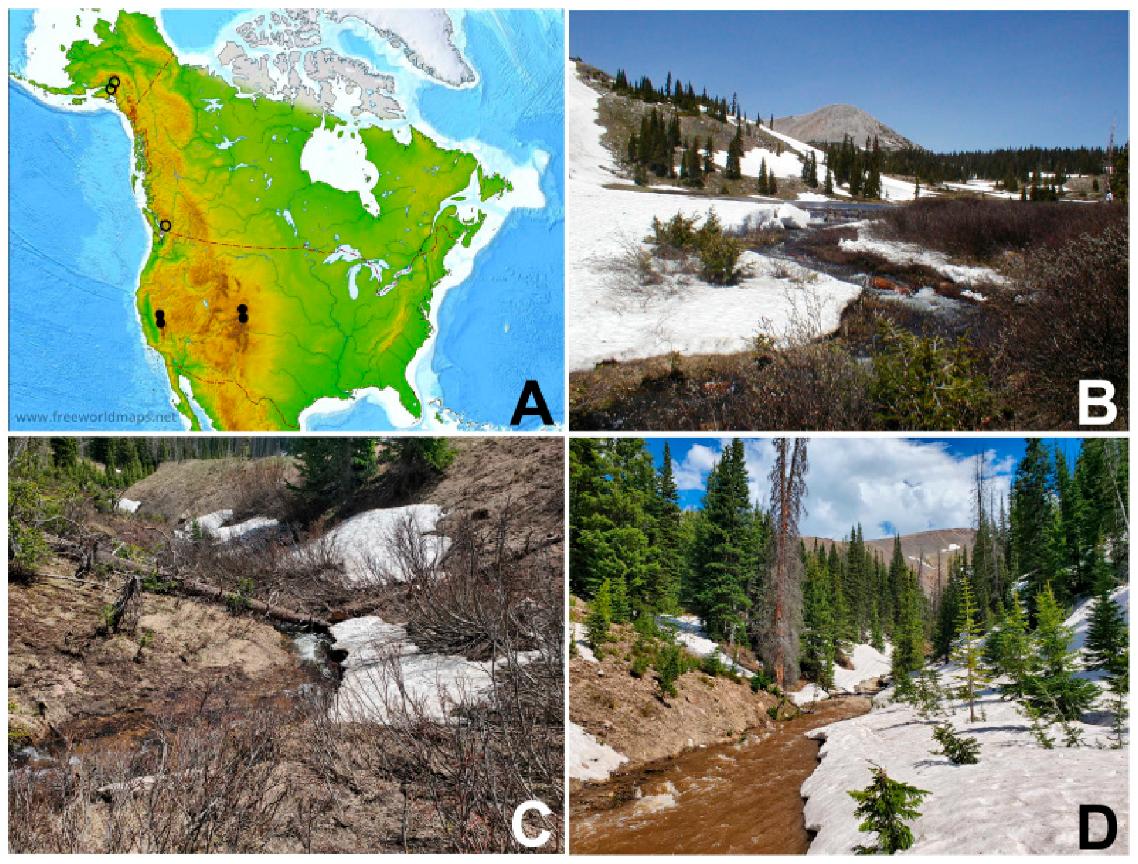
2.1. Collection Sites

At the type locality of the new species in Wyoming (Figure 1A,B), larvae and pupae were collected in the North Fork drainage basin of the Little Laramie River in Albany County (Table 1). Larvae and pupae were taken from trailing vegetation and stones in 2007 and 2008 and from black plastic bags anchored in the stream on 1 June 2008. Holes in the ice allowed access to the stream. The plastic bags were used as attachment substrates for larvae and particularly for pupae that are typically concealed in sediment. The plastic was checked weekly through the third week of July. Larvae were collected into Carnoy’s fixative (3 parts 99% ethanol: 1 part glacial acetic acid) and transferred to fresh fixative 2–4 h later. Pupae were collected by cutting the plastic around them and placing them in Petri dishes with moist filter paper. Adults were allowed to emerge from the pupae in the laboratory (elevation 2184 m) and were dispatched 12–24 h later by freezing. Pupal exuviae and cocoons were associated with each adult.
Larvae in Colorado were collected from the Michigan River and Cache La Poudre drainages in Jackson County (Figure 1C,D) in June 2022 and fixed in Carnoy’s fixative, which was refreshed 4–8 h later (Table 1). Plastic bags were placed in the streams in June and retrieved in July 2022, but no larvae or pupae were attached to them.
In addition to material collected in Colorado and Wyoming, we also examined larval material from California (Figure 1A) currently housed, as Prosimulium esselbaughi, in the Clemson University Arthropod Collection (Clemson, SC, USA). Because larvae of P. minifulvum Adler, Currie and Wood are not morphologically distinguishable from those of so-called P. esselbaughi from California, we examined only those larvae that had originally been identified chromosomally.

2.2. Chromosomes

The posterior half of each larval abdomen was removed, and the chromosomes and gonads were Feulgen-stained, dissected from the abdomen, and analyzed under oil immersion [13]. Diagnostic chromosomal sequences were photographed with a Jenoptik ProgRes® SpeedXT Core 5 digital camera mounted on an Olympus BX40 light microscope. The images were imported into Adobe® PhotoShop® Elements 8 to assemble and label the chromosome maps.
The band sequences of the long (L) and short (S) arms of each of the three chromosomes (I, II, and III) were compared with the standard sequence for the genus Prosimulium [14,15]. Chromosomal rearrangements previously found in other taxa were named according to their original designations [14]. New rearrangements were assigned unique numbers. Enhanced bands (i.e., heterobands) relative to the standard are identified by the chromosome arm and section number (e.g., IIIS hb80). Fixed inversions in the text and on the maps are italicized; polymorphic rearrangements appear in non-italic type. We also indicate on our maps the following landmarks that are homologous throughout the Simuliidae and that have been used throughout the history of simuliid chromosomal studies [11,14,15]: CI, CII, CIII (centromeres of chromosomes I, II, and III, respectively), NO (nucleolar organizer), Pb (parabalbiani), and RB (ring of Balbiani, a lightly stained area that varies among nuclei and individuals from little or no puffing (e.g., Figure 2C) to well-expressed puffing).

2.3. Morphology

Selected specimens for light microscopy were dissected in 80% ethanol. Larval structures and adult legs were temporarily slide-mounted in a drop of 50% acetic acid. Pupal gills were removed and moved to depression slides in glycerin. Adult heads and terminalia were heated for ca. 2 min in 85% lactic acid, transferred to a drop of glycerin in a depression slide, further dissected into component parts, and oriented for interpretation and imaging.
Structures were photographed at multiple focal planes with a Jenoptik ProgRes® SpeedXT Core 5 digital camera mounted on an Olympus BX40 light microscope. Helicon Focus (version 7.7.5) stacking software was used to form composite images from multiple focal planes. All morphological images were made from specimens collected at the type locality.
Adults in ethanol were chemically dried using hexamethyldisilazane and then pinned through the thorax with a minuten. The pupal exuviae and cocoon were placed in a microvial with glycerin and pinned beneath the associated adult. Dissected parts were pinned in a separate microvial beneath the adult. Descriptions of colors were based on pinned specimens.
Larvae and pupae for scanning electron microscopy were dehydrated through an ethanol series, dried in hexamethyldisilazane, mounted on conductive stubs with double-sided adhesive tape, sputter-coated for 3 min with platinum, and imaged with a Hitachi TM-3000 Scanning Electron Microscope (composite mode, 15 kV, and full vacuum).
Terminology for structures follows that of Adler et al. [11] and, for the larval mandible, Chance [16].

2.4. Type Depositories

After larvae were prepared for chromosomal study they were transferred from Carnoy’s fixative to 80% ethanol. The holotype and most paratypes were deposited in the United States National Museum (USNM), Washington, DC. Additional paratypes (adults with pupal exuviae) were deposited in the Canadian National Collection (CNC), Ottawa.

3. Results

Prosimulium supernum Adler and Reeves, n. sp.
Prosimulium esselbaughi: [11] (part: Alpine and Mariposa Counties, California, possibly Nevada).

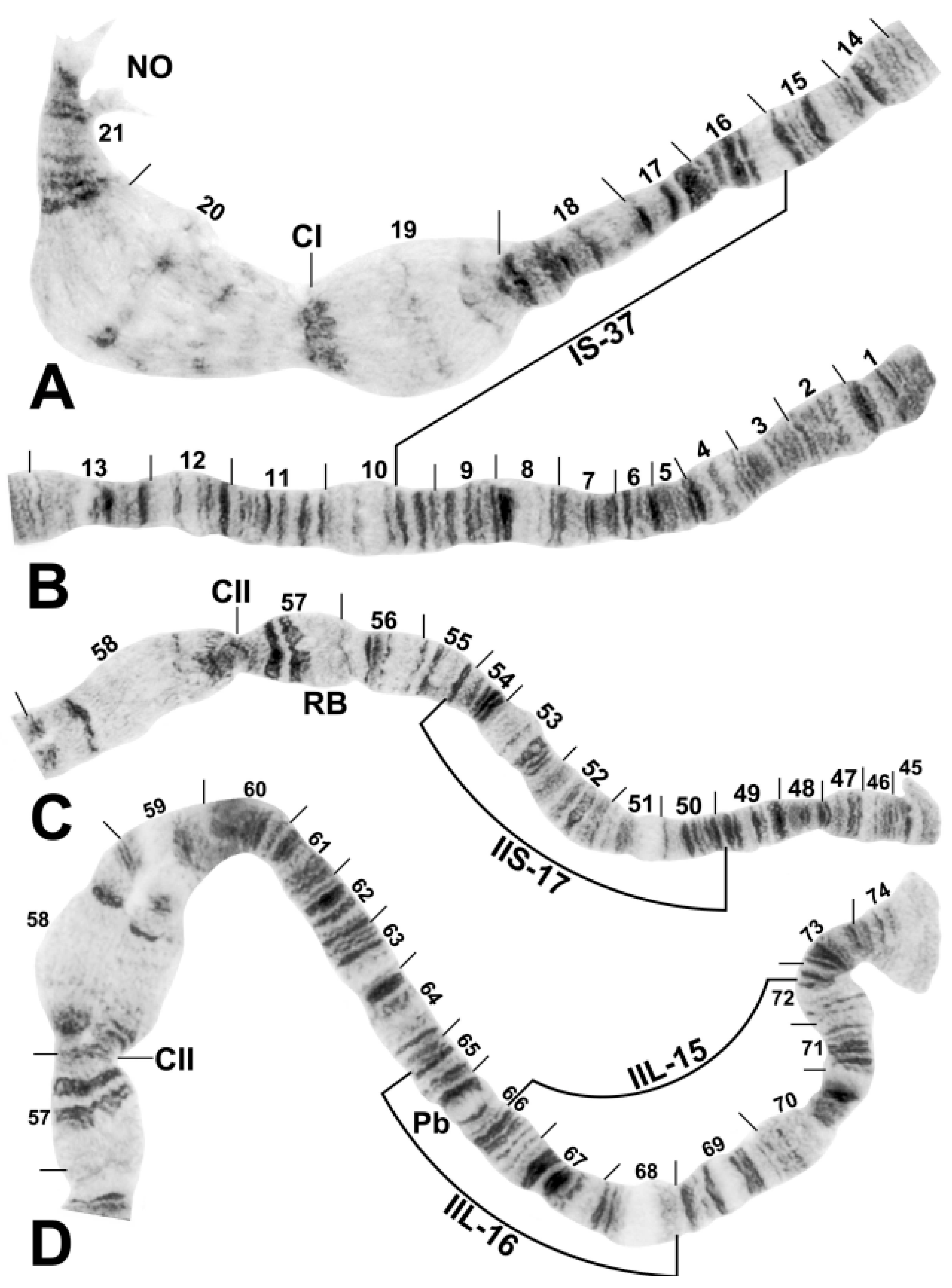
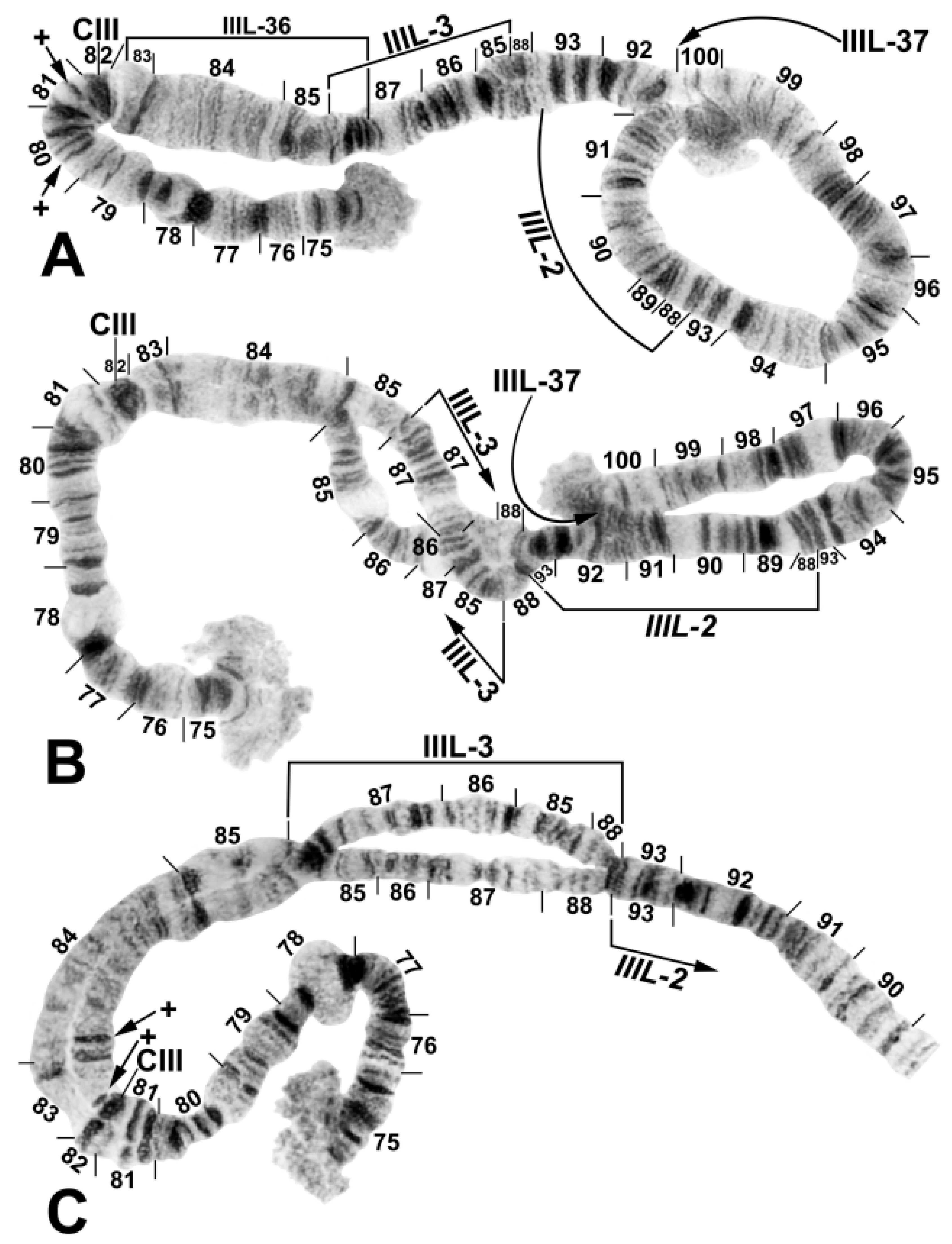
3.1. Chromosomal Description

Chromosomal sequences of all 18 larvae (10 females, 8 males) prepared for analysis were read completely. The chromosomal complement (haploid number = 3) had standard arm associations and the homologues were tightly paired. The nucleolar organizer was in the standard position (chromosome section 22) for the P. hirtipes group (Figure 2A). The centromere region of chromosome I was standard (i.e., not transformed) (Figure 2A), and that of chromosome II was not expanded beyond the standard for the genus (Figure 2C,D). A chromocenter was absent.
All chromosome arms had the standard banding sequence for the genus Prosimulium, except IIIL, which was fixed for IIIL-2 (Figure 3). Sex determination was located on the IIIL arm. Fundamentally, the X sequence carried IIIL-3 (Figure 3A). We interpreted the Y sequence as standard for IIIL-3, with differential expression of bands in sections 85 to 87 (Figure 3B,C). We use “differential band expression” as a descriptive term meaning that the bands in one homologue are well-stained and distinct but in the other homologue the same bands are diffuse and weakly stained. Loops or knots were not expressed in the IIIL-3 region of males. Rather, the homologues in sections 85–87 showed consistent repulsion. Additional rearrangements were frequently associated with the sex arm. Half the larvae in the Colorado populations were heterozygous for IIIL-37 (Figure 3A,B). Differential band expression also occurred outside the IIIL-3 region, in chromosome section 83, in Wyoming larvae (Figure 3C).
Six autosomal polymorphisms were found (Table 2): IS-37, IIS-17, IIL-15, IIL-16 (Figure 2), IIIS hb80, and IIIS hb81 (Figure 3A).

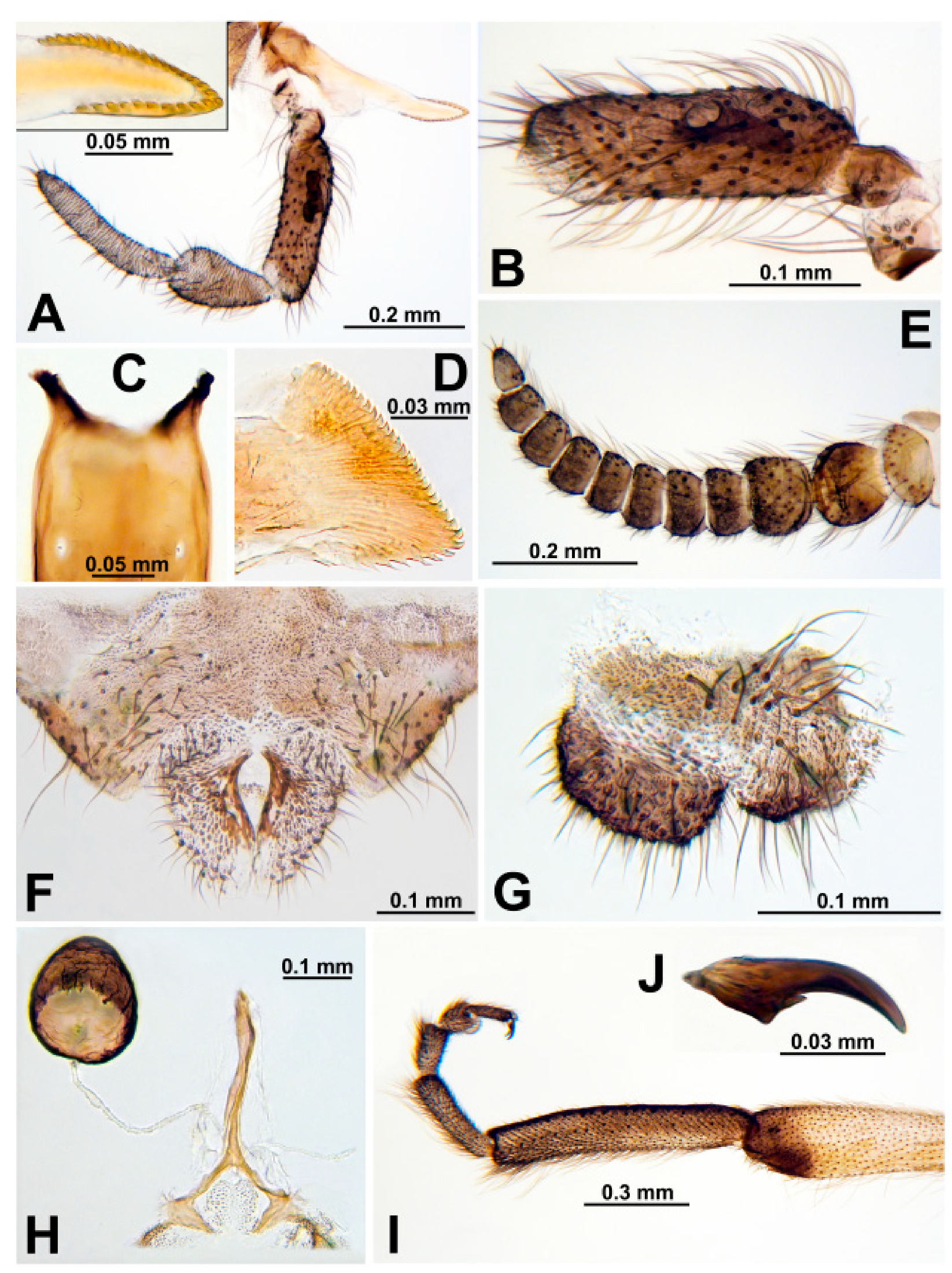
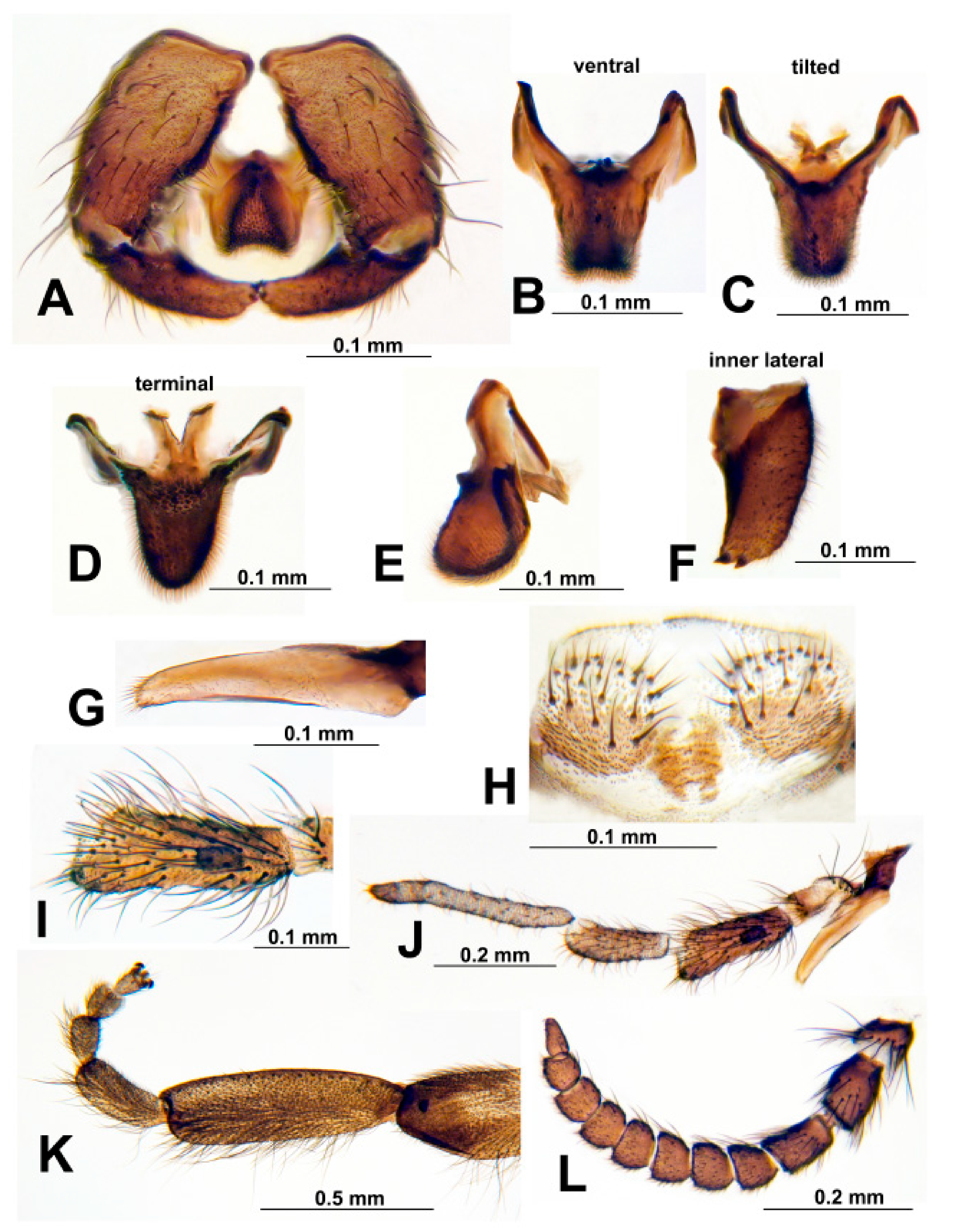
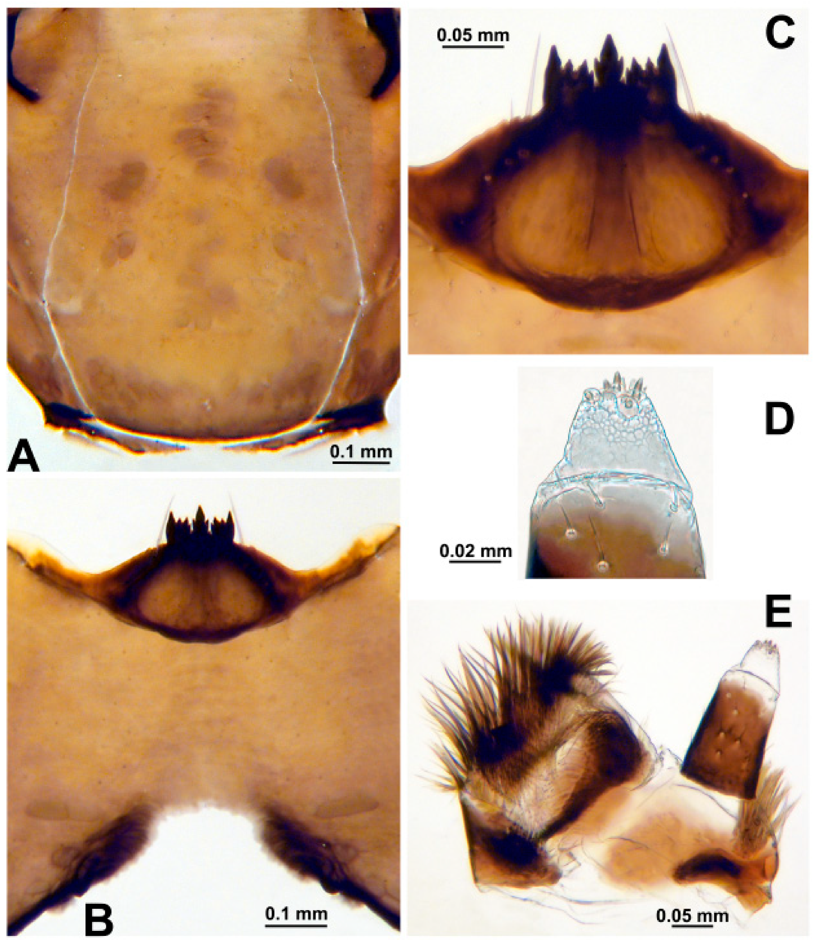
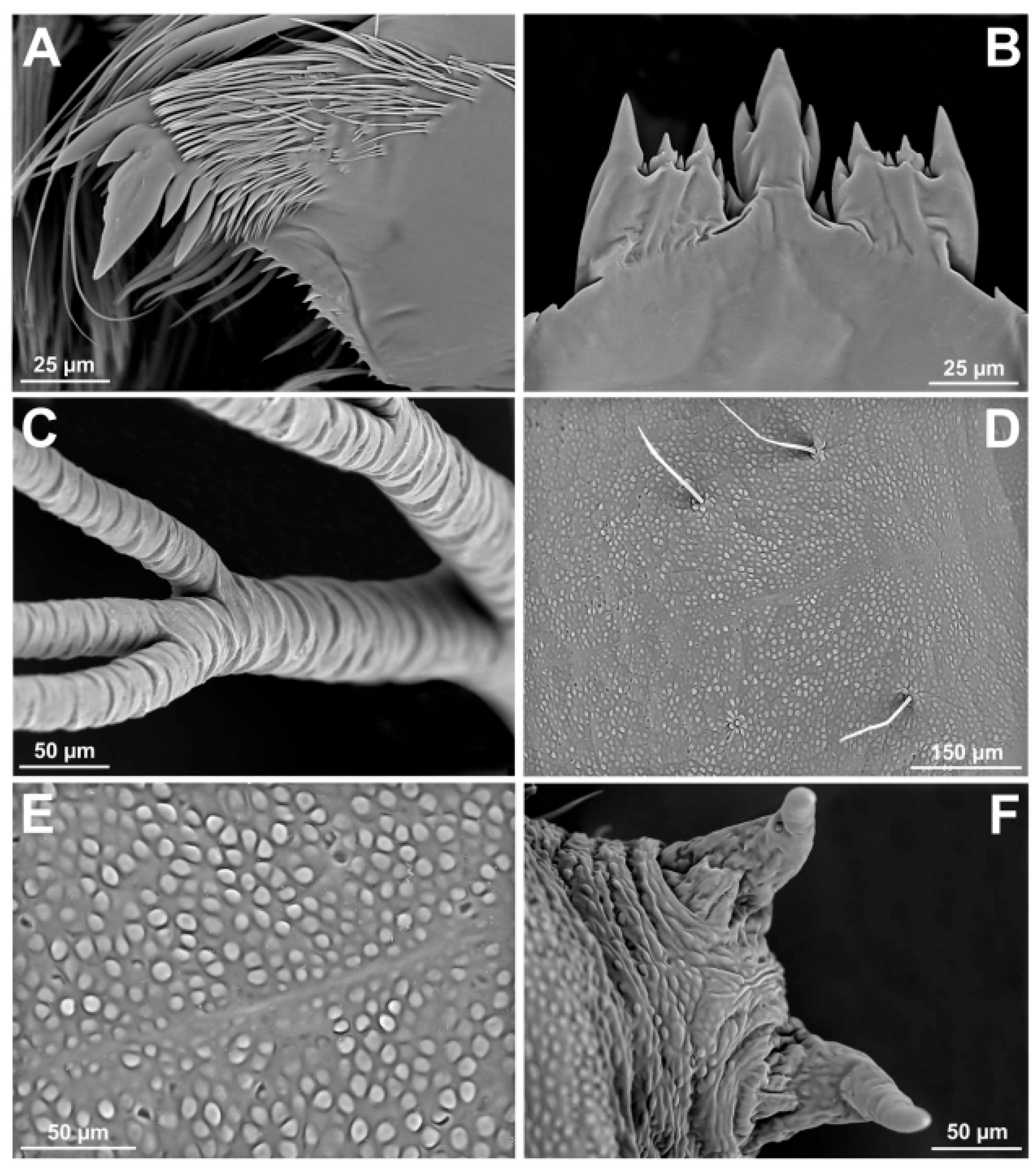
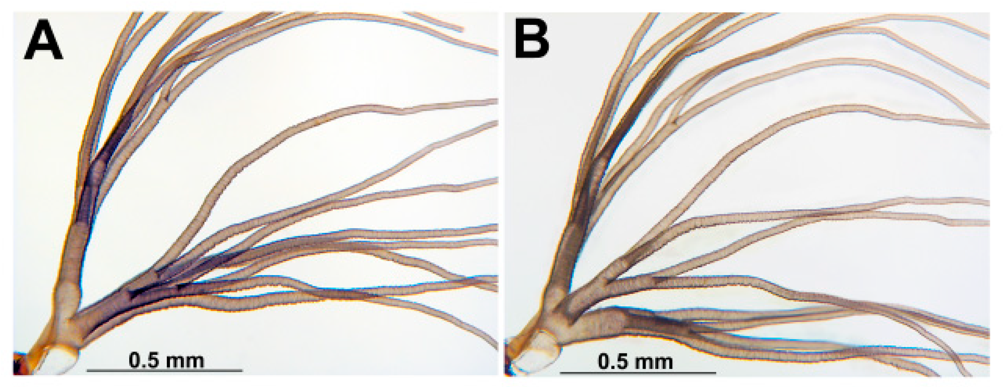
3.2. Morphological Description

Female. Thorax length 1.4–1.7 mm (mean = 1.5 mm, n = 7). Body grayish brown, pollinose, except pronotum, postpronotal lobes, and scutellum pale yellowish brown. All hair yellowish to golden. Head about 0.6 times as wide as thorax. Frons and clypeus well-haired; frons 0.3 times as wide as head. Labrum slightly shorter than clypeus. Antenna (Figure 4E) brownish, paler basally, with scape, pedicel, and 9 flagellomeres; proportional lengths of pedicel, first flagellomere, and second flagellomere 1.7: 1.6: 1.0. Maxillary palp (Figure 4A) brownish, with proportional lengths of third, fourth, and fifth palpomeres 1.5: 1.0: 1.6; sensory vesicle (Figure 4B) elongated, slender, about 0.4 times length of third palpomere, with short neck and wide mouth about 0.25 times length of vesicle. Lacinia (Figure 4A) with 27–29 teeth. Mandible (Figure 4D) with 23–25 inner teeth and 7 or 8 outer teeth. Cibarium (Figure 4C) at junction with pharynx smooth, unarmed, shallow, broadly U-shaped. Pleural membrane, katepisternum, and postnotum bare. Precoxal bridge incomplete. Legs yellowish, except coxae, trochanters, apices of femora, and tibiae pale brownish; tarsi brownish except basal ¾ of posterior margin of hind basitarsus brownish yellow. Hind leg with basitarsus (Figure 4I) nearly parallel-sided, about 5.3 times as long as wide, and 0.7 times as wide as greatest width of hind tibia; calcipala and pedisulcus absent. Claw (Figure 4J) unarmed or with minute basal tooth. Wing 3.6–4.2 mm long (mean = 4.0 mm, n = 6). Costa, subcosta, and radius with fine setae dorsally and ventrally. Halter grayish white. Segment VIII (Figure 4F) with sclerotized sternal plate; other segments lacking sclerotized sternal plate. Hypogynial valves (ovipositor lobes) (Figure 4F) gently curved toward midline, obliquely truncated posteromedially, membranous except inner margin of each valve sclerotized; inner margins concave, creating teardrop-shaped space. Genital fork (Figure 4H) with stem and arms slender, well sclerotized; space between arms mitre-shaped; each arm expanded into slender triangular lateral plate directed posteromedially. Anal lobe in lateral view (Figure 4G) narrow anteriorly, expanded posteroventrally as broadly rounded lobe extended to, or slightly beyond, anterior margin of cercus. Cercus in lateral view (Figure 4G) short, subrectangular, rounded posteriorly, about twice as wide as long. Spermatheca (Figure 4H) broadly tapered apically, about as long as basal width, wrinkled, heavily pigmented except broad basal area completely devoid of pigment; spermathecal duct and both accessory ducts unpigmented.
Male. Thorax length 1.3–1.4 mm (n = 2). Body dark brown to matte black, except scutellum pale brown. All hair pale golden. Head 0.8 times as wide as thorax. Antenna (Figure 5L) brownish, with scape, pedicel, and 9 flagellomeres; proportional lengths of pedicel, first flagellomere, and second flagellomere 1.7: 1.5: 1.0. Maxillary palp (Figure 5J) brownish, with proportional lengths of third, fourth, and fifth palpomeres 1.2:1.0:2.0; sensory vesicle (Figure 5I) small, about 0.2 times as long as third palpomere, with small, round mouth. Lacinia (Figure 5G) with small apical set of hairs. Katepisternum, pleural membrane, and postnotum bare. Legs pale brownish, except coxae, trochanters, and apices of femora and tibiae brown. Hind basitarsus (Figure 5K) 3.2 times as long as its greatest width, 0.75 times as wide as greatest width of hind tibia; calcipala and pedisulcus absent. Wing 3.4–3.5 mm long (n = 2). Costa and radius with fine setae; subcosta with fine setae ventrally. Halter grayish brown. Gonocoxite in ventral view (Figure 5A) about 1.2 times longer than gonostylus. Gonostylus in ventral view (Figure 5A) smoothly curved toward midline, gradually tapered, with 2 apical spinules; in inner lateral view (Figure 5F) about 1.6 times as long as its greatest width. Ventral plate in ventral view (Figure 5A) subquadrate, slightly tapered, with minute setae in broad triangular pattern; anterior margin rather straight (although dorsal wall in one specimen with protuberance of dark cuticle) and posterior margin slightly concave; body of plate slenderer and more tapered in progressively tilted views (Figure 5B,C); arms somewhat divergent from each other and forming broad U-shape; in lateral view (Figure 5E) convex posteriorly; in terminal view (Figure 5D) subtriangular, strongly rounded distally. Median sclerite (Figure 5D) short, bifurcated apically. Paramere subquadrate, with slender anterior projection, without spines or setae. Dorsal plate absent. Aedeagal membrane with fine setae. Abdominal tergite X (Figure 5H) minute, subrectangular, with or without anterior and posterior incisions. Cercus (Figure 5H) small, rounded, with 23 or 24 setae.
Pupa. Length (excluding gills) (n = 7) 4.2–5.1 mm, mean = 4.6 mm. Cephalic plate with dense covering of minute, rounded microgranules and 1 pair of unbranched facial trichomes. Thorax (Figure 6D,E) superficially wrinkled (most prominent laterally), densely covered with minute, rounded microgranules; 3 or 4 unbranched dorsal trichomes per side. Gill (Figure 7A,B) about 0.5–0.6 times as long as pupa, with 16 (rarely 15) slender, grayish filaments in 3 groups arising from short basal stalk about as long as wide; stalks of all 3 groups about as long as to 3 times longer than wide; branching pattern: dorsal group with 8 (rarely 7) filaments arranged as [2 + (1 + 2)] + (1 + 2) or [1 + (1 + 2)] + (1 + 2), lateral and ventral groups each with 2 petiolate pairs of filaments; in lateral view, dorsal group often separated from lateral and ventral groups (Figure 7A); filaments furrowed (Figure 6C). Abdomen densely covered with minute, rounded microgranules, dorsally with postscutellar bridge bearing 4 small unbranched setae per side; segment I with 3 or 4 small unbranched setae per side; segment II with 5–7 small unbranched setae per side; segments III and IV each with 4 recurved hooks and 2 or 3 small unbranched setae per side; segments V–IX each with spine comb and 1–5 small unbranched setae per side; segment IX (Figure 6F) with pair of long terminal spines. Abdomen laterally with pleurites each bearing 1 or 2 small unbranched setae per side; striate membrane with 0–3 small unbranched setae per side; segments V and VI each with 1 short, stout seta in tiny sclerite per side. Abdomen ventrally with segment IV bearing pair of slender hooks on each side; segments V–VII each with pair of stout, bifid or trifid hooks per side. Cocoon sac-like, without definitive structure, densely woven, typically covering pupa and part of gill.
Larva. Length (n = 4) 6.0–7.6 mm, mean = 6.6 mm. Body (in Carnoy’s fixative) grayish brown to pale brownish. Head capsule (Figure 8A) yellowish brown to chestnut brown, palest anteriorly; head spots pale brown, often obscure or faint; anteromedial and sometimes first anterolateral spots typically most conspicuous. Venter of head capsule (Figure 8B) brown, with horizontal long spot and round spot on each side of postgenal cleft brownish. Antenna about as long as, or slightly shorter than, labral fan stalk, with basal and medial articles hyaline and distal article dark brown; proportional lengths of proximal, medial, and distal articles 1.0: 1.7: 1.2. Labral fan (n = 15) with 17–21 (mean = 19.5) primary rays (25–28 for California larvae). Mandible (Figure 6A) with 5 apical teeth, numerous spinous teeth, and 14 or 15 marginal teeth. Maxillary palp about 2 times as long as basal width, with fine, colorless setae along its length (Figure 8D,E). Hypostoma (Figure 8C) with median tooth extended anteriorly beyond all other teeth; sublateral teeth posterior to lateral teeth and extended to same level as tines of median tooth (or beyond if teeth are tilted dorsally; Figure 6B); with 3–5 lateral serrations and 3 or 4 sublateral setae per side. Postgenal cleft (Figure 8B) short, with anterior margin truncate or slightly arched, about 0.3 times as long as postgenal bridge (measured from anterior margin of anterior tentorial pits to hypostomal groove). Cervical sclerites (Figure 8A) minute, enclosed within occiput. Gill histoblast of 16 long, thread-like filaments. Lateral plate of thoracic proleg well-sclerotized, slender, L-shaped. Abdominal cuticle with short, colorless, unbranched setae. Rectal papillae of 3 finger-like lobes. Anal sclerite rectangular, with anterior arms about 1.3–1.4 times as long as posterior arms. Posterior circlet with 67–74 rows of 11–13 hooklets per row.

3.3. Diagnosis

The following characters place P. supernum n. sp. in the P. hirtipes group: chromosomes fixed for inversion IIIL-2 (Figure 3); female with laciniae and mandibles toothed and spermatheca about as long as, or longer than, wide; male with gonostylus bearing 2 apical spinules; pupa with 10–16 filaments; and larva with abdomen rather abruptly expanded at segment V.
Chromosomally, P. supernum n. sp. can be distinguished from all other species of the P. hirtipes group in western North America by the centromere region of chromosome I in standard (not transformed) configuration (Figure 2A), X-linkage of IIIL-3 (Figure 3), absence of a chromocenter, and absence of fixed inversions other than IIIL-2; however, females of P. supernum n. sp. and P. esselbaughi both carry IIIL-2 and IIIL-3 and cannot be distinguished from one another.
Morphologically, the female of P. supernum n. sp. cannot be reliably distinguished from the females of most other western species in the P. hirtipes group, such as P. esselbaughi and P. daviesi Peterson and Defoliart. The male of P. supernum n. sp. can be distinguished from those of 5 of the 13 species in the P. hirtipes group with known males (that of P. idemai Adler, Currie and Wood is unknown) by having a dark brown to black (rather than orange) scutum, a ventral plate (Figure 5A) with a weakly concave posterior margin and long basal arms (in ventral view), and yellowish femora. Prosimulium supernum n. sp. is not reliably distinguished from the remaining 7 species (P. daviesi, P. doveri Sommerman, P. esselbaughi, P. fulvithorax Shewell, P. minifulvum, P. rusticum Adler, Currie and Wood, and P. travisi Stone). Prosimulium opleri Peterson and Kondratieff, known from a single specimen (male) collected in Rocky Mountain National Park, Colorado, within 14 km of our nearest collection site for P. supernum n. sp., is currently in synonymy with P. shewelli Peterson and DeFoliart. It differs most prominently by the following characters: first flagellomere distinctly longer than pedicel (rather than about as long as), sensory vesicle slightly less than half as long palpomere III (rather than only about 0.2 times as long as), costal base and stem vein with black (rather than pale golden) setae, and terminalia “unusually small” sensu [17] for the P. hirtipes group (rather than of typical size). The pupa of P. supernum n. sp. with its 16 (rarely 15) filaments can be separated from those of other 16-filamented western species in the P. hirtipes group by the clustering of the lateral and ventral branches apart from the dorsal branch (Figure 7A); however, if this configuration is not expressed (Figure 7B), the pupa becomes inseparable from those of species with the branches rather evenly spaced. The larva of P. supernum n. sp. can be distinguished from all western species of Prosimulium, except P. esselbaughi, P. idemai, and P. minifulvum by the middle hypostomal tooth extended well beyond the lateral teeth (Figure 6B and Figure 8C). The head spots of P. supernum n. sp. are generally paler than those of P. idemai, which has 13 or 14 filaments (rather than 15 or 16) in its gill histoblast.
Overall, P. supernum n. sp. is structurally and chromosomally most like P. esselbaughi. The most consistent and reliable diagnostic structural difference between the two species is the configuration of the pupal gill, best appreciated in lateral view. In P. supernum n. sp., the lateral and ventral branches either run parallel, thus appearing as a single cluster separate from the dorsal branch (Figure 7A), or all three branches appear separate (Figure 7B), whereas in P. esselbaughi, the dorsal and lateral branches typically run parallel and present a cluster separate from the ventral branch [18,19].

3.4. Type Material

Holotype (USNM): Male (pinned) with dissected genitalia (in associated glycerin vial) and pupal exuviae and cocoon (in associated glycerin vial), Wyoming, Albany County, Snowy Range Pass, Libby Creek, 41°21′07′′ N 106°17′01′′ W, 3233 m asl, 7 July 2008, collected by W. K. Reeves. Paratypes (USNM and CNC): Same location and collector as holotype, 11 June 2007, 13 larvae; 12 June 2008, 2 larvae; 7 July 2008, 7 pupae, 1 male and 5 females (pinned with pupal exuviae in glycerin vials); 17 July 2008, 4 pupae, 2 females (pinned with pupal exuviae in glycerin vials). Colorado, Jackson County, near Cameron Pass, snowmelt tributary of Michigan River, 40°30′31′′ N 105°53′05′′ W, 3089 m asl, 11 June 2022, W. K. Reeves, 6 larvae; tributary of Michigan River, 40°30′56′′ N 105°53′11′′ W, 3139 m asl, 20 June 2022, W. K. Reeves, 2 larvae; Cameron Pass, Michigan Ditch, 40°31′13′′ N 105°53′32′′ W, 3135 m asl, 20 June 2022, W. K. Reeves, 2 larvae.

3.5. Additional Specimens Examined

California, Alpine Co., Rt. 4, trickle 1.2 mi. W of Raymond Meadow Creek bridge 24 June 1991, P. H. Adler, 2 larvae; Mariposa County, Rt. 41, Rail Creek, 12 May 1997, P. H. Adler, 7 larvae; Rt. 41, 1 mi. E of Big Meadow Overlook, 14 May 1997, P. H. Adler, 3 larvae; Rt. 41, 2 mi. W of 5000′ elev. marker, 11 May 1997, P. H. Adler, 3 larvae; Rt. 41, 1.7 mi. N of Avalanche Creek, 12 May 1997, P. H. Adler, 5 larvae; Rt. 41, 4 mi. N of tunnel, 14 May 1997, P. H. Adler, 1 larva; Mono Co., Rt. 108, ca. 2 mi. east of border between Mono County and Tuolumne County, 11 June 1990, P. H. Adler, 1 larva.

3.6. Distribution

Prosimulium supernum n. sp. is confirmed from the Rocky Mountains of Colorado and Wyoming. We also ascribe populations from the Sierra Nevada of California to this species, based on chromosomal and morphological diagnostic characters. A sample of three larvae from Nevada (Pine County, Lehman Creek, 25 August 1966) with chromosomes matching P. esselbaughi are possibly those of P. supernum n. sp., but the larvae were females; the two species are chromosomally indistinguishable as females. Our map (Figure 1A) shows only localities for which we had both morphological and chromosomal data and could, therefore, make accurate identifications of the new species and P. esselbaughi sensu stricto. We suspect that all previous literature records of P. esselbaughi sensu lato from Alaska and Canada (i.e., Alberta, British Columbia, and the Yukon) [11,19] pertain to P. esselbaughi in the strict sense, based on the distributions and morphological characters (particularly of the pupal gill) that accompanied the records. Literature records of P. esselbaughi sensu lato from Montana, Oregon, and Washington [11] might apply to P. esselbaughi sensu stricto, P. supernum new species, or both; this area possibly represents the transition of the ranges of the two species.

3.7. Bionomics

Larvae of P. supernum n. sp., like those of the closely related P. esselbaughi [18], are found in fast, cold streams about a meter or more wide. Whereas P. esselbaughi has been found from sea level to above the timberline in the area around Anchorage, Alaska [18], P. supernum n. sp. is a specialist of high elevations, having been found only at elevations above 3000 m in the Rocky Mountains and above 1500 m in the Sierra Nevada. The larvae of P. supernum n. sp. have been collected in association with Helodon susanae (Peterson), P. daviesi, Simulium carbunculum Adler, Currie and Wood, and the S. arcticum complex. Like all species of Prosimulium, the new species is univoltine. One larva from Mariposa County, California, was infected with an unidentified mermithid nematode.
Topotypical females emerged with their abdomen replete with fat body, suggesting autogeny, at least in the first gonotrophic cycle, as discovered [18] for P. esselbaughi. The females are inferred to feed on mammals, based on their fully functional biting mouthparts and claws adapted for mammal feeding. The inference is bolstered by a few records available for P. esselbaughi, indicating that large mammals (e.g., sheep and humans) are hosts [18,20].

3.8. Etymology

The species name supernum is from Latin, meaning celestial, high, or lofty, in reference to the high-elevation habitat of the species.

4. Discussion

East–west differentiation of North American taxa is a common, long-recognized pattern; the distinction is usually defined by the Rocky Mountains. In the simuliid genus Prosimulium, only one of the 38 nominal species, the trans-arctic P. ursinum (Edwards), is shared between eastern and western North America [11]. In contradistinction, most nominal species in the group are continuously distributed along a north–south axis [11]. Yet, given the history of glaciation, availability of refugia, dissected topography, and consequent opportunities for isolation, north–south differentiation should be a prominent feature of organisms in western North America. Accordingly, phylogeographic analyses have revealed north–south differentiation in taxa as evolutionarily diverse as birds [21,22], mammals [23], and plants [24].
During the Last Glacial Maximum, the North American Ice Sheet separated the western half of the continent into two major refugia, Beringia in the north and most of the continent to the south of present-day Canada [25], with smaller hypothesized refugia along the Pacific Northwest coast [26]. The biological implication of this vast ice sheet is that populations to its north and south should express disparities in life history, structure, and genetics. This trend is apparent within all nominal species of western Prosimulium that are distributed from Alaska southward into the Rockies and Sierra Nevada [11], each of which consists of genetically different northern (e.g., Alaskan) and southern (e.g., central Rocky Mountain) populations. Prosimulium frohnei Sommerman and P. fulvum (Coquillett) lack a chromocenter in southern populations but both are chromocentric in Alaska; P. fulvum also expresses differential sex-chromosome linkage in northern versus southern populations [11,27]. Alaskan and Yukon populations of P. neomacropyga Peterson and P. travisi show substantial cytogenetic and molecular divergence from Colorado populations, suggesting cryptic species [11,12,15,28]. Northern and southern populations, formerly treated as cytoforms of P. doveri [27], were recently recognized as distinct species: the northern P. doveri and southern P. daviesi [11]. The taxonomic implication is that new species will need to be described, and in other cases some names currently held in synonymy will need to be revalidated. Further insights into these north–south relationships could be gained from molecular analyses, such as the DNA barcoding that was conducted for P. travisi and P. neomacropyga [12].
Chromosomally and structurally, P. supernum n. sp. most closely resembles the more northern P. esselbaughi first described from Alaska. It was originally identified as P. esselbaughi [11] and is here considered a separate species, differing largely in the pupal gill configuration and chromosomal rearrangements linked to sex. In addition, the floating rearrangement profiles are unique; none of the 17 total floating rearrangements are shared between the two species. Prosimulium esselbaughi and P. supernum n. sp., thus, represent the sixth example of north–south differentiation of Prosimulium in the Western Cordillera and the second case among western Prosimulium species in which northern and southern populations are accorded separate species status.
Other simuliid taxa of the Western Cordillera also express north–south disparity. The sister-species Metacnephia sommermanae (Stone) and M. coloradensis Peterson and Kondratieff, for example, differ cytogenetically and morphologically [11,29]. Why the trend has not been more frequently observed among the Simuliidae might reflect limited north–south genetic investigations and the subtle nature of morphological differences often attributed to intraspecific variation.
Overlain on the large-scale pattern of differentiated populations to the north and south of the North American Ice Sheet is the possibility of further differentiation in northern and southern populations, reflecting isolation, reduced population sizes, and local adaptation in historical microrefugia [30]. Prosimulium supernum n. sp. from the Sierra Nevada of California, for instance, might be expected to show different chromosomal rearrangement profiles, although a detailed investigation was not possible for the material now stored in ethanol. Genetically unique populations of other taxa, such as birds and mammals, have been found in the Sierra Nevada [21,31]. Post-glacial factors also might be at work in determining current population genetics at high elevations, such as limited dispersal and local adaptation. Even between the Colorado and Wyoming populations of P. supernum n. sp., which are separated by 100 km, chromosomal differences in autosomal polymorphism profiles are stark—only one of nine floating rearrangements is shared.
Post-glacial range expansions of populations north and south of the ice sheet would be expected to bring the differentiated populations into closer proximity, eventually including a zone of overlap in some cases. The species pair, P. doveri Sommerman to the north and P. daviesi to the south, overlap in the Coast Range of northwestern Washington [11]. A zone of overlap has not been found for P. supernum n. sp. and P. esselbaughi, although genetic sampling has been limited. Prosimulium esselbaughi, however, extends in a cytogenetically homogenous north–south band more than 2100 km, from at least the Anchorage area of southern Alaska to Vancouver in southern British Columbia [15] (as P. hirtipes “2 (Alaska)”). The Alaskan population might represent Beringian survival, whereas the British Columbian population is perhaps the result of post-glacial movements southward from the Beringian refugium. The Alaskan and British Columbian populations not only share both fixed inversions (IIIL-2 and IIIL-3), but also the sex inversion and three of the five other floating inversions [15].
The influence of glaciation and subsequent divergence on host use, parasites, and potential vector relationships of P. supernum n. sp. and P. esselbaughi is unknown. The mammalian hosts of each species and any associated pathogens and parasites transmitted during blood-feeding also would have been subjected to the influences of glaciation. Perhaps, too, the non-vector-transmitted symbiotes (e.g., mermithid nematodes) also carry a signature of Pleistocene isolation. Contemporary populations to the north and south of the great North American Ice Sheet provide ideal subjects for examining potential co-differentiation of black flies and their parasites.

Author Contributions

Conceptualization, P.H.A.; Methodology, P.H.A. and W.K.R.; Validation, P.H.A.; Formal Analysis, P.H.A.; Investigation, P.H.A.; Resources, P.H.A. and W.K.R.; Data Curation, P.H.A.; Writing—Original Draft Preparation, P.H.A.; Review, P.H.A. and W.K.R.; Visualization, P.H.A.; Supervision, P.H.A. and W.K.R.; Project Administration, P.H.A.; Funding Acquisition, P.H.A. and W.K.R. All authors have read and agreed to the published version of the manuscript.

Funding

The research by P.H.A. was supported by NIFA/USDA under project number SC-1700596 and is Technical Contribution No. 7112 of the Clemson University Experiment Station. W.K.R was supported by USDA-ARS project 5410-32000-016-00D.

Institutional Review Board Statement

Not applicable.

Informed Consent Statement

Not applicable.

Data Availability Statement

All data supporting reported results are included in the text.

Acknowledgments

We thank A. G. Wheeler for helpful comments on the manuscript and K. Taylor, L. DeBrey, A. Fabian, and B. Yarnell for logistical assistance in collecting, with additional project support by D. A. Strickman.

Conflicts of Interest

The authors declare no conflict of interest. The funders had no role in the design of the study; in the collection, analyses, or interpretation of data; in the writing of the manuscript; or in the decision to publish the results.

References

  1. Schweikert, R.A.; Bogan, N.L.; Girty, G.H.; Hanson, R.E.; Merguerian, C. Timing and structural expression of the Nevadan Orogeny, Sierra Nevada, California. Geol. Soc. Am. Bull. 1984, 95, 967–979. [Google Scholar] [CrossRef]
  2. McMillan, M.E.; Heller, P.L.; Wing, S.L. History and causes of post-Laramide relief in the Rocky Mountain orogenic plateau. Geol. Soc. Am. Bull. 2006, 118, 393–405. [Google Scholar] [CrossRef]
  3. Clark, P.U.; Dyke, A.S.; Shakun, J.D.; Carlson, A.E.; Clark, J.; Wohlfarth, B.; Mitrovica, J.X.; Hostetler, S.W.; McCabe, A.M. The Last Glacial Maximum. Science 2009, 325, 710–714. [Google Scholar] [CrossRef]
  4. Hewitt, G. The genetic legacy of the Quaternary ice ages. Nature 2000, 405, 907–913. [Google Scholar] [CrossRef]
  5. Brunsfeld, S.J.; Sullivan, J.; Soltis, D.E.; Soltis, P.S. A comparative phylogeography of northwestern North America: A synthesis. In Integrating Ecology and Evolution in a Spatial Context; Silvertown, J., Antonovics, J., Eds.; Blackwell Science: Oxford, UK, 2001; pp. 319–339. [Google Scholar]
  6. Ehlers, J.; Gibbard, P. Quaternary glaciation. In Encyclopedia of Snow, Ice and Glaciers; Singh, V.P., Singh, P., Haritashya, U.K., Eds.; Springer: New York, NY, USA, 2011; pp. 873–882. [Google Scholar] [CrossRef]
  7. Adler, P.H.; Cheke, R.A.; Post, R.J. Evolution, epidemiology, and population genetics of black flies (Diptera: Simuliidae). Infect. Genet. Evol. 2010, 10, 846–865. [Google Scholar] [CrossRef] [PubMed]
  8. Adler, P.H. World blackflies (Diptera: Simuliidae): A comprehensive revision of the taxonomic and geographical inventory [2022]. 2022, p. 145. Available online: http://biomia.sites.clemson.edu/pdfs/blackflyinventory.pdf (accessed on 11 November 2022).
  9. Weinmann, C.J.; Anderson, J.R.; Longhurst, W.M.; Connolly, G. Filarial worms of Columbian black-tailed deer in California. 1. Observations in the vertebrate host. J. Wildl. Dis. 1973, 9, 213–220. [Google Scholar] [CrossRef] [PubMed]
  10. Schulz-Key, H.; Wenk, P. The transmission of Onchocerca tarsicola (Filarioidea: Onchocercidae) by Odagmia ornata and Prosimulium nigripes (Diptera: Simuliidae). J. Helminthol. 1981, 55, 161–166. [Google Scholar] [CrossRef] [PubMed]
  11. Adler, P.H.; Currie, D.C.; Wood, D.M. The Black Flies (Simuliidae) of North America; Cornell University Press: Ithaca, NY, USA, 2004. [Google Scholar]
  12. Rivera, J.; Currie, D.C. Identification of Nearctic black flies using DNA barcodes (Diptera: Simuliidae). Mol. Ecol. Resour. 2009, s1, 224–236. [Google Scholar] [CrossRef] [PubMed]
  13. Adler, P.H.; Kúdelová, T.; Kúdela, M.; Seitz, G.; Ignjatović-Ćupina, A. Cryptic biodiversity and the origins of pest status revealed in the macrogenome of Simulium colombaschense (Diptera: Simuliidae), history’s most destructive black fly. PLoS ONE 2016, 11, e0147673. [Google Scholar] [CrossRef]
  14. Basrur, P.K. The salivary gland chromosomes of seven segregates of Prosimulium (Diptera: Simuliidae) with a transformed centromere. Can. J. Zool. 1959, 37, 527–570. [Google Scholar] [CrossRef]
  15. Basrur, P.K. The salivary gland chromosomes of seven species of Prosimulium (Diptera: Simuliidae) from Alaska and British Columbia. Can. J. Zool. 1962, 40, 1019–1033. [Google Scholar] [CrossRef]
  16. Chance, M.M. The functional morphology of the mouthparts of blackfly larvae (Diptera: Simuliidae). Quaest. Entomol. 1970, 6, 254–284. [Google Scholar]
  17. Peterson, B.V.; Kondratieff, B.C. The black flies (Diptera: Simuliidae) of Colorado: An annotated list with keys, illustrations and descriptions of three new species. Mem. Am. Entomol. Soc. 1995, 42, 1–121. [Google Scholar]
  18. Sommerman, K.M. Prosimulium esselbaughi n. sp., the Alaskan P. hirtipes 2 (Diptera: Simuliidae). Proc. Entomol. Soc. Wash. 1964, 66, 141–145. [Google Scholar]
  19. Peterson, B.V. The Prosimulium of Canada and Alaska. Mem. Entomol. Soc. Can. 1970, 69, 1–216. [Google Scholar]
  20. Mason, P.G.; Shemanchuk, J.A. Black flies. Agric. Can. Publ. 1990, 1499/E, 1–19. [Google Scholar]
  21. Hull, J.M.; Keane, J.J.; Savage, W.K.; Godwin, S.A.; Shafer, J.A.; Jepsen, E.P.; Gerhardt, R.; Stermer, C.; Ernest, H.B. Range-wide genetic differentiation among North American great gray owls (Strix nebulosa) reveals a distinct lineage restricted to the Sierra Nevada, California. Mol. Phylogen. Evol. 2010, 56, 212–221. [Google Scholar] [CrossRef]
  22. Bayard de Volo, S.; Reynolds, R.T.; Sonsthagen, S.A.; Talbot, S.L.; Antolin, M.F. Phylogeography, postglacial gene flow, and population history of North American Northern Goshawks (Accipiter gentilis). Auk 2013, 130, 342–354. [Google Scholar] [CrossRef]
  23. Krejsa, D.M.; Talbot, S.L.; Sage, G.K.; Sonsthagen, S.A.; Jung, T.S.; Magoun, A.J.; Cook, J.A. Dynamic landscapes in northwestern North America structured populations of wolverines (Gulo gulo). J. Mammal. 2021, 102, 891–908. [Google Scholar] [CrossRef]
  24. Nadeau, S.; Godbout, J.; Lamothe, M.; Gros-Louis, M.-C.; Isabel, N.; Ritland, K. Contrasting patterns of genetic diversity across the ranges of Pinus monticola and P. strobus: A comparison between eastern and western North American postglacial colonization histories. Am. J. Bot. 2015, 102, 1342–1355. [Google Scholar] [CrossRef]
  25. Dulfer, H.E.; Margold, M.; Darvill, C.M.; Stroeven, A.P. Reconstructing the advance and retreat dynamics of the central sector of the last Cordilleran Ice Sheet. Quatern. Sci. Rev. 2022, 284, 107465. [Google Scholar] [CrossRef]
  26. Colella, J.P.; Lan, T.; Talbot, S.L.; Lindqvist, C.; Cook, J.A. Whole-genome resequencing reveals persistence of forest-associated mammals in Late Pleistocene refugia along North America’s North Pacific Coast. J. Biogeogr. 2021, 48, 1153–1169. [Google Scholar] [CrossRef]
  27. Rothfels, K.H. Cytotaxonomy of black flies (Simuliidae). Annu. Rev. Entomol. 1979, 24, 507–539. [Google Scholar] [CrossRef]
  28. Madahar, D.P. Cytogenetic study in triploidy in Prosimulium ursinum (Simuliidae: Diptera). Genetics 1973, 74 (Suppl. S2), 170–171. [Google Scholar]
  29. Finn, D.; Adler, P.H. Population genetic structure of a rare high-elevation black fly, Metacnephia coloradensis, occupying Colorado lake outlet streams. Freshwat. Biol. 2006, 51, 2240–2251. [Google Scholar] [CrossRef]
  30. Mee, J.A.; Moore, J.-S. The ecological and evolutionary implications of microrefugia. J. Biogeogr. 2014, 41, 837–841. [Google Scholar] [CrossRef]
  31. Latch, E.K.; Heffelfinger, J.R.; Fike, J.A.; Rhodes, O.E., Jr. Species-wide phylogeography of North American mule deer (Odocoileus hemionus): Cryptic glacial refugia and postglacial recolonization. Mol. Ecol. 2009, 18, 1730–1745. [Google Scholar] [CrossRef] [PubMed]
Figure 1. Collecting sites for Prosimulium supernum n. sp. (A) Map of North America. Solid black circles represent P. supernum n. sp. and open circles represent chromosomally confirmed records of its near relative P. esselbaughi; map by Daniel Feher, https://www.freeworldmaps.net/about.html (accessed on 20 December 2022). (B) Type locality of P. supernum n. sp., Libby Creek, Wyoming, 12 June 2008. (C) Cameron Pass, snow melt tributary of Michigan River, Colorado, 11 June 2022 (site 2, Table 1). (D) Cameron Pass, Michigan Ditch, Colorado, 20 June 2022 (site 4, Table 1).
Diversity 15 00212 g001
Figure 2. Chromosomes of Prosimulium supernum n. sp. from Colorado (male larvae), showing standard sequence. (A). IS basal half, including standard (non-transformed) centromere region; bracket indicates breakpoints of autosomal inversion IS-37; CI, centromere of chromosome I; NO, nucleolar organizer. (B). IS distal half. (C). IIS; bracket indicates breakpoints of autosomal inversion IIS-17; CII, centromere of chromosome II; RB, ring of Balbiani. (D). IIL; brackets indicate breakpoints of autosomal inversions IIL-15 and IIL-16; Pb, parabalbiani.
Diversity 15 00212 g002
Figure 3. Chromosome III of Prosimulium supernum n. sp.; CIII, centromere. (A). Sequence of female larva from Colorado, showing the presence of homozygous IIIL-2 and IIIL-3 and heterozygous IIIL-37 (arrow indicates breakpoints). Limits of IIIL-36 (not present) indicated by brackets; locations of heterobands IIIS hb80 and IIIS hb81 (neither present) indicated by a plus sign (+). (B). Sequence of male larva from Colorado, showing the presence of homozygous IIIL-2 and heterozygous IIIL-3 and IIIL-37 (arrow indicates breakpoints). (C). Sequence of male larva from Wyoming, showing the presence of homozygous IIIL-2, heterozygous IIIL-3, and heterobands (+) IIIS hb83 and IIIS hb84.
Diversity 15 00212 g003
Figure 4. Female of Prosimulium supernum n. sp. (A) Maxillary palp with 5 palpomeres and lacinia; inset shows apex of lacinia. (B) Maxillary palpomeres I, II, and III, showing sensory vesicle. (C) Cibarium, anterior. (D) Mandible, apex. (E) Antenna. (F) Sternite VIII and hypogynial valves. (G) Anal lobe and cercus, lateral view. (H) Genital fork and spermatheca. (I) Hind tibia (apical portion only), tarsus, and acropod with claws. (J) Hind claw.
Diversity 15 00212 g004
Figure 5. Male of Prosimulium supernum n. sp. (A) Genitalia, ventral view. (B) Ventral plate, ventral view (slightly tilted). (C) Ventral plate, ventral view (moderately tilted to show median sclerite). (D) Ventral plate and median sclerite, terminal view. (E) Ventral plate, lateral view. (F) Gonostylus, inner lateral view. (G) Lacinia. (H) Tergite X and cerci. (I) Maxillary palpomere III, showing sensory vesicle. (J) Maxillary palp with 5 palpomeres and lacinia. (K) Hind tibia (apex only), tarsus, and acropod with claws. (L) Antenna.
Diversity 15 00212 g005
Figure 6. Scanning electron micrographs of Prosimulium supernum n. sp. (A) Larval mandible, apex, aboral surface. (B) Hypostoma, ventral view (teeth tilted dorsally). (C) Pupal gill filaments, showing surface sculpture. (D) Pupal thoracic sculpture and trichomes, dorsal view; the ecdysial line is faintly visible, running obliquely from right to left. (E) Pupal thoracic sculpture, dorsal view along the ecdysial line. (F) Pupal segment IX, showing terminal spines, dorsal view.
Diversity 15 00212 g006
Figure 7. Pupal gill of Prosimulium supernum n. sp. (A,B) Lateral views with slight differences in orientation; apices of filaments not shown.
Diversity 15 00212 g007
Figure 8. Larva of Prosimulium supernum n. sp. (A) Frontoclypeal apotome. (B) Head capsule, ventral view. (C) Hypostoma, ventral view. (D) Maxillary palp, apex. (E) Maxilla with maxillary palp, lateral view.
Diversity 15 00212 g008
Table 1. Sites from which type material was collected for Prosimulium supernum n. sp. in the Rocky Mountains of Colorado and Wyoming.
SiteLocation (Stream Width)Latitude LongitudeElevation (m above Sea Level)DateLife Stage
1aWY, Albany County, Snowy Range Pass, Libby Creek (2.0–2.5 m)41°21′07″ N 106°17′01″ W323311 June 200713 larvae (4 chromosome preparations)
1b 12 June 20082 larvae (2 chromosome preparations)
1c 7 July 20087 pupae, 2 males and 5 females with pupal exuviae
1d 17 July 20084 pupae, 2 females with pupal exuviae
2CO, Jackson County, near Cameron Pass, snowmelt trib. Michigan River (0.5–1.0 m)40°30′31″ N 105°53′05″ W308911 June 20226 larvae (6 chromosome preparations)
3CO, Jackson County, trib. Michigan River (0.5–1.0 m)40°30′56″ N 105°53′11″ W313920 June 20222 larvae (2 chromosome preparations)
4CO, Jackson County, Cameron Pass, Michigan Ditch (2.0–2.5 m)40°31′13″ N 105°53′32″ W313520 June 20222 larvae (2 chromosome preparations)
Table 2. Frequency of homologues with chromosomal rearrangements in larvae of Prosimulium supernum n. sp. from the Rocky Mountains of Colorado and Wyoming.
Site
1a 11b 1234
Female:Male3:30:24:22:01:1
IS-3720.08
IIS-170.250.25
IIL-150.42
IIL-160.25
IIIS hb800.25
IIIS hb811.001.000.08
IIIL-231.001.001.001.001.00
IIIL-3 4*****
IIIL-360.170.25
IIIL-370.170.500.25
1 Site 1a/1b corresponds to the type locality. 2 A dash (–) indicates a frequency of 0.00. 3 Italics indicate the inversion was fixed. 4 An asterisk (*) indicates that IIIL-3 was linked to the X chromosome; the Y chromosome was standard for IIIL-3.
Disclaimer/Publisher’s Note: The statements, opinions and data contained in all publications are solely those of the individual author(s) and contributor(s) and not of MDPI and/or the editor(s). MDPI and/or the editor(s) disclaim responsibility for any injury to people or property resulting from any ideas, methods, instructions or products referred to in the content.

