Article

A Review of the Asexual Mite Genus Paralycus Womersley, 1944 (Acari: Oribatida: Pediculochelidae), with Description of Three New Species and A Key to Species of the World †

by
Vasiliy B. Kolesnikov
1,2,
Barry OConnor
3,
Sergey G. Ermilov
2 and
Pavel B. Klimov
2,4,*
1
Federal Public Budgetary Scientific Institution All-Russian Research Institute of Plant Protection, 396030 Voronezh, Russia
2
Institute of Environmental and Agricultural Biology (X-BIO), Tyumen State University, 625003 Tyumen, Russia
3
Department of Ecology & Evolutionary Biology, University of Michigan, 3600 Varsity Drive, Ann Arbor, MI 48109, USA
4
Department of Biological Sciences, Purdue University, Lilly Hall of Life Sciences, West Lafayette, IN 47907, USA
*
Author to whom correspondence should be addressed.
urn:lsid:zoobank.org:pub:DEC3BA3E-F98B-4321-90AD-5F8CFA809003.
Diversity 2023, 15(2), 160; https://doi.org/10.3390/d15020160
Submission received: 24 December 2022 / Revised: 19 January 2023 / Accepted: 19 January 2023 / Published: 22 January 2023
(This article belongs to the Special Issue Diversity, Biogeographic, and Evolutionary Research in Acarology)

Abstract

:
Mites of the genus Paralycus (Oribatida: Pediculochelidae) are minute, asexual, paedomorphic oribatids that have been largely overlooked by major biodiversity surveys. Here, we review this genus and describe three new species based on adult females: Paralycus persephone sp. n. and Paralycus daeira sp. n. from deep soil in Colorado, USA and Paralycus pricei sp. n. from South Africa. We provide the first complete ontogenetic series in the family using P. daeira sp. n. as a model, clarify the species boundaries in several species, and compile an annotated checklist and key to species of Paralycus of the World. Our work suggests that the pediculochelid biodiversity is underappreciated as these mites may be common in subterranean habitats/deep soil. Further discoveries of the Paralycus diversity in these habitats are anticipated.

Graphical Abstract

1. Introduction

The oribatid mite family Pediculochelidae Lavoipierre, 1946 (Acari: Oribatida), comprises 11 species belonging to a single genus, Paralycus Womersley, 1944 (our data). The genus is worldwide in distribution in temperate and tropical regions; however, its records are rare [1,2], and it is likely that most soil meiofauna surveys have overlooked these soil mites due to their small sizes and weak coloration. All known species lack males, possibly indicating a long-term maintenance of asexual reproduction [3]. Pediculochelids are found in the soil [2,3,4,5,6,7,8], on bees [4,9,10], and rarely in other habitats [4,11,12,13,14,15].
The family name Pediculochelidae was proposed by Lavoipierre (1946) for Pediculochelus raulti Lavoipierre, 1946 [9]. However, a species belonging to Pediculochelidae had been described earlier as Alicus pyrigerus Berlese, 1905 in family Alycidae [5]. For this species, Womersley (1944) [16] proposed a new genus, Paralycus (based on the differences in the body shape and bothridial setae), thus making Pediculochelus a junior synonym [3,4]; however, as per ICZN rules, this did not affect the validity of the family name Pediculochelidae.
Pediculochelids have a paedomorphic morphology, whereby adults retain juvenile traits, thus making it challenging to compare their traits with those of other mites. The position of the family on the mite tree of life or among major mite lineages has been a subject of debates. When first proposed, Lavoipierre (1946) [9] noted some resemblance to the family Tarsonemidae, a highly derived lineage in Prostigmata. Other researchers placed this family either in Astigmata [11] or Endeostigmata [17]. However, Norton et al. (1983) [3], based on a cladistic analysis, convincingly showed that Pediculochelidae is a member of the oribatid superfamily Protoplophoroidea in Enarthronota, and this placement has been later confirmed by multigene molecular analyses [18,19].
Although taxonomic treatments of pediculochelids are available, the species diversity, species boundaries, and ontogeny are still poorly understood. For example, a recent key to species [13] contains inaccuracies and omits an important but insufficiently described species, Paralycus pyrigerus. In addition, the taxonomic scope of Paralycus raulti is uncertain as it included specimens with remarkably variable morphologies [4]. Furthermore, we have very limited knowledge of the pediculochelid ontogeny and life cycle [4,14], which can provide important phylogenetically informative characters [20].
Here, we address aspects related to the biodiversity, species boundaries, and ontogenetic development of Paralycus. We report two new species from deep soil in Colorado, USA collected by a flotation technique [21], one new species from South Africa, which was formerly confused with P. raulti, and re-described the true P. raulti. We also describe all ontogenetic instars for P. daeira, and we provide an annotated check list and identification key of Paralycus species of the world.

2. Materials and Methods

Mites were collected from deep soil (0.9 m) using a soil washing and flotation method [21], but heptane was replaced with kerosene. Mites were sorted under a dissection microscope and individually slide-mounted in Hoyer’s medium [22]. Specimens were examined and photographed using a Leica DM 2500 LED microscope and Leica DMC4500 digital camera. Images were taken from multiple focal planes and assembled in the software Helicon Focus 7.6.4 Pro (algorithm B, rarely A) with subsequent manual editing (retouching) to add missing fine detail from the individual focal planes. Parts of the layered images were combined in Adobe Photoshop 22.2.0. Line drawings were made in Photoshop 22.2.0 using microphotographs as the background.
Body length was measured from its tip to the posterior edge of the notogaster. Notogastral width refers to its maximum widths in dorsal aspect. For leg solenidia, palp ω and bothridial setae he, measurements are given as length × width of the widest part. Formulas for leg solenidia are given in brackets for genu-tibia-tarsus. All measurements are given in micrometers (µm).
The morphological terminology used in this paper follows that of F. Grandjean as interpreted by Norton et al. (1983) [3]: Prodorsum: ro, le, in, exa, exp, bs = rostral, lamellar, interlamellar, anterior exobothridial, posterior exobothridial, and bothridial setae, respectively. Gastronotum: C, DE, F, H, PS = segments; c = gastronotal setae c-row; d = gastronotal setae d-row; e = gastronotal setae e-row; f = gastronotal setae f-row; h = gastronotal setae h-row; p = gastronotal setae p-row (pseudanal setae); tf = transverse furrows; ia, im, ip, ih, ips = cupules. Gnathosoma: a, m, h = subcapitular setae; or = adoral setae; sup, inf, d, acm, ul, sul, vt, lt = palp setae; ω = palp solenidion; ep = postpalpal setae; cha, chb = cheliceral setae. Epimeral and lateral podosomal regions: 1a, 1b, 1c, 2a, 2b, 3a, 3b, 3c, 4a, 4b, 4c = epimeral setae; eI = supracoxal setae. Anogenital region: g, eg, an, ad = genital, eugenital, anal, and adanal setae, respectively; trv = cavities of genital tracheae. Legs: Tr, Fe, Ge, Ti, Ta = trochanters, femora, genua, tibiae, and tarsi, respectively; ω, φ, σ = solenidia (leg tarsus, tibia, genu, respectively); ɛ = famulus; d, l, v, bv, ev, ft, p, u, a, s, it, tc, pv = leg setae (dorsal, lateral, ventral, basiventral, fastigial, proral, unguinal, anterolateral, subunguinal, iteral, tectal and primiventral, respectively). Instars: L, PN, DN, TN, AD = larva, protonymph, deutonymph, tritonymphs and adult, respectively.
All specimens are deposited at the University of Michigan, Museum of Zoology (UMMZ).

3. Results

3.1. Taxonomy of Paralycus

Family Pediculochelidae Lavoipierre, 1946
Pediculochelidae Lavoipierre 1946: 130.
Genus Paralycus Womersley, 1944
Paralycus Womersley 1944: 134 (type species: Alicus pyrigerus Berlese, 1905 by original designation); Price 1973; Norton et al. 1983: 506; Norton et al. 2001: 97; Subías 2022: 29.
Pediculochelus Lavoipierre 1946: 130 (type species: Pediculochelus raulti Lavoipierre, 1946 by original designation); Price, 1973: 302 (synonymized by Norton et al. 1983).
Diagnosis. Adult. They are very small, elongated mites with a weakly sclerotized, striated cuticle. They have a subcapitilum with 3–4 pairs of setae (h, a, m1, m2, the latter seta is present or absent) and 2 pairs of adoral setae. They have palps with 4 free segments; the palp femora and genua are separated by an incomplete suture. Chelicerae chelate, with 2 setae, prodorsal shield does not cover chelicerae. They have a palp trochanter and genua without setae; a palp femur with 1 (sup) or 2 (sup and inf) setae; and a palp tarsus with solenidion ω and setae acm, ul, sul, vt′, lt′ always present while setae vt″, lt″, and cm are present in some species (see Remarks). They have a propodosoma with a shield in the mid-dorsal region having simple rostral and lamellar setae. The dorsolateral areas of propodosoma have 3 pairs of simple setae (interlamellar, anterior, and posterior exobothridial) and 1 pair of clavate bothridial setae. The gastronotum is divided into 4 regions by three transverse dorsal sutures. There are 16 pairs of simple gastronotal setae: four pairs in c row; two pairs in each d, e, and f rows; and three pairs in each h and p rows. Notogastral cupules are not observed. The setal formula of epimera is: 3-2-3-2(or 3). The supracoxal setae are triangular. There are 3, 4, or 5 pairs of genital, 2 pairs of eugenital, 2 pairs of anal, and 3 pairs of adanal setae. The aggential setae is absent. There are 2 pairs of genital papillae; a third pair of genital papillae is not added in tritonymphs and adults. Genital, anal, and adanal plates are absent. The pharyngeal cupola is long. The legs are short; the claws are reduced on all tarsi, each tarsus with a minute empodial vestige and caruncle-like membrane. The setal formula of the trochanters is 0-0-0(or 1)-0, of the femora is 2-2-2-2, of the genua is 3(or 4)-2(or 3)0-0, of the tibiae is 2-3-2-2 [1-0-1-0], and of the tarsi is 9-6-5-5 [1-1-0-0].
Remark 1.Paralycus laviopierrei (Price, 1973) and P. nortoni Xu, Zhu, Wu et Zhang, 2020 have seta inf on the palp femur and lack setae vt″, lt″, and cm on the palp tarsus [3,4,13]; P. parvulus (Price, 1973) has setae inf and cm but lacks setae vt″ and lt″ [4]. Paralycus daeira and P. persephone lack inf, but they have setae cm, lt″, and vt″ (in all juvenile instars in P. daeira). In P. daeira and P. persephone, there are three setae on the palp tarsus (acm, sul, and ul′) have expanded tips (vs. only two setae, acm and sul have expanded tips in P. laviopierrei, P. parvulus, and P. nortoni).

3.2. An Annotated Checklist of Species of the Genus Paralycus

1. Paralycus chongqingensis Fan, Li et Xuan, 1996
Paralycus chongqingensis Fan et al. 1996: 174, Figures 1 and 2; Smelansky 2003: 181; Xu et al. 2020: 486; Subías 2022: 29.
Type depository. Holotype (female) and 16 paratypes (8 females, 1 larva, 1 protonymph, 6 deutonymph) are in the Department of Plant Protection, College of Plant Protection, Southwest University, Chongqing, China (confirmed by the curator in this institution).
Known instars. Female, larva (not described), protonymph and deutonymph [14].
Distribution. China: Chongqing (type locality) [14].
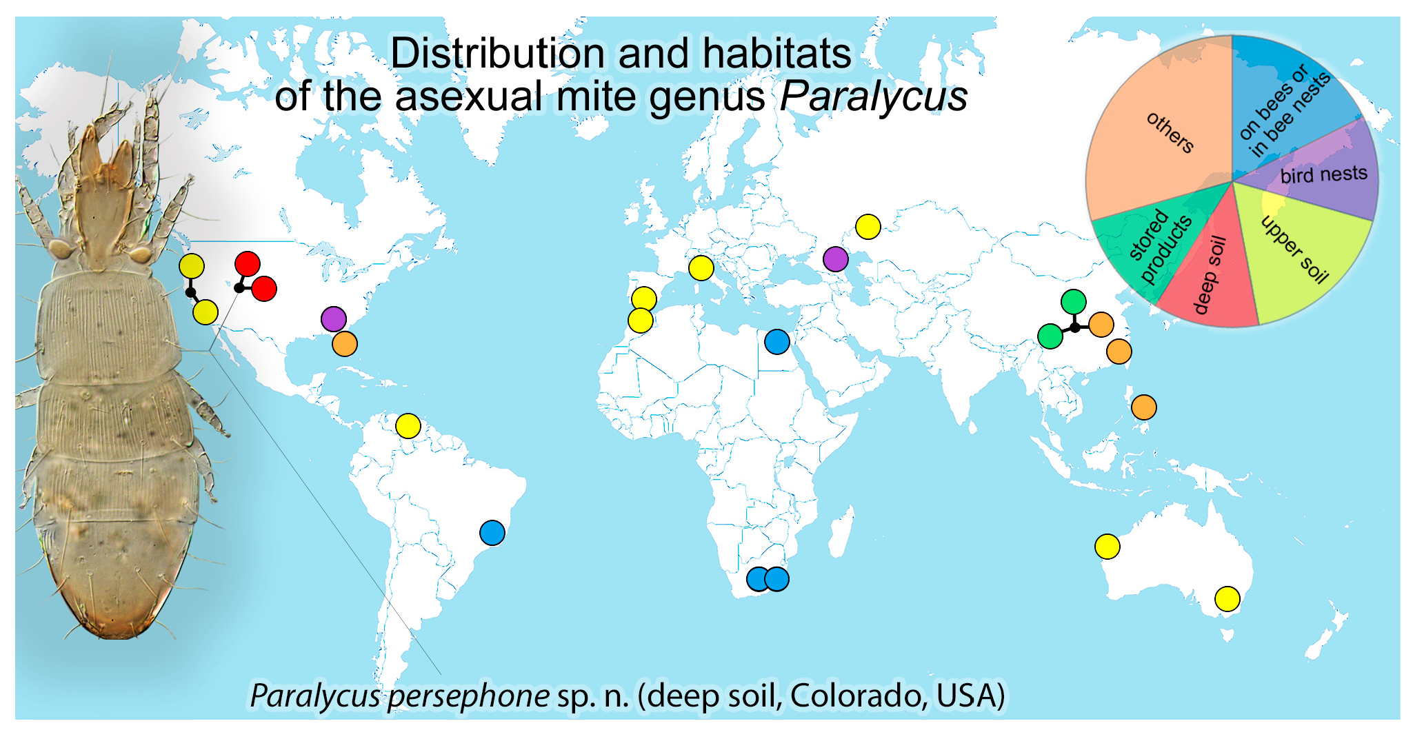
Habitat. Stored products, such as walnut, star anise, chili, garlic, beans, dried kelp (Figure 1) [14].
2. Paralycus daeira sp. n. (see below)
3. Paralycus lavoipierrei (Price, 1973)
Pediculochelus lavoipierrei Price 1973: 305, Figures 6–12.
Paralycus lavoipierrei: Norton et al. 1983: 493, Figures 3–11; Marshall et al. 1987: 28; Lebedeva and Poltavskaya 2013: 107; Xu et al. 2020: 486; Subías and Shtanchaeva 2021: 72; Subías 2022: 29.
Paralycus cf. lavoipierrei: Smelansky 2003: 181
Type depository. Holotype (female) and 4 paratypes (females) are in the U. S. National Museum; 15 paratypes (13 females, 1 larva, 1 deutonymph) are in the Entomology Museum, University of California, Berkeley, USA.
Known instars. Female, larva and deutonymph [4].
Distribution. USA: California [3,4,23] (type locality); Australia: Western Australia, Cape Range [3]; Russia: Orenburg Oblast [2], Stavropol’sky Kray [24]; Venezuela [25].
Habitat. Grassland soil [4], carbonate soil [2], nest of rosy starling (Figure 1) [24].
4. Paralycus longior Fan, Li et Xuan, 1996
Paralycus longior Fan et al. 1996: 175, Figures 3 and 4; Smelansky 2003: 181; Xu et al. 2020: 486; Subías 2022: 29.
Type depository. Holotype (female) and 7 paratypes (6 females, 1 larva) are in the Department of Plant Protection, College of Plant Protection, Southwest University, Chongqing, China (formerly the Department of Plant Protection, Southwest Agricultural University) (not found by the curator in this institution).
Known instars. Female and larva [14].
Distribution. China: Chongqing (type locality) [14].
Habitat. Stored products, such as Auricularia fungi, lily bulbs, and tangerine cake (Figure 1) [14].
5. Paralycus nortoni Xu, Zhu, Wu et Zhang, 2020
Paralycus nortoni Xu et al. 2020: 482, Figures 1–3; Subías 2022: 29.
Type depository. Holotype (female) and paratype (females) are in the National Zoological Museum of China, Institute of Zoology, Chinese Academy of Sciences, Beijing and Department of Plant Protection, Fujian Agriculture and Forestry University, China.
Known instars. Female [13].
Distribution. China: Fujian Province (type locality) [13].
Habitat. Under bark of Pinus massoniana infested by longhorn beetles Monochamus alternatus (Figure 1) [13].
6. Paralycus parasiti Zhang et Li, 2001
Paralycus parasiti Zhang and Li 2001: 317, Figures 1 and 2; Xu et al. 2020: 486; Subías 2022: 29.
Type depository. Holotype (female) is in the Department of Plant Protection, Southwest Agricultural University, China.
Known instars. Female [12].
Distribution. China: Chongqing (type locality) [12].
Habitat. From Coccinella septempunctata (Figure 1) [12].
7. Paralycus parvulus (Price, 1973)
Pediculochelus parvulus Price 1973: 306, Figures 13–15.
Paralycus parvulus: Norton et al. 1983: 493; Marshall et al. 1987: 28; Smelansky 2003: 181; Xu et al. 2020: 486; Subías 2022: 29.
Type depository. Holotype (female) are in U. S. National Museum; 2 paratype (2 females) are in the Entomology Museum, University of California, Berkeley, USA.
Known instars. Female [4].
Distribution. USA: California (type locality) [4,23].
Habitat. Grassland soil (Figure 1) [4,23].
8. Paralycus persephone sp. n. (see below)
9. Paralycus pricei sp. n. (see below)
10. Paralycus pyrigerus (Berlese, 1905) (see below)
11. Paralycus raulti (Lavoipierre, 1946) (see below)
Unidentified specimens
One specimen is from the nest of a pigeon in Atlanta, USA [4]; several specimens were from Australia [6] and the USA [15]. “Paralycus raulti” was reported from Florida (USA) and Samar (Philippines) on rats and chickens [11]. Baker and Wharton (1952, Figure 254) from [11] illustrated a single specimen presumably from one of these locations. Based on this figure, their specimen differs from both Price’s specimens identified by him as P. raulti by the long rostral setae (as in P. raulti but not P. pricei) and short cheliceral seta cha (as in P. pricei, but P. raulti). A reexamination is needed to accurately identify Baker and Wharton’s specimens. The one species is a Paralycus sp. Brazil: Minas Gerais, Sabará, managed nest of Melipona marginata (Hymenoptera: Apidae) 19°54′50.1″ S 43°49′35.4″ W BMOC 15-0104-030 (this mite species is similar to P. longior, but cannot be described without studying the types of P. longior first).

3.3. Descriptions of Species

Paralycus daeira sp. n.
Paralycus sp. Pepato and Klimov, 2015: 8 (included in a molecular phylogeny); Klimov et al., 2017: 109 (included in a molecular phylogeny).
Diagnosis (female). The rostral setae are not reaching half the length of the chelicera; the lamellar setae are situated close to each other. The cheliceral setae cha are shorter than half the length of the chelicera. Gastronotal setae c, d, e, and f are not reaching bases of the next row of setae; h1 reached the bases of p1; p2 is shorter than p1. The epimeral setae 4a is absent. Three pairs of genital setae are present; the distance is g1g2 > g2g3. The leg trochanteral formula is 0-0-0-0; the genua I had 4 setae (d, l′, l″, v) while the genua II had 2 setae (l′ and l″); the solenidion ω of tarsi I had not expanded in the middle; and the solenidion φ of tibiae III is long (about 1/4 of the length of d).
Description. Female.Measurements. Idiosomal length 219, width 56.
Integument. The body is colorless. The prodorsal shield (except its posterior part) is smooth. The legs, chelicerae, and coxae are smooth. The dorsum (except segment P), ventrum, and ovipositor are striated.
Gnathosoma. The subcapitulum (30 × 22) had 3 pairs of filiform, smooth setae (a 9; m 4; h 4) and 2 pairs of filiform, and smooth adoral setae (4–5). There are 20 palps, and the setal formula is 0-1-0-1-9+ω. Of the setae, 3 setae (sul, acm, ul′) are with expanded tips. The inf is absent, and the postpalpal setae (ep 2) is blunt. The chelicerae is large (24) with 2 filiform and smooth setae (cha 5; chb 4); the cha is shorter than half of the length of the chelicera. The pharyngeal cupola is long, reaching the level of exp.
The prodorsum is covered with a shield-shaped plate in mid-dorsal region. The plate had 2 pairs of setae (ro 7; le 19). The setae ro does not reach half of the length of the chelicera. The bases of setae le are close to each other; the setae in (33) and exa (7) filiform are smooth, the exp is very short (2) and simple, and the bothridial setae (bs 12 × 7) is clavate and smooth.
Gastronotum. Segment C had 4 pairs of setae: c1 (6), c2 (11), c3 (9), and cp (19). Segment DE had 4 pairs of setae: d1 (7), d2 (10), e1 (6), and e2 (10). Segment F had 2 pairs of setae: f1 (20) and f2 (18). Segments H and P fused with 6 pairs of setae: h1 (24), h2 (25), h3 (10), p1 (30), p2 (28), and p3 (5). All gastronotal setae filiform are smooth; the setae f, h, p1, and p2 expanded at the bases; the c1 does not reach the bases of the cp; the d1 does not reach the bases of e; the e1 does not reach the bases of f; the f1 does not reach the bases of h1; the h1 reached the bases of p1; and the p2 is shorter than p1.
Epimeral and podosomal regions. The setal formula of the epimera is 3-2-3-2; the setae 1a (2), 1b (5), 1c (3), 2a (27), 2b (8), 3a (3), 3b (4), 3c (2), 4b (2), and 4c (3) filiform are smooth; the 2a reached the bases of 1a; the bases of 3a are situated close to each other; the 4a is absent. The supracoxal setae (3) is triangular with a rounded tip.
Anogenital region. There are 3 pairs of genital setae: g1-2 (4) and g3 (7). The distance g1g2 > g2g3. All genital setae are situated medially; the eugenital setae are minute (2). There are 3 pairs of adanal setae: ad1 (14), ad2 (30), and ad3 (7) and 2 pairs of anal setae: an1 (6) and an2 (3). All anogenital setae filiform are smooth. The genital tracheae are reduced and represented by short cavities.
Legs. The leg chaeto- and solenidiotaxy is reported: I 0-2-4-2-9 (0-1-1), II 0-2-2-3-6 (0-0-1), III 0-2-0-2-5 (0-1-0), and IV 0-2-0-2-5 (0-0-0). The famulus of tarsi I baculiform is thin and expanded at end; other setae filiform are smooth. The solenidion ω of tarsi I 6 × 1 is not expanded in the middle; the ω of tarsi II 3 × 1 is smaller and not expanded in middle; the φ of tibiae I elongate is attenuate; and the φ of tibiae III 5 is not shorter than half the length of tibia III and baculiform. The length of the tibial seta is d 19 (Figure 2, Figure 3 and Figure 4 and Table 1).
Male. Unknown.
Juvenile instars.Measurements. The body length of the larva is 144 (excluding gnathosoma), protonymph is 160, deutonymph is 200, tritonymph is 214; body width of larva is 45, protonymph is 53, deutonymph is 55, and tritonymph is 56.
Integument is similar to the female but with segment P transversely striated dorsally.
Gnathosoma is similar to the female except the measurements. Subcapitulum is as follows: L 24 × 18, PN 23 × 19, DN 27 × 21, TN 29 × 23. The length of setae h is: L 3, PN and DN 4, TN 5; a L and PN 6, DN and TN 7; m L and PN 2, DN and TN 3. The adoral setae is: L and PN 3, DN and TN 4–5. The palps length is: L and PN 15, DN 17, TN 20. The chelicerae length is: L 18, PN 20, DN 22, TN 23. The cheliceral setae cha is: L and PN 2, DN 3, TN 4; chb L and PN 2, DN and TN 3.
Prodorsum is similar to the female except the bases of lele are well separated (L, PN) or close (DN, TN). The length of the setae are: ro L 4, PN 5, DN 6, TN 7; le L 14, PN 14, DN and TN 16; in L 23, PN 24, DN and TN 30; exa L 5, PN and DN 6, TN 7; exp L 1, PN 2, DN and TN 3; bs L 13 × 6, PN 14 × 6, DN and TN 14 × 7.
Gastronotum is similar to the female, but transverse furrows are weakly developed in L, PN, and DN. The tf3 is absent in L; the gastronotal setae p is short and situated around the anal opening in L and the posterior anal opening (except p3) in other stages. The length of the setae are: c1 5; c2 L and PN 7, DN and TN 9; c3 L 6, PN and DN 9, TN 12; cp L and PN 13, DN 18, TN 19; d1 L 3, PN, DN and TN 4; d2 L 7, PN and DN 8, TN 9; e1 L 6, PN, DN and TN 7; e2 L 8, PN and DN 9, TN 11; f1 18–20; f2 L and PN 18, DN and TN 20; h1 L 15, PN 17, DN and TN 20; h2 L 18, PN 19, DN and TN 24; h3 L 3, PN 6, DN 7, TN 12; p1 L 2, PN 9, DN 16, TN 25; p2 L 2, PN 17, DN 25, TN 27; and p3 L 2, PN and DN 5, TN 6.
For the epimeral and podosomal regions, the claparède’s organs are absent in L and other instars. The chaetotaxy of the epimera is: L 3-1-2-0; PN 3-2-3-1; DN 3-2-3-2; TN 3-2-3-2. All setae filiform are smooth. The length of the setae is: 1a 2; 1b L 3, PN, DN, and TN 5; 1c L 2, PN, DN, and TN 3; 2a L, PN, and DN 20, TN 25; 2b PN 4, DN, and TN 6; 3a L and PN 2, DN and TN 3; 3b 4; 3c PN, DN, and TN 2; 4b 2; 4c 3. The supracoxal setae L is 1, PN, DN, and TN 2.
The anogenital region showed larva without genital, anal, and adanal setae. The protonymph had 1 pair of genital (3) and 3 pairs of adanal (3) setae; the anal setae are absent. The deutonymph had 2 pairs of genital (4) and 3 pairs of adanal (ad1 7, ad2 23, ad3 5) setae, and the anal setae an2 (1) are simple while the an1 are represented by alveoli. The tritonymph had 3 pairs of genital (3–5), 3 pairs of adanal (ad1 11, ad2 28, ad3 7), and 2 pairs of anal (6) setae; the eugenital setae are absent. All setae in all juvenile instars filiform (except an1 is alveoli) are smooth. The eugenital setae and other genital structures are absent.
The legs are similar to the female, but the legs IV are absent in L. The leg setae and solenidia are as follows: L I 0-2-3-2-9 (0-1-1), II 0-2-2-3-6 (0-0-1), III 0-2-0-2-5 (0-1-0); PN I 0-2-3-2-9 (0-1-1), II 0-2-2-3-6 (0-0-1), III 0-2-0-2-5 (0-1-0), IV 0-0-0-0-5 (0-0-0); DN and TN I 0-2-4-2-9 (0-1-1), II 0-2-2-3-6 (0-0-1), III 0-2-0-2-5 (0-1-0), IV 0-2-0-2-5 (0-0-0). (Figure 5, Figure 6, Figure 7, Figure 8, Figure 9, Figure 10, Figure 11 and Figure 12 and Table 1).
Type material. The holotype (female) and paratypes (1 larva, 1 protonymph, 2 deutonymphs, 1 tritonymph, 1 female) came from the USA: Colorado, Weld Co., 2 mi NE Masters, 40°20′02.4″ N 104°13′49.13″ W, 1371m, sandy soil, 90 cm depth, kerosene flotation, 12 Oct 2008, B.M. OConnor & J. Wilke, BMOC 08-1012-006. They are deposited at UMMZ.
DNA voucher. AD1370 BMOC 08-1012-006 is used with the same data with the following GenBank sequence accession ids: KY922457 (Cytochrome oxidase subunit I (COXI) gene); KY922705 (Heat shock protein cognate 5 (Hsc70-5) gene); KY922585 (Signal recognition particle protein 54k (Srp54k) gene); KY922330 (Elongation factor 1-alpha (Ef1alpha100E) gene); KP325061 (Small subunit ribosomal RNA (18S) gene); KY922209 (Small subunit ribosomal RNA (18S) gene); KP325023 (large subunit ribosomal RNA (28S) gene); KY922080 (large subunit ribosomal RNA (28S) gene).
Type deposition. UMMZ.
Additional material. We used numerous specimens with the same data, preserved for DNA work as the frozen tissue samples at UMMZ (accession BMOC 08-1012-006).
Etymology. Daeira is an underworld nymph and companion of the goddess Persephone (Greek mythology).
Remark 2. Females of Paralycus daeira are similar to those of P. lavoipierrei by the following character states: the rostral and cheliceral setae do not reach half of the length of chelicera; the bases of setae le are adjacent; epimeral setae 2a are long; gastronotal setae c, d, e, and f are not protruding beyond the bases of setae in the subsequent rows; setae f, h, p1, and p2 are slightly widened at their bases; there are 3 pairs of genital setae; solenidion ω of tarsus I is not expanded. However, P. daeira differs from P. lavoipierrei by the absence of epimeral setae 4a and seta v′ on leg trochanters III (present in P. lavoipierrei); solenidion φ on leg tibiae III is longer than half the length of tibia III (distinctly shorter in P. lavoipierrei); the absence of palpal seta inf and presence of setae cm, lt″ and vt″ (inf present; cm, lt″ and vt″ are absent in P. lavoipierrei); the striated posterior part of the propodosomal shield (vs. with only one medial line is present in P. lavoipierrei). Furthermore, P. daeira is similar to P. chongqingensis by the cheliceral setae cha not reaching half the length of the chelicera, gastronotal setae c1, d1 and e not protruding beyond the bases of setae in the subsequent rows, and by the presence of three pairs of genital setae; but differs from P. chongqingensis by the absence of epimeral setae 4a, setae v′ on leg trochanters III and setae v on leg genua II (all these setae are present in P. chongqingensis), and by the adjacent bases of the lamellar setae le (slightly distant in P. chongqingensis).
The larvae and deutonymphs of P. daeira and P. laviopierrei are very similar. They differ by solenidion φ of tibiae III longer than half the length of tibia III in P. daeira (distinctly shorter in P. laviopierrei); in addition, deutonymphs of these two species can be distinguished by the absence of leg seta v′ on trochanters III in P. daeira (present in P. laviopierrei).
Paralycus persephone sp. n.
Diagnosis (female). The rostral setae are long, reaching half the length of chelicerae; the bases of the lamellar setae are separate. The cheliceral setae cha are shorter than half the length of chelicerae. The gastronotal setae are not expanded at the bases; setae c1, d1, and e1 not reaching bases of next row of setae; setae f1 reached the bases of h1; setae h1 reached bases of p1; p2 are shorter than other dorsal gastronotal setae. The epimeral setae 4a are absent. There are 3 pairs of genital setae present, distance g1g2 > g2g3. The leg trochanteral formula is 0-0-0-0; genua I had 3 setae (d and l), and genua II had 2 setae (l). The solenidion ω of tarsi I are large and expanded in the middle; solenidion φ of tibiae III are short.
Description. Female.Measurements. The body length is 191 (excluding gnathosoma); the width is 64.
Integument. The body is colorless. The shield of prodorsum had one short medial strip in the posterior part and legs, and the coxae I and II are smooth. The dorsum (except segment P and part of segment H), ventrum, chelicerae (basal part), and ovipositor are striated.
Gnathosoma had a subcapitulum 29 × 22 with 3 pairs of filiform, smooth setae (a 10; m 4; h 5), and 2 pairs of filiform and smooth adoral setae (3–5). It had 24 palps; the setal formula is 0-1-0-1-9+ω; 3 setae had expanded tips (sul, acm, ul′); inf is absent; and the postpalpal setae (ep 2) is blunt. The chelicerae are 21 with 2 filiform and smooth setae (cha 6; chb 5). The cha is shorter than half of the length of the chelicera. The pharyngeal cupola is long, reaching the level of the exa.
Prodorsum is covered with a shield-shaped plate in mid-dorsal region, bearing 2 pairs of filiform and smooth setae (ro 12; le 16); ro reached half of the cheliceral length. The bases of le are distant from each other; in (25) and exa (10) filiform is smooth; exp is very short (3) and simple; and bothridial setae (12 × 9) clavate is smooth.
Gastronotum. Segment C had 4 pairs of setae: c1 (10), c2 (13), c3 (12), and cp (26). Segment DE had 4 pairs of setae: d1 (8), d2 (13), e1 (20), and e2 (25); segment F had 2 pairs of setae: f1 (30) and f2 (26). Segments H and P fused with 6 pairs of setae: h1 (30), h2 (28), h3 (20), p1 (23), p2 (25), and p3 (10). All gastronotal setae filiform are smooth and not expanded at the base; c1 does not reach the base of cp; d1 does not reach the base of e; e1 does not reach the base of f; f1 reached the base of h1; h1 reached the base of p1, and p2 is shorter than other dorsal gastronotal setae.
For the epimeral and podosomal regions, the setal formula of epimera is 3-2-3-2; the setae 1a (4), 1b (6), 1c (5), 2a (8), 2b (6), 3a (4), 3b (6), 3c (3), 4b (3), and 4c (4) filiform are smooth; 2a does not reach the bases of 1a, the bases of 3a are situated close to each other; and 4a is absent. The supracoxal setae (2) are triangular with a rounded tip.
In the anogenital region, there are 3 pairs of genital setae (g1 4, g2 5, g3 6). The distance g1g2 > g2g3; g1 and g3 are medial, and g2 is slightly more lateral than g1 and g3. Eugenital setae are minute (2). There are 3 pairs of adanal setae: ad1 (14), ad2 (23), and ad3 (5), and 2 pairs of anal setae: an1 (6) and an2 (3). All anogenital setae filiform are smooth. Genital tracheae are reduced and represented by short cavities.
For the legs, the chaeto- and solenidotaxy are: I 0-2-3-2-9 (0-1-1), II 0-2-2-3-6 (0-0-1), III 0-2-0-2-5 (0-1-0), and IV 0-2-0-2-5 (0-0-0). Famulus of tarsi I baculiform are thin and expanded at the end; other setae filiform are smooth. Solenidion ω of tarsi I are large (6 × 3) and distinctly widened in the middle; ω of tarsi II (3 × 1) are shorter and had a widened middle; φ of tibiae I are elongated and attenuated; φ of tibiae III 2 are short and expanded at the end. The length of tibial seta d is 13 (Figure 13, Figure 14 and Figure 15 and Table 2).
Male. Unknown.
Type material. Holotype (female), 1 paratype female, 2 paratype deutonymphs, 1 paratype protonymph: USA: Colorado, Weld Co., 2 mi NE Masters, 40°20′02.4″ N 104°13′49.13″ W, 1371m, sandy soil, 90 cm depth, kerosene flotation, 12 Oct 2008, B.M. OConnor & J. Wilke, BMOC 08-1012-006. Deposited at UMMZ.
Type deposition. UMMZ.
Additional material. Numerous specimens, same data, preserved for DNA work as frozen tissue samples at UMMZ.
Etymology. Persephone is the goddess of the underworld (Greek mythology).
Remark 3.Paralycus persephone is similar to P. parvulus in having long gastronotal setae cp, f and h distinctly extending beyond the bases of setae in the subsequent rows, the rostral setae reaching half the length of the chelicera; the position of setae le (bases are distinctly separated from each other), the absence of epimeral seta 4a, the presence of 3 pairs of genital setae, and the absence of setae v′ on leg trochanters III. However, P. persephone differs from P. parvulus as follows: gastronotal setae c1, d, and e shorter, not reaching the bases of the following pair of setae (extending beyond the bases of the next row setae in P. parvulus); seta v on leg genua I are absent (present in P. parvulus); and the greatly widened solenidion ω on leg tarsi I (weakly widened in P. parvulus). P. pyrigerus also has long gastronotal setae f, h, and p, but P. persephone has shorter setae c2, d, and e1, not reaching the bases of the next pair of setae (extending beyond these bases in P. pyrigerus) and the long, distinctly widened solenidion ω on tarsi I, longer than half of the length of tarsi I (not widened and shorter than half of the length of tarsi I in P. pyrigerus).
Paralycus pricei sp. n.
Pediculochelus raulti Price 1973 (specimen no. 1): 302, Figures 3–5 (misidentification).
Diagnosis (females). The rostral setae does not reach half the length of the chelicera; the bases of lamellar setae are distinctly separated from each other. The subcapitulum has four pairs of setae (h, a, m1, m2). The cheliceral setae cha are shorter than half the length of chelicera. The gastronotal setae d, e, f, and h1 does not reach the bases of setae in the next row; p2 is the longest. The epimeral setae 4a are present. There are 4 pairs of genital setae (g1–4). The leg trochanteral formula is 0-0-1-0. The genu I has 3 setae (d and l); the genu II has 2 setae (l); and the solenidion ω of tarsus I is simple and not widened.
Description. Female. Idiosoma 180 x 58.
Integument. The body is colorless. The shield of prodorsum, legs, and coxae are smooth; the dorsal and ventral sides of the body are striated.
Gnathosoma has subcapitulum (28 × 22) with 4 pairs of filiform and smooth setae (a 11; m1 and m2 4; h 10) and 2 pairs of filiform, smooth adoral setae (4). The palps length is 24. The chelicerae are large (22) with 2 filiform and smooth setae (cha 5; chb 4); the cha is shorter than half the length of the chelicera.
The prodorsum has a shield-shaped plate in mid-dorsal region, bearing 2 pairs of setae (ro 6; le 28); ro does not reach half the length of the chelicera. The bases of le are adjacent; in (38), exa (16), and exp (2) filiform are smooth; bothridial setae (13 × 6) clavate are smooth.
For the Gastronotum, Segment C has 4 pairs of setae: c1 (13), c2 (13), c3 (25), and cp (25). Segment DE has 4 pairs of setae: d1 (10), d2 (12), e1 (12), and e2 (16). Segment F has 2 pairs of setae: f1 (20) and f2 (18). Segments H and P fused and bear 6 pairs of setae: h1 (14), h2 (16), h3 (7), p1 (14), p2 (30), and p3 (8). All setae filiform are smooth and not widened at the bases; c1 nearly reached the base of cp, d1 does not reach the bases of e1, d2 does not reach the bases of e2; e1 does not reach the bases of f; f1 does not reach the bases of h1; h1 does not reach the bases of p1; and p2 is longer than other dorsal gastronotal setae.
In the epimeral and podosomal regions, chaetotaxy of epimera are 3-2-3-3; setae 1a (7), 1b (8), 1c (6), 2a (11), 2b (11), 3a (5), 3b (12), 3c (8), 4a (6), 4b (5), and 4c (4) are filiform and smooth; 2a does not reach the bases of 1a, and the bases of 3a are adjacent.
In the anogenital region, there are 4 pairs of genital setae (g1 4, g2 4, g3 5, g4 9); the distance is g1g2 > g2g3; g1 and g2 are medial, and g3 and g4 are slightly more lateral than g1 and g2. There are 3 pairs of adanal setae: ad1 (12), ad2 (26), and ad3 (8); 2 pairs of anal setae: an1 (6) and an2 (5); and all anogenital setae are filiform and smooth.
For the legs, the genua I has 3, and the genua II has 2 setae; the trochanteral formula is 0-0-1-0. All setae are filiform and smooth; solenidion ω of tarsus I 5 × 1 is slightly expanded at the tip and situated in the basal part of tarsus.
Male. Unknown.
Type material. The holotype (female) (=paratype of P. raulti designated by Price (1973) [4] is specimen no. 1 from the personal collection of Dr. M. Lavoipierre) has the following data: South Africa, Durban, 1940 from bees Amegilla fallax and Apis mellifera adansonii (as Anthophora fallax and Apis mellifera adansonii).
Type deposition. It is probably deposited at the University of California, Berkeley [26].
Etymology. The species is named after Dr. Douglas W. Price who suggested that specimen no. 1 from M. Lavoipierre’s collection could represent an undescribed species.
Remark 4. Price (1973) [4] did not give information about the structure of the ovipositor, palp, and legs as the specimen was old, and these structures could not be studied in detail. The differences between P. pricei (specimen no. 1) and P. raulti (specimen no. 2) were given previously [4]; see also the key to species below.
Paralycus pricei is similar to P. parasiti by the well separated bases of the lamellar setae; the dorsal gastronotal setae (c1, d, e, f, h) not reaching the bases of the setae in the next row; and the presence of epimeral setae 4a and 4 pairs of genital setae g1g4. However, P. pricei differs from P. parasiti by the rostral setae ro not reaching half the length of the chelicerae (reaching in P. parasiti). The leg genua I has 3 setae: l, d (4 setae: v, l, d in P. parasiti); the leg genua II has 2 l setae, and setae v are absent (3 setae: v, l in P. parasiti).
Paralycus pyrigerus (Berlese, 1905)
Alycus pyrigerus Berlese 1905: 232; Berlese 1910: 13, Figure 18 from [27].
Paralycus pyrigerus: Womersley 1944: 134; Price 1973: 302; Norton et al. 1983: 493; Ruiz et al. 1991: 59; Gil-Martín et al. 1992: 55; Smelansky 2003: 181; Xu et al. 2020: 486; Subías 2022: 29.
Diagnostic description (female). The rostral setae reaches half the length of chelicera. Gastronotal setae c1 do not reach the bases of cp setae, c2 reaches the bases of cp; setae cp, d, e, f, and h are distinctly protruding beyond the bases of setae in the subsequent rows. Tarsus I is more than 2 times longer than wide. Solenidion ω of tarsus I is slightly expanded in the middle, distinctly shorter than half length of tarsus I.
Male. Unknown.
Type material. Holotype (female): Italy: Monte Pisano, chestnut forest, soil/decaying wood.
Type deposition. Holotype: CREA Research Centre for Plant Protection and Certification, Florence, Italy.
Known instars. Female (Berlese 1905).
Distribution. Italy: near Pisa (type locality) [5]; Spain: Andalusia [8]; Morocco [7].
Habitat. Soil (Figure 1) [5,7,8].
Remark 5. This species is inadequately described [5], making it difficult to identify P. pyrigerus based on the original description. In 1965, Donald E. Johnston examined the single holotype specimen preserved in the Berlese collection at the Agricultural Station near Florence, Italy, and found that it is congeneric with Pediculochelus [3], but he did not re-describe it. We examined this specimen as images provided by Dr. Sauro Simoni and Silvia Guidoni, and we report that it is in poor condition, thus preventing a detailed study (Figure 16).
However, here, we report some diagnostic characters that could be observed. Paralycus pyrigerus and P. parvulus are similar species, both having long rostral setae reaching half the length of the chelicera and the dorsal notogastral setae distinctly protruding beyond the bases of setae in the subsequent rows. Based on the original figure of Berlese (1910) [27] and our study of the photos of the type, P. pyrigerus has setae c1 not protruding beyond the bases of cp, and c1 are subequal to ro (vs. setae c1 reaching the level of bases of setae cp and are longer than ro in P. parvulus). We were not able to study the entire ventral side of the type specimen (except setae g1 and g2, which were clearly seen; however, additional genital setae are probably present as well). Because the original description of P. pyrigerus lacks important diagnostic detail, we suggest that all subsequent reports of this species in the literature need confirmation.
Paralycus raulti (Lavoipierre, 1946)
Pediculochelus raulti Lavoipierre 1946: 130, Figure 1 (part.); Price 1973 (specimen no. 2): 302, Figures 1–5; Abou Senna 1997: 667; Baker and Wharton 1952: 325, Figure 254 from [11] (specimens need to be examined to confirm identification).
Paralycus raulti: Krantz 1970: 145; Norton et al. 1983: 493; Smelansky 2003: 181; Xu et al. 2020: 486; Subías 2022: 29
Diagnostic description (female). The rostral setae reaches half the length of chelicera; the bases of lamellar setae are separated from each other. The cheliceral setae cha is longer than half the length of the chelicera. The gastronotal setae d1, e, and f does not reach the bases of the setae in the next row. Setae h1 reaches the bases of p1; p2 is the longest among the dorsal gastronotal setae. The epimeral setae 4a is present. There are 5 pairs of genital setae. The leg trochanteral formula is 0-0-1-0; the genual formula is 4-2-0-0 [3]. Solenidion ω of tarsus I slightly expanded in the middle (after [4,9]).
Known instars. Female [4,9].
Distribution. South Africa: Durban (type locality) [9]; Egypt [10].
Habitat. It lives on adult bees Amegilla fallax and Apis mellifera adansonii [9] and in tentorial pits on the head of the honeybee Apis mellifera (Figure 1) [10].
Type material. Holotype female and 3 female paratype (including specimen no. 2 designated by [4]) are from: South Africa, Durban, 1940 from bees Amegilla fallax and Apis mellifera adansonii (as Anthophora fallax, Apis adansonii).
Type depository. Holotype (female) and 2 paratypes (females) are in the National Museum of Natural History, Paris, France; 1 paratype (female, specimen 2) and 1 paratype female specimen 1 (identified here as P. pricei) are probably at the University of California, Berkeley [26].
Remark 6. Price (1973) [4] studied two paratypes of P. raulti from Lavoipierre’s personal collection and found that both paratypes differ in a number of character states. He suggested that two separate species were involved but did not formally propose a new species for one of them. Here, we propose a new species, P. pricei, based on Price’s original description of specimen no. 1 [4]. Unfortunately, Price (1973) [4] did not study the type of specimens deposited in the National Museum of Natural History, France, which include the holotype. We contacted this institution but received no response. Here, we identify P. raulti based on the specimen figured by Lavoipierre (1946) [9] as it is likely to be the holotype.
Our study of P. raulti was based on the original description and figures of [3,4,9]. Price (1973) [4] reported that the paratype studied by him was in poor condition, so the gnathosoma and legs could not be observed in detail. The original description [9] shows that P. raulti has long cha, which is consistent with specimen no. 2 in [4]. Price figured leg genua I with 3 setae, but Norton et al. (1983) [3], who studied the same specimens reported 4 setae. Paralycus raulti is very similar to P. longior but cheliceral setae cha are longer than half the length of the chelicera in P. raulti (vs. shorter in P. longior).

3.4. Ontogenetics of Paralycus

Previously, some juvenile instars have been briefly described in Paralycus but never in a complete series—larvae and deutonymphs (originally identified as nymphs) in P. laviopierrei [4] and protonymphs and deutonymphs in P. chongqingensis [14]. Here, we report a complete life-cycle for the deep-soil mite P. daeira: larva, protonymph, deutonymph, tritonymph, adult (see above). A summary of essential ontogenetic transformations is presented below. The bases of the lamellar setae le are well separated from each other in larvae and protonymphs or adjacent in the other instars. Larvae have two transverse furrows tf1-2 while the other instars have three furrows tf1-3. Pseudanal setae p1-3 are short (as long as the epimeral setae, except 2a), not widened, and situated around the anal opening in larvae while in in protonymphs, pseudanal setae p1 and p2 are widened at the bases, shifted to the posterior end of the idiosoma, and are longer than epimeral setae (except 2a). In deutonymphs–adults, setae p1 are shifted to the dorsal part of idiosoma while setae p2 do not change its position as compared to the protonymph. Larvae lack the epimeral setae 2b, 3c, 4b, and 4c; of them, coxal setae 2b, 3c, and 4b are added in protonymphs, and 4c are added in deutonymphs. Genital setae g1 are added in protonymphs while g2 are added in deutonymphs, and g3 are added in tritonymphs. Short adanal setae ad1-3 are added in protonymphs; in other subsequent instars, these setae are long, longer than most epimeral setae (except 2a). Anal setae an are added in deutonymphs; of them, an2 are minute and an1 are represented by alveoli while in tritonymphs and adults, anal setae an are all filiform. Larvae are without legs IV and Claparède’s organs. Ontogenetic changes in the leg chaetotaxy and solenidiotaxy are shown in Table 2.
Adults of P. daeira lack coxal setae 4a and seta v′ on trochanters III, but these setae are present in P. laviopierrei and P. chongqingensis. In P. laviopierrei, setae v′ on trochanter III are added in deutonymphs.
The family Pediculochelidae is a sister group to the family Haplochthoniidae Hammen, 1959. In the family Haplochthoniidae, ontogenetic series are known for Amnemochthonius taeniophorus Grandjean, 1948 (only protonymph, tritonymphs and adults) [28] and Haplochthonius simplex Willmann, 1930 (all instars) [29,30,31,32]. The ontogenetic development of these two species is very similar to that of P. daeira, although there are differences (summarized in Table 3).

4. Discussion

Habitat and biodiversity assessment of Paralycus.
Known species of Paralycus has been recorded from a variety of habitats but these records are rare (Figure 1B). In many cases, these records are based on a small number of specimens, so it is possible that these mites could have simply migrated from their main habitat (such as soil) to numerous peripheral habitats. However, there has been some consistency in reports of mites from the same habitats. P. raulti and P. pricei have been found phoretic on adult apid bees, Amegilla fallax and Apis mellifera [9,10], and a large colony of Paralycus sp. has been found in the nest of a stingless bee, Melipona marginata (Lepeletier, 1836) in Brazil (our data, unpublished). Of these bees, A. fallax is a solitary bee, constructing nests in clay-rich soils [33], while A. mellifera and M. marginata are eusocial bees, constructing large, above-ground nests lasting for many generations of bees [33]. Inside the nest of M. marginata, Paralycus lives on the moldy nest walls and other nest material/debris. One can suppose that moldy spill-over debris, such as discarded pollen and nest material, in the nest of honeybees can also serve as a habitat for Paralycus as this mite is known to feed on fungal mycelium [15]. Two records were from bird nests [4,24]. The upper soil is another habitat were species of Paralycus have been recorded: P. lavoipierrei, P. parvulus, P. pyrigerus [2,4,5,7,8]. We found two abundant species in deep soil, P. persephone and P. daeira, but unfortunately, the feeding habit of these two species is unknown.
There are single records of Paralycus from other habitats, such as from chickens and rats [4,11], a ladybird beetle [12], in bark of pine trees [13], stored products [14], and fungal mycelia growing on historical books [15]. Because there was no independent confirmation of these records in the literature, we consider these habitats as marginal, where mites cannot sustainably maintain their population sizes for many generations or they represent mites which migrated from their adjacent main habitat, for example, soil.
Although upper soil and nests of social bees consistently but rarely harbor Paralycus, based on the great abundance of Paralycus found in our deep soil, it is most likely these mites are actually widespread in deep soil around the world. One can expect further discoveries of a new dimension of Paralycus diversity in this habitat.
Key to ontogenetic instars of Paralycus.
(based on P. daeira, males are absent in Paralycus)
1 Three pairs of legs. Genital setae (g1-3) and genital papillae absent. Adanal setae ad1-3 absent. Epimeral setae 2b and 3c absent. Gastronotal transverse furrow tf3 absent…Larva
- Four pairs of legs. Genital setae (g) and genital papillae present. Adanal setae ad1-3 present. Epimeral setae 2b and 3c present; gastronotal transverse furrow tf3 present…2
2 One pair of genital setae g1 and one pair of genital papillae present; one pair of setae 4b on epimeres IV; anal setae an1-2 absent…Protonymph
- Two or more pairs of genital setae; two pairs of genital papillae; two pairs of setae, 4b and 4c on epimeres IV; anal setae an1-2 present (an1 alveolar or filiform) …3
3 Two pairs of genital setae g1-2; one pair of genital papillae; anal setae an1 represented by alveolae…Deutonymph
- Three pairs of genital setae g1-3; two pairs of genital papillae; anal setae an1 filiform …4
4 Genital structures, cavities of anterior genital tracheae, and eugenital setae eg1-2 absent…Tritonymph
- Genital structures, cavities of anterior genital tracheae, and eugenital setae eg1-2 present…Adult female
Key to species of the genus Paralycus
Females
1 Gastronotal setae d1, e1, f1 reach bases of setae in the next row …2
- Gastronotal setae d1, e1, f1, or some of them, not reaching bases of setae in the next row …3
2 Gastronotal setae c1 reach the level of setae cp. USA (grassland soil)… P. parvulus (Price, 1973)
- Gastronotal setae c1 not reaching the level of setae cp. Italy, Spain, Morocco (soil)…P. pyrigerus (Berlese, 1905)
3 Epimeral setae 4a present …4
- Epimeral setae 4a absent…9
4 Five pairs of genital setae…5
- Four or three pairs of genital setae…6
5 Cheliceral setae cha longer than half length of chelicera. Africa (bees)…P. raulti (Lavoipierre, 1946)
- Cheliceral setae cha shorter than half length of chelicera. China (stored products)…P. longior Fan, Li et Xuan, 1996
6 Four pairs of genital setae…7
- Three pairs of genital setae…8
7 Rostral setae longer than half length of chelicera. China (ladybird beetle)…P. parasiti Zhang & Li, 2001
- Rostral setae shorter than half length of chelicera. Africa (bees)…P. pricei sp. n.
8 Rostral setae longer than half length of chelicera; gastronotal setae f1 reach bases of h1; genual chaetotaxy 4-3-0-0. China (stored products)…P. chongqingensis Fan, Li et Xuan, 1996
- Rostral setae shorter than half the length of chelicera; gastronotal setae f1 not reaching bases of h1; genual setation 4-2-0-0. USA, Australia, Russia, Venezuela (soil)…P. lavoipierrei (Price, 1973)
9 Trochanteral chaetotaxy 0-0-1-0; five pairs of genital setae. China (pine trees)…P. nortoni Xu, Zhu, Wu et Zhang, 2020
- Trochanteral chaetotaxy 0-0-0-0; three pairs of genital setae…10
10 Gastronotal setae f1 reach bases of h1; solenidion ω of tarsus I distinctly widened in the middle; bases of lamellar setae le well separated; dorsal gastronotal setae not widened; epimeral setae 2a not reaching bases of 1a. USA (deep soil)…P. persephone sp. n.
- Gastronotal setae f1 not reaching bases of h1; solenidion ω of tarsus I not widened in the middle; bases of lamellar setae le adjacent; setae f, h, p1, and p2 widened at bases; epimeral setae 2a reach bases of 1a. USA (deep soil)…P. daeira sp. n.
Notes. Xu et al. (2020) erroneously indicated in their key that P. parvulus has four pairs of genital setae. Paralycus pyrigerus is included in the key based on a few characters observed in a poorly presered type specimen. Xu et al. (2020) indicated in their key that P. lavoipierrei has 7 setae on tarsus I, but Norton et al. (1983) showed that there are 8 setae (excluding famulus).

Author Contributions

Conceptualization, V.B.K. and P.B.K.; methodology, P.B.K. and V.B.K.; validation, P.B.K., S.G.E. and B.O.; formal analysis, P.B.K.; investigation, P.B.K. and V.B.K.; resources, P.B.K. and B.O.; data curation, V.B.K. and P.B.K.; writing—original draft preparation, V.B.K. and P.B.K.; writing—review and editing, P.B.K., S.G.E. and B.O.; visualization, V.B.K. and P.B.K.; supervision, P.B.K. and V.B.K.; project administration, P.B.K.; funding acquisition, P.B.K. All authors have read and agreed to the published version of the manuscript.

Funding

This research was funded by the Ministry of Science and Higher Education of the Russian Federation within the framework of the Federal Scientific and Technical Program for the Development of Genetic Technologies for 2019–2027 (agreement No. 075-15-2021-1345, unique identifier RF----193021X0012).

Institutional Review Board Statement

Not applicable.

Data Availability Statement

The data presented in this study are available on request from the corresponding author.

Acknowledgments

The authors thank Roy A. Norton (The State University of New York, College of Environmental Science and Forestry, Syracuse, USA) for help. Jeffrey D. Wilke greatly assisted in the collection of the deep soil specimens from Colorado, USA. Sauro Simoni and Silvia Guidoni (Agricultural Station near Florence, Italy) greatly assisted in the P. pyrigerus type study and photography. Zi-Ying Wang (Southwest University, Chongqing, China) and Qing-Hai Fan (Ministry for Primary Industries, Plant Health & Environment Laboratory, Auckland, New Zealand) helped to verify the presence of the types of Paralycus longior and P. chongqingensis at the Southwest University.

Conflicts of Interest

The authors declare no conflict of interest.

References

  1. Subías, L.S. Listado sistemático, sinonímico y biogeográfico de los ácaros oribátidos (Acariformes: Oribatida) del mundo (excepto fósiles). Monogr. Electrónicas S.E.A. 2022, 12, 1–538. [Google Scholar]
  2. Smelansky, I.E. New records of oribatid mites from the families Gehypochthoniidae and Pediculochelidae (Acari, Oribatida) in Northern Eurasia. Euroasian Entomol. J. 2003, 2, 181–183. [Google Scholar]
  3. Norton, R.A.; OConnor, B.M.; Johnston, D.E. Systematic relationships of the Pediculochelidae (Acari: Acariformes). Proc. Entomol. Soc. Wash. 1983, 27, 97–99. [Google Scholar] [CrossRef]
  4. Price, D.W. Genus Pediculochelus (Acarina: Pediculochelidae), with notes on P. raulti and descriptions of two new species. Ann. Entomol. Soc. Am. 1973, 66, 302–307. [Google Scholar] [CrossRef]
  5. Berlese, A. Acari nuovi. Materiali pel Manipulus V. Redia 1905, 2, 231–238. [Google Scholar]
  6. Nobble, J.C.; Whitford, W.G.; Kaliszweski, M. Soil and litter microarthropod populations from two contrasting ecosystems in semi-arid eastern Australia. J. Arid. Environ. 1996, 32, 329–346. [Google Scholar] [CrossRef]
  7. Gil-Martín, J.; Subías, L.S.; Arillo, A. Oribatidos de Marruecos y Sahara Occidental I: O. Inferiores (Acari, Oribatida, Macropylina). Graellsia 1992, 48, 53–63. [Google Scholar]
  8. Ruiz, E.; Subías, L.S.; Kahwash, A.M. Oribatidos Inferiores (Acari, Oribatida, Macropylina) de Andalucia, con description de tres nuevas especies. Eos 1991, 67, 55–65. [Google Scholar]
  9. Lavoipierre, M. A new acarine parasite of bees. Nature 1946, 158, 130. [Google Scholar] [CrossRef]
  10. Abou Senna, F.M. A new record of phoretic mites on honey bee Apis mellifera L. in Egypt. J. Egypt. Soc. Parasitol. 1997, 27, 667–680. [Google Scholar]
  11. Baker, E.W.; Wharton, G.W. An Introduction to Acarology; Macmillan: New York, NY, USA, 1952; pp. 1–465. [Google Scholar]
  12. Zhang, A.-H.; Li, Y.-R. A new species of the genus Paralycus from Chongqing, China. J. Southwest Agric. Univ. 2001, 23, 317–318. [Google Scholar]
  13. Xu, Y.; Zhu, Y.-Z.; Wu, J.-Q.; Zhang, F.-P. A new species of the genus Paralycus from Fujian, China. Acarologia 2020, 60, 481–487. [Google Scholar] [CrossRef]
  14. Fan, Q.-H.; Li, L.-S.; Xuan, J.-Y. Two new species of the genus Paralycus from Sichuan, China (Acari: Pediculochelidae). Acta Zootaxonomica Sin. 1996, 21, 174–178. [Google Scholar]
  15. Norton, R.A.; Florian, M.E.; Manning, L.E. Ecdysial cleavage line in Paralycus sp. (Acari: Oribatida: Pediculochelidae). Int. J. Acarol. 2001, 27, 97–99. [Google Scholar] [CrossRef]
  16. Womersley, H. Australian Acarina, families Alycidae and Nanorchestidae. Trans. R. Soc. South Aust. 1944, 68, 133–143. [Google Scholar]
  17. Krantz, G.W. A Manual of Acarology. Oregon State University Book Stores, Inc.: Corvallis, OR, USA, 1970; pp. 1–335. [Google Scholar]
  18. Klimov, P.B.; OConnor, B.M.; Chetverikov, P.E.; Bolton, S.J.; Pepato, A.R.; Mortazavi, A.L.; Tolstikov, A.V.; Bauchan, G.R.; Ochoa, R. Comprehensive phylogeny of acariform mites (Acariformes) provides insights on the origin of the four-legged mites (Eriophyoidea), a long branch. Mol. Phylogenetics Evol. 2017, 119, 105–117. [Google Scholar] [CrossRef]
  19. Pepato, A.R.; Klimov, P.B. Origin and higher-level diversification of acariform mites—Evidence from nuclear ribosomal genes, extensive taxon sampling, and secondary structure alignment. BMC Evol. Biol. 2015, 15. [Google Scholar] [CrossRef] [PubMed] [Green Version]
  20. Norton, R.A.; Ermilov, S.G. Catalogue and historical overview of juvenile instars of oribatid mites (Acari: Oribatida). Zootaxa 2014, 3833, 1–132. [Google Scholar] [CrossRef] [Green Version]
  21. Kethley, J. A procedure for extraction of microarthropods from bulk soil samples with emphasis on inactive stages. Agric. Ecosyst. Environ. 1991, 34, 193–200. [Google Scholar] [CrossRef]
  22. Walter, D.E.; Krantz, G.W. Chapter 7: Collecting, rearing, and preparing specimens. In A Manual of Acarology; Krantz, G.W., Walter, D.E., Eds.; Tech University Press: Lubbock, TX, USA, 2009; pp. 83–96. [Google Scholar]
  23. Marshall, V.G.; Reeves, R.M.; Norton, R.A. Catalogue of the Oribatida of continental USA and Canada. Mem. Entomol. Soc. Can. 1987, 139, 418. [Google Scholar]
  24. Lebedeva, N.V.; Poltavskaya, M.P. Oribatid mites (Acari, Oribatida) of plain area of the Southern European Russia. Zootaxa 2013, 3709, 101–133. [Google Scholar] [CrossRef] [Green Version]
  25. Subías, L.S.; Shtanchaeva, U. Contribución al conocimiento de la distribución de los ácaros oribátidos (Acari, Oribatida) tropicales. Rev. Iber. De Aracnol. 2021, 38, 69–80. [Google Scholar]
  26. Norton, R.A.; State University of New York, College of Environmental Science and Forestry, Syracuse, New York, USA. Personal Communication, 2022.
  27. Berlese, A. Acari nuovi. Manipulus V–VI. Redia 1910, 6, 199–234. [Google Scholar]
  28. Grandjean, F. Les Enarthronota (acariens) (2e série). Ann. Des Sci. Nat. Zool. 1949, 10, 29–58. [Google Scholar]
  29. Grandjean, F. Les Enarthronota (acariens). Première série. Ann. Des Sci. Nat. Zool. 1946, 8, 213–248. [Google Scholar]
  30. Grandjean, F. Formules anales, gastronotiques, génitales et aggénitales du développement numérique des poils chez les Oribates. Extr. Du Bull. De La Société Zool. De Fr. 1949, 74, 201–225. [Google Scholar]
  31. Knülle, W. Morphologische und entwicklungsgeschichtliche Untersuchungen zum phylogenetischen System der Acari: Acariformes Zachv. I. Oribatei: Malaconothridae. Mitt. Aus Dem Zool. Mus. Berl. 1957, 33, 97–213. [Google Scholar] [CrossRef]
  32. Seniczak, S.; Seniczak, A. Differentation of body form of Protoplophoroidea (Acari: Oribatida) in the light of ontogeny of three species. J. Nat. Hist. 2010, 44, 389–419. [Google Scholar] [CrossRef]
  33. Michener, C.D. The Bees of the World; The Johns Hopkins University Press: Baltimore, MD, USA; London, UK, 2000; pp. 1–913. [Google Scholar] [CrossRef]
Figure 1. Geographic distribution of Paralycus (A), habitats of Paralycus species (B). Symbols: red circle—type locality; yellow circle—non-type locality; 1—P. chongqingensis; 2—P. daeira sp. n.; 3—P. laviopierrei; 4—P. longior; 5—P. nortoni; 6—P. parasiti; 7—P. parvulus; 8—P. persephone sp. n.; 9—P. pricei sp. n.; 10—P. pyrigerus; 11—P. raulti; no number—unidentified species.
Diversity 15 00160 g001
Figure 2. Paralycus daeira sp. n., female, light microscope images: (A)—dorsal view; (B)—ventral view; (C)—solenidion ω I; (D)—solenidion φ III. Scale bar 100 μm.
Diversity 15 00160 g002
Figure 3. Paralycus daeira sp. n., female: (A)—dorsal view; (B)—ventral view. Scale bar 100 μm.
Diversity 15 00160 g003
Figure 4. Paralycus daeira sp. n., female: (A)—leg I, right, dorsal view; (B)—tarsus I, right, ventral view; (C)—leg II, left, dorsal view; (D)—leg III, left, paraxial view; (E)—leg IV, left, paraxial view; (F)—genitals; (G)—palp, left, dorsal view. Scale bar 50 μm (AF), 10 μm (G).
Diversity 15 00160 g004
Figure 5. Paralycus daeira sp. n., larva (A,B) and protonymph (C,D), light microscope images: (A,C)—dorsal view; (B,D)—ventral view. Scale bar 100 μm.
Diversity 15 00160 g005
Figure 6. Paralycus daeira sp. n., deutonymph (A,B) and tritonymph (C,D), light microscope images: (A,C)—dorsal view; (B,D)—ventral view. Scale bar 100 μm.
Diversity 15 00160 g006
Figure 7. Paralycus daeira sp. n., larva: (A)—dorsal view; (B)—ventral view. Scale bar 100 μm.
Diversity 15 00160 g007
Figure 8. Paralycus daeira sp. n., protonymph: (A)—dorsal view; (B)—ventral view. Scale bar 100 μm.
Diversity 15 00160 g008
Figure 9. Paralycus daeira sp. n., deutonymph: (A)—dorsal view; (B)—ventral view. Scale bar 100 μm.
Diversity 15 00160 g009
Figure 10. Paralycus daeira sp. n., tritonymph: (A)—dorsal view; (B)—ventral view. Scale bar 100 μm.
Diversity 15 00160 g010
Figure 11. Paralycus daeira sp. n., larva (AE) and protonymph (FH): (A)—leg I, left, dorsal view; (B)—tarsus I, left, ventral view; (C)—leg II, left, dorsal view; (D)—leg III, left, paraxial view; (E)—palp, left, dorsal view; (F)—palp, left, dorsal view; (G)—leg I, left, dorsal view; (H)—leg IV, left, paraxial view. Scale bar 50 μm (AD,G,H), 10 μm (E,F).
Diversity 15 00160 g011
Figure 12. Paralycus daeira sp. n., tritonymph: (A)—leg I, left, dorsal view; (B)—tarsus I, left, ventral view; (C)—leg II, left, dorsal view; (D)—leg III, right, paraxial view; (E)—leg IV, right, paraxial view; (F)—palp, left, dorsal view. Scale bar 50 μm (AE), 10 μm (F).
Diversity 15 00160 g012
Figure 13. Paralycus persephone sp. n., female, light microscope images: (A)—dorsal view; (B)—ventral view; (C)—solenidion ω I; (D)—genitals. Scale bar 100 μm.
Diversity 15 00160 g013
Figure 14. Paralycus persephone sp. n., female: (A)—dorsal view; (B)—ventral view. Scale bar 100 μm.
Diversity 15 00160 g014
Figure 15. Paralycus persephone sp. n., female: (A)—leg I, left, paraxial view; (B)—leg I, right, dorsal view; (C)—leg II, right, dorsal view; (D)—leg III, right, paraxial view; (E)—leg IV, right, paraxial view; (F)—genitals; (G)—palp, right, dorsal view. Scale bar 50 μm (AF), 10 μm (G).
Diversity 15 00160 g015
Figure 16. Paralycus pyrigerus (Berlese, 1905), holotype, light microscope images: (A)—dorsal view; (B)—ventral view (inset). Scale bar 100 μm.
Diversity 15 00160 g016
Table 1. Ontogenetic development of leg setae and solenidia (first appearance) in Paralycus daeira sp. n.
LegInstarsTrFeGeTiTa
ILarva-bv″, d(l), dl′, v′, φ(ft), a″, s, (u), (p), ω, ɛ
Protonymph-----
Deutonymph--v--
Tritonymph-----
Adult-----
IILarva-bv″, d(l)l′, v′, d(ft), (u), (p), ω
Protonymph-----
Deutonymph-----
Tritonymph-----
Adult-----
IIILarva-ev′, d-v′, dφft″, (u), (p)
Protonymph-----
Deutonymph-----
Tritonymph-----
Adult-----
IVProtonymph----ft″, (u), (p)
Deutonymph-ev′, d-v′, d-
Tritonymph-----
Adult-----
Note: Roman letters refer to normal setae, Greek letters refer to solenidia (except ɛ = famulus); dφ—seta and solenidion coupled. Single prime (′) marks setae on the anterior and double prime (″)—setae on the posterior sides of a given leg segment. Parentheses refer to a pair of setae (′ and ″). Setae/solenidia are listed only for the stage in which they first appear.
Table 2. Leg setae and solenidia of adult female Paralycus persephone sp. n.
LegTrFeGeTiTa
I-bv″, d(l), dl′, v′, φ(ft), a″, s, (u), (p), ω, ɛ
II-bv″, d(l), dl′, v′, d(ft), (u), (p), ω
III-ev′, d-v′, dφft″, (u), (p)
IV-ev′, d-v′, dft″, (u), (p)
Note: See Table 1 footnotes.
Table 3. Ontogeny of three species of Enarthronota. For Amnemochthonius taeniophorus, larvae and deutonymphs are unknown.
CharactersParalycus daeiraAmnemochthonius taeniophorusHaplochthonius simplex
Adoral setae (or1-3), pairs2 (L–AD, or3 absent)3 (PN, TN, AD)2 (L, or3 absent), 3 (PN–AD)
Palpal seta infabsent (L–AD)absent (PN), present (TN, AD)absent (L, PN), present (DN–AD)
Palpal seta aabsent (L–AD)absent (PN, TN, AD)absent (L), present (PN–AD)
Transverse furrows tf1-3tf1-2 (L), tf1-3 (PN–AD)absent (PN, TN), present (AD)absent (L–TN), present (AD)
Gastronotal dorsal stripeslongitudinal (L–AD)transverse (PN, TN), absent (AD)transverse (L–TN), absent (AD)
Setae p4 and h4absent (L–AD)absent (PN, TN, AD)present (L), absent (PN–AD)
Genital setae, pairs0 (L), 1 (PN), 2 (DN), 3 (TN, AD)1 (PN), 3 (TN), 4 (AD)0 (L), 1 (PN), 3 (DN), 5 (TN), 7 (AD)
Adanal setae, pairs0 (L), 3 (PN–AD)3 (PN, TN, AD)0 (L), 4 (PN–AD)
Anal setae, pairs0 (L–PN), 2 (DN–AD)0 (PN), 3 (TN–AD)0 (L–PN), 4 (DN–AD)
Genital papillae, pairs0 (L), 1 (PN), 2 (DN–AD)1 (PN), 2 (TN–AD)0 (L), 1 (PN), 2 (DN), 3 (TN–AD)
Anterior genital tracheaabsent (L–TN), short cavities (AD)present (PN, TN, AD)absent (L), present (PN–AD)
Posterior genital tracheaabsent (L–AD)absent (PN, TN, AD)absent (L), present (PN–AD)
Claparede’s organabsentunknownpresent (minute)
Trochanter setae0-0-0-0 (L–AD)0-0-0-0 (PN), 1-0-1-0 (TN, AD)0-0-0-0 (L), 1-0-0-0 (PN), 1-0-1-0 (DN), 1-1-2-1 (TN, AD)
Femoral setae l I-IIIabsent (L–AD)absent (PN, TN, AD)absent (L–DN), present (TN, AD)
Genual seta v Iabsent (L–PN), present (DN–AD)absent (PN, TN, AD)absent (L–TN), present (AD)
Genual seta v IIabsent (L–AD)absent (PN, TN, AD)absent (L–TN), present (AD)
Tarsal seta it′ Iabsent (L–AD)absent (PN, TN, AD)absent (L–TN), present (AD)
Tarsal seta it″ Iabsent (L–AD)absent (PN, TN, AD)absent (L–DN), present (TN–AD)
Tarsal seta it″ IIabsent (L–AD)absent (PN, TN, AD)absent (L–TN), present (AD)
Tarsal seta s IVabsent (L–AD)absent (PN), present (TN, AD)absent (L–PN), present (DN–AD)
Tarsal seta tc′ IVabsent (L–AD)absent (PN), present (TN, AD)absent (L–TN), present (AD)
Tarsal seta tc″ IVabsent (L–AD)absent (PN), present (TN, AD)absent (L–DN), present (TN–AD)
Tarsal setae a and ft′ IVabsent (L–AD)absent (PN, TN, AD)absent (L–PN), present (DN–AD)
Disclaimer/Publisher’s Note: The statements, opinions and data contained in all publications are solely those of the individual author(s) and contributor(s) and not of MDPI and/or the editor(s). MDPI and/or the editor(s) disclaim responsibility for any injury to people or property resulting from any ideas, methods, instructions or products referred to in the content.

