Article

Seasonal and Interannual Variability in the Phenolic Content of the Seagrass Nanozostera noltei: Characterization of Suitable Candidates for the Monitoring of Seagrass Health

by
Micheline Grignon-Dubois
* and
Bernadette Rezzonico
Environnements et Paléoenvironnements Océaniques et Continentaux (EPOC, UMR 5805), Université de Bordeaux, F-33600 Pessac, France
*
Author to whom correspondence should be addressed.
Diversity 2023, 15(12), 1210; https://doi.org/10.3390/d15121210
Submission received: 11 September 2023 / Revised: 4 December 2023 / Accepted: 6 December 2023 / Published: 11 December 2023

Abstract

:
Developing early warning indicators to accurately detect ecosystem disturbances is vital for enhancing ecosystem management. The seasonal and interannual variability of the phenolic content of Nanozostera noltei from Arcachon Bay, France, was explored over 47 consecutive months to identify suitable early indicators of the state of seagrass beds. Five phenolic acid derivatives and eight flavonoids were fully characterized using chromatographic and spectroscopic techniques; a caffeic tetramer was described for the first time as a metabolite of N. noltei and of the genus Nanozostera. The individual phenolic concentrations in each of the 47 collections were determined by quantitative HPLC and analyzed as a function of year and season. The variability of the phenolic content in the rhizomes of N. noltei from Arcachon Bay was also determined over one year, as well as rhizomes of N. noltei from three other locations in the Atlantic and Mediterranean for comparison purposes. In addition, the phenolic fingerprints of Z. marina rhizomes were also characterized for the first time. The results show that leaf phenolic chemistry could be used to signify changes in the ecological health of N. noltei. In particular, it appears that diosmetin 7-sulfate, rosmarinic acid and zosteranoic acid could be reliable and easy-to-use indicators for monitoring N. noltei meadows. From a phytochemical point of view, this work is the first report of zosteranoic acid in the leaves and the rhizomes of N. noltei and in the rhizomes of Z. marina.

1. Introduction

Seagrasses comprise a group of 73 species of rooted vascular plants which evolved three to four times from terrestrial ancestors and successfully returned to the sea. They belong to five families, including Cymodoceae, Hydrocharitaceae, Posidoniaceae, Zosteraceae and Ruppiaceae [1]. Seagrasses grow in vast marine meadows, providing valuable habitats. Their contribution to ocean biodiversity has become increasingly recognized in recent decades, and seagrass meadows are considered the world’s most widespread and productive coastal zones, offering numerous ecosystem services [2,3,4]. Seagrass beds are biodiversity hotspots recognized as important socio-ecological systems worldwide and of particular importance to local communities and fisheries. In addition, they constitute important blue carbon ecosystems [5,6,7]. Seagrass meadows are also among the most rapidly declining coastal habitats worldwide, as a result of either natural or anthropogenic disturbances.
The key factor for seagrass growth is the availability of light, while poor water quality (particularly high levels of nutrients) caused by pollution is the biggest threat to seagrasses around the world [8]. In many coastal areas, human and/or natural deterioration of underwater light availability often results in the large-scale loss of seagrasses [9,10,11]. It is estimated that up to 50% of all seagrass habitats has been lost along the developed east coast of the USA during the past century [1]. In Europe, about one third of the area of seagrasses was lost between 1869 and 2016 [12,13].
Seagrass monitoring has become essential to inform adaptive management and remedy the widespread decline of seagrass ecosystems. Positive changes resulting from regulation and management are beginning to be observed in a few places, although gains in seagrass areas and biomass are not yet reversing the general trend of decline [13]. Monitoring programs use a variety of indicators to assess the ecological quality of European coastal waters and the conservation status of European seagrass meadows. These indicators may vary across species and regions, depending on the type and location of seagrass meadows [14,15]. Stress needs to be identified as early as possible to avoid the permanent loss of seagrass beds, but response times vary considerably from one indicator to another. Physiological and biochemical indicators have proved to be better candidates for the early warning of changes than structural and morphological parameters (see [16,17] as examples).
Most of the biochemical indicators are primary metabolites. In contrast, the specialized metabolites of seagrasses have rarely been used as indicators to detect environmental degradation or improvement. Of the 73 species of seagrass, only a few have their specialized metabolite fingerprints described, and their absence as biochemical indicators is probably due to this lack of data. This is especially true in the case of phenolic compounds, which form the basis of defensive mechanisms in plants, in which they have various functions. They play a major role in growth and reproduction and provide protection against pathogens and predators [18]. They are common in marine ecosystems where competition is intense, and their role in the chemical ecology of marine angiosperms has been reviewed (see [19,20,21] as examples). Many are antimicrobial agents, presumed to protect marine macrophytes against pathogen attacks [22], to reduce plant palatability toward grazers [23] or to inhibit microbial settlement or growth [24,25,26,27].
The specialized metabolites of many seagrasses remain understudied and numerous reports of phenolic-mediated interactions involving seagrasses suffer from the lack of reliable phytochemical data in the literature. Often, total phenolic compounds are measured using colorimetric methods, which cannot distinguish among constituents and have serious limitations. This could explain, at least in part, the absence of phenolic compounds in panels of biochemical indicators of seagrass health, in spite of their high potential. Phenolic fingerprinting of seagrass beds could enable the early detection of environmental degradation before the decline begins. Documenting the presence of those compounds in living tissues, and how they seasonally vary in abundance, is crucial to understanding the large-scale response of the plant to potential pathogens, herbivore outbreaks or other ecological processes. Efforts to explore the phenolic chemistry of seagrasses appear to be essential and would help those engaged in chemical ecology and the monitoring of seagrass beds.
Of the five species of strictly marine Magnoliophytes found in Europe, one is endemic to the Mediterranean (Posidonia oceanica), three are found in the Atlantic and Mediterranean (Cymodocea nodosa, Zostera marina and Nanozostera noltei) and one is a Lessepsian immigrant (Halophila stipulacea). Nanozostera noltei (Hornem.) Toml. and Posl. (basionym: Zostera noltei Hornem., common name dwarf eelgrass, Zosteraceae family) belong to the genus Nanozostera, which also comprises three other species, namely N. japonica, N. mucronata, and N. muelleri [28] (see Supplementary Materials for details about Zosteraceae). N. noltei is found in shallow coastal waters in northwestern Europe, the Mediterranean Sea, the Black Sea, the Caspian Sea, the Aral Sea, and on islands in the Atlantic off the coast of northwest Africa [29,30]. These species often co-occur with Zostera marina. Both have been included in the IUCN Red List of Threatened Species [31].
N. noltei has been shown to contain both flavonoids and phenolic acids. Concerning the former, diosmetin- and luteolin 7-sulfates have been reported for specimens from the Isles of Wight (GB) and Algeciras (ES) [32,33]. Diosmetin, diosmetin 7-O-glucoside and luteolin-7-O-glucoside have been mentioned, but not unambiguously characterized, for specimens from the Black Sea and the Adriatic Sea [34,35]. The 7-sulphates of apigenin, luteolin and diosmetin and the 7-glucosides of apigenin and luteolin have been reported in variable amounts for specimens collected in the coastal waters of France [25,36,37], western Norway [38] and Spain [39]. Analysis of the flavonoid content of fifteen populations of N. noltei over a broad spatial scale covering the Mediterranean Sea and the north-eastern Atlantic coast has enabled us to identify three geographically distinct flavonoid chemotypes on the basis of their respective major compounds [36,37]. One is characterized by apigenin 7-sulfate (the eastern part of the Gulf of Cadiz), one by diosmetin 7-sulfate (the French Atlantic coast and Mediterranean Sea), and the third contains similar quantities of the above two compounds (Mauritania and South Portugal) (Figure 1 and Figure S2).
Rosmarinic acid and zosteric acid were seasonally quantified from fresh detrital leaves collected monthly over the course of one year [40,41]. The presence and abundance of rosmarinic, caffeic and zosteric acids were also quantified in N. noltei from four different meadows located across the Atlantic and the Mediterranean coast, namely the Bay of Cadiz, Sa Menorca, Alfacs Bay and Arcachon Bay [42]. Interestingly, the concentration of phenolic acid was found to be 2 to 13 times lower in the meadow of Alfacs Bay, which was exposed to high levels of nutrient discharge at the time of the study (May–July 2004). We observed similar effects with Z. marina, whose phenolic content in the Baltic Sea, which is heavily affected by human pressures, was found to be significantly lower than that in the Atlantic, Pacific and Mediterranean, falling to values 10 to 20 times lower in Puck Bay, which underwent large-scale degradation in the latter part of the 20th century [43]. Analysis of the variability in the phenolic chemistry of Z. marina along environmental gradients in Norway has identified the flavonoid/rosmarinic acid ratio as a possible molecular index of seagrass ecosystem health [44].
Arcachon Bay is home to one of Europe’s largest N. noltei beds. Mapping of the area colonized by N. noltei in 2019 was compared with those from 1989 and 2012. The results show that, after a period of strong regression from the 2000s onwards, the surface area of these intertidal meadows was relatively stable between 2012 and 2019, and the regression process now seems to be slowing down or even reversing [45]. Similar results were also observed in the Bay of Santander, an estuary located in the north of Spain [46]. The relative stability of N. noltei meadows in Arcachon Bay and our previous work on this species led us to select Arcachon Bay as a suitable site for analyzing the seasonal and interannual variability of the seagrasses’ phenolic content. Our aim was to provide a picture of the variability of polyphenol production in a seagrass bed that has not been subjected to major environmental disturbance, in order to establish a baseline reference for the state of the meadow, and to identify suitable early warning indicators that can be routinely used by managers responsible for seagrass monitoring.
Long-term monitoring, which involves repeated observations, provides valuable information, notably by generating reference data. They are necessary to establish trends and dynamics, allowing a clear understanding of the seasonal variations in seagrasses’ phenolic chemistry. In the present work, living tissue of N. noltei from Arcachon Bay was collected monthly over a four-year period. Extracts were prepared from the fresh leaf material and analyzed to determine similarities or differences among collections. Individual phenolics were isolated, structurally identified and quantified to analyze how these compounds vary between seasons and years. Rhizomes were also analyzed over one year, as well as specimens from other Atlantic and Mediterranean sites, for comparative purposes.

2. Materials and Methods

2.1. Chemicals

Analytical-grade water was obtained from Sodipro Company (Echirolles, France). Trifluoroacetic acid (TFA) and all the solvents used (HPLC-grade) were purchased from Aldrich Chemical Company (St. Louis, MO, USA). Standards were purchased from Extrasynthèse (Genay, France). C18 reverse-phase silica gel, HPLC columns and TLC aluminum sheets (silica gel layer) were purchased from Macherey-Nagel (Düren, Germany).

2.2. General Experimental Procedures

Ultrasound-assisted extraction experiments were performed with a Sonorex Digitec H 512 sonicator, 35 kHz (Bandelin, Berlin, Germany), centrifugation was performed with a Jouan B4i centrifuge (Thermo electron, Waltham, MA, USA), and lyophilization was performed using a manifold freeze-dryer (Christ Alpha 1–2/LD, Gefriertrocknungsanlagen, Osterode am Harz, Germany). 1H, 13C NMR and 2D NMR spectra were recorded on Bruker Avance 300 and 600 MHz instruments in DMSO-d6 or aceton-d6 (Euriso-Top, Gif-Sur-Yvette, France). Chemical shifts are expressed in δ (ppm) values relative to the residual undeuterated solvent signal as the internal reference. The following abbreviations were used to describe the multiplicities: s = singlet, d = doublet, dd = doublet of doublet. Coupling constants are reported in hertz (Hz). 13C NMR assignments were made by 2D HSQC and HMBC for Dept and JMod experiments. Ultraviolet (UV) spectra were recorded on a V-630 UV-VIS Jasco spectrophotometer (Easton, Los Angeles, CA, USA) in HPLC-grade methanol. High performance liquid chromatography (HPLC) combined with diode array detection (DAD) was performed on a Thermo Electron liquid chromatography system. High-resolution mass spectrometry (HRMS) and low-resolution mass spectrometry (LRMS) were performed using electro spray ionization (ESI). HRMS was performed using an AccuTOF™ LC-plus (Jeol, Peabody, MA, USA). LC/MS was performed using a HP1100 (Hewlett-Packard, Palo Alto, CA, USA) equipped with an Agilent MSD 1946B simple quad mass spectrometer and HP Chemstation software (B.04.2.3.).

2.3. Study Sites

Arcachon Bay (Figure 1 and Figures S4–S6) is a 155 km2 mesotidal system on the Atlantic coast of southwest France (44.58 N–1.24 W). The bay is connected to the Atlantic Ocean by two channels (2–3 km wide and 12 km length) that allow for significant water exchange. The inner bay is mainly composed of tidal flats, while the Arguin sandbank is bordered by a wide inlet and faces the wave-dominated Bay of Biscay plateau.
The semi-diurnal tidal range varies from 1.1 to 4.9 m, with the large tidal mudflats (115 km2) drained at low tide by the shallow tidal creeks of the inner bay (156 km2). At high tide, the surface water temperature and salinity fluctuate annually from 1 to 30 °C and from 22 to 32 psu, respectively. N. noltei meadows are found on intertidal mudflats between −0.3 m and +3.1 m above the lowest tide. The most obvious feature of the back-barrier lagoon landscape is the surface variability linked to ebb and flood tides and resulting from the emergence of extensive intertidal features during low tide phases [47]. Around two-thirds of the lagoon’s total volume flows in and out with each tidal cycle. The tidal prism (384 million cubic meters) is directly linked to a mesotidal range (varying from 2.1 to 4.8 m), and the water surface increases by over 200% between low spring tides (40 km2) and high tides (172 km2). Annual freshwater inflows come from the Leyre in the southeastern part of the bay, the Canal du Porge and many little streams. At low tide, the surface of the lagoon presents a complex network of anastomosing channels (57 km2) isolating vast expanses of permeable muddy sand (117 km2), home to N. noltei meadows [48].

2.4. Collection

Above-ground biomass was collected monthly from January 2017 to December 2020 from an intertidal N. noltei meadow in the intertidal zone of Taussat (Figure 1A, Figures S4–S6 and Table 1). A total of 47 sampling campaigns were conducted in triplicate along a permanent transect within a homogeneous section of the meadow. The GPS coordinates were recorded. Rhizomes were also required to compare their phenolic content with that of leaves. However, to reduce the number of destructive samplings, whole plants (i.e., with above- and belowground tissues) were sampled only 6 times between May 2019 and October 2020. In addition, samples of N. noltei leaves and Z. marina rhizomes from other sites throughout the Mediterranean and Atlantic were considered (Table 1). Identification of the species was based on morphology, plant anatomy and comparison with identification keys reported in the literature [49] (see Section S2 and Figure S7).
All samples taken were cleaned on site in tide pools or channels, and then with lightly salted water from one of the many small streams flowing into the lagoon. Then, collected material was hand-picked to remove associated debris when present. Plant material was air-dried at room temperature to a constant weight in a ventilated room at a constant temperature of 25 °C for around 20 h. The plant material was then subjected to two rounds of microwaving (two minutes each at 360 W) in a domestic rotary-heat microwave oven. After drying, unused plant material was kept at −80 °C until further studied. Dried material was cut into small pieces (0.2–0.5 cm) prior to extraction. Specimens of N. noltei collected in Arcachon Bay are representative of the Dios-7-S chemotype, found in the Mediterranean and the entire Atlantic coast of Europe, with the exception of the Ria Formosa and the Gulf of Cadiz (Figure 1B, Figures S2 and S3) [37].
Table 1. Nanozostera noltei study sites and collection data.
BioregionCountrySampling SiteCoordinatesSampling Date
Atlantic OceanFranceArcachon Bay, Taussat44°42′ N, 1°4′ Wmonthly from January 2017 to December 2020
other sites
FranceArcachon Bay, Arguin Bank44°34′ N,
1°14′ W
20 November 2019
FranceHossegor Lake43°66′ N,
1°40′ W
10 September 2011
SpainCadiz Bay36°23′ N,
6°10′ W
24 June 2010
MoroccoMerja Zerga34°84′ N,
6°27′ W
15 November 2011
PortugalRia Formosa37°01′ N,
7°51′ W
27 June 2010
MauritaniaPointe de l’Etoile21°01′ N,
17°00′ W
29 April 2014
Mediterranean SeaSloveniaStrunjan45°53′ N,
13°60′ E
30 October 2014
TunisiaBizerte37°13′ N,
9°55′ E
07 July 2011
FranceSalses Leucate Lagoon42°50′ N,
3°37′ E
16 August 2010
FranceThau Lagoon43°45′ N,
3°65′ E
30 June 2010
FranceBerres Lagoon43°28′ N,
5°10′ E
31 August 2010

2.5. Extraction

Air-dried leaves (10 g) were sequentially extracted in a Soxhlet-type apparatus by refluxing methanol (120 mL, 24 h) and then 50% aqueous methanol (120 mL, 24 h). Then, the extracts were pooled together and thoroughly homogenized under sonication, and fifteen ml of the extract were set aside for HPLC. The remaining material was evaporated under vacuum until the methanol was completely eliminated, and then the aqueous solution was freeze-dried. The chemical content of the extracts was analyzed by NMR and HPLC. Phenolic compounds were identified based on NMR spectra, UV-visible (UV-vis) spectra, HPLC retention time, online DAD spectra and MS molecular ions in comparison with authentic standards and synthetic samples.

2.6. Purification of Phenolic Compounds for Structural Assignment

Thirty-five grams of a crude MeOH/MeOH-H2O extract of the air-dried plant (150 g) were diluted with water, adjusted to pH 3 and serially extracted in a separating funnel with methylene chloride, ethyl acetate, and then n-butanol. The process was monitored by TLC (SiO2) and HPLC. Only the butanolic fraction was found to contain significant amounts of phenolics. Analysis of the final aqueous phase showed the presence of salt and sugars and the absence of flavonoids. Individual phenolic compounds were isolated from the butanolic fraction by repeated column chromatography on C18 reverse-phase silica gel. Gradient elution was performed with H2O-MeOH as the mobile phase proceeding from 99:1 to 20:80 (v/v). Fractionation was monitored by HPLC and NMR. Structure elucidation of the isolated compounds was based on UV, NMR and MS spectroscopies and confirmed by co-injection and comparison to authentic standards when available. They were unambiguously assigned as compounds 113 (Figure 2). Their spectral data are reported in the supplementary materials (see Section S5).

2.7. Acid Hydrolysis of the Crude Extracts

One hundred milligram samples of crude extract were separately dissolved in 100 mL of methanol and stirred with 5 mL of TFA at room temperature until total disappearance of the sulfated flavonoids as monitored by HPLC. After the evaporation of methanol under vacuum, the reaction mixture was partitioned between n-butanol and water. Addition of BaCl2 to the aqueous layer gave a white precipitate of BaSO4. The hydrolysate was also controlled for sugars against a standard mixture (HPTLC; toluene-n-BuOH-pyridine-H20, 2:5:3:3). Analysis of the reaction mixture (HPLC, LC/MS, UV, and comparison to standard) shows the complete disappearance of zosteric acid (peak 1), while the other phenolic acids were recovered unchanged. Five peaks assigned as flavonoid derivatives (610) disappeared, while the peak intensity of the aglycones (1113) increased. The results showed the large dominance of diosmetin (13) along with luteolin (11) and apigenin (12) in variable amounts.

2.8. Purification of the Aglycone Mixtures

Ten grams of crude extract from the Arcachon Bay were hydrolyzed as described above. After the evaporation of MeOH under reduced pressure, the aglycone mixture was extracted with ethyl acetate. The ethyl acetate residue was chromatographed on C18 silica gel to give pure samples of luteolin (11), apigenin (12) and diosmetin (13). Their identity was unambiguously confirmed by NMR and comparison to standards.

2.9. Synthesis of the Sulfated Flavonoid Standards

We used the dicyclohexylcarbodiimide-mediated sulfation of flavones with tetrabutylammonium hydrogen sulfate as described by Barron and Ibrahim for luteolin, apigenin and diosmetin [50]. Spectral data for the resulting sulfated products were identical to those described in the literature (Table S2) [32,50].

2.10. Qualitative and Quantitative HPLC Analyses

The separation and quantification of phenolic compounds in each of the crude extracts of N. noltei (three biological samples for each of the 47 collections) were performed using a liquid chromatography system (Thermo electron, Waltham, MA, USA) equipped with a SCM 1000 solvent degasser, a thermostatically controlled column compartment, an AS 3000 autosampler with a 100 μL loop, a PDA UV6000LP detector and a Chromquest Chromatography Workstation. A Nucleodur C18 sphinx RP column (110 Å pore size, 5-μm particle size, 250 × 4.6 mm i.d.) was used for all the HPLC analysis. Separations were carried out at 40 °C and the analytes were eluted at a flow rate of 1 mL/min using the binary gradient 0.1% (v/v) TFA in water (A) and methanol (B). The following linear gradient was used: 0 min, 80% A; 60 min, 80% B; run time, 50 min; stop time, 50 min; post time, 10 min. UV spectra were collected over the range of 220–380 nm. The injection volume was 20 μL. Data were integrated using Chromquest software 5.0. In addition, they were processed to create a chromatogram, in which each peak represents the absorbance of the eluting substance at its λ max (max-plot chromatogram). Stock solutions of the crude extracts were prepared in HPLC-grade dmso/water (4:1, v/v). All solutions were filtered prior to analysis through a 0.20 μm syringe filter. They were injected three times into the HPLC, and the results were averaged. Chromatograms were systematically recorded at 270, 328 and 350 nm, which allowed a clear distinction between benzoate, hydroxycinnamate, and flavonoid derivatives. The chromatographic peaks of the crude extracts were checked for peak purity and identification. Quantitative determinations of flavonoids were carried out by peak area measurements at 350 nm (diosmetin and luteolin derivatives) or 338 nm (apigenin derivatives) using an external calibration of their respective authentic aglycon dissolved in DMSO. The curve was established over seven data points, covering the concentration range of 0.0619–0.00619 mg/mL. Linear regressions on the HPLC analyses gave R2 values of 0.9996 (luteolin), 0.9995 (apigenin) and 0.9997 (diosmetin). Quantitative determinations of caffeic (2), coumaric (3), and rosmarinic acids (4) were carried out by peak area measurements at 328 nm, using a calibration curve of their respective authentic sample at the same wavelength (caffeic acid: R2 0.9995, 6 points; coumaric acid, R2 0.9995, 7 points; rosmarinic acid 0.9995, 7 points). Quantitative determinations of zosteric acid (1) were carried out by peak area measurements at 280 nm, using a calibration curve of coumaric acid [41] and those of compound 5 used the calibration curve of rosmarinic acid. Data are expressed in mg per gram of dry matter of plant tissue (mg/g plant DW; mean ± standard deviation (SD) of three determinations). The HPLC analysis of standards was performed using exactly the same conditions as for the extracts. The concentration of each individual compound was calculated using the following equation:
%W = Windividual phenol compound = (C × 100)/Cs
where C is the concentration of the tested phenolic compound (mg/mL) in the analyzed crude extract, calculated from peak areas and linear regression and Cs is the concentration of the sample (mg/mL) diluted in dmso/deionized water 4:1 (v/v).
The results are summarized in Table 2, Table 3, Table 4 and Table 5, which show the average concentrations of the harvest replicates for each of the collection dates considered.

2.11. LC/MS Analysis

LC/MS was performed using an HP1100 (Hewlett-Packard) equipped with an Agilent MSD 1946B single quad mass spectrometer and HP Chemstation software. Positive mode ESI spectra of the column eluate were recorded in the range of m/z 120–1000 a.m.u. Absorbance was measured at 280 nm. Compounds were separated using a Zorbax poroshell C18 column (Agilent, Santa Clara, CA, USA): 100 mm × 3 mm i.d., 2.7 μm particle sizes. The analytes were eluted at a flow rate of 0.2 mL/min using the binary gradient (v/v) formic acid in water (pH = 2.55, A) and methanol (B). The following linear gradient was used: 5% B to 100% B (15 min). Separation of the analytes was carried out at 50 °C. The injection volume was 2 μL. For mass spectrometric analysis, compounds were detected using the following conditions: nebulizing gas pressure, 60 psi; drying gas flow rate, 12 l/min; drying gas temperature, 350 °C; capillary voltage, 4000 V; temperature source, 350 °C. Data were acquired in full scan mode (m/z 100–1000) at a fragmentor voltage of 70 V.

2.12. Statistical Analyses

Significant differences in phenolic compound compositions (p < 0.05) between seasons were assessed with one-way analysis of variance (ANOVA) followed by a post-hoc Tukey’s test. Principal component analyses (PCA) based on correlation matrices were conducted with the mean phenolic concentration values to analyze the relationships between the collection dates (observations) and the phenolic compositions (variables). Agglomerative hierarchical clustering (AHC) was automatically run after the PCA using the Ward algorithm [51] and the Euclidean distance. The variables were also subjected to cluster analysis. Statistical analyses were computed using XLSTAT 2022.5.1 software (Addinsoft Corp., Paris, France).

3. Results

3.1. Determination of the Phenolic Content of N. noltei from Arcachon Bay

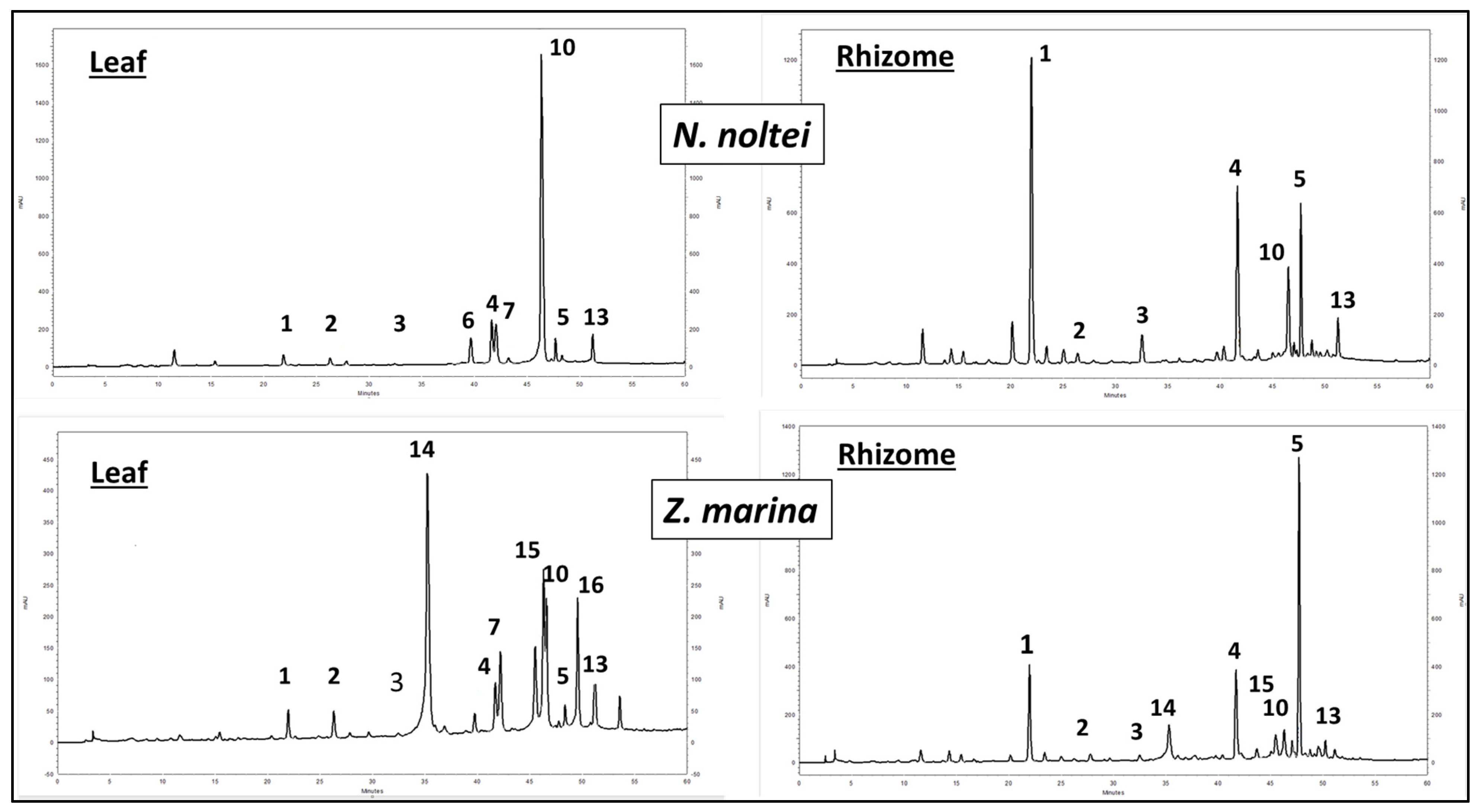
Eleven individual compounds (four phenolic acid derivatives and seven flavonoids) were detected with variable intensity in all collections of leaves. The phenolic acids were identified as zosteric (1), caffeic (2) and rosmarinic acids (4) and a caffeic tetramer (5), of which rosmarinic acid was always predominant in all collections (Figure 2 and Figure 3). This is the first report of compound 5 in N. noltei. The eight flavonoids were identified as luteolin 7-O-glucoside (6), luteolin 7-sulfate (7), apigenin 7-O-glucoside (8), apigenin 7-sulfate (9), diosmetin 7-sulfate (10), luteolin (11) and diosmetin (13), of which diosmetin 7-sulfate was largely predominant in all collections. Two minor products, identified as coumaric acid (3) and apigenin (12), were also found in some samples. Unlike in the leaves, the rhizomes were found to contain mainly phenolic acids 15, along with flavonoids in relatively small variable amounts (Figure 2 and Figure 3). The structure assignment was based on LC/MS analyses, online UV spectra, NMR data and comparison to standards (Tables S1 and S2; Figures S2 and S8–S10). Total acid hydrolysis of the crude extracts also confirmed the structure assignments. The process resulted in the complete disappearance of zosteric acid (peak 1), while the other phenolic acids (25) were recovered unchanged. The five peaks assigned as hydrolysable flavonoids (610) disappeared, while the peak intensity of the aglycones (1113) increased.

3.2. Seasonal and Interannual Dynamics of the Individual Phenolic Concentrations in N. Noltei

Quantitative amounts of compounds 14 are expressed in mg per g of N. noltei plant DW, and those of compounds 5 to 10 in mg of standard equivalents per g of N. noltei plant DW, with the standards used being, respectively, 5: rosmarinic acid, 67: luteolin, 89: apigenin and 10: diosmetin.

3.2.1. Phenolic Concentrations in N. noltei Leaves

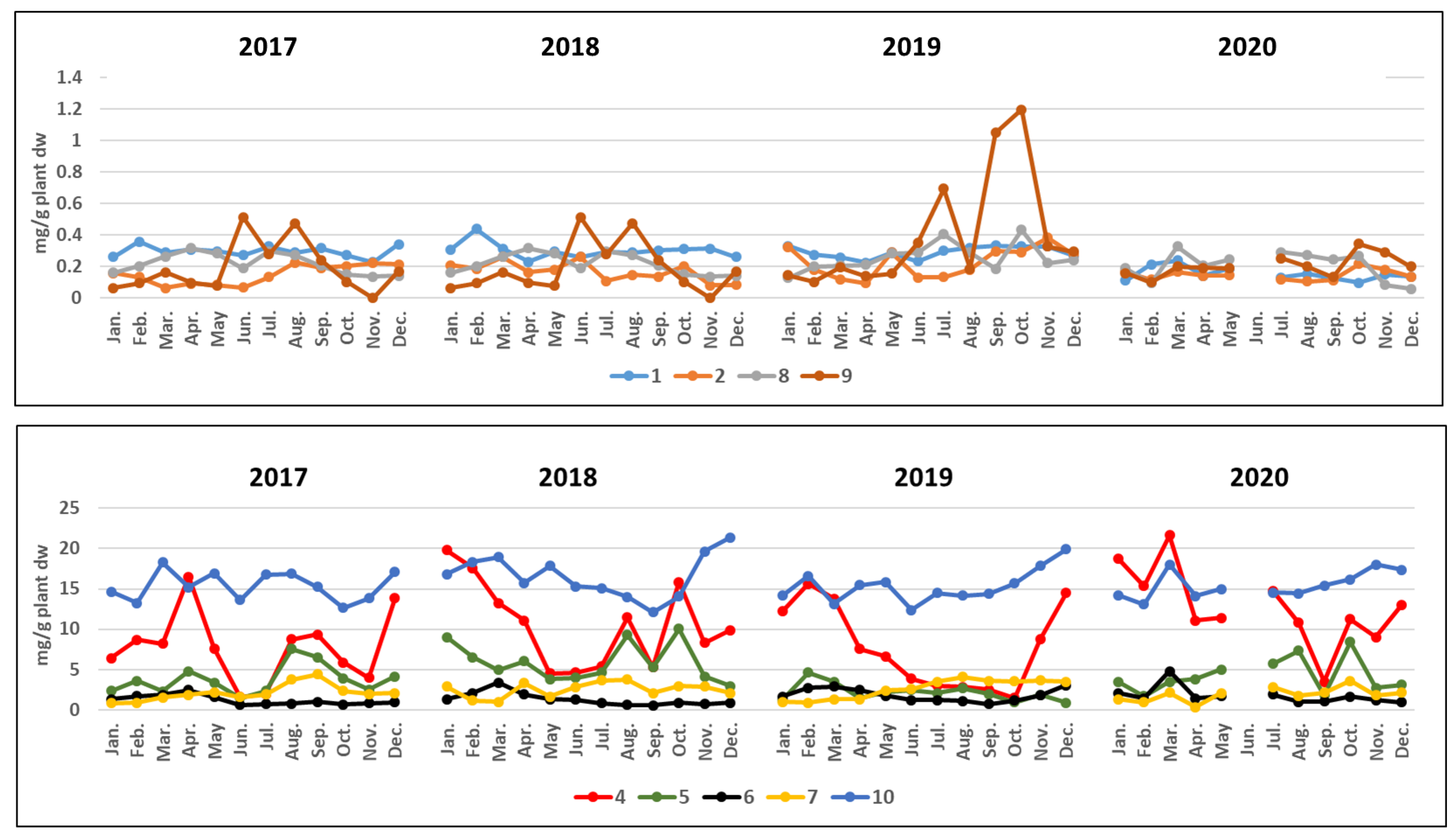
The quantitative HPLC dataset of the leaf tissues is displayed in Table 2 and Figure 4 and Figure S11 (average of the three collection replicates expressed as mg per g of plant dry weight (mg/g plant DW). The total amounts of phenolic acids (evaluated as the sum of all individuals quantified by HPLC) vary respectively in the range of 3.46–18.51 (2017), 8.80–34.26 (2018), 1.47–15.63 (2019) and 5.58–25.55 mg/g plant DW (2020) (Table 2). Rosmarinic acid (4) was by far the most abundant whatever the season and year, and compound 5 was second in abundance (Figure 4). Only four exceptions were observed, in which the respective values of these two compounds were either inversed (July 2017 and August 2019) or about the same (September 2018 and August 2019). The production of rosmarinic acid (4) shows a marked seasonality, as shown by the year-to-year range variations (1.61–16.51 (2017), 4.51–19.82 (2018), 1.47–15.63 (2019) and 3.52–21.63 (2020) mg/g plant DW), with the highest concentration always observed during the cold season.
Table 2. Quantitative amounts (mg/g plant DW, mean values ± SD) of individual phenolic compounds in leaves of N. noltei collected monthly over four years. TPA: total phenolic acids; TF: total flavonoids; TP: total phenolic compounds. For each class of compound (phenolic acid or flavonoid), individual substances are given in order of elution. Compounds are coded as in Figure 2. The quantitative amounts of compounds 1 to 4 are expressed in mg per g of N. noltei DW, and those of compounds 5 to 10 in mg of standard equivalents per g of N. noltei DW, with the standards used being, respectively, 5: rosmarinic acid, 67: luteolin, 89: apigenin and 10: diosmetin.
2017CompoundsJanuaryFebruaryMarchAprilMayJuneJulyAugustSeptemberOctoberNovemberDecemberConcentration Ranges
Phenolic
acids
10.26 ± 0.040.36 ± 0.020.29 ± 0.010.31 ± 0.010.30 ± 0.010.27 ± 0.010.33 ± 0.010.29 ± 0.010.32 ± 0.010.27 ± 0.010.23 ± 0.010.34 ± 0.020.23–0.36
20.19 ± 0.010.13 ± 0.010.07 ± 0.010.09 ± 0.010.08 ± 0.010.07 ± 0.010.13 ± 0.010.22 ± 0.010.19 ± 0.010.20 ± 0.020.22 ± 0.010.22 ± 0.020.07–0.22
30000.03 ± 0.01000.02 ± 0.01000000–0.03
46.42 ± 0.098.72 ± 0.028.18 ± 0.0116.51 ± 0.017.59 ± 0.121.61 ± 0.011.98 ± 0.028.76 ± 0.019.35 ± 0.025.90 ± 0.013.97 ± 0.0113.82 ± 0.031.61–16.51
52.33 ± 0.033.58 ± 0.022.28 ± 0.024.78 ± 0.013.41 ± 0.141.51 ± 0.012.34 ± 0.017.56 ± 0.016.52 ± 0.013.90 ± 0.012.53 ± 0.154.13 ± 0.011.51–7.56
TPA9.2012.7910.8221.7211.383.464.8016.8316.3810.276.9518.513.4618.51
% of TP32%42%31%50%33%16%18%42%43%38%28%46%1650%
Flavonoids61.31 ± 0.011.73 ± 0.051.88 ± 0.012.46 ± 0.021.61 ± 0.010.63 ± 0.010.73 ± 0.010.79 ± 0.011.01 ± 0.010.70 ± 0.010.86 ± 0.010.96 ± 0.010.7–2.46
70.85 ± 0.030.86 ± 0.021.56 ± 0.011.86 ± 0.022.21 ± 0.011.60 ± 0.011.90 ± 0.013.75 ± 0.014.40 ± 0.012.34 ± 0.012.0 ± 0.022.10 ± 0.010.85–4.40
80.16 ± 0.010.20 ± 0.010.27 ± 0.010.32 ± 0.010.28 ± 0.010.19 ± 0.010.30 ± 0.010.27 ± 0.010.21 ± 0.010.15 ± 0.010.14 ± 0.010.14 ± 0.010.14–0.32
90.06 ± 0.010.09 ± 0.010.16 ± 0.010.10 ± 0.010.08 ± 0.020.51 ± 0.010.28 ± 0.020.48 ± 0.010.24 ± 0.010.10 ± 0.0100.17 ± 0.010–0.51
1014.65 ± 0.0413.25 ± 0.0318.31 ± 0.0115.21 ± 0.0116.94 ± 0.0313.62 ± 0.0216.80 ± 0.0216.89 ± 0.0215.26 ± 0.0112.63 ± 0.0113.84 ± 0.0117.09 ± 0.0212.63–18.31
110.27 ± 0.010.22 ± 0.010.46 ± 0.010.37 ± 0.010.58 ± 0.010.30 ± 0.010.46 ± 0.010.34 ± 0.010.17 ± 0.010.16 ± 0.020.40 ± 0.010.26 ± 0.010.16–0.58
120000000000000
132.04 ± 0.021.6 ± 0.021.45 ± 0.021.27 ± 0.021.37 ± 0.010.88 ± 0.011.54 ± 0.020.80 ± 0.010.43 ± 0.010.68 ± 0.010.97 ± 0.011.39 ± 0.020.43–2.04
TF19.3417.9524.0921.5923.0717.7322.0123.3221.7216.7618.2122.1118.2124.09
% of TP68%58%69%50%67%84%82%58%57%62%72%54%5084%
TP28.5430.7434.9143.3134.4521.1926.8140.1538.1027.0325.1640.6221.1943.31
2018CompoundsJanuaryFebruaryMarchAprilMayJuneJulyAugustSeptemberOctoberNovemberDecemberConcentration
ranges
Phenolic
acids
10.31 ± 0.010.44 ± 0.010.31 ± 0.010.23 ± 0.010.29 ± 0.010.26 ± 0.020.29 ± 0.020.29 ± 0.010.30 ± 0.020.31 ± 0.020.31 ± 0.020.26 ± 0.020.23–0.44
20.21 ± 0.010.19 ± 0.010.26 ± 0.010.16 ± 0.010.18 ± 0.020.26 ± 0.020.11 ± 0.010.15 ± 0.020.14 ± 0.010.20 ± 0.020.08 ± 0.020.08 ± 0.020.08–0.26
30000000000000
419.82 ± 0.0217.57 ± 0.1613.26 ± 0.0211.06 ± 0.024.51 ± 0.014.63 ± 0.025.36 ± 0.0111.44 ± 0.205.30 ± 0.0215.87 ± 0.028.33 ± 0.039.86 ± 0.034.51–24.74
59.0 ± 0.026.52 ± 0.014.94 ± 0.026.03 ± 0.013.82 ± 0.034.0 ± 0.034.63 ± 0.029.27 ± 0.035.29 ± 0.0210.05 ± 0.024.14 ± 0.012.95 ± 0.023.82–10.05
TPA29.3424.7218.7717.488.809.1510.3921.1511.0326.4312.8613.158.8029.34
% of TP56%52%43%44%28%31%33%52%41%56%34%34%2856%
Flavonoids61.34 ± 0.012.11 ± 0.013.38 ± 0.011.93 ± 0.021.35 ± 0.021.29 ± 0.020.82 ± 0.010.63 ± 0.020.55 ± 0.020.90 ± 0.020.71 ± 0.010.89 ± 0.020.55–3.38
72.92 ± 0.021.14 ± 0.011.01 ± 0.013.35 ± 0.021.66 ± 0.022.82 ± 0.023.64 ± 0.023.77 ± 0.022.10 ± 0.022.96 ± 0.022.87 ± 0.022.06 ± 0.021.01–3.77
80.17 ± 0.010.09 ± 0.010.41 ± 0.020.32 ± 0.020.19 ± 0.010.29 ± 0.010.21 ± 0.010.16 ± 0.010.11 ± 0.010.11 ± 0.010.09 ± 0.020.13 ± 0.020.09–0.41
90.11 ± 0.010.10 ± 0.010.12 ± 0.010.08 ± 0.010.16 ± 0.010.25 ± 0.020.21 ± 0.010.13 ± 0.020.21 ± 0.020.13 ± 0.020.10 ± 0.020.13 ± 0.010.08–0.25
1016.84 ± 0.0218.35 ± 0.0218.91 ± 0.0115.73 ± 0.0217.88 ± 0.0215.29 ± 0.0215.08 ± 0.0314.0 ± 0.0212.10 ± 0.0214.07 ± 0.0219.65 ± 0.1721.37 ± 0.1312.10–21.37
110.31 ± 0.010.12 ± 0.010.17 ± 0.010.31 ± 0.010.22 ± 0.010.22 ± 0.020.19 ± 0.010.41 ± 0.010.37 ± 0.011.71 ± 0.010.72 ± 0.020.12 ± 0.010.12–1.71
120000000000000
131.40 ± 0.011.21 ± 0.011.36 ± 0.010.70 ± 0.020.73 ± 0.010.65 ± 0.020.51 ± 0.020.62 ± 0.020.74 ± 0.010.51 ± 0.020.92 ± 0.020.92 ± 0.010.51–1.40
TF23.0923.1225.3622.4222.1920.8120.6619.7216.1820.3925.0625.6216.1825.62
% of TP44%48%57%56%72%69%67%48%59%44%66%66%4472%
TP52.4347.8444.1339.9030.9929.9631.0540.8727.2146.8237.9238.7727.2152.43
2019CompoundsJanuaryFebruaryMarchAprilMayJuneJulyAugustSeptemberOctoberNovemberDecemberConcentration ranges
Phenolic
acids
10.33 ± 0.020.28 ± 0.020.26 ± 0.020.23 ± 0.010.29 ± 0.010.23 ± 0.020.30 ± 0.020.32 ± 0.020.33 ± 0.020.33 ± 0.020.33 ± 0.020.27 ± ±0.020.23–0.33
20.32 ± 0.010.18 ± 0.020.12 ± 0.020.10 ± 0.020.29 ± 0.010.13 ± 0.020.13 ± 0.020.18 ± 0.020.30 ± 0.020.29 ± 0.020.38 ± 0.010.28 ± 0.020.10–0.38
3000000.04 ± 0.0100.04 ± 0.010.05 ± 0.010.04 ± 0.010.05 ± 0.010.04 ± 0.010–0.047
412.22 ± 0.0315.63 ± 0.0213.79 ± 0.037.56 ± 0.026.63 ± 0.023.85 ± 0.033.00 ± 0.022.86 ± 0.022.53 ± 0.021.47 ± 0.028.73 ± 0.0214.47 ± 0.011.47–15.63
51.20 ± 0.024.64 ± 0.023.49 ± 0.021.51 ± 0.012.19 ± 0.012.45 ± 0.022.08 ± 0.022.69 ± 0.021.96 ± 0.020.95 ± 0.021.87 ± 0.020.87 ± 0.020.87–4.64
TPA14.0720.7317.669.409.406.705.516.095.173.0811.3615.933.0820.73
% of TP44%49%47%29%29%27%21%23%20%12%31%36%2149%
Flavonoids61.64 ± 0.012.74 ± 0.022.90 ± 0.022.48 ± 0.021.74 ± 0.011.19 ± 0.021.24 ± 0.011.13 ± 0.010.72 ± 0.021.18 ± 0.021.79 ± 0.023.06 ± 0.020.72–3.06
71.02 ± 0.020.91 ± 0.021.31 ± 0.011.30 ± 0.022.43 ± 0.022.55 ± 0.033.5 ± 0.034.11 ± 0.023.61 ± 0.023.53 ± 0.023.67 ± 0.023.49 ± 0.020.91–4.11
80.13 ± 0.010.20 ± 0.010.21 ± 0.010.21 ± 0.010.28 ± 0.020.29 ± 0.020.41 ± 0.020.29 ± 0.010.19 ± 0.020.43 ± 0.030.22 ± 0.010.24 ± 0.020.13–0.43
90.15 ± 0.020.10 ± 0.020.20 ± 0.020.14 ± 0.010.16 ± 0.010.35 ± 0.020.69 ± 0.010.19 ± 0.021.05 ± 0.021.20 ± 0.020.33 ± 0.020.29 ± 0.020.10–1.20
1014.16 ± 0.0316.61 ± 0.0313.16 ± 0.0415.47 ± 0.0315.83 ± 0.0212.36 ± 0.0214.52 ± 0.0214.16 ± 0.0214.4 ± 0.0315.71 ± 0.0217.87 ± 0.0219.92 ± 0.0212.36–19.92
110.06 ± 0.020.09 ± 0.020.28 ± 0.010.75 ± 0.020.65 ± 0.010.51 ± 0.020.32 ± 0.020.28 ± 0.010.17 ± 0.020.19 ± 0.020.18 ± 0.020.33 ± 0.010.06–0.75
120000.05 ± 0.0100.18 ± 0.010.08 ± 0.01000.04 ± 0.010.08 ± 0.010.14 ± 0.0100–0.18
130.59 ± 0.030.83 ± 0.021.64 ± 0.023.03 ± 0.021.76 ± 0.020.72 ± 0.020.59 ± 0.010.53 ± 0.020.61 ± 0.010.57 ± 0.020.68 ± 0.020.90 ± 0.010.59–3.03
TF17.7521.4819.7023.4322.8518.1521.3520.6920.7922.8924.8828.2317.7528.23
% of TP56%51%53%71%71%73%79%77%80%88%69%64%5188%
TP31.8242.2137.3632.8332.2524.8526.8626.7825.9625.9736.2444.1624.8544.16
2020CompoundsJanuaryFebruaryMarchAprilMayJuneJulyAugustSeptemberOctoberNovemberDecemberConcentration
ranges
Phenolic
acids
10.11 ± 0.010.21 ± 0.020.24 ± 0.020.14 ± 0.010.19 ± 0.01 0.13 ± 0.020.15 ± 0.020.13 ± 0.020.10 ± 0.020.15 ± 0.020.14 ± 0.020.10–0.24
20.17 ± 0.010.12 ± 0.010.17 ± 0.010.14 ± 0.010.15 ± 0.01 0.12 ± 0.010.11 ± 0.020.11 ± 0.010.22 ± 0.020.18 ± 0.010.14 ± 0.020.11–0.22
300000 0000000
418.78 ± 0.0215.34 ± 0.0221.63 ± 0.0211.08 ± 0.0111.41 ± 0.01 14.79 ± 0.0110.82 ± 0.023.52 ± 0.0211.23 ± 0.029.04 ± 0.0113.05 ± 0.039.04–21.63
53.52 ± 0.011.69 ± 0.013.51 ± 0.023.82 ± 0.025.01 ± 0.01 5.73 ± 0.017.33 ± 0.021.82 ± 0.018.44 ± 0.022.74 ± 0.013.09 ± 0.021.69–8.44
TPA22.5817.3625.5515.1816.76 20.7718.415.5819.9912.1116.425.5825.55
% of TP55%50%48%47%45% 50%50%22%47%36%43%2255%
Flavonoids62.06 ± 0.011.45 ± 0.014.81 ± 0.011.41 ± 0.011.71 ± 0.02 1.94 ± 0.020.98 ± 0.011.06 ± 0.021.67 ± 0.011.20 ± 0.010.97 ± 0.020.97–4.81
71.31 ± 0.020.95 ± 0.022.12 ± 0.010.32 ± 0.012.07 ± 0.02 2.86 ± 0.021.78 ± 0.022.15 ± 0.013.63 ± 0.021.84 ± 0.022.16 ± 0.020.32–3.63
80.19 ± 0.020.10 ± 0.010.33 ± 0.010.21 ± 0.010.24 ± 0.02 0.29 ± 0.010.27 ± 0.020.25 ± 0.020.27 ± 0.010.08 ± 0.020.06 ± 0.010.06–0.33
90.16 ± 0.020.10 ± 0.020.20 ± 0.010.19 ± 0.020.19 ± 0.01 0.25 ± 0.020.20 ± 0.010.13 ± 0.010.35 ± 0.020.29 ± 0.020.20 ± 0.020.10–0.35
1014.20 ± 0.0213.07 ± 0.0218.02 ± 0.0214.12 ± 0.0214.96 ± 0.02 14.52 ± 0.0314.43 ± 0.0215.42 ± 0.0216.16 ± 0.0118.02 ± 0.0217.35 ± 0.0213.07–18.02
110.09 ± 0.010.28 ± 0.020.30 ± 0.010.17 ± 0.010.27 ± 0.01 0.45 ± 0.020.21 ± 0.010.15 ± 0.020.33 ± 0.020.11 ± 0.010.19 ± 0.020.09–0.45
1200000 0000000
130.71 ± 0.011.36 ± 0.021.68 ± 0.020.71 ± 0.020.96 ± 0.01 0.73 ± 0.020.51 ± 0.020.40 ± 0.020.45 ± 0.010.43 ± 0.030.74 ± 0.010.40–1.68
TF18.7217.3127.4617.1320.40 21.0418.3819.5622.8621.9721.6717.1327.46
% of TP45%50%52%53%55% 50%50%78%53%64%57%4578%
TP41.3034.6753.0132.3137.16 41.8136.7925.1442.8534.0838.0925.14–53.01
Seven flavonoids (711 and 13; Figure 2 and Figure 3 and Table 2) were detected with variable intensity in all samples. Diosmetin 7-sulfate (10) was the most abundant regardless of season and year (12.10–21.37 mg/g plant DW over the four years). Compared to rosmarinic acid, the variation in its concentrations was three- to seven-fold less. Luteolin 7-O-glucoside (6) and luteolin 7-sulfate (7) were systematically present, ranging from 0.55 to 4.81 and 0.85 to 4.40 mg/g plant DW. Apigenin 7-O-glucoside (8) and apigenin 7-sulfate (9), when they were present, occurred in considerably lower amounts over the four years (respectively, 0.04–0.43 and 0.08–1.20 mg/g plant DW) (Figure 4A). Variable amounts of luteolin (11: 0.06–1.71 mg/g plant DW) and diosmetin (13: 0.40–3.03 mg/g plant DW) were also detected in all the samples. Although they differ greatly in abundance, sulfated flavones 7 and 10 showed similar seasonal and year-to-year variability, with a moderate production maximum observed during November and December.

3.2.2. Phenolic Concentrations in N. noltei Rhizomes

To reduce the number of destructive samplings, whole plants (i.e., with above- and belowground tissues) were sampled only six times between May 2019 and October 2020. Crude extracts from rhizomes were prepared under the same conditions as for leaves and quantitatively compared by HPLC to leaf extracts from the same plant sample (Table 3 and Table 4 and Figure 3).
Table 3. Quantitative amounts (mg/g plant DW, mean values ± SD) of individual phenolic compounds in rhizomes of N. noltei collected in Arcachon Bay. TPA: total phenolic acids; TF: total flavonoids; TP: total phenolic compounds. Concentrations of compounds 14 are expressed in mg per g of N. noltei plant DW, and those of compounds 5 to 10 in mg of standard equivalents per g of N. noltei DW, with the standards used being, respectively, 5: rosmarinic acid, 67: luteolin, 89: apigenin and 10: diosmetin.
CompoundsMay 2019June 2019July 2019April 2020August 2020October 2020Concentration Ranges
Phenolic acids10.72 ± 0.020.26 ± 0.020.24 ± 0.010.13 ± 0.020.62 ± 0.020.50 ± 0.020.13–0.72
20.08 ± 0.020.09 ± 0.010.05 ± 0.010.02 ± 0.010.05 ± 0.010.07 ± 0.020.02–0.09
30.21 ± 0.021.14 ± 0.020.20 ± 0.010.22 ± 0.010.06 ± 0.010.07 ± 0.010.06–1.14
43.28 ± 0.021.85 ± 0.011.65 ± 0.022.35 ± 0.021.39 ± 0.011.79 ± 0.021.39–3.28
50.92 ± 0.020.74 ± 0.021.21 ± 0.021.57 ± 0.021.47 ± 0.011.75 ± 0.010.74–1.75
TPA5.214.083.354.293.594.183.355.21
Flavonoids60.09 ± 0.010.07 ± 0.010.05 ± 0.020.05 ± 0.010.08 ± 0.010.06 ± 0.010.05–0.09
70.11 ± 0.020.05 ± 0.02000.02 ± 0.010.03 ± 0.010–0.05
800000.04 ± 0.0100–0.04
90.05 ± 0.010.03 ± 0.010.02 ± 0.010000–0.05
100.71 ± 0.020.20 ± 0.010.17 ± 0.0100.86 ± 0.020.26 ± 0.010–0.86
110.11 ± 0.020.11 ± 0.0100.04 ± 0.02000–0.11
1200000.02 ± 0.010.01 ± 0.000–0.01
131.22 ± 0.020.79 ± 0.020.59 ± 0.020.67 ± 0.010.34 ± 0.020.15 ± 0.020.59–1.221
TF2.291.260.830.761.360.510.512.29
TP7.505.344.185.054.954.694.187.50
Table 4. Relative content (%) of each compound in respect to its compound class (phenolic acid or flavonoid). TPA: total phenolic acids; TF: total flavonoids.
CompoundsMay 2019June 2019July 2019April 2020August 2020October 2020
Phenolic
acids
114%6%7%3%17%12%
21%2%1%1%2%2%
34%28%6%5%2%2%
463%45%49%55%39%43%
518%18%36%36%41%42%
Flavonoids64%6%6%6%6%12%
75%4% 2%5%
8 3%
92%3%3%
1031%16%20% 63%52%
115%9% 5%
12 1%1%
1353%63%71%89%25%30%
TPA69%76%80%85%72%89%
TF31%24%20%15%28%11%
Unlike leaves, rhizomes were found to contain mainly the phenolic acids 15, along with flavonoids in relatively small variable amounts (Table 3). Total phenolic compounds were four- to nine-fold less in rhizomes than in leaves. This is mainly due to the virtual absence of flavonoid sulfates (ten- to forty-six-fold less in rhizomes than in leaves) and, to a lesser extent, to the lower amount of phenolic acids (two- to five-fold less in rhizomes than in leaves). All the samples were dominated by rosmarinic acid (4, 1.39–3.28 mg/g plant DW, 39–63% of the phenolic acids) and compound 5 (0.74–1.75 mg/g plant DW, 18–42% of the phenolic acids). The abundance of zosteric acid in rhizomes was higher than in leaves (1: 0.13–0.72 mg/g plant DW, 3–17%), and coumaric acid (3), virtually absent in the leaves, was found in significant amounts (0.06–1.14 mg/g plant DW, 2–28%) in the rhizomes. Given that zosteric acid is the sulfated form of coumaric acid, these results suggest that the sulfation step may take place in the belowground tissues. Diosmetin 7-sulfate (10), although only slightly present (0–0.86 mg/g plant DW), remains the dominant flavonoid of the rhizomes (16–63% of the total flavonoids). Globally, total phenolic acids and total flavonoids respectively account for 69–89% and 11- 31% of the total phenolics (Table 4).
Compound 5 was previously undescribed in the plant kingdom, until its recent identification in detrital and fresh leaves of Z. marina [43,52]. Structurally, it is close to salvianolic acid B, differing only in the absence of the two hydrogen atoms at the 7″ and 8″ positions (Figure 2). Nevertheless, rather than refer to it as 7″,8″-didehydrosalvianolic acid B, as has previously been done [43,52], we propose to adopt zosteranoic acid as its common name. Indeed, it has only been described in two species of Zosteraceae, which are in no way related to the genus Salvia from which salvianolic acid B was first isolated and named.

3.2.3. Phenolic Concentrations in Z. marina Rhizomes and N. noltei Samples from Other Sites in the Atlantic and Mediterranean

The presence of zosteranoic acid (5) in both above- and belowground tissues is of particular interest. This prompted us to examine the phenolic content of Z. marina rhizome samples from Arcachon Bay, Hossegor Lake and Thau Lagoon (Figure 1B and Table 1). In addition, we have reanalyzed crude extracts of N. noltei leaves from seagrass beds representative of the geographical distribution of the species. The results are summarized in Table 5 and Table S3.
Table 5. Quantitative amounts of zosteranoic acid, 5, from specimens collected at other sites in the Atlantic and Mediterranean. Values are expressed as mg rosmarinic acid equivalents ± standard deviation (SD) per gram of plant DW.
SitesN. noltei LeavesN. noltei RhizomesZ. marina Rhizomes
Arguin3.48 ± 0.02 3.07 ± 0.02
Hossegor1.08 ± 0.011.37 ± 0.027.54 ± 0.02
Thau2.59 ± 0.021.13 ± 0.015.47 ± 0.02
Berres1.86 ± 0.02
Salses0.22 ± 0.01
Strunjan1.80 ± 0.02
Bizerte1.57 ± 0.02
Ria Formosa3.16 ± 0.020.54 ± 0.01
Pointe de l’Etoile3.31 ± 0.02
Cadiz bay2.37 ± 0.02
Merja Zerga1.03 ± 0.02
Concentration ranges0.22–3.480.54–1.373.07–7.54
Regarding N. noltei leaves from other sites, the same individual flavonoids were present, and their respective concentrations were consistent with the three geographically distinct chemotypes previously highlighted [37]. As with specimens from Arcachon Bay, zosteric and caffeic acids were present in moderate and variable amounts, coumaric acid was absent, and rosmarinic acid was the major phenolic acid. Zosteranoic acid, which was not detected in the previous work, was found at all locations.
As with the leaves, the results obtained with N. noltei rhizomes from the three localities (Hossegor, Thau and Ria Formosa) were similar to those from Arcachon Bay. All the samples were dominated by rosmarinic acid (respectively, 1.38, 2.27 and 3.55 mg/g plant DW), and zosteranoic acid was found in all samples (respectively, 1.37, 1.13 and 0.54 mg/g plant DW). Flavonoids were present in relatively low amounts ranging, respectively, from 0.01 to 0.77, 0 to 0.33 and 0.07 to 0.73 mg/g plant DW. Globally, total phenolic acids versus total flavonoids at Hossegor, Thau and Ria Formosa respectively accounted for 73 vs. 27%, 83 vs. 17% and 74 vs. 26% of the total phenolics.
Similar results following the same tendency were observed with the rhizomes of Z. marina, for which high concentrations of zosteranoic acid are especially noteworthy (Arguin, 3.07; Hossegor, 7.54; Thau, 5.48 mg/g plant DW). These results show that Z. marina and N. noltei contain the same phenolic acids, unlike their flavonoids, which clearly distinguish the two species. This is the first characterization of the phenolic fingerprints of Z. marina rhizomes and the first report of zosteranoic acid in Z. marina belowground tissues.

3.3. Principal Component Analysis and Hierarchical Cluster Analysis

Principal component analyses (PCAs) were carried out with the five datasets obtained from N. noltei (leaves and rhizomes) and Z. marina rhizomes (see Section 3.2.1, Section 3.2.2 and Section 3.2.3). Data were also subjected to a hierarchical cluster analysis (HCA). Sample similarities were calculated based on the Euclidean distance, and the Ward hierarchical agglomerative method [51]. Automatic truncation based on entropy parameters was used [53]. The results are depicted in Figure 5 and Figure S13.

3.3.1. N. noltei Leaves from Arcachon Bay

The relationships among the 47 collection dates (observations) and the phenolic compound concentrations determined by HPLC (variables) were analyzed. The PCAs showed a cumulative variance of 40.24% up to the second principal component with values of F1 and F2 equal to 23.53% and 16.71%, respectively (Figure 5(1A)). The first principal component is strongly correlated with flavonoids 7 and 9 (positive associations) and with flavonoid 13 and rosmarinic acid (4) (negative associations). The second principal component increases with flavonoids 6 and 8 (positive associations). Rosmarinic acid (4) is positively correlated with zosteranoic acid (5) and luteolin 7-O-glucoside (6) and negatively correlated with apigenin 7-sulfate (9). Diosmetin 7-sulfate (10) is only weakly correlated with the other phenolics. The dendrogram obtained from the cluster analysis of the 47 collections in relation to the concentrations of chemical constituents is presented in Figure 5(1B). The dotted line on the chart represents the automatic truncation based on entropy parameters [53]. It identified two clusters (C1 and C2), which occur at a dissimilarity level of approximately 300. Cluster C1 comprises 32 collections, with a predominance of late spring, summer and autumn, while cluster C2 gathers 15 collections, mainly representative of the cold seasons (especially January, February and March for the years 2018, 2019 and 2020, and December of 2017, 2019 and 2020). At a dissimilarity level of about 250, cluster C1 consists of four subclusters (late spring, summer and autumn) and cluster C2 consists of two subclusters representative of the cold seasons.
The dendrogram obtained from the cluster analysis of the variables identifies two clusters (Figure 5(1C)). Cluster C1 groups together nine of the twelve phenolic compounds, while cluster C2 is bifolious, gathering rosmarinic acid (4) and diosmetin 7-sulfate (10), which are the main products whatever the season or year. At a dissimilarity level around 15, cluster C1 distinguishes zosteranoic acid (5) as a simplicifolius from the minor phenolics 7, 11, 9, 1, 2, 8, 6, and 11.

3.3.2. N. noltei Rhizomes from Arcachon Bay
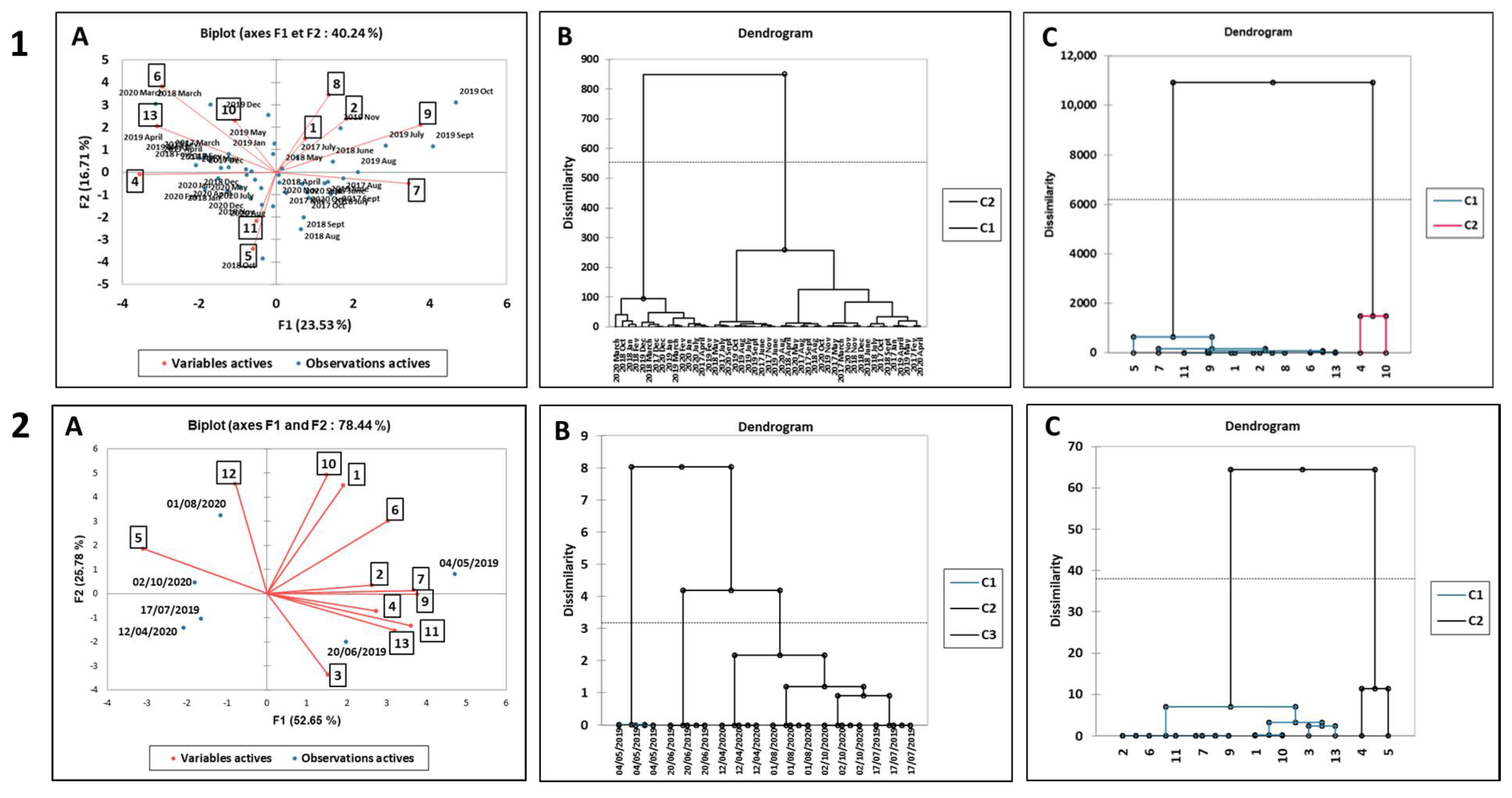
The relationships among the six collection dates (observations) and the phenolic compound concentrations determined by HPLC (variables) are depicted in Figure 5(2A). It shows a cumulative variance of 78.44% up to the second principal component with values of F1 and F2 equal to 52.65% and 25.78%, respectively. The first principal component is strongly correlated with flavonoids 7, 9, 11, and 13 and rosmarinic acid (4) (positive associations) and with zosteranoic acid (5) (negative association). The second component has large positive associations with the other flavonoids 10 and 11 and with zosteric acid (1). Rosmarinic acid (4) and zosteranoic acid (5), which were moderately and positively correlated to each other in the leaves, were found to be negatively correlated in the rhizomes. This suggests that the formation of zosteranoic acid might take place in the rhizomes via rosmarinic acid dimerization. A negative correlation was also observed between zosteric acid (1) and coumaric acid (3), which reinforces the suggestion, based on their concentrations, that zosteric acid biosynthesis may take place in the rhizomes (see Section 3.2.2). Diosmetin 7-sulfate (10), which was only weakly correlated with the other phenolics in the leaves, shows strong positive correlations with zosteric acid (1), luteolin 7-O-glucoside (6) and apigenin (12) in the rhizomes.
The dendrogram obtained from the cluster analysis of the six collections of rhizomes in relation to the concentrations of chemical constituents is presented in Figure 5(2B). The automatic truncation identified three clusters (C1–C3), which occur at a dissimilarity level of approximately 3.2. Cluster C1 identifies the 4 May 2019 collection as a simplicifolius and and C3 as a bifolious clade with C2 constituted of one collection (20 June 2019) and C3 of four (the three collections of 2020 and the one on 17 July 2019).
The dendrogram obtained from the cluster analysis of the variables identifies two clusters (Figure 5(2C)). Cluster C1 groups together nine of the eleven phenolic compounds, namely three phenolic acids (13) and six flavonoids (67, 911 and 13), while cluster C2 includes rosmarinic acid (4) and zosteranoic acid (5), which are the major products in the rhizomes.
Figure 5. Principal component and hierarchical cluster analysis for: (1)-N. noltei leaves from Arcachon Bay and (2)-N. noltei rhizomes from Arcachon Bay. In each case: (A)-PCA biplot graph based on PC1 and PC2 scores; (B)-dendrogram based on the phenolic composition; and (C)-dendrogram obtained from the cluster analysis of the variables. Compound numbers and collection dates are the same as in Table 2 and Table 3 and Figure 2 and Figure 3.
Diversity 15 01210 g005

4. Discussion

To date, little attention has been paid to seasonal variability in the phenolic content of N. noltei. The seasonal variation of flavonoids found in the leaves of N. noltei from Norwegian coastal waters was analyzed bi-monthly over one year [38], and those of rosmarinic and zosteric acids in detrital leaves of N. noltei from Arcachon Bay were analyzed monthly over one year [40,41]. Longer-term observations were needed to more accurately assess the relationship between phenolic profiles and seasons, given that the response of an individual species could be used to predict how populations may expand or decline in the future.
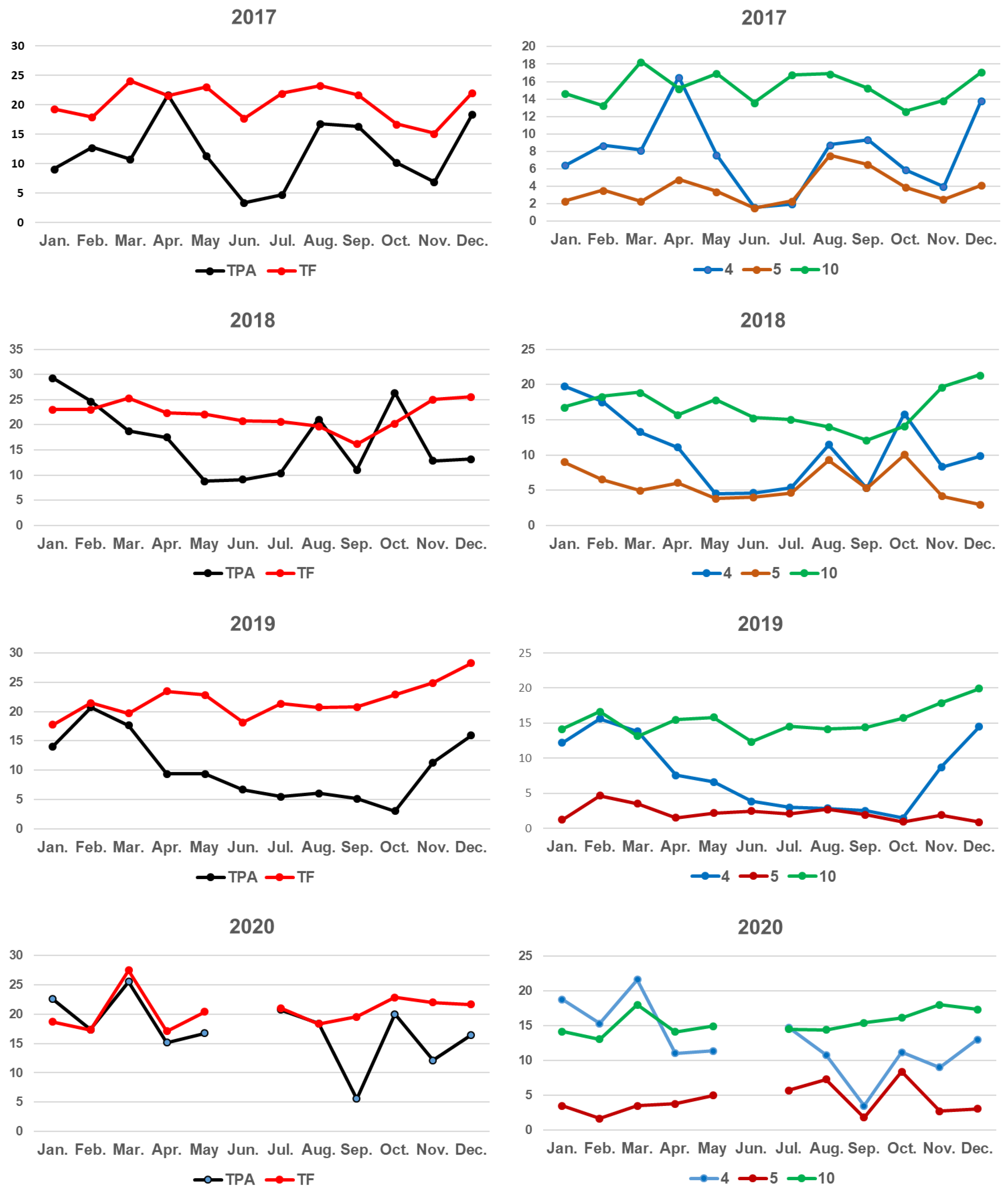
The present study took place in Arcachon Bay, which is home to one of Europe’s largest N. noltei beds. For the first time, the variability of the phenolic chemistry of a N. noltei meadow was analyzed in detail over 47 consecutive months from 2017 to 2020. All the individual phenolics were identified and quantified over time. The different collections were found to be quite homogeneous, showing in all cases a similar HPLC profile, with variability in the concentration of individual compounds according to the seasons. The quantitative variation of flavonoids and phenolic acids was found to be relatively consistent from year to year as shown in Figure 4, which illustrates the month-to-month variation in the concentrations of phenolics over the four years of the study. The phenolic acid pool in both leaves and rhizomes was always dominated by rosmarinic acid (4), and zosteranoic acid (5) was the second phenolic acid in abundance (Table 2 and Table 3 and Figure S11). The flavonoid pool in leaves was largely dominated by diosmetin 7-sulfate, followed by luteolin 7-sulfate, while flavonoids are only present as minor compounds in the rhizomes (Table 3 and Table 4). The values for total flavonoids are usually higher than those for total phenolic acids and show a lower variability over seasons and from year to year than the total of phenolic acids (Figure 6, left column). The right column in Figure 6 compares the seasonal and year-to-year variations of rosmarinic acid (4), zosteranoic acid (5) and diosmetin 7-sulfate (10).
Comparison of the summer extracts showed similar HPLC profiles and a weak variability in the phenolic concentrations. For all leaf samples, the flavonoid pattern was found to be largely dominated by diosmetin 7-sulfate (10), representing 41–52% of the flavonoid pool. Luteolin 7-sulfate was the second flavonoid in abundance. Low amounts of caffeic (1) and coumaric acids (5) were found in all the samples (0.08–0.16 mg/g and 0.03–0.04 mg/g, respectively). These two phenolic acids are very common in seagrasses [54]. A strong seasonality was observed for rosmarinic acid (4), whose concentrations were higher in late autumn and winter than in late spring and summer (Table 2). In contrast, the variability of diosmetin 7-sulfate (10) seems to be relatively unaffected by temperature.
Based on the concentration amounts in the 47 collections, the phenolic compounds of N. noltei leaf tissue rank in the following order, whatever the season and the year:
-
Phenolic acids: Rosmarinic > Zosteranoic >>> Zosteric ~ Caffeic >> Coumaric.
-
Flavonoids: Diosmetin 7-sulfate >>> Luteolin 7-sulfate ~ Luteolin 7-O-glucoside > Apigenin 7-O-glucoside ~ Apigenin 7-sulfate.
With the exception of zosteric and coumaric acids, the other phenolic acids present in N. noltei and Z. marina are exclusively based on the caffeic acid building block, with the compounds being formed from two or four caffeic acid units. We had previously isolated rosmarinic acid from Z. marina and Phyllospadix torreyi, which also belongs to the Zosteraceae family [40,41,55]. The presence of zosteranoic acid (5) in both above- and belowground tissues is of particular interest. Chemically, compound 5 is a dimer of rosmarinic acid, which, in turn, is a dimer of caffeic acid and 3,4-dihydroxyphenyl lactic acid, linked by an ester bond. This is the first report of compound 5 in the leaves and rhizomes of N. noltei and in the rhizomes of Z. marina. In vivo, rosmarinic acid is localized in the vacuoles, but it also occurs in the cytoplasm as an anion and cannot pass any membrane by diffusion [56]. The rosmarinic acid biosynthesis pathway has been thoroughly explored. It has been shown that it may be further converted into salvianolic acid B, but the detailed biosynthetic pathway of this conversion has not yet been completely described [57]. It is therefore plausible that in Zosteraceae, a similar process may convert rosmarinic acid into zosteranoic acid. The question arises as to why the product of this conversion differs between marine and terrestrial plants (absence of two hydrogen atoms at the 7″ and 8″ positions, Figure 2). Molecular mechanic calculations of the energies of formation of these two compounds show that zosteranoic acid is thermodynamically more stable than salvianolic acid B (84.22 and 86.89 kJ/M, respectively). Nevertheless, the difference in structure probably reflects the adaptation of the plant metabolism to the marine environment.
The predominance of rosmarinic and zosteranoic acids in the rhizomes of N. noltei and Z. marina suggests their possible role as defense compounds. Root exudates play an important role in induced plant defense mechanisms [18]. The roots of Ocimum basilicum (Lamiaceae) secrete rosmarinic acid when challenged by the pathogenic fungus Pythium ultimum [58]. In the case of Z. marina, lesions caused by Labyrinthula protists were found to be positively correlated with the rosmarinic acid content of the seagrass [59]. Rosmarinic acid and sulfated flavonoids have been identified as inhibitors of microfouling on the surface of Z. marina [24,26]. The algicidal effects of Z. marina and N. noltei extracts on the neurotoxic bloom-forming dinoflagellate Alexandrium catenella have been demonstrated [25].
Seagrasses and algae may appear superficially similar, but in many ways, they are very different organisms. The transition of plants from sea to land required adaptation to a very different physical and chemical environment [60,61]. As a result, species composition and diversity vary dramatically between marine, freshwater and terrestrial habitats. Compared to seagrasses, which possess five families and a total of 73 species, terrestrial angiosperms are approximated to comprise about 416 families and a total of 300,000 confirmed species [62]). There is a higher degree of diversity within the latter group, making it challenging to draw comparisons. However, seagrasses and salt marsh plants are more similar taxonomically and physiologically to terrestrial plants than to other marine autotrophs [19]. Most of the specialized metabolites found in seagrass beds originate from their ancestral terrestrial life. This is especially the case with phenolic compounds.
Large amounts of rosmarinic acid are found in over 160 species belonging to many families, especially Lamiaceae [63]. Studies of the seasonal variations are most often limited to the growing season and/or only measure the total amounts of compound classes and not the quantities of individually characterized secondary metabolites. The lowest level of rosmarinic acid in the leaves of Salvia abrotanoides and S. yangii was found during flowering [64]. With Thymus longicaulis C. Presl., the lowest amount was found in July and the highest in October [65]. However, other species of Thymus L. showed the highest amounts during budding and full flowering, and then their amounts decreased during fruiting, and they displayed the lowest levels at the end of their vegetation [66].
Sulfated flavonoids are present in angiosperms and have been identified in eudicotyledonous and monocotyledonous plants [32,67,68,69]. A survey of 50 Fluviales species showed that over 50% have either flavone or caffeic acid sulfates present. Flavone sulfates were detected in 16% of the samples, and the 7-sulfates of luteolin, apigenin, diosmetin and chrysoeriol and the 7,3′-disulphate of luteolin were identified variously in Thalassia, Zannichellia and Zostera species [32]. The sulfate component is believed to represent a marine adaptation [67]. Sulfate is the third highest ion in concentration in seawater, and hydrogen sulfide is commonly found in anoxic marine sediment. Flavonoids are essential for various biological processes, principally plant development, growth and maturation. They help plants to withstand various biotic and abiotic stresses, such as viruses, fungi, bacteria and herbivores, while acting as chemical messengers in conjunction with mycorrhizae and bacteria. Flavonoids also contribute to flower color, attract pollinators and have allelopathic functions. Some studies indicate that sulfated flavonoids are involved in the detoxification of reactive oxygen species and regulation of plant growth [70].
Over 5000 naturally occurring flavonoids have been characterized from various plants. It is estimated that 150 sulfated flavonoids of natural occurrence have been identified. A survey of fourty-three species of seagrass showed that five of the twelve genera, including some members of the Zosteraceae family, contained flavonoid sulfates, namely Phyllospadix, Zostera, Enhalus, Thalassia and Halophila [32,33,55]. The flavonoid profile of P. torreyi, composed exclusively of sulfates, appears to be unique among seagrasses [55].
Very little is known of the accumulation of flavonoid sulfates in seagrass tissues, and their functional roles remain unclear. Harborne [71] suggested that conjugation with seagrass flavonoids might be a possible route for the inactivation or storage of inorganic sulfates. A sulfate conjugate is more water soluble than the unsulfated molecule or even the corresponding glycoside, thus facilitating transport to various cellular compartments and exudation in the water column. The possibility of flavonoid sulfates having a dynamic function in salt uptake and metabolism has also been suggested. Taxonomic and ecological implications were evoked by McMillan et al. [33]. Luteolin 7-O-D-glucopyranosyl-2-sulfate has been shown to chemically defend the tropical seagrass Thalassium testidinum against zoosporic fungi [72] and pathogenic Labyrinthula sp. [73]. The ability of flavonoid sulfate extracts from Zostera spp. to inhibit the growth of the HAB Alexandrium catenella has been demonstrated [25].

5. Conclusions

In the present study, we monthly investigated the biochemical changes in the phenolic content of Nanozostera noltei leaf tissue from Arcachon Bay over 47 consecutive months (2017–2020). It was especially important to identify all the phenolic compounds likely to play a specific role in the plants’ response to stressors. It was also crucial to quantify the relative changes in abundance of different phenolic compounds. This work has enabled the characterization, identification and quantification of the phenolic content of N. noltei over time. Seasonal and interannual chemical variability among collections has been analyzed for each of the individual phenolics characterized in leaf tissue. The results show the same phenolic composition throughout the study and a relative stability in the concentrations of individual compounds from year to year. Our results provide a significant dataset for understanding how a seagrass’ phenolic chemistry functions over time, and for predicting the variability of its components. Of the thirteen phenolic compounds characterized and quantified as part of this long-term monitoring, three appear to be good candidates for use as early warning indicators of changes in seagrass meadows and appear suitable for use routinely by environmental managers. The selected individuals are diosmetin 7-sulfate (10), rosmarinic acid (4) and zosteranoic acid (5). All are always present in significant quantities, are recognized as defensive compounds and, according to our results, their identification is simple and reproducible by HPLC alone, using their distinctive online UV spectra (Figures S2, S10 and S13). Variations in phenolic acids and flavonoids in Z. marina leaves along environmental gradients has recently been reported [44]. Interestingly, the flavonoid/rosmarinic acid ratio was detected as a possible molecular index of Z. marina health. These data are in good agreement with our selection of putative indicators for N. noltei. Monitoring is essential to establish trends and understand the spatial and temporal dynamics of seagrass beds. From these results, efforts to explore seagrasses’ phenolic chemistry appear to be essential and capable of helping those engaged in chemical ecology and the monitoring of seagrass beds. Further studies focusing solely on the three putative bioindicators selected on the basis of our results will be needed to refine these conclusions. Future projects should also include studies of other seagrass species.

Supplementary Materials

The following supporting information can be downloaded at: https://www.mdpi.com/article/10.3390/d15121210/s1, Figure S1. Distribution map of Nanozostera noltei Hornem.; Figure S2. Top: HPLC profile (280 nm) of the three flavonoid chemotypes of N. noltei; middle: on line UV spectra of each flavonoid peak; bottom: mass spectra of flavonoid 7-sulfates 7, 9, and 10; Figure S3. Map showing the geographic range of the flavonoid variants of N. noltei; Figure S4. Morpho-bathymetric map of Arcachon Bay showing the underwater relief; Figure S5. Mapping of Zostera noltei meadows in the Bay of Arcachon by hyperspectral imagery (Rigouin, L., Trut, G., Bajjouk, T., Rebeyrol, S., Liabot, P.O., Ganthy, F., Auby, I. (2022); Figure S6. Location of study site off the Taussat coast: 44°42′56.02″ N; 1°4′44.98″ W; elevation 0.28 m; Figure S7. Photograph of a N. noltei sample from the study site (Arcachon Bay); Figure S8. NMR spectra (300 MHz, Aceton-d6); Figure S9. Mass spectra of compound 5 in positive and negative mode; Figure S10. UV spectra of compound 5; Figure S11. Histograms showing the variation of the quantitative amounts of phenolic compounds in N. noltei leaves over months and years; Figure S12. Principal component and hierarchical cluster analysis; Figure S13. HPLC profile of Nanozostera noltei crude extract from leaf recorded at 280, 330 and 350 nm; Table S1. 1H and 13C NMR data for compound 5 (Aceton-d6); Table S2. 1H and 13C-NMR chemical shifts for flavonoid sulfates 7, 9, and 10 (δ ppm, DMSO-d6); Table S3. Quantitative amounts (mg/g plant dried weight, mean values ± SD) of individual phenolic compounds from specimen collected at other sites.

Author Contributions

Conceptualization, M.G.-D.; methodology, M.G.-D. and B.R.; software, M.G.-D. and B.R.; validation, M.G.-D. and B.R.; formal analyses, M.G.-D. and B.R.; writing—original draft preparation, M.G.-D.; writing—review and editing, M.G.-D. All authors have read and agreed to the published version of the manuscript.

Funding

This research received no external funding.

Institutional Review Board Statement

Not applicable.

Data Availability Statement

The data presented in this study are available in this published article and Supplementary Material.

Conflicts of Interest

The authors declare no conflict of interest.

References

  1. Short, F.T.; Short, C.A.; Novak, A. Seagrasses. In The Wetland Book: II: Distribution, Description and Conservation; Finlayson, C.M., Milton, G.R., Prentice, R.C., Davidson, N.C., Eds.; Springer Science: Berlin/Heidelberg, Germany, 2016. [Google Scholar]
  2. Mtwana Nordlund, L.; Koch, E.W.; Barbier, E.B.; Creed, J.C. Seagrass ecosystem services and their variability across genera and geographical regions. PLoS ONE 2016, 11, e0163091. [Google Scholar] [CrossRef] [PubMed]
  3. Apostoloumi, C.; Malea, P.; Kevrekidis, T. Principles and concepts about seagrasses: Towards a sustainable future for seagrass ecosystems. Mar. Pollut. Bull. 2021, 173, 112936. [Google Scholar] [CrossRef] [PubMed]
  4. do Amaral Camara Lima, M.; Bergamo, T.F.; Ward, R.D.; Joyce, C.B. A review of seagrass ecosystem services: Providing nature-based solutions for a changing world. Hydrobiologia 2023, 850, 2655–2670. [Google Scholar] [CrossRef]
  5. Duarte, C.M. Reviews and syntheses: Hidden forests, the role of vegetated coastal habitats in the ocean carbon budget. Biogeosciences 2017, 14, 301–310. [Google Scholar] [CrossRef]
  6. Fourqurean, J.W.; Duarte, C.M.; Kennedy, H.; Marbà, N.; Holmer, M.; Mateo, M.A.; Apostolaki, E.T.; Kendrick, G.A.; Krause-Jensen, D.; McGlathery, K.J.; et al. Seagrass ecosystems as a globally significant carbon stock. Nat. Geosci. 2012, 5, 505–509. [Google Scholar] [CrossRef]
  7. Ricart, A.M.; York, P.H.; Bryant, C.; Rasheed, M.A.; Ierodiaconou, D.; Macreadie, P.I. High variability of Blue Carbon storage in seagrass meadows at the estuary scale. Sci. Rep. 2020, 10, 5865. [Google Scholar] [CrossRef]
  8. Orth, R.J.; Luckenbach, M.L.; Marion, S.R.; Moore, K.A.; Wilcox, D.J. Seagrass recovery in the Delmarva coastal bays, USA. Aquat. Bot. 2006, 84, 26–36. [Google Scholar] [CrossRef]
  9. Short, F.T.; Wyllie-Echeverria, S. Natural and human-induced disturbance of seagrasses. Environ. Conserv. 1996, 23, 17–27. [Google Scholar] [CrossRef]
  10. Waycott, M.; Duarte, C.M.; Carruthers, T.J.B.; Orth, R.J.; Dennison, W.C.; Olyarnik, S.; Calladine, A.; Fourqurean, J.W.; Heck, K.L., Jr.; Hughes, A.R.; et al. Accelerating loss of seagrasses across the globe threatens coastal ecosystems. Proc. Natl. Acad. Sci. USA 2009, 106, 12377–12381. [Google Scholar] [CrossRef]
  11. Duffy, J.E.; Benedetti-Cecchi, L.; Trinanes, J.; Muller-Karger, F.E.; Ambo-Rappe, R.; Boström, C.; Buschmann, A.H.; Byrnes, J.; Coles, R.G.; Creed, J.; et al. Toward a coordinated global observing system for seagrasses and marine macroalgae. Front. Mar. Sci. 2019, 6, 317. [Google Scholar] [CrossRef]
  12. Green, A.E.; Unsworth, R.K.F.; Chadwick, M.A.; Jones, P.J.S. Historical analysis exposes catastrophic seagrass loss for the United Kingdom. Front. Plant Sci. 2021, 12, 629962. [Google Scholar] [CrossRef] [PubMed]
  13. de los Santos, C.B.; Krause-Jensen, D.; Alcoverro, T.; Marbà, N.; Duarte, C.M.; van Katwijk, M.M.; Pérez, M.; Romero, J.; Sánchez-Lizaso, J.L.; Roca, G. Recent trend reversal for declining European seagrass meadows. Nat. Commun. 2019, 10, 3356. [Google Scholar] [CrossRef] [PubMed]
  14. Marbà, N.; Krause-Jensen, D.; Alcoverro, T.; Birk, S.; Pedersen, A.; Neto, J.M.; Orfanidis, S.; Garmendia, J.M.; Muxika, I.; Borja, A. Diversity of European seagrass indicators: Patterns within and across regions. Hydrobiologia 2013, 704, 265–278. [Google Scholar] [CrossRef]
  15. Roca, G.; Alcoverro, T.; Krause-Jensen, D.; Balsby, T.J.S.; van Katwijk, M.M.; Marbà, N.; Santos, R.; Arthur, R.; Mascaró, O.; Fernández-Torquemada, Y.; et al. Response of seagrass indicators to shifts in environmental stressors: A global review and management synthesis. Ecol. Indic. 2016, 63, 310–323. [Google Scholar] [CrossRef]
  16. Roca, G.; Alcoverro, T.; de Torres, M.; Manzanera, M.; Martínez-Crego, B.; Bennett, S.; Farina, S.; Pérez, M.; Romero, J. Detecting water quality improvement along the Catalan Coast (Spain) using stress-specific biochemical seagrass indicators. Ecol. Indic. 2015, 54, 161–170. [Google Scholar] [CrossRef]
  17. Griffiths, L.L.; Melvin, S.D.; Connolly, R.M.; Pearson, R.M.; Brown, C.J. Metabolomic indicators for low-light stress in seagrass. Ecol. Indic. 2020, 114, 106316. [Google Scholar] [CrossRef]
  18. Kumar, S.; Abedin, M.M.; Singh, A.K.; Das, S. Role of Phenolic Compounds in Plant-Defensive Mechanisms. In Plant Phenolics in Sustainable Agriculture; Lone, R., Shuab, R., Kamili, A., Eds.; Springer: Singapore, 2020; pp. 517–532. [Google Scholar] [CrossRef]
  19. Sieg, R.D.; Kubanek, J. Chemical ecology of marine angiosperms: Opportunities at the interface of marine and terrestrial systems. J. Chem. Ecol. 2013, 39, 687–711. [Google Scholar] [CrossRef]
  20. Subhashini, P.; Dilipan, E.; Thangaradjou, T.; Papenbrock, J. Bioactive natural products from marine angiosperms: Abundance and functions. Nat. Prod. Bioprospect. 2013, 3, 129–136. [Google Scholar] [CrossRef]
  21. Zidorn, C. Secondary metabolites of seagrasses (Alismatales and Potamogetonales; Alismatidae): Chemical diversity, bioactivity, and ecological function. Phytochemistry 2016, 124, 5–28. [Google Scholar] [CrossRef]
  22. Vergeer, L.H.T.; Develi, A. Phenolic acids in healthy and infected leaves of Zostera marina and their growth-limiting properties towards Labyrinthula zosterae. Aquat. Bot. 1997, 58, 65–72. [Google Scholar] [CrossRef]
  23. Martinez-Crego, B.; Arteaga, P.; Tomas, F.; Santos, R. The role of seagrass traits in mediating Zostera noltei vulnerability to mesograzers. PLoS ONE 2016, 11, e0156848. [Google Scholar] [CrossRef]
  24. Guan, C.; Parrot, D.; Wiese, J.; Sönnichsen, F.D.; Saha, M.; Tasdemir, D.; Weinberger, F. Identification of rosmarinic acid and sulfated flavonoids as inhibitors of microfouling on the surface of eelgrass Zostera marina. Biofouling 2017, 33, 867–880. [Google Scholar] [CrossRef] [PubMed]
  25. Laabir, M.; Grignon-Dubois, M.; Masseret, E.; Rezzonico, B.; Soteras, G.; Rouquette, M.; Rieuvilleneuve, F.; Cecchi, P. Algicidal effects of Zostera marina L. and Zostera noltii Hornem. extracts on the neuro-toxic bloom-forming dinoflagellate Alexandrium catenella. Aquat. Bot. 2013, 111, 16–25. [Google Scholar] [CrossRef]
  26. Papazian, S.; Parrot, D.; Burýšková, B.; Weinberger, F.; Tasdemir, D. Surface chemical defence of the eelgrass Zostera marina against microbial foulers. Sci. Rep. 2019, 9, 3323. [Google Scholar] [CrossRef] [PubMed]
  27. Todd, J.; Zimmerman, R.C.; Crews, P.; Alberte, R.S. The antifouling activity of natural and synthetic phenolic acid sulfate esters. Phytochemistry 1993, 34, 401–404. [Google Scholar] [CrossRef]
  28. Sullivan, B.K.; Short, F.T. Taxonomic revisions in Zosteraceae (Zostera, Nanozostera, Heterozostera and Phyllospadix). Aquat. Bot. 2023, 187, 103636. [Google Scholar] [CrossRef]
  29. Green, E.P.; Short, F.T. World Atlas of Seagrasses; University of California Press: Berkeley, CA, USA, 2003. [Google Scholar]
  30. Diekmann, O.E.; Gouveia, L.; Perez, J.A.; Gil-Rodriguez, C.; Serrão, E.A. The possible origin of Zostera noltii in the Canary Islands and guidelines for restoration. Mar. Biol. 2010, 157, 2109–2115. [Google Scholar] [CrossRef]
  31. Short, F.T.; Carruthers, T.J.R.; Waycott, M.; Kendrick, G.A.; Fourqurean, J.W.; Callabine, A.; Kenworthy, W.J.; Dennison, W.C. Zostera noltii. The IUCN Red List of Threatened Species. 2010: E.T173361A6999224. Available online: https://www.iucnredlist.org/species/173361/6999224 (accessed on 3 September 2023).
  32. Harborne, J.B.; Williams, C. Occurrence of sulphated flavones and caffeic acid esters in members of the fluviales. Biochem. Syst. Ecol. 1976, 4, 37–41. [Google Scholar] [CrossRef]
  33. McMillan, C.; Zapata, O.; Escobar, L. Sulphated phenolic compounds in seagrasses. Aquat. Bot. 1980, 8, 267–278. [Google Scholar] [CrossRef]
  34. Milkova, T.; Petkova, R.; Christov, R.; Popov, S.; Dimitrova-Konaklieva, S. Chemical composition of Zostera marina and Zostera nana Roth. from the Black Sea. Bot. Mar. 1995, 38, 99–101. [Google Scholar] [CrossRef]
  35. Males, Z.; Plazibat, M. Investigation of chemical composition of Zostera noltii Hornem. Farm. Glas. 2000, 56, 109–118. [Google Scholar]
  36. Grignon-Dubois, M.; Rezzonico, B. First phytochemical evidence of chemotypes for the seagrass Zostera noltii. Plants 2012, 1, 27–38. [Google Scholar] [CrossRef] [PubMed]
  37. Grignon-Dubois, M.; Rezzonico, B. Phenolic chemistry of the seagrass Zostera noltei Hornem. Part 1: First evidence of three infraspecific flavonoid chemotypes in three distinctive geographical regions. Phytochemistry 2018, 146, 91–101. [Google Scholar] [CrossRef]
  38. Enerstvedt, K.H.; Lundberg, A.; Sjøtun, K.; Fadnes, P.; Jordheim, M. Characterization and seasonal variation of individual flavonoids in Zostera marina and Zostera noltii from Norwegian coastal waters. Biochem. Syst. Ecol. 2017, 74, 42–50. [Google Scholar] [CrossRef]
  39. Manck, L.; Quintana, E.; Rocio, S.; Brun, F.G.; Hernández, I.; Ortega, M.J.; Zubia, E. Profiling of phenolic natural products in the seagrass Zostera noltei by UPLC-MS. Nat. Prod. Commun. 2017, 12, 687–690. [Google Scholar] [CrossRef]
  40. Achamlale, S.; Rezzonico, B.; Grignon-Dubois, M. Rosmarinic acid from beach waste: Isolation and HPLC quantification in Zostera detritus from Arcachon lagoon. Food Chem. 2009, 113, 878–883. [Google Scholar] [CrossRef]
  41. Achamlale, A.; Rezzonico, B.; Grignon-Dubois, M. Evaluation of Zostera detritus as a potential new source of zosteric acid. J. Appl. Phyc. 2009, 21, 347–352. [Google Scholar] [CrossRef]
  42. Grignon-Dubois, M.; Rezzonico, B.; Alcoverro, T. Regional scale patterns in seagrass defences: Phenolic acid content in Zostera noltii. Estuar. Coast Shelf Sci. 2012, 114, 18–22. [Google Scholar] [CrossRef]
  43. Grignon-Dubois, M.; Rezzonico, B. Phenolic chemistry of the seagrass Zostera marina Linnaeus: First assessment of geographic variability among populations on a broad spatial scale. Phytochemistry 2023, 213, 113788. [Google Scholar] [CrossRef]
  44. Dybsland, S.C.; Bekkby, T.; Enerstvedt, H.K.; Kvalheim, O.M.; Rinde, E.; Jordheim, M. Variation in phenolic chemistry in Zostera marina seagrass along environmental gradients. Plants 2021, 10, 334. [Google Scholar] [CrossRef]
  45. Rigouin, L.; Trut, G.; Bajjouk, T.; Rebeyrol, S.; Liabot, P.O.; Ganthy, F.; Auby, I. Caractérisation de la Qualité Biologique des Masses d’Eau Côtières: Cartographie des Herbiers de Zostera noltei du Bassin d’Arcachon (MEC FRFC06–Arcachon Amont) par Imagerie Hyperspectrale; ODE/LITTORAL/LERAR/22.16; Ifermer: Plouzané, France, 2022; 72p, Available online: https://archimer.ifremer.fr/doc/00795/90675/ (accessed on 4 August 2023).
  46. Calleja, F.; Galván, C.; Silió-Calzada, A.; Juanes, J.A.; Ondiviela, B. Long-term analysis of Zostera noltei: A retrospective approach for understanding seagrasses’ dynamics. Mar. Environ. Res. 2017, 130, 93–105. [Google Scholar] [CrossRef] [PubMed]
  47. Bertrand, F. The Arcachon Bay estuary: A “collage” of landscapes. In Landscapes and Landforms of France; Fort, F.M., André, M.F., Eds.; World Geomorphological Landscapes; Springer: Berlin/Heidelberg, Germany, 2014; pp. 71–80. [Google Scholar] [CrossRef]
  48. Plus, M.; Dumas, F.; Stanisière, J.Y.; Maurer, D. Hydrodynamic characterization of the Arcachon Bay using model-derived descriptors. Cont. Shelf Res. 2009, 29, 1008–1013. [Google Scholar] [CrossRef]
  49. den Hartog, C.; Kuo, J. Taxonomy and biogeography of seagrasses. In Seagrasses: Biology, Ecology and Conservation; Larkum, A.W.D., Orth, R.J., Duarte, C.M., Eds.; Springer: Dordrecht, The Netherlands, 2006; pp. 1–23. [Google Scholar] [CrossRef]
  50. Barron, D.; Ibrahim, R.K. Hydrochloric acid and aryl-sulphatase as reagents for UV-spectral detection of 3- and 4′-sulphated flavonoids. Phytochemistry 1988, 27, 2335–2338. [Google Scholar] [CrossRef]
  51. Ward, J.H. Hierarchical grouping to optimise on objective function. J. Amer. Stat. Assoc. 1963, 58, 236–244. [Google Scholar] [CrossRef]
  52. Li, Y.; Rárová, L.; Scarpato, S.; Çiçek, S.S.; Jordheim, M.; Štenclová, T.; Strnad, M.; Mangoni, A.; Zidorn, C. Seasonal variation of phenolic compounds in Zostera marina (Zosteraceae) from the Baltic Sea. Phytochemistry 2022, 196, 113099. [Google Scholar] [CrossRef] [PubMed]
  53. Casado, M.A.; Ramírez-Sanz, L.; Castro, I.; de Miguel, J.M.; de Pablo, C.L. An Objective method for partitioning dendrograms based on entropy parameters. Plant Ecol. 1997, 131, 193–197. [Google Scholar] [CrossRef]
  54. Zapata, O.; McMillan, C. Phenolic acids in seagrasses. Aquat. Bot. 1997, 7, 307–317. [Google Scholar] [CrossRef]
  55. Grignon-Dubois, M.; Rezzonico, B.; Blanchet, H. Phenolic fingerprints of the Pacific seagrass Phyllospadix torreyi–Structural characterization and quantification of undescribed flavonoid sulfates. Phytochemistry 2022, 201, 113256. [Google Scholar] [CrossRef]
  56. Häusler, E.; Petersen, M.; Alfermann, A.W. Isolation of protoplasts and vacuoles from cell suspension cultures of Coleus blumei Benth. Plant Cell Rep. 1993, 12, 510–512. [Google Scholar] [CrossRef]
  57. Trócsányi, E.; György, Z.; Zámboriné-Németh, É. New insights into rosmarinic acid biosynthesis based on molecular studies. Curr. Plant Biol. 2020, 23, 100162. [Google Scholar] [CrossRef]
  58. Bais, H.P.; Walker, T.S.; Schweizer, H.P.; Vivanco, J.M. Root specific elicitation and antimicrobial activity of rosmarinic acid in hairy root cultures of Ocimum basilicum. Plant Physiol. Biochem. 2002, 40, 983–995. [Google Scholar] [CrossRef]
  59. McKone, K.L.; Tanner, C.E. Role of salinity in the susceptibility of eelgrass Zostera marina to the wasting disease pathogen Labyrinthula zosterae. Mar. Ecol. Progr. Ser. 2009, 377, 123–130. [Google Scholar] [CrossRef]
  60. Webb, T.J. Marine and terrestrial ecology: Unifying concepts, revealing differences. Trends Ecol. Evol. 2012, 27, 535–541. [Google Scholar] [CrossRef] [PubMed]
  61. Steele, J.H.; Brink, K.H.; Scott, B.E. Comparison of marine and terrestrial ecosystems: Suggestions of an evolutionary perspective influenced by environmental variation. ICES J. Mar. Sci. 2019, 76, 50–59. [Google Scholar] [CrossRef]
  62. Grosberg, R.K.; Vermeij, G.J.; Wainwright, P.C. Biodiversity in water and on land. Cur. Biol. 2012, 22, R900–R903. [Google Scholar] [CrossRef]
  63. Guan, H.; Luo, W.; Bao, B.; Cao, Y.; Cheng, F.; Yu, S.; Fan, Q.; Zhang, L.; Wu, Q.; Shan, M. A Comprehensive Review of Rosmarinic Acid: From Phytochemistry to Pharmacology and Its New Insight. Molecules 2022, 27, 3292. [Google Scholar] [CrossRef]
  64. Stafiniak, M.; Slusarczyk, S.; Pencakowski, B.; Matkowski, A.; Rahimmalek, M.; Bielecka, M. Seasonal variations of rosmarinic acid and its glucoside and expression of genes related to their biosynthesis in two medicinal and aromatic species of Salvia subg. Perovskia. Biology 2021, 10, 458. [Google Scholar] [CrossRef] [PubMed]
  65. Galasso, S.; Pacifico, S.; Kretschmer, N.; Pan, S.P.; Marciano, S.; Piccolella, S.; Monaco, P.; Bauer, R. Influence of seasonal variation on Thymus longicaulis C. Presl. Chemical composition and its antioxidant and anti-inflammatory properties. Phytochemistry 2014, 107, 80–90. [Google Scholar] [CrossRef]
  66. Raudone, L.; Zymone, K.; Raudonis, R.; Vainoriene, R.; Motiekaityte, V.; Janulis, V. Phenological changes in triterpenic and phenolic composition of Thymus L. species. Ind. Crops. Prod. 2017, 109, 445–451. [Google Scholar] [CrossRef]
  67. Harborne, J.B. Flavonoid sulphates: A new class of sulphur compounds in higher plants. Phytochemistry 1975, 11, 1147–1155. [Google Scholar] [CrossRef]
  68. Barron, D.; Varin, L.; Ibrahim, R.K.; Harborne, J.B.; Williams, C.A. Sulphated flavonoids—An update. Phytochemistry 1988, 27, 2375–2395. [Google Scholar] [CrossRef]
  69. Teles, Y.C.F.; Souza, M.S.R.; Souza, M.F.V. Sulphated flavonoids: Biosynthesis, structures, and biological activities. Molecules 2018, 23, 480. [Google Scholar] [CrossRef] [PubMed]
  70. Varin, L.; Marsolais, F.; Richard, M.; Rouleau, M. Biochemistry and molecular biology of plant sulfotransferases. FASEB J. 1997, 11, 517e525. [Google Scholar] [CrossRef] [PubMed]
  71. Harborne, J.B. The Plant and Its Biochemical Adaptation to the Environment. In Introduction to Ecological Biochemistry; Academic Press: London, UK, 1997; pp. 1–32. [Google Scholar] [CrossRef]
  72. Jensen, P.R.; Jenkins, K.M.; Porter, D.; Fenical, W. Evidence that a new antibiotic flavone glycoside chemically defends the sea grass Thalassia testudinum against zoosporic Fungi. Appl. Environ. Microbiol. 1998, 64, 1490–1496. [Google Scholar] [CrossRef]
  73. Trevathan-Tacketta, S.M.; Lane, A.L.; Bishop, N.; Ross, C. Metabolites derived from the tropical seagrass Thalassia testudinum are bioactive against pathogenic Labyrinthula sp. Aquat. Bot. 2015, 122, 1–8. [Google Scholar] [CrossRef]
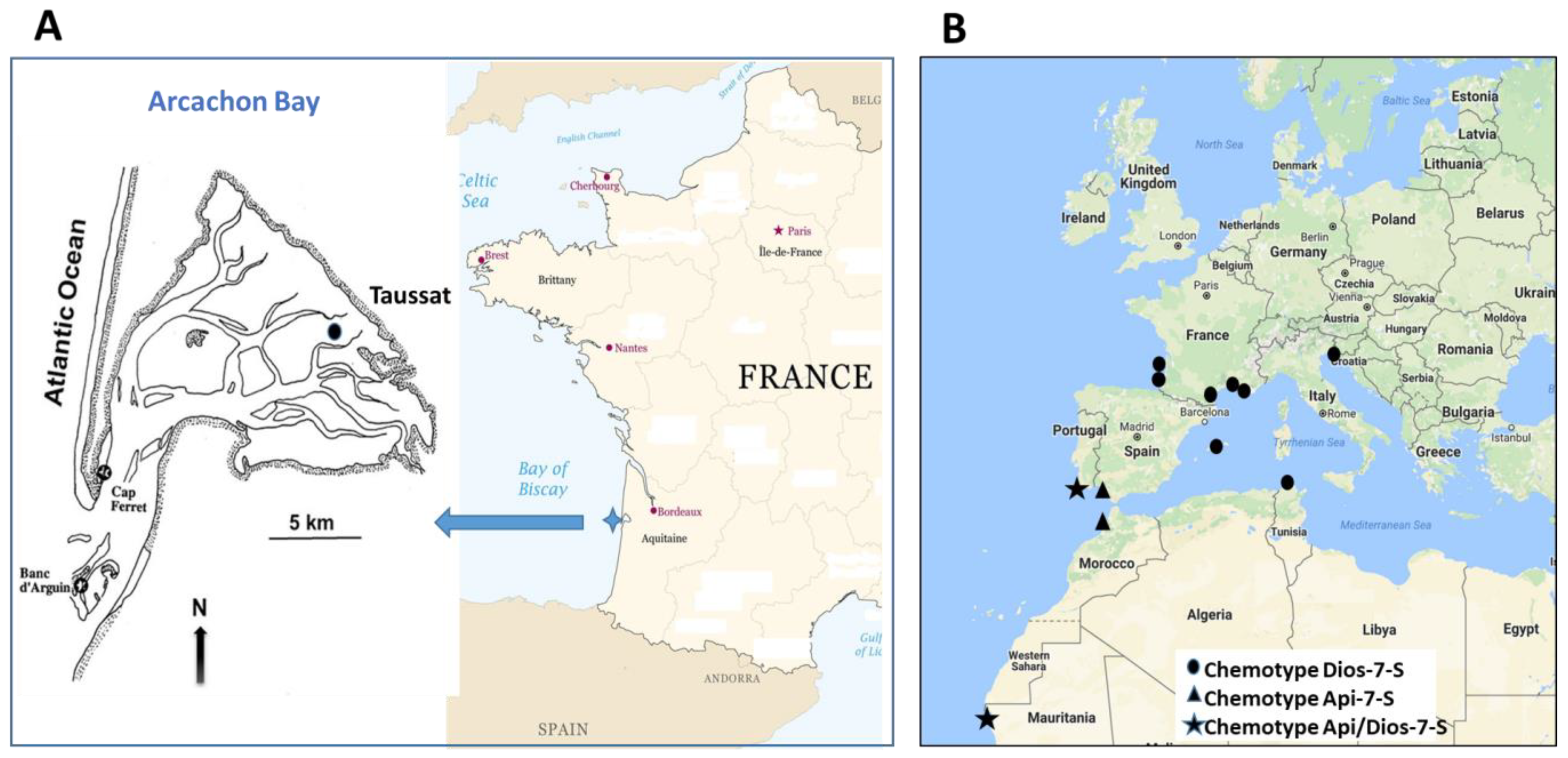
Figure 1. Map showing the location of sampling sites. (A) Arcachon Bay; (B) other sites in the Atlantic and Mediterranean (see Table 1 for GPS coordinates).
Diversity 15 01210 g001
Figure 2. Structures of the phenolic compounds found in Nanozostera noltei and Zostera marina. 1: zosteric acid; 2: caffeic acid; 3: coumaric acid; 4: rosmarinic acid; 5: zosteranoic acid; 6: luteolin 7-glucoside; 7: luteolin 7-sulfate; 8: apigenin 7-glucoside; 9: apigenin 7-sulfate; 10: diosmetin 7-sulfate; 11: luteolin; 12: apigenin; 13: diosmetin; 14: luteolin 3′-disulfate; 15: chrysoeriol 7-sulfate; 16: luteolin 3′,4′-dimethyl ether 7-sulfate.
Diversity 15 01210 g002
Figure 3. Top: HPLC profile of Nanozostera noltei crude extract (recorded at 280 nm) from leaves (left) and rhizomes (right). Bottom: HPLC profile of Zostera marina crude extract (recorded at 280 nm) from leaves (left) and rhizomes (right). Peak number assignments—1: zosteric acid; 2: caffeic acid; 3: coumaric acid; 4: rosmarinic acid; 5: zosteranoic acid; 6: luteolin 7-glucoside; 7: luteolin 7-sulfate; 10: diosmetin 7-sulfate; 13: diosmetin; 14: luteolin 7,3′-disulfate; 15: chrysoeriol 7-sulfate; 16: dimethoxyluteolin 7-sulfate. Compounds are coded as in Table 2, Table 3, Table 4 and Table 5 and Figure 2.
Diversity 15 01210 g003
Figure 4. Variation of the quantitative amounts of phenolic compounds in N. noltei leaves over months and years. (Top)—minor compounds. 1: zosteric acid; 2: caffeic acid; 8: apigenin 7-O-glucoside; 9: apigenin 7-sulfate. (Bottom)—major compounds. 4: rosmarinic acid; 5: zosteranoic acid; 6: luteolin 7-O-glucoside; 7: luteolin 7-sulfate; 10: diosmetin 7-sulfate. Compounds are coded as in Table 2, Table 3, Table 4 and Table 5 and Figure 2. Quantitative amounts of compounds 14 are expressed in mg per g of N. noltei–plant DW, and those of compounds 5 to 10 in mg of standard equivalents per g of N. noltei DW, with the standards used being, respectively, 5: rosmarinic acid, 67: luteolin, 89: apigenin and 10: diosmetin. Compared to rosmarinic acid, the seasonality of the caffeic tetramer 5 is less marked and the production peak tends to occur regardless of the season (1.51–7.56 (2017), 3.82–10.05 (2018), 0.87–4.64 (2019) and 1.69–8.44 mg/g plant DW (2020)). Zosteric (1) and caffeic (2) acids are much less abundant than 4 and 5 and show a relatively low variability from season to season and year to year (respectively, 0.10–0.43 and 0.07–0.38 mg/g plant DW over the four years of monitoring). Coumaric acid (3) was only found in six samples and in very small amounts (0.024–0.048 mg/g plant DW) (Figure 5).
Diversity 15 01210 g004
Figure 6. Variation of the quantitative amounts of phenolic compounds in N. noltei leaves over months and years—(left column): comparison between total phenolic acids (TPA, black lines) and total flavonoids (TF, red lines); (right column): comparison between levels of rosmarinic acid (1, blue lines), zosteranoic acid (5, brown lines) and diosmetin 7-sulfate (10, green lines). Concentrations are expressed in mg per g of N. noltei plant DW.
Diversity 15 01210 g006
Disclaimer/Publisher’s Note: The statements, opinions and data contained in all publications are solely those of the individual author(s) and contributor(s) and not of MDPI and/or the editor(s). MDPI and/or the editor(s) disclaim responsibility for any injury to people or property resulting from any ideas, methods, instructions or products referred to in the content.