Article

A Rare Fish Amphistome Revisited: The Phylogenetic Position of Kalitrema kalitrema (Trematoda: Cladorchiidae) Found in Hypostomus spp. (Siluriformes: Loricariidae) from Brazil

by
Hudson Alves Pinto
1,*,
Camila Pantoja
1,
Jordana Costa Alves de Assis
1,
Danimar López-Hernández
1,
Fabio Vieira
2,
José Luis Luque
3 and
Philippe Vieira Alves
4
1
Laboratório de Biologia de Trematoda, Instituto de Ciências Biológicas, Universidade Federal de Minas Gerais, Belo Horizonte 31270-901, MG, Brazil
2
Departamento de Zoologia, Instituto de Ciências Biológicas, Universidade Federal de Minas Gerais, Belo Horizonte 31270-901, MG, Brazil
3
Departamento de Parasitologia Animal, Universidade Federal Rural do Rio de Janeiro, Seropédica 23890-000, RJ, Brazil
4
Setor de Parasitologia, Departamento de Biodiversidade e Bioestatística, Instituto de Biociências, Universidade Estadual Paulista-UNESP, Distrito de Rubião Júnior, Botucatu 18618-689, SP, Brazil
*
Author to whom correspondence should be addressed.
Diversity 2023, 15(10), 1034; https://doi.org/10.3390/d15101034
Submission received: 31 August 2023 / Revised: 18 September 2023 / Accepted: 23 September 2023 / Published: 26 September 2023
(This article belongs to the Special Issue Taxonomy, Biodiversity and Ecology of Parasites of Aquatic Organisms)

Abstract

:
Despite recent advances in the molecular knowledge of amphistome trematodes, most genera known from fish remain to be genetically characterized. This is the case for Kalitrema, a genus of the speciose family Cladorchiidae and the type of Kalitrematinae. The type and only species of this genus, Kalitrema kalitrema Travassos, 1933, was originally proposed based on two specimens found in an armored suckermouth catfish from Brazil, and its phylogenetic position has not been evaluated. In this study, paramphistomes found in Hypostomus alatus (2/9; 22.2%) and Hypostomus francisci (4/143; 2.8%) from the Paraobepa River (São Francisco River basin), Minas Gerais, Brazil, between December 2019 and November 2021, were subjected to morphological study. The parasites were identified in low intensity of infection [1.2 (1–2)] and redescribed as K. kalitrema. This species exhibits unique features such as a linguiform body with a circular ridge near the anterior end and a deep, median notch present at the posterior extremity of the body, apparently dividing the body into two lobes. A subset of specimens was further subject to phylogenetic analyses based on the most densely sampled markers, the nuclear ribosomal RNA (28S and ITS2) and mitochondrial cox1, which revealed the inclusion of K. kalitrema in a Neotropical clade of fish paramphistomes. The most comprehensive phylogenetic tree, based on the 28S dataset, confirmed K. kalitrema as an independent, early diverging lineage among Neotropical fish cladorchiids. However, the monophyly of Kalitrematinae was not sustained, given that species of the other kalitrematine genera Pseudocladorchis and Iquitostrema included in the phylogenetic analysis fell in a distinct clade with other fish cladorchiids. As a result, we propose here a narrower concept for Kalitrematinae sensu stricto, accommodating only Kalitrema (type genus) until a more natural subfamilial or familial classification is provided.

Graphical Abstract

1. Introduction

The Neotropical region holds the highest diversity of freshwater and marine fishes in the world, attracting the attention of conservationists and environmental policymakers to this biodiversity hotspot [1,2]. However, much less attention has been paid to the parasitic metazoans associated with these vertebrates, which usually live hidden in the gut of their more well-known hosts. Even though parasites are often neglected in biodiversity management and protection programs, a vast body of literature has consistently confirmed that parasites are pivotal elements of ecosystems and that a healthy environment is where parasites thrive in diversity [3,4,5], and even global parasite conservation plans have been recently proposed [6].
Amphistome trematodes of the family Cladorchiidae Fischoeder, 1901 are cosmopolitan, speciose gut parasites of all groups of vertebrates but birds [7,8]. They are characterized by several features, such as having the acetabulum at or close to the posterior extremity while an oral sucker is absent (features also shared with other paramphistomoids), paired primary pharyngeal sacs, two testes, cirrus-sac are usually present, ventral pouch absent, and ovary post-testicular, or very rarely intertesticular [8]. The group has greatly diversified in South America—in fact, most species are known from fish in this subcontinent—primarily in the two dominant groups of freshwater fishes, i.e., Characiformes and Siluriformes [9]. With the recent descriptions of new species by Pantoja et al. [10,11], fish cladorchiids stand out as one of the most diverse groups of fish trematodes in South America, with 38 valid species [11,12]. Despite that, the knowledge of the actual diversity of these paramphistomes is largely underappreciated, a fact that is accentuated by the paucity of helminthological surveys in the literature, with less than 5% of the ichthyofauna of the region scrutinized for parasites [12]. Moreover, despite these signals of advances in the use of molecular data to characterize fish paramphistomes, most species, including key ones for taxonomic resolution, have not been sequenced so far.
Kalitrema kalitrema Travassos, 1933 is the type and only species of Kalitrema Travassos, 1933. The species was originally described based only on two specimens found in the Suckermouth catfish Hypostomus punctatus Valenciennes (=Plecostomus punctatus; Loricariidae) in the Cachimbal stream, Paraiba do Sul River basin, Brazil [13]. Travassos [13] regarded the morphology of the species as ‘curious’, which did not correspond to any known species or even cladorchiid genera described at the time. The most distinctive morphological features of K. kalitrema are the linguiform body with a circular ridge near the anterior end and a deep, median notch present at the posterior extremity of the body, apparently dividing it into two lobes. These distinctive traits led Travassos to propose the subfamily Kalitrematinae, a rank maintained in the more recent classification of paramphistomes [8]. Despite its key role in validating the subfamilial status of Kalitrematinae, molecular sequences for K. kalitrema have not been generated so far.
During a long-term investigation carried out under the scope of a multidisciplinary project for monitoring the local biodiversity in the Paraobepa River, São Francisco River, a paramphistome species was found parasitizing armored catfishes of the genus Hypostomus Lacépède. The species, morphologically identified as K. kalitrema, is redescribed here. Moreover, the phylogenetic position of this poorly known fish parasite was evaluated for the first time.

2. Materials and Methods

2.1. Collection of Samples

A total of 202 specimens armored catfishes (Siluriformes, Loriicaridae) belonging to 4 species, i.e., Hypostomus alatus Castelnau (n = 9), Hypostomus francisci (Lütken) (n = 143), Hypostomus freire Penido, Pessali & Zawadzki (n = 7), Hypostomus guajupia Penido, Pessali & Zawadzki (n = 20), and specimens identified at the genus level, Hypostomus sp. (n = 23) were collected over six samplings points distributed along the Paraobepa River, Upper São Francisco River basin), between the municipality of Brumadinho (20°11′48.9″ S; 44°7′22.8″ W) and Pompeu (18°52′15.4″ S; 44°47′13.2″), Minas Gerais, Brazil. The sampling is part of a long-term monitoring carried out in Paraobepa River, and the fish evaluated for this study were caught between December 2019 and November 2021. After capture using fishing nets, fish were euthanized and immediately transported to the laboratory for dissection. Host capture carried out within the framework of the monitoring project was authorized by the Instituto Estadual de Florestas (IEF) (GPFAP/DFAU/IEF Nº 003/2019). Moreover, all procedures followed the recommendations of the local ethics committee on animal experimentation, CEUA, at UFMG (protocol 295/2019).
The newly collected trematodes were removed from the hosts’ intestines, placed in saline (0.9% NaCl solution), cleaned from the intestinal content, and gently flattened between glass slides, followed by fixation in 10% formalin. Some specimens were fixed in 95% ethanol for molecular analyses.

2.2. Morphological Data

Specimens for morphological identification were stained with alum acetocarmine, dehydrated in an increasing series of ethanol, clarified in beechwood creosote and mounted on permanent slides with Canada balsam. Whole-mounted paramphistomes were evaluated using a Leica DM 750 optical microscope (Leica Microsystems, Heerbrugg, Switzerland) with a differential phase contrast system and photographed with a camera coupled to the microscope. Images were analyzed in the Leica Application Suite software (LAZ EZ), v.2.0 (Heerbrugg, Switzerland). Measurements were taken with the aid of a micrometer eyepiece. Morphometric data are presented in micrometers (unless otherwise indicated) as the mean, followed by the standard deviation and range in parentheses. For comparison, photomicrographs of the type material (syntypes) of Kalitrema kalitrema deposited at the Helminthological Collection of the Oswaldo Cruz Institute (CHIOC-10279a and CHIOC-10279b) were studied.
Voucher specimens were deposited in the Collection of Trematodes of UFMG (UFMG-TRE 134-136) and the CHIOC (CHIOC-40218- 40220).

2.3. Molecular Data

Total genomic DNA was extracted from fragments of two amphistome specimens (one from each species of Hypostomus studied) using a QIAamp Mini kit (QIAGEN) following the manufacturer’s instructions. The concentration and quality of extracted DNA were evaluated using the microvolume spectrophotometer NanoDrop® Lite (Thermo Fisher Scientific, Waltham, MA, USA). Partial regions of the 28S (~1200 bp; primers Dig-12/1500R), ITS1-5.8S-ITS2 (~1000 bp; primers BD1/BD2), and cox1 (~800 bp; primers JB3/COI-R Trema) were amplified by PCR, using the conditions previously described [14,15,16]. PCR reagents, electrophoresis, amplicon purifications and sequencing were previously described in the works of our research team [17]. Chromatograms obtained were assembled and inspected for errors in ChromasPro v.2.0.1 software (Technelysium Pty Ltd., South Brisbane, Australia) and consensus sequences were used for downstream phylogenetic analyses.
The sequences generated de novo were assembled into three alignments, one for each marker, but only considering the ITS2 in the case of the internal transcribed spacer due to the availability of data for comparison. Representatives of paramphistomes from cold-blooded vertebrates, i.e., families Cladorchiidae and Diplodiscidae, with available data in the Genbank, were included in the alignments. Sequences of these two families were chosen as the ingroup because the monophyly of Cladorchiidae is not sustained with the inclusion of certain Diplodiscidae, as observed in recent phylogenetic reconstructions [18]. Only unique haplotypes were considered in the phylogenetic analysis. The alignments were built using default parameters of the MAFFT algorithm [19] implemented in the GUIDANCE2 server [20]. Unreliable positions in the single-gene alignments were identified and removed using the Gblock web server (https://ngphylogeny.fr/ (accessed on 17 February 2023) [21] with less stringent settings.
Phylogenetic reconstructions were performed with the maximum likelihood (ML) and Bayesian inference (BI) criteria using the evolutionary models implemented in ModelFinder [22] within IQ-TREE [23], based on the small sample size corrected Akaike Information Criterion (AICc). The search for the BI was restricted to those models allowed in MrBayes v.3.2 [24] using the “-mset” option. The models used in the ML analyses were TVM + F+I + G4, TVM + F+G4, and TIM2 + F+I + G4 for the 28S, ITS2, and cox1 datasets, respectively, while in the BI analyses, the models chosen were GTR + F+I + G4, GTR + F+G4, HKY + F+I + G4 for the same molecular markers. Zygocotyle lunata (Diesing, 1836) (Zygocotylidae) was chosen as the outgroup for all analyses based on the phylogenies provided by Alves et al. [17].
BI analyses were conducted in MrBayes v.3.2, running two independent MC3 runs of four chains (one cold, three heated) for 5 million generations, sampling tree topologies every 1000th generation, with the first 25% of samples discarded as burn-in. Tracer v.1.7 [25] was used to check the convergence and mixing of different parameters and to confirm that the effective sample size (ESS) of each parameter was adequate to provide reasonable estimates of the variance in model parameters (i.e., ESS values > 200). The ML trees were generated via IQTREE, and clade supports were estimated with 1000 replicates of the ultrafast bootstrap (UFBoot—[26]) and an SH-aLRT test with 1000 replicates [27]. To avoid overestimation of UFBoot, we used a hill climbing nearest neighbor interchange (NNI), as recommended by Hoang et al. [28]). Clades with support values of both UFBoot ≥ 95 and SH-aLRT ≥ 80 for the ML analyses and posterior probability (PP) ≥ 0.99 for the BI ones were considered strongly supported. All the above analyses were run on the computational resource CIPRES [29]. Genetic divergences were calculated as uncorrected p-distances using MEGA 7.0 [30].

3. Results

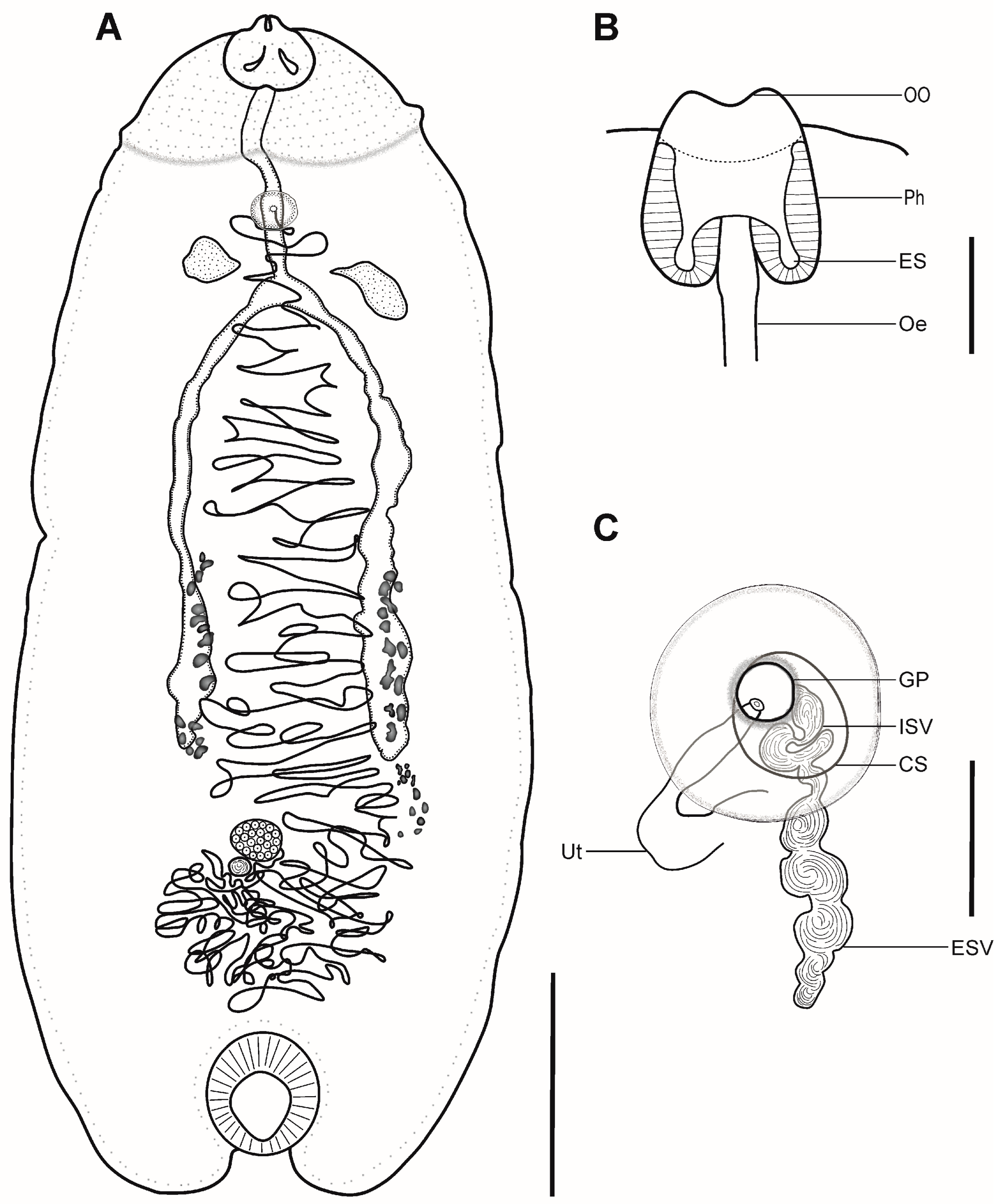
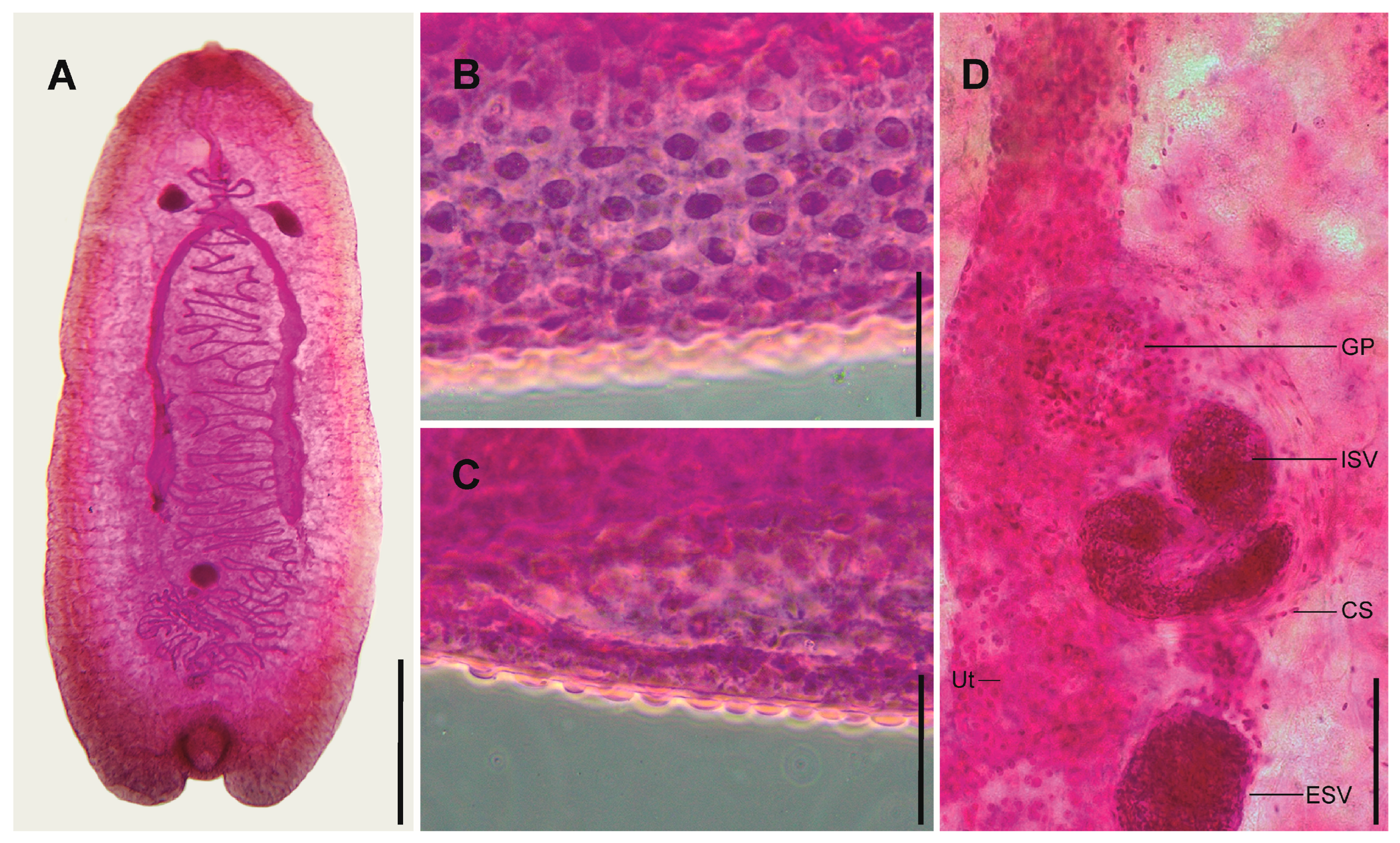
3.1. Kalitrema Kalitrema Travassos, 1933 (Figure 1 and Figure 2)

Type host and locality: Hypostomus punctatus Valenciennes (=Plecostomus punctatus) (Siluriformes: Loricariidae), Cachimbal stream (Paraiba do Sul River basin), Pinheiral, Rio de Janeiro, Brazil.
New hosts: Hypostomus alatus Castelnau, Hypostomus francisci (Lütken)
Prevalence: 2/9 (22.2%) of Hypostomus alatus, 4/143 (2.8%) of H. francisci.
Mean intensity of infection: 1.2 (12).
Site of infection: Anterior intestine.
Localities: Paraobepa River; Upper São Francisco River basin, at its portion at the municipalities of Brumadinho and São Joaquim de Bicas, state of Minas Gerais, Brazil.
Voucher material: UFMG-TRE134–136 and CHIOC 40218–40220
Representative DNA sequences: Two isolates, one recovered from each host species: partial 28S (OR541121 and OR541122, identical sequences, 943 bp long); partial ITS (OR555847 and OR555848, identical sequences, 803 bp long); and partial cox1 (OR537891 and OR537892, identical sequences, 753bp long).
Redescription (Figure 1 and Figure 2) (measurements of syngenophores in Table 1 and hologenophore in the description): Body relatively large, linguiform, dorso-ventrally flattened, 8250. Maximum width at distal caeca, 3400. Body surface with small, dome-shaped, non-ciliated tegumental papillae (about 11–13 in diameter) (Figure 2B,C). Collar-like circular ridge at the anterior part of the body. Posterior extremity with a median notch. Oral opening terminal. Pharynx muscular, well developed, with extramural sacs, subspherical, elongated-oval or transversely oval, 416 long and 572 wide (Figure 1B). Oesophagus long, narrow, curved or straight, 1843 long. Oesophageal bulb absent. Intestinal bifurcation in the anterior half of the body, posterior to the genital pore. Distance from intestinal bifurcation to anterior extremity, 2463 (30% of body length). Caeca blind, with thin walls and narrow lumen, slightly sinuous, extending up to posterior half of body, far short of ventral sucker. Acetabulum (ventral sucker) small in relation to body size, muscular, well developed, subspherical, elongate-oval or transversely oval, 922 long, 953 wide. Testes two, small, subspherical, elongate-oval or transversely oval, entire or slightly lobed, symmetrical or subsymmetrical, separated, irregular, extracaecal or slightly overlapping caeca, in the anterior half of body close to intestinal bifurcation. Distance of testes from anterior end 2238, 27% of body length. Right testis, 454 long, 223 wide; left testis, 367 long, 215 wide. External seminal vesicle thin-walled, elongate, partitioned, 434 long, 124 wide. Cirrus sac subspherical or elongate-oval, 286 long, 206 wide, contains internal seminal vesicle proximally. Internal seminal vesicle thin-walled, elongate, coiled, 372 long, 73 wide (Figure 1C). True genital sucker absent. Genital pore surrounded by glandular tissue, median, posterior to pharynx, prebifurcal. Ovary small, spherical, subspherical or transversely oval, entire, submedian, posterior to caecal ends, at some distance from the acetabulum, 261 long, 291 wide. Post-ovarian region 2700, 33% of body length. Laurer’s canal was not observed. Seminal receptacle subspherical, elongate-oval or transversely oval, immediately posterior to the ovary, 136 long, 163 wide. Vitelline follicles in narrow lateral fields, usually caecal, from mid-caeca, extending slightly posterior to caecal ends. Uterus long, coiled, descending limb encroaches close to ventral sucker, ascending limb intercaecal. Eggs were absent in the specimens examined. Excretory vesicle and duct not observed.
Figure 1. Kalitrema kalitrema ex Hypostomus alatus from Brazil: (A) complete specimen, ventral view; (B) detail of the pharynx, ventral view; (C) details of the terminal genitalia. Scale bars (A): 2 mm, (B,C): 250 μm. Abbreviations: CS—Cirrus sac, ES—extramural sac, ESV—External seminal vesicle, GP—genital pore, ISV—internal seminal vesicle, Oe—Oesophagus, OO—oral opening, Ph—pharynx, Ut—uterus.
Diversity 15 01034 g001

Remarks

Specimens evaluated in the present study correspond well to the generic diagnosis of Kalitrema provided by Travassos [13] and Jones [8] in having extracaecal testes, linguiform body with anterior end demarcated by a circular ridge, notched posterior extremity, and small acetabulum not papillate on the luminal surface. Therefore, the identification of the amphistome as Kalitrema kalitrema, the type and only species of the genus, was straightforward. The specimens also matched the morphometrics of the original material, e.g., body length (6175–9900 vs. 7000–8700), body width (2100–4400 vs. 2600–3000), acetabulum length × width (601–1095 × 658–969 vs. 760 × 780) and in ovary length × width (221–373 × 281–450 vs. 350 × 360). Even though K. kalitrema was originally described as possessing a genital sucker, evaluation of the type and newly collected material revealed that a true genital sucker is absent; in fact, numerous glandular cells are present surrounding the genital pore in a slightly elevated area (see Figure 2). Furthermore, the detailed description of the terminal genitalia revealed, for the first time, the presence of a cirrus sac, which was previously overlooked. In the original description, Travassos [13] did not assign any specimen as the holotype, so the syntype CHIOC-10279b is designated herein as lectotype, i.e., the name-bearer of the taxon when a holotype has not been originally determined [31]. Mandatorily, the specimen CHIOC-10279a becomes the paralectotype.
An interesting finding related to the host-parasite interrelationship in the infection site that deserves to be mentioned is that the worms were found with the lateral parts ventrally bent, occupying much of the intestinal lumen and distending it (Supplementary Material—Video).
Figure 2. Kalitrema kalitrema ex Hypostomus alatus from Brazil: (A) Whole view of a stained specimen. (B) Dorsal and (C) lateral view of small dome-shaped tegumental papillae. (D) Detail of cirrus sac. Scale bars (A): 2 mm, (B,C): 50 μm, (D): 100 μm. Abbreviations: CS—Cirrus sac, ESV—External seminal vesicle, GP—genital pore, ISV—internal seminal vesicle, Ut—uterus.
Diversity 15 01034 g002

3.2. Molecular Analyses

Two sequences for each marker (28S, ITS, cox1) were successfully generated for the isolates of K. kalitrema (one from each host species, H. alatus and H. francisci). Since all sequences of individual markers were identical, only one of each fragment was used in the downstream phylogenetic analyses.
The alignment of 39 sequences using the 28S dataset comprised 1072 positions, including 227 parsimony informative sites. The trees resulting from the ML and BI were identical in topology (Figure 3), except that in the BI tree, the clade of Pseudocladorchis Daday, 1907 emerged in a polytomy. Kalitrema kalitrema appeared as an early diverging lineage of a large, well-supported clade (UFBoot = 96; SH-aLRT = 92.8; PP = 1), sister to all remaining Neotropical cladorchiids parasitizing fish; Catadiscus marinholutzi Freitas and Lent, 1939 (Diplodiscidae), known from anurans in Brazil, is placed as sister to all fish Neotropical cladorchiids yet with no statistical support. The entire Neotropical clade is sister to an assemblage comprising the Palaearctic species Ophistodiscus diplodiscoides Cohn, 1904 (Cladorchiidae) + Diplodiscus spp. (Diplodiscidae) parasites of amphibians; both clades are, in turn, sisters to a highly eclectic, earliest-diverging clade, composed of the fish helostomatine Helostomatis cf. helostomatis (MacCallum, 1905) from the Indomalayan region + the fish dadayiines Amurotrema dombrowskajae Achmerow, 1959 and Pisciamphistoma stunkardi (Holl, 1929) from the Palaearctic and Nearctic regions, respectively, + the dugong solenorchiine Solenorchis travassosi Hilmy, 1949 from the Australasian region + two unidentified species of the anuran schizamphitomine genus Megalodiscus Chandler, 1923 from the Nearctic. No cladorchiid subfamily with more than one representative, i.e., Dadayiinae, Kalitrematinae, and Schizamphistominae, were found as monophyletic in the phylogenetic analysis. The Cladorchiidae itself is paraphyletic relative to the inclusion of diplodiscid members. Pairwise comparison shows that K. kalitrema diverged by 5.2 to 7.5% (44–61 nt difference) from other Neotropical taxa and by 8.8 to 12.6% (70–106 nt difference) from the remaining species.
The ITS2 alignment included only 11 sequences of cladorchiid and diplodiscid paramphistomes and comprised 289 positions with 77 parsimony informative sites. The small number of taxa used in the analysis decreases the explanatory power of this dataset. Nevertheless, in the ML tree (Figure 4A), K. kalitrema appeared in a virtual polytomy (due to the presence of unsupported internal branches, collapsed in the BI tree) together with the Neotropical fish dadayiine Doradamphistoma spp. and Dadaytrema spp. (representatives of both genera are placed in a well-supported clade), and species of Diplodiscus from the frog Rana dybowskii Günther in the Palaearctic realm. The genetic divergence between K. kalitrema and its relatives from the Neotropical region ranged from 14.8 to 18.4% (38–47 nt difference), while from Diplodiscus spp. the divergence ranged from 21.6 to 22% (55–56 nt difference).
The cox1 alignment included 47 sequences of the selected paramphistomes and comprised 265 positions with 143 parsimony informative sites. Despite a better taxon coverage than for the ITS2 dataset, the overlapping sites among the sequences are not comprehensive, so a large portion of the alignment was excluded after the Gblock analysis. The trees obtained from the ML and BI analyses show similar interrelationships—considering the terminal clades—among the paramphistomid taxa. Three major clades can be recognised (Figure 4B): the earliest diverging is composed of the anuran schizamphistomine Megalodiscus temperatus (Stafford, 1905) and the rodent cladorchiine Neostichorchis subtriquetrus (Rudolphi, 1814) (=Stichorchis subtriquetrus), both from the Palaearctic realm; the second clade is formed by paramphistomes of anurans, i.e., diplodiscid species of Diplodiscus (three taxa) and the schizamphistomine O. diplodiscoides, all from the Palaearctic region; the third clade encompasses only Neotropical taxa of the Dadayiinae and Kalitrematinae (both non-monophyletic) parasitising fish. In this later clade, K. kalitrema emerged as a sister to another kalitrematine species, Iquitostrema papillatum Pantoja, Scholz, Luque and Jones, 2018, but the lack of branch support prevents any assumption of a close relationship between these taxa. Kalitrema kalitrema diverged by 15.2 to 21.1% (34–56 nt difference) from other Neotropical taxa and by 19.6 to 24.9% (51–66 nt difference) from the remaining species.

4. Discussion

Amphistome trematodes, commonly referred to as “rumen flukes,” pose a substantial threat to a wide range of wildlife and domestic animals, particularly ruminants. The presence of these parasites leads to considerable economic losses in the livestock industry. In light of this, extensive scientific research has been conducted to explore the host-parasite interactions associated with these trematodes, aiming to identify potential targets for treatment and control strategies [32,33]. Many of these studies rely on molecular data to address pertinent questions, and a substantial number of sequences are already accessible in the GenBank database. Conversely, fish paramphistomoids have received considerably less research focus, and the molecular characterization of most of these species is still lacking [10,11]. This knowledge gap hinders our ability to confidently unravel the evolutionary relationships of this highly diverse group and assess species boundaries, especially in South American drainages. In this study, the integration of molecular and morphological analyses made it possible to effectively redescribe K. kalitrema, a rare Neotropical fish amphistome, and elucidate its evolutionary relationships. Additionally, this study evaluated the systematic significance of key morphological features traditionally used to characterize distinct taxa at higher levels of classification.
While Travassos [13] acknowledged the distinct morphology of K. kalitrema, certain features were either misinterpreted or overlooked, preventing a comprehensive characterization. For instance, we confirmed the presence of a cirrus-sac in the species, as observed in other cladorchiids; this feature is even used to differentiate paramphistomes of mammals from those of cold-blooded vertebrates, including the Cladorchiidae [7,8]. In the differential diagnoses of both Kalitrematinae and Kalitrema, Jones [8] expressed uncertainties regarding the presence of a genuine genital sucker. Our findings have confirmed the absence of this feature in K. kalitrema. Instead, the species exhibits a glandular area that is slightly elevated and surrounds the genital pore. It is noteworthy that many Neotropical cladorchiids previously described as possessing a genital sucker may not have this structure. A more detailed morphological evaluation, as observed in recent studies [10, 11, present study], could provide further evidence supporting this prediction.
The findings from the multi-loci molecular analyses provide support for recent phylogenetic studies [11,17,18] in several aspects: (i) the inclusion of Diplodiscidae renders Cladorchiidae as non-monophyletic; (ii) Dadayiinae, Kalitrematinae, and Schizamphistominae are artificial assemblages of unrelated taxa; (iii) Neotropical fish cladorchiids are so far monophyletic; (iv) zoogeography likely played a significant role in the evolutionary history of paramphistomes, as all Neotropical taxa, including C. marinholutzi from anurans, consistently clustered together in the same clade, albeit with low support. It is evident that comprehensive revisions in the classification of Paramphistomoidea are necessary to accurately reflect the evolutionary relationships among its members. For instance, the subfamilial classification scheme of the highly diverse Cladorchiidae, currently comprising ten taxa, requires a critical reassessment due to the non-monophyly observed in most groups for which molecular data are available. Features such as the number (one or two) and position (intercaecal, caecal or extracaecal) of testes are of limited systematic value and should be avoided in the new scheme. However, among the 69 genera within Cladorchiidae, many of which are monotypic, only 14 have representative sequences available for comparison. Therefore, taxonomic actions should be approached cautiously, given the current state of knowledge. Considering these factors, we propose a refined concept of Kalitrematinae sensu stricto, encompassing only Kalitrema (the type genus) and its sole species. Other members of Kalitrematinae sensu lato that have not yet undergone molecular characterization, i.e., Betamphistoma Thatcher and Jégu, 1996, Brevicaecum McClelland, 1957, Micramphistoma Thatcher, 1992, and Nicollodiscus Srivastava, 1939, or even representatives of different subfamilies may demonstrate close relationships to K. kalitrema and should be included within this revised scheme.
The host spectrum of fish cladorchiids varies among taxa. While many species are known only from one species (oioxenous specificity), others have been found infecting multiple hosts even from different orders (euryxenous specificity), with a particular emphasis on serrasalmids (Characiformes), and pimelodid and doradid catfishes (Siluriformes) [7,10,11]. Kalitrema kalitrema is documented for the first time in H. francisci and H. alatus, indicating a stenoxenous specificity, meaning it can be restricted to congeners. On the other hand, the recent report of the species in Loricaria cataphracta L. from Amazonia during an ecological study [34] indicates that this fish amphistome could infect other loricariid genera. However, a detailed morphological characterization and molecular comparison with the data presented here are necessary to confirm this hypothesis. Nevertheless, yet originally described from the Paraiba do Sul River basin, the occurrence of K. kalitrema in the São Francisco River basin is not unexpected due to the complex geomorphological processes that have occurred in these hydrological drainages, resulting in shared ichthyofauna [35,36] and likely shared parasites.

Supplementary Materials

The following supporting information can be downloaded at https://www.mdpi.com/article/10.3390/d15101034/s1, Video—Kalitrema kalitrema in the intestinal lumen of Hypostomus francisci from Brazil.

Author Contributions

Conceptualization, H.A.P. and P.V.A.; methodology, D.L.-H., J.C.A.d.A. and C.P.; formal analysis, P.V.A. and C.P.; resources H.A.P., F.V. and J.L.L. writing—original draft preparation H.A.P., C.P. and P.V.A.; writing—review and editing; supervision, H.A.P.; funding acquisition, H.A.P. and J.L.L.; project administration, H.A.P. and F.V. All authors have read and agreed to the published version of the manuscript.

Funding

Fish sampling was based on financial resources provided by Vale S.A. Molecular analysis was funded by the National Council for Scientific and Technological (CNPq, grant, 404083/2021-8). P.V.A. was supported by the São Paulo Research Foundation (FAPESP; grant 2021/12593-2). H.A.P. and J.L.L. were supported by CNPq research scholarships. C.P. was supported by Coordination for the Improvement of Higher Education (CAPES-Print post-doc scholarship). D.L.H. was supported by CNPq (post-doc scholarship). J.C.A.d.A. was supported by Amplo Engenharia.

Institutional Review Board Statement

Part of the data presented is the result of the Environmental Damage Diagnosis Program on the Biotic Environment (PDD), which was prepared, coordinated and managed by Amplo Engenharia and Project Management, based on financial resources provided by Vale S.A., approved by the State Forestry Institute of Minas Gerais—IEF. We thank Vale S.A., IEF and Amplo for their support. This is product number 0002 of the series of PDD publications. Sampling and transport of the hosts were granted by the Instituto Estadual de Florestas (IEF) (GPFAP/DFAU/IEF Nº 003/2019). All procedures followed recommendations of the local ethics committee on animal experimentation, CEUA, UFMG (protocol 295/2019).

Data Availability Statement

Not applicable.

Acknowledgments

We are much obliged to Marcelo Knoff (Curator of CHIOC), Daniela de Almeida Lopes and Alessandra Gomes da Cruz (CHIOC’s staff) for kindly providing photomicrographs of the syntypes of Kalitrema kalitrema and assistance in the specimen deposit process.

Conflicts of Interest

The authors declare no conflict of interest. The funders had no role in the design of the study, in the collection, analyses, or interpretation of data, in the writing of the manuscript, or in the decision to publish the results.

References

  1. Reis, R.E.; Alberts, J.S.; Di Dario, F.; Mincarone, M.M.; Petry, P.; Rocha, L.A. Fish biodiversity and conservation in South America. J. Fish Biol. 2016, 89, 12–47. [Google Scholar] [CrossRef] [PubMed]
  2. Myers, N.; Mittermeier, R.A.; Mittermeier, C.G.; Da Fonseca, G.A.; Kent, J. Biodiversity hotspots for conservation priorities. Nature 2000, 403, 853–858. [Google Scholar] [CrossRef] [PubMed]
  3. Brooks, D.R.; Hoberg, E.P. Triage for the biosphere: The need and rationale for taxonomic inventories and phylogenetic studies of parasites. Comp. Parasitol. 2000, 67, 1–25. [Google Scholar]
  4. Marcogliese, D.J. Parasites of the superorganism: Are they indicators of ecosystem health? Int. J. Parasitol. 2005, 35, 705–716. [Google Scholar] [CrossRef]
  5. Hudson, P.J.; Dobson, A.P.; Lafferty, K.D. Is a healthy ecosystem one that is rich in parasites? Trends Ecol. Evol. 2006, 21, 381–385. [Google Scholar] [CrossRef] [PubMed]
  6. Carlson, C.J.; Hopkins, S.; Bell, K.C.; Doña, J.; Godfrey, S.S.; Kwak, M.L.; Lafferty, K.D.; Moir, M.L.; Speer, K.A.; Strona, G.; et al. A global parasite conservation plan. Biol. Conserv. 2020, 250, 108596. [Google Scholar] [CrossRef]
  7. Sey, O. CRC Handbook of the Zoology of Amphistomes, 1st ed.; CRC Press: Boca Raton, FL, USA, 1991. [Google Scholar]
  8. Jones, A. Family Cladorchiidae Fischoeder, 1901. In Keys to the Trematoda; Jones, A., Bray, R.A., Gibson, D.I., Eds.; CAB International & The Natural History Museum: Wallingford, UK, 2005; Volume 2, pp. 257–317. [Google Scholar]
  9. Tonella, L.H.; Ruaro, R.; Daga, V.S.; Garcia, D.A.Z.; Vitorino, O.B.; Magalhães, T.L.; Reis, R.E.; Di Dario, F.; Petry, A.C.; Mincarone, M.M.; et al. Neotropical freshwater fishes: A dataset of occurrence and abundance of freshwater fishes in the Neotropics. Ecology 2023, 104, e3713. [Google Scholar] [CrossRef]
  10. Pantoja, C.; Scholz, T.; Luque, J.L.; Jones, A. New genera and species of paramphistomes (Digenea: Paramphistomoidea: Cladorchiidae) parasitic in fishes from the Amazon basin in Peru. Syst. Parasitol. 2018, 95, 611–624. [Google Scholar] [CrossRef]
  11. Pantoja, C.; Scholz, T.; Luque, J.L.; Jones, A. First molecular assessment of the interrelationships of cladorchiid digeneans (Digenea: Paramphistomoidea), parasites of Neotropical fishes, including descriptions of three new species and new host and geographical records. Folia Parasitol. 2019, 66, 011. [Google Scholar] [CrossRef]
  12. Choudhury, A.; Aguirre-Macedo, M.L.; Curran, S.S.; Ostrowski de Núñez, M.; Overstreet, R.M.; Pérez-Ponce de León, G.; Santos, C.P. Trematode diversity in freshwater fishes of the globe II: ‘New World’. Syst. Parasitol. 2016, 93, 271–282. [Google Scholar] [CrossRef]
  13. Travassos, L. Sur un nouveau trematode de poisons de la vallée du fleuve Parahyba. CR Soc. Biol. Paris 1933, 114, 839–840. [Google Scholar]
  14. Tkach, V.V.; Littlewood, D.T.J.; Olson, P.D.; Kinsella, J.M.; Swiderski, Z. Molecular phylogenetic analysis of the Microphalloidea Ward, 1901 (Trematoda: Digenea). Syst. Parasitol. 2003, 56, 1–15. [Google Scholar] [CrossRef]
  15. Luton, K.; Walker, D.; Blair, D. Comparisons of ribosomal internal transcribed spacers from two congeneric species of flukes (Platyhelminthes: Trematoda: Digenea). Mol. Biochem. Parasitol. 1992, 56, 323–327. [Google Scholar] [CrossRef]
  16. Miura, O.; Kuris, A.M.; Torchin, M.E.; Hechinger, R.F.; Dunham, E.J.; Chiba, S. Molecular-genetic analyses reveal cryptic species of trematodes in the intertidal gastropod, Batillaria cumingi (Crosse). Int. J. Parasitol. 2005, 35, 793–801. [Google Scholar] [CrossRef] [PubMed]
  17. Alves, P.V.; Assis, J.C.A.; López-Hernández, D.; Pulido-Murillo, E.A.; Melo, A.L.; Locke, S.A.; Pinto, H.A. A phylogenetic study of the cecal amphistome Zygocotyle lunata (Trematoda: Zygocotylidae), with notes on the molecular systematics of Paramphistomoidea. Parasitol. Res. 2020, 119, 2511–2520. [Google Scholar] [CrossRef] [PubMed]
  18. Benovics, M.; Mikulíček, P.; Žákovicová, Z.; Papežík, P.; Pantoja, C. Hidden in plain sight: Novel molecular data reveal unexpected genetic diversity among paramphistome parasites (Digenea: Paramphistomoidea) of European water frogs. Parasitology 2022, 149, 1425–1438. [Google Scholar] [CrossRef] [PubMed]
  19. Katoh, K.; Standley, D.M. MAFTT multiple sequence alignment software Version 7: Improvements in performance and usability. Mol. Biol. Evol. 2013, 30, 772–780. [Google Scholar] [CrossRef] [PubMed]
  20. Sela, I.; Ashkenazy, H.; Katoh, K.; Pupko, T. GUIDANCE2: Accurate detection of unreliable alignment regions accounting for the uncertainty of multiple parameters. Nucleic Acids Res. 2015, 1, W7–W14. [Google Scholar] [CrossRef]
  21. Dereeper, A.; Guignon, V.; Blanc, G.; Audic, S.; Buffet, S.; Chevenet, F.; Dufayard, J.F.; Guindon, S.; Lefort, V.; Lescot, M.; et al. Phylogeny.fr: Robust phylogenetic analysis for the non-specialist. Nucleic Acids Res. 2008, 36, W465–W469. [Google Scholar] [CrossRef]
  22. Kalyaanamoorthy, S.; Minh, B.Q.; Wong, T.K.; Von Haeseler, A.; Jermiin, L.S. ModelFinder: Fast model selection for accurate phylogenetic estimates. Nat. Methods 2017, 14, 587–589. [Google Scholar] [CrossRef]
  23. Minh, B.Q.; Schmidt, H.A.; Chernomor, O.; Schrempf, D.; Woodhams, M.D.; Von Haeseler, A.; Lanfear, R. IQ-TREE 2: New models and efficient methods for phylogenetic inference in the genomic era. Mol. Biol. Evol. 2020, 37, 1530–1534. [Google Scholar] [CrossRef]
  24. Ronquist, F.; Teslenko, M.; Van Der Mark, P.; Ayres, D.L.; Darling, A.; Höhna, S.; Larget, B.; Liu, L.; Suchard, M.A.; Huelsenbeck, J.P. MrBayes 3.2: Efficient Bayesian Phylogenetic Inference and Model Choice Across a Large Model Space. Syst. Biol. 2012, 61, 539–542. [Google Scholar] [CrossRef] [PubMed]
  25. Rambaut, A.; Drummond, A.J.; Xie, D.; Baele, G.; Suchard, M.A. Posterior summarisation in Bayesian phylogenetics using Tracer 1.7. Syst. Biol. 2018, 67, 901–904. [Google Scholar] [CrossRef] [PubMed]
  26. Minh, B.Q.; Nguyen, M.A.T.; Von Haeseler, A. Ultrafast approximation for phylogenetic bootstrap. Mol. Biol. Evol. 2013, 30, 1188–1195. [Google Scholar] [CrossRef]
  27. Guindon, S.; Dufayard, J.F.; Lefort, V.; Anisimova, M.; Hordijk, W.; Gascuel, O. New algorithms and methods to estimate maximum-likelihood phylogenies: Assessing the performance of PhyML 3.0. Syst. Biol. 2010, 59, 307–321. [Google Scholar] [CrossRef] [PubMed]
  28. Hoang, D.T.; Chernomor, O.; Von Haeseler, A.; Minh, B.Q.; Vinh, L.S. UFBoot2: Improving the ultrafast bootstrap approximation. Mol. Biol. Evol. 2018, 35, 518–522. [Google Scholar] [CrossRef]
  29. Miller, M.A.; Pfeiffer, W.; Schwartz, T. The CIPRES science gateway: A community resource for phylogenetic analyses. In Proceedings of the Gateway Computing Environments Workshop (GCE), New Orleans, LA, USA, 14 November 2010. [Google Scholar]
  30. Kumar, S.; Stecher, G.; Tamura, K. MEGA7: Molecular evolutionary genetics analysis version 7.0 for bigger datasets. Mol. Biol. Evol. 2016, 33, 1870–1874. [Google Scholar] [CrossRef]
  31. ICZN International Code of Zoological Nomenclature, 4th ed.; 1999; Available online: http://www.iczn.org (accessed on 15 March 2023).
  32. Huson, K.M.; Oliver, N.A.; Robinson, M.W. Paramphistomosis of ruminants: An emerging parasitic disease in Europe. Trends Parasitol. 2017, 33, 836–844. [Google Scholar] [CrossRef]
  33. Tandon, V.; Roy, B.; Shylla, J.A.; Ghatani, S. Amphistomes. In Digenetic Trematodes; Toledo, R., Fried, B., Eds.; Springer: Cham, Switzerland, 2019; Volume 1154, pp. 255–277. [Google Scholar]
  34. Virgilio, L.R.; Martins, W.M.O.; Lima, F.S.; Takemoto, R.M.; Camargo, L.M.A.; Meneguetti, D.U.O. Endoparasite fauna of freshwater fish from the upper Juruá River in the Western Amazon, Brazil. J. Helminthol. 2022, 96, e55. [Google Scholar] [CrossRef]
  35. Cherem, L.F.S.; Varajão, C.A.C.; Braucher, R.; Bourlés, D.; Salgado, A.A.R.; Varajão, A.C. Long-term evolution of denudational escarpments in southeastern Brazil. Geomorphology 2012, 173, 118–127. [Google Scholar] [CrossRef]
  36. Anjos, M.S.; Queiroz, L.J.; Penido, I.S.; Bitencourt, J.A.; Barreto, S.B.; Sarmento-Soares, L.M.; Batalha-Filho, H.; Affonso, P.R.A.M. A taxonomically complex catfish group from an underrepresented geographic area: Systematics and species limits in Hypostomus Lacépède, 1803 (Siluriformes, Loricariidae) from Eastern South America. J. Zool. Syst. Evol. Res. 2021, 59, 1994–2009. [Google Scholar] [CrossRef]
Figure 3. Maximum likelihood (ML) phylogram based on the 28S dataset of the selected Cladorchiidae and Diplodiscidae. GenBank accession numbers are followed by the taxon and host names. The newly obtained sequence is in bold. Branch length scale bar indicates the number of substitutions per site. The species names are colored according to the current subfamilial or familial morphology-based classification.
Diversity 15 01034 g003
Figure 4. Maximum likelihood (ML) phylogram of the selected Cladorchiidae and Diplodiscidae as inferred from the sequences of ITS2 (A) and cox1 (B). GenBank accession numbers are followed by the taxon and host names. The newly obtained sequence is in bold. Branch length scale bar indicates the number of substitutions per site. The species names are colored according to the current subfamilial or familial morphology-based classification.
Diversity 15 01034 g004
Table 1. Comparative metrical data of Kalitrema kalitrema Travassos, 1933 from Hypostomus spp. from Brazil.
Source Present StudyTravassos (1933)
Host Hypostomus alatusHypostomus punctatus
Hypostomus francisci
N N = 6N = 2
BodyL7729 (6175–9900)7000, 8700
W2979 (2100–4400)2600, 3000
Body length/body width 1:2.59 (1:2.25–3.34)
PharynxL421 (260–595)
W493 (355–691)
OesophagusL1314 (1035–1759)780,1300
DTRAE 1117 (821–1386)800, 900
AcetabulumL823 (601–1095)760
W797 (631–969)780
DIBAE 1810 (1453–2271)
Right testisL300 (190–417)570
W315 (211–485)390, 440
Left testisL331 (216–382)570
W346 (140–797)390, 440
External seminal vesicleL378 (243–478)
W61 (45–93)
Cirrus-sacL252 (110–348)
W191 (105–367)
Internal seminal vesicleL304 (206–382)
W73 (50–95)
GRSGPL363 (276–553)310, 390
W369 (230–558)310, 390
Genital pore openingL81 (62–140)
W100 (61–209)
OvaryL299 (221–373)350
W354 (281–450)360
Seminal receptacleL165 (122–208)
W175 (148–211)
DBOT 3400 (2537–4150)
Post-ovarian region%31 (23–34)
EggLn/a160, 168
Wn/a96, 112
Abbreviations: DTRAE = distance from transverse ridge to anterior extremity, DIBAE = distance from intestinal bifurcation to anterior extremity, DBOT = distance between ovary and testes, GRSGP = Glandular region surrounding genital pore L = Length, W: width, n/a = not available.
Disclaimer/Publisher’s Note: The statements, opinions and data contained in all publications are solely those of the individual author(s) and contributor(s) and not of MDPI and/or the editor(s). MDPI and/or the editor(s) disclaim responsibility for any injury to people or property resulting from any ideas, methods, instructions or products referred to in the content.

