Communication

Bacteria Release from Microplastics into New Aquatic Environments

Department of Biological Applications and Technology, University of Ioannina, 45110 Ioannina, Greece
*
Author to whom correspondence should be addressed.
Diversity 2023, 15(1), 115; https://doi.org/10.3390/d15010115
Submission received: 25 November 2022 / Revised: 21 December 2022 / Accepted: 11 January 2023 / Published: 13 January 2023
(This article belongs to the Special Issue Occurrence and Molecular Biology of Water Bacteria and Protozoa)

Abstract

:
Microplastics are considered the most common waste in aquatic ecosystems, and studying them along with their interactions with biota are considered a priority. Here, results on the role of microplastics in the dispersion of microbes from terrestrial to aquatic ecosystems are presented. Data were obtained from microcosm experiments in which microplastics (plastic bags (BA), polyethylene bottles (BO), acrylic beads (BE), and cigarette butts (BU)) with their attached natural bacterial communities were inoculated in filtered and autoclaved lake water. The bacterial abundance on microplastics was estimated before inoculation using a protocol for the enumeration of sediment bacteria and ranged between 1.63 (BA) and 203.92 (BE) × 103 cells mm−2. Bacteria were released in the new medium, and their growth rates reached 5.8 d−1. In the attached communities, Beta- (21.4%) and Alphaproteobacteria (18.6%) were the most abundant classes, while in the free-living communities Gammaproteobacteria dominated (48.07%). Abundant OTUs (≥1%) of the free-living communities were associated with the genera Acinetobacter, Pseudomonas, Ecidovorax, Delftia, Comamonas, Sphingopyxis, and Brevundimonas and members of the FCB group. Members of these genera are known to degrade natural or man-made organic compounds and have recently emerged as opportunistic pathogens. Thus, besides trophic transmission, microplastics can directly release bacteria in the environment, which could affect the health of humans, animals, and ecosystems.

1. Introduction

Plastic plays an essential role in modern life due to its properties (e.g., buoyancy and density) and the potential to be processed with additives that ameliorate its characteristics and lifespan. However, the same properties constitute an ecological issue, as the rate of the accumulation of plastic in the environment is much greater than the rate of its decomposition, resulting in ever-increasing pollution and the deterioration of ecosystems [1]. Plastic and its small derivatives, microplastics, enter aquatic environments through treated and untreated sewage waters, direct disposal, or air currents [2], and horizontal advection transport and turbulent mixing affect their dispersion and distribution in the new environment [3].
Microplastics are of increasing ecological concern since they can affect aquatic life in different ways in remote areas of the globe and far from the original site of production or disposal. They can be consumed by metazoa and retained in the gut or translocated to other tissues or systems of the consumer or its predators [4,5]. Their ingestion may have direct or indirect (through associations with other pollutants) detrimental effects on the physiology of species (e.g., reduced reproduction, growth, and fitness) [6]. Furthermore, there is evidence that the formation of biofilm (plastisphere [7]) on microplastics inhibits the ability of grazers to distinguish plastic and live prey [8] and makes them more attractive for grazers [9]. This may result in the transfer of microplastics and chemicals adsorbed on their surfaces up the food chain with the potential for bioaccumulation and biomagnification [10]. Microplastics can also act as vectors for the attached microbes, including pathogenic bacteria, harmful algae, and invaders [11,12]. For example, the freshwater fish pathogen Aeromonas salmonicida has been found on microplastics in the Northern Adriatic Sea [13]. It has also been suggested that the plastisphere may include some opportunistic animal or human pathogens such as vibrios [7]. Potential harmful algae on the plastic debris include members of dinoflagellates (e.g., Alexandrium sp.) and cyanobacteria (e.g., Dolichοspermum flos-aque) [7,14,15].
It is well known that the microbial community of the plastisphere is distinct from that found in the surrounding waters [16]. Here, we present the results of a preliminary study aiming to investigate whether members of the plastisphere are released in the water column when microplastics reach a new environment. For this, we performed microcosm experiments using microplastics made of some of the most common plastic polymers (polyethylene terephthalate—PET, low-density polyethylene—LPDE, and acrylic) as well as cellulose acetate, which is found in cigarette butts. We hypothesized that some members of the plastisphere can switch from the attached to the free-living lifestyle and grow in the water medium. At the same time, microbes living on land can be attached to microplastics and then transferred to aquatic ecosystems, with the microplastics acting as inoculators for the aquatic environment.

2. Materials and Methods

2.1. Collection and Processing of Microplastics

In the microcosm experiments, different microplastics, with their attached bacterial communities (both naturally occurring and those made in experimental conditions), were used as inocula in liquid cultures. First, a blue transparent plastic bag (BA) was collected from the shore of Pamvotis lake (Ioannina, NW Greece) using sterilized equipment. The plastic bag was stored in an autoclaved glass jar at 4 °C in the dark until further processing. In the lab, it was cut under aseptic conditions into ~500 square membranes with an average side dimension of 4.53 ± 1.22 mm and an average surface area of 19.97 ± 5.91 mm2 (membrane thickness was regarded as negligible). Before inoculation, the fragments were washed with autoclaved milli-Q water to remove loosely attached microorganisms. Another set of microplastics was prepared in the lab from fresh, store-bought plastics: a polyethylene bottle (BO), commercial acrylic beads (BE), and unused cigarette butts (BU), which were mainly made of cellulose acetate [17]. The plastic bottle and the cigarette butts were cut into smaller segments with 15.24 mm2 and 57.84 mm2 outer surfaces, respectively. The acrylic beads had 15.71 mm2 surfaces. For sterilization, these microplastics were transferred to glass Petri dishes containing an aqueous solution of H2O2 (30% v/v) for 5–15 min, washed with autoclaved milli-Q water [18], and dried in a lab oven at 60 °C for 18 h. The microplastics that were to be used in control cultures were stored at 4 °C in the dark. The rest were buried in soil at a depth of 15 cm in a sub-urban area for 26 days. After this incubation period, soil and other debris were removed manually from the microplastics, which were then immersed, under gentle agitation, in separate beakers containing milli-Q water to remove the fine sediment and determine the bacterial abundance.

2.2. Determination of Bacterial Abundance on Microplastics

Microplastics were checked using microscopy for the presence of attached bacteria, and their abundance was estimated using the protocol for the enumeration of sediment bacteria [19]. First, microplastics (65 microplastics for each different type) were transferred in 10 mL centrifuge tubes and fixed with formol (10 mL, 0.37% final concentration). Duplicate samples were prepared for each microplastic. Samples were sonicated using an Omni Sonic Ruptor (Power 30%, Timer 3/4, Pulser 50/60 at 20 kHz) to detach the bacteria. Subsamples (1 mL) were taken after gentle vortexing and transferred in Eppendorf tubes containing 1 mL of autoclaved milli-Q water. To determine the bacterial abundance, 200 μL were transferred onto Whatman® Nuclepore™ Track-Etched Membranes (0.2 μm, diameter 25 mm), and after staining with DAPI (4,6-diamidino-2-phenylindole, 10 μg/mL) the sample was washed with milli-Q water (2 mL) and filtered under low vacuum (<5 mmHg). Bacteria were counted under an epifluorescence microscope (LEICA DM LS2) at ×1000 magnification. At least 50 fields were measured in each sample. The same initial sample of microplastics was also used to estimate the dimensions of different types of microplastics.

2.3. Microcosm Experiment

The culture medium for microcosm experiments was prepared with the consecutive filtering of Pamvotis lake water through 180 μm and 20 μm nylon meshes and finally through 0.7 μm Sartorius™ Glass Microfiber Discs (47 mm diameter) under low vacuum (≤5 mmHg). The water was divided equally into 500 mL Erlenmeyer flasks that were autoclaved at 121 °C for 20 min. Adequate quantities of different categories of microplastics (BA, BO, BE, and BU) were added to different Erlenmeyer flasks so that the total surface area in each flask was ~52 (BA) or ~46 (BO, BE, and BU) cm2. Duplicate microcosms were prepared for BE, BO, and BU. Triplicates were prepared for BA.
Microcosms were incubated in an MRC Orbital Shaker Incubator at 22 °C in the dark. Samples (2–5 mL) were taken daily from each replicate microcosm to investigate the occurrence of bacteria in the culture medium and determine the bacterial abundance. For this, samples were fixed with a 37% v/v formol solution, stained with DAPI, and filtered on 0.2 μm (25 mm diameter) Whatman® Nuclepore™ Black Track-Etched Polycarbonate Membrane Filters. Bacteria were counted under an epifluorescence microscope (LEICA DM LS2) at ×1000 magnification. At least 50 fields were measured in each sample. The bacterial growth rates (μ) in the microcosms were calculated using the linear part of the exponential growth curve, while the doubling time was estimated from the equation td = ln(2)/μ. A one-way ANCOVA was performed using PAST.3 software to investigate statistically significant differences between the slopes of the exponential growth curves (ln-transformed time data).

2.4. Bacterial Diversity on the Plastic Bag (BA) Microcosms

The bacterial diversity of the initial BA-attached community, as well as that of the free-living community at the end of the experiment, was assessed through sequencing of the 16S rDNA V3–V4 region. For the attached community, DNA was extracted from 20 randomly selected BA membranes from the pool prepared (as described in Section 2.1) to be used in the experiment. To investigate the diversity of the free-living bacteria, at the end of the experiment, culture media from triplicate BA microcosms (total of 170 mL) were mixed and filtered through 0.2 μm Isopore Membrane Filters (diameter 47 mm) using a vacuum pump at <5 mmHg. Although fine particles that passed through a 0.7 μm filter may have provided a surface for the attachment of microbes, we considered all bacteria retained on a 0.2 μm filter as free-living [20]. Both the microplastics and the filter were stored in cryovials at −20 °C. DNA was extracted from the samples using a Qiagen DNeasy PowerSoil Kit (Hilden, Germany) following the manufacturer’s protocol. The DNA concentration (ranging from 3 to 25.8 ng μL−1) and the absorbance ratio at 260/280 nm (ranging from 1.51 to 2.12) were measured with a Q3000 Quawell DNA/Protein Analyser, Quawell Technology Inc, CA, USA, using the software Q3000 V4.2.1. The 16S rDNA gene V3–V4 variable region PCR primers S-D-Bact-0341-b-S-17/S-D-Bact-0785-a-A-21 [21], with a barcode on the forward primer, were used in a 30-cycle PCR (94 °C for 3 min; 28 cycles of 94 °C for 30 s, 53 °C for 40 s, and 72 °C for 1 min; and a final elongation step at 72 °C for 5 min) using a HotStarTaq Plus Master Mix Kit (Qiagen, Gaithersburg, MD, USA). After amplification, pooled and purified PCR products were used to prepare the DNA library. Library preparation and sequencing were performed at MR DNA (http://www.mrdnalab.com, Shallowater, TX, USA, Accessed on 1 December 2022) on a MiSeq following the manufacturer’s guidelines. Sequences were processed using MOTHUR software v1.30, clustered in operational taxonomic units (OTUs) at 97% sequence similarity, and classified using BLASTn against a curated database derived from the Ribosomal Database Project (RDPII, http://rdp.cme.msu.edu, Accessed on 1 December 2022) and the National Center for Biotechnology Information (www.ncbi.nlm.nih.gov, Accessed on 1 December 2022) that was implemented through the analysis pipeline developed by MR DNA. Sequences were submitted to SRA with accession number PRJNA633010. Diversity indices and statistically significant differences between Shannon index values were calculated/investigated using PAST.3 software. A Venn analysis (using Venny 2.1) was performed to investigate unique and shared OTUs between samples.

3. Results

Epifluorescence microscopy showed the colonization of all types of microplastics by bacteria. The bacterial concentrations in the suspension media, after sonication, ranged between 4.24 × 10⁵ (BA) and 1.00 × 10⁷ cells ml−1 (BO). When converted to cell number per microplastic surface unit, the abundance was 100-fold higher on beads (BE, 203.92 × 10³ cells mm−2) compared to the plastic bag membranes (BA, 1.63 × 10³ cells mm−2). For the bottle fragments (BO) and the cigarette butts (BU), the respective values were 41.54 × 10³ and 75,94 × 10³ cells mm−2.
Bacteria were released from the microplastics straight after inoculation (Figure 1). Their abundances at the beginning of the experiment were on the order of 103 cells ml−1 and reached 105–106 cells ml−1 in different microcosms. In BA microcosm bacteria underwent a lag phase in the first ~20 h of the experiment. No delay in the exponential growth was observed in BE, BO, or BU. The growth rates varied from 0.128 (BE) to 0.243 (BO) h−1, but a one-way ANCOVA did not show statistically significant differences (p > 0.05) in the growth rates between microcosms inoculated with different microplastics. The respective doubling times were 5.42 and 2.86 h.
The analysis of the V3–V4 region of the 16S rDNA indicated that 23 bacterial phyla were represented in the BA-membrane-attached community. All but four phyla (Aquificae, Tenericutes, Chlamydiae, and Fibrobacteres) were also found in the respective water media at the end of the experiment. Three phyla (Proteobacteria, Bacteroidetes, and Cyanobacteria) represented ~88% of the total relative abundance of the attached community. Acidobacteria, Firmicutes, Verrucomicrobia, Planctomycetes, and Actinobacteria had relative abundances between 1 and 3%. Proteobacteria and Bacteroidetes contributed 98.5% to the total relative abundance of free-living bacteria, and all other phyla contributed ≤0.6%. The attached bacterial community comprised members of 50 classes, and 39 out of these 50 classes were also found in the culture medium. Ten and five classes were found in relative abundances >1% in the attached and free-living community and represented >94% and 97% of the total relative abundance, respectively. In the attached community, Beta- (21.4%) and Alphaproteobacteria (18.6%) were the most abundant classes, while in the free-living community Gammaproteobacteria dominated (48.07%). In total, 2097 OTUs were found attached on plastic bag (BA) membranes, and 1294 were found free-living at the end of the experiment. Few OTUs showed relative abundances ≥1% (19 and 17 OTUs, respectively; Figure 2). The majority (>92%, 1938 and 1211, respectively) were considered rare (<0.1%). The two samples shared only two abundant OTUs. OTU1 was classified as Acinetobacter johnsonii, and OTU19 was classified as an uncultured Flavobacterium sp. In total, the two samples shared 904 OTUs (36% of total richness). The Shannon index showed higher diversity for the attached community (H = 5.33) compared to the free-living community (H = 3.47), and the difference was statistically significant (diversity t test, p < 0.01). Dominance (1-Simpson index) was low in both samples (≤0.1). The Chao 1 index was 3198 for the BA-attached bacterial community and 2591 for the free-living community.

4. Discussion

The measurement of bacterial abundance on the microplastics after sonication and using epifluorescence microscopy confirmed the colonization of all surfaces by bacteria. Abundance ranged from 103 to 105 cells mm−2, with the lowest values corresponding to bag membranes (BA) that were collected from the shore of Pamvotis lake. The three other types of microplastics (BO, BU, and BE), which were buried in soil for ~1 month prior to the experiment, had bacterial abundances mm−2 10- to 100-fold higher than BA. Between the three materials, the minima corresponded to cigarette butts, although their porous nature would theoretically allow for higher abundances. Overall, the high abundances in these recently buried microplastics are probably indicative of the presence of primocolonizers and actively growing bacteria in the newly formed biofilm [16]. Early attached bacteria are more susceptible to processes or substances that may remove them [22]. Thus, bacteria on these materials might have been detached more easily through sonication. The results of the bacterial abundance of the attached communities were in the range of values found previously (102–104 cells mm−2 [16]). However, since there is no universal protocol for the determination of abundance, the necessity of evaluating and choosing the most adequate and convenient method is raised here.
It is well known that bacteria can switch between planktonic and sessile lifestyles [23], and according to [24] these bacteria, not specialized to a specific lifestyle, can be considered generalist species. In our experiment, bacteria moved from the microplastics into the water medium and were able to grow with rates that reached 5.8 d−1. It has previously been shown that plastic leachates can promote bacterial growth [25]. Furthermore, we consider that free-living bacteria were not under grazing pressure, although we did not check if eukaryotic predators were released from the plastisphere. The particle-attached lifestyle offers many advantages, such as protection from predators, environmental pressures, and antibiotics and access to resources [26], but different top-down (e.g., predation) or bottom-up (e.g., the leaching of organic matter and nutrients from surfaces) factors can trigger a habitat shift and bacterial movement into the water [27].
Particles, due to their microniches, host a high level of bacterial diversity compared to the surrounding waters [23]. For plastic debris in particular, several studies have shown high evenness but lower richness compared to their natural aquatic environment [28]. Furthermore, several studies have shown that microplastics harbor a community that is distinct from that found in the surrounding waters. In this study, the bacterial richness on microplastics was lower than that of the free-living community in the microcosms, but diversity (Shannon, Chao_1, and evenness) was higher. This diverse bacterial community was represented by 23 phyla. Four phyla were not detected in the water. Among these, Aquificae, Tenericutes, and Chlamydiae were previously found in soil and/or marine ecosystems [29,30], and Aquificae in particular flourishes in soils enriched with nitrogen [31]. Members of the fourth phylum, Fibrobacteres, are known as plant polymer degraders and, similar to other cellulolytic bacteria, attachment to plant biomass is considered a prerequisite for hydrolytic activity [32]. In line with previous findings, Proteobacteria, Bacteroidetes, and Cyanobacteria were the most abundant phyla in the attached community [28,33]. The Cyanobacteria abundance in the water medium was low because of the incubation conditions (dark), but Proteobacteria and Bacteroidetes represented ~75% and ~24% of the total relative abundance, respectively. Gammaproteobacteria, represented mainly by two OTUs (OTU1 ~33% and OTU9 ~7%), both classified to Acinetobacter, dominated the microplastic-attached bacterial assemblage. Acinetobacter (Table 1) is widely distributed in natural soil and water ecosystems [34] as well as in nosocomial environments [35] and sites that are polluted with nutrients, heavy metals, and antibiotics from domestic sewage or wastewater treatment plants (WWTP) [36]. A previous study [37] found Acinetobacter on microplastics from both raw sewage and WWTP effluents. Considering that microplastics harboring Acinetobacter could be detected up to 2 km downstream of a WWTP, they suggested that microplastics could transport this potentially pathogenic taxon within rivers. Gammaproteobacteria were also represented by an OTU associated with Pseudomonas migulae, which can degrade phenanthrene [38], a polycyclic aromatic hydrocarbon used in plastics industry. Other abundant bacteria (relative abundance ≥1%) were associated with members of the FCB group (Flavobacterium spp., Emticicia sp., and Chryseobacterium indoltheticum). Members of this group are chemoorganotrophs that are able to remineralize organic matter in soils, sediments, and marine and freshwaters [39]. Abundant Betaproteobacteria (Table 1) were associated with the species Acidovorax spp, Delftia tsuruhatensis, and Comamonas jiangduensis. Acidovorax species have been found to be significant members of the Microcystis phycosphere that are able to degrade microcystins and algal-derived carbon such as glycolate as well as complex organic compounds such as polycyclic aromatic hydrocarbons [40,41]. D. tsuruhatensis has been isolated from environmental samples and is under investigation for bioremediation applications since it has been found to assimilate terephthalate [42] and degrade phenols [43]. Recently, D. tsuruhatensis was isolated from human patients and has emerged as an opportunistic healthcare-associated pathogen [44]. Finally, two abundant free-living OTUs were found to be associated with Alphaproteobacteria Sphingopyxis chilensis and Brevundimonas sp. (Table 1). Bacteria belonging to Sphingopyxis can be found in diverse natural environments, including those under anthropogenic pressures resulting from hydrocarbon, pesticide, and heavy metal contamination [45]. Sphingopyxis chilensis has been found to degrade chlorophenols [46], which are often used as pesticides, herbicides, and disinfectants. Brevundimonas has been isolated from numerous soil and aquatic habitats. It has been used for the bioremediation of hydrocarbon-polluted environments, but recently it has also emerged as an opportunistic pathogen [47].
In conclusion, this preliminary experiment indicates that microplastics have the potential to transfer and release bacteria in new environments. Habitat properties and biotic interactions, as well as the presence of contaminants not investigated here, may put selective forces against or for the growth of specific microbes and will determine their effects on microbial communities and organic matter and nutrient cycling. Therefore, the plastisphere has implications for the dispersion of bacteria through their direct release in the environment, which could affect the health of humans, animals, and ecosystems.

Author Contributions

Conceptualization, H.K.; methodology, formal analysis, and investigation, I.N., O.P., S.M. and H.K.; writing—original draft preparation, I.N. and H.K.; writing—review and editing, I.N., O.P., S.M. and H.K. All authors have read and agreed to the published version of the manuscript.

Funding

O.P. was supported by a University of Ioannina Research Committee (ELKE) Ph.D. Scholarship.

Institutional Review Board Statement

Not applicable.

Data Availability Statement

Sequences were submitted to SRA with accession number PRJNA633010.

Conflicts of Interest

The authors declare no conflict of interest.

References

  1. Barnes, D.; Galgani, F.; Thompson, R.; Barlaz, M. Accumulation and fragmentation of plastic debris in global environments. Philos. Trans. R. Soc. Lond. B Biol. Sci. 2009, 364, 1985–1998. [Google Scholar] [CrossRef] [PubMed] [Green Version]
  2. Ryan, P.G.; Moore, C.J.; Van Franeker, J.A.; Moloney, C.L. Monitoring the abundance of plastic debris in the marine environment. Philos. Trans. R. Soc. Lond. B Biol. Sci. 2009, 364, 1999–2012. [Google Scholar] [CrossRef] [PubMed] [Green Version]
  3. Li, Υ.; Zhang, H.; Tang, C. A review of possible pathways of marine microplastics transport in the ocean. Anthr. Coasts 2019, 3, 6–13. [Google Scholar] [CrossRef] [Green Version]
  4. Browne, M.A.; Galloway, T.; Thompson, R. Microplastic—An Emerging Contaminant of Potential Concern? Integr. Environ. Assess. Manag. 2007, 3, 559–561. [Google Scholar] [CrossRef]
  5. Farrell, P.; Nelson, K. Trophic level transfer of microplastic: Mytilus edulis (L.) to Carcinus maenas (L.). Environ. Pollut. 2013, 177, 1–3. [Google Scholar] [CrossRef] [PubMed]
  6. Horton, A.; Walton, A.; Spurgeon, D.J.; Lahive, E.; Svendsen, C. Microplastics in freshwater and terrestrial environments: Evaluating the current understanding to identify the knowledge gaps and future research priorities. Sci. Total Environ. 2017, 586, 127–141. [Google Scholar] [CrossRef] [PubMed] [Green Version]
  7. Zettler, E.; Mincer, T.; Amaral-Zettler, L. Life in the “Plastisphere”: Microbial Communities on Plastic Marine Debris. Environ. Sci. Technol. 2013, 47, 7137–7146. [Google Scholar] [CrossRef]
  8. Amaral-Zettler, L.A.; Zettler, E.R.; Slikas, B.; Boyd, G.D. The biogeography of the plastisphere: Implications for policy. Front Ecol. Environ. 2015, 13, 541–546. [Google Scholar] [CrossRef] [Green Version]
  9. Carson, H.S. The incidence of plastic ingestion by fishes: From the prey’s perspective. Mar. Pollut. Bull. 2013, 74, 170–174. [Google Scholar] [CrossRef]
  10. Da Costa, J.P.; Santos, P.S.M.; Duarte, A.C.; Rocha-Santos, T. (Nano)plastics in the environment—Sources, fates and effects. Sci. Total Environ. 2016, 566–567, 15–26. [Google Scholar] [CrossRef]
  11. Gregory, M.R. Environmental implications of plastic debris in marine settings—Entanglement, ingestion, smothering, hangers-on, hitch-hiking and alien invasions. Philos. Trans. R. Soc. Lond. B, Biol. Sci. 2009, 364, 2013–2025. [Google Scholar] [CrossRef] [Green Version]
  12. Caruso, G. Microplastics in Marine Environments: Possible Interactions with the Microbial Assemblage. J. Pollut. Eff. Cont. 2015, 3, e111. [Google Scholar] [CrossRef] [Green Version]
  13. Viršek, M.K.; Lovšinb, M.N.; Korena, S.; Kržanc, A.; Peterlina, M. Microplastics as a vector for the transport of the bacterial fish pathogen species Aeromonas salmonicida. Mar. Pollut. Bull. 2017, 125, 301–309. [Google Scholar] [CrossRef]
  14. Maso, M.; Garces, E.; Pages, F.; Camp, J. Drifting plastic debris as a potential vector for dispersing Harmful Algal Bloom (HAB) species. Sci. Mar. 2003, 67, 107–111. [Google Scholar] [CrossRef] [Green Version]
  15. Yokota, K.; Waterfield, H.; Hastings, C.; Davidson, E.; Kwietniewski, E.; Wells, B. Finding the missing piece of the aquatic plastic pollution puzzle: Interaction between primary producers and microplastics. Limnol. Oceanogr. Lett. 2017, 2, 91–104. [Google Scholar] [CrossRef] [Green Version]
  16. Odobel, C.; Dussud, C.; Philip, L.; Derippe, G.; Lauters, M.; Eyheraguibel, B.; Burgaud, G.; Ter Halle, A.; Meistertzheim, A.-L.; Bruzaud, S.; et al. Bacterial Abundance, Diversity and Activity During Long-Term Colonization of Non-biodegradable and Biodegradable Plastics in Seawater. Front. Microbiol. 2021, 12, 734782. [Google Scholar] [CrossRef]
  17. Benavente, J.M.; Arévalo Caballero, M.J.; Silvero, G.; López-Coca, I.; Gómez Escobar, V. Cellulose Acetate Recovery from Cigarette Butts. Multidiscip. Digit. Publ. Inst. Proc. 2018, 2, 1447. [Google Scholar] [CrossRef] [Green Version]
  18. Masura, J.; Baker, J.; Foster, G.; Arthur, C.; Herring, C.; Technical Editor. Laboratory Methods for the Analysis of Microplastics in the Marine Environment: Recommendations for Quantifying Synthetic Particles in Waters and Sediments; NOAA Technical Memorandum NOS-OR&R-48; 2015. [Google Scholar]
  19. Andresen, M.; Kristensen, E. The importance of bacteria and microalgae in the diet of the deposit-feeding polychaete Arenicola marina. Ophelia 2002, 56, 179–196. [Google Scholar] [CrossRef]
  20. Oberbeckmann, S.; Kreikemeyer, B.; Labrenz, M. Environmental Factors Support the Formation of Specific Bacterial Assemblages on Microplastics. Front. Microbiol. 2018, 8, 2709. [Google Scholar] [CrossRef] [Green Version]
  21. Klindworth, A.; Pruesse, E.; Schweer, T.; Peplies, J.; Quast, C.; Horn, M.; Glockner, F.O. Evaluation of general 16S ribosomal RNA gene PCR primers for classical and next-generation sequencing-based diversity studies. Nucleic Acids Res. 2013, 41, e1. [Google Scholar] [CrossRef]
  22. Fu, J.; Zhang, Y.; Lin, S.; Zhang, W.; Shu, G.; Lin, J.; Li, H.; Xu, F.; Tang, H.; Peng, G.; et al. Strategies for Interfering With Bacterial Early Stage Biofilms. Front. Microbiol. 2021, 12, 675843. [Google Scholar] [CrossRef] [PubMed]
  23. Crespo, B.G.; Pommier, T.; Fernández-Gómez, B.; Pedrós-Alió, C. Taxonomic composition of the particle-attached and free-living bacterial assemblages in the Northwest Mediterranean Sea analyzed by pyrosequencing of the 16S rRNA. MicrobiologyOpen 2013, 2, 541–552. [Google Scholar] [CrossRef] [PubMed]
  24. Hu, Y.; Xie, G.; Jiang, X.; Shao, K.; Tang, X.; Gao, G. The Relationships Between the Free-Living and Particle-Attached Bacterial Communities in Response to Elevated Eutrophication. Front. Microbiol. 2020, 11, 423. [Google Scholar] [CrossRef] [PubMed]
  25. Sheridan, E.A.; Fonvielle, J.A.; Cottingham, S.; Zhang, Y.; Dittmar, T.; Aldridge, D.C.; Tanentzap, A.J. Plastic pollution fosters more microbial growth in lakes than natural organic matter. Nat. Commun. 2022, 13, 4175. [Google Scholar] [CrossRef] [PubMed]
  26. Fei, C.; Ochsenkühn, M.A.; Shibl, A.A.; Isaac, A.; Wang, C.; Amin, S.A. Quorum sensing regulates ‘swim-or-stick’ lifestyle in the phycosphere. Environ. Microbiol. 2020, 22, 4761–4778. [Google Scholar] [CrossRef]
  27. Villalba, L.A.; Kasada, M.; Zoccarato, L.; Wollrab, S.; Grossart, H.P. Differing Escape Responses of the Marine Bacterium Marinobacter adhaerens in the Presence of Planktonic vs. Surface-Associated Protist Grazers. Int. J. Mol. Sci. 2022, 23, 10082. [Google Scholar] [CrossRef]
  28. Amaral-Zettler, L.A.; Zettler, E.R.; Mincer, T.J. Ecology of the plastisphere. Nat. Rev. Microbiol. 2020, 18, 139–151. [Google Scholar] [CrossRef]
  29. Mhete, M.; Eze, N.P.; Rahube, O.T.; Akinyemia, F.O. Soil properties influence bacterial abundance and diversity under different land-use regimes in semi-arid environments. Sci. Afr. 2020, 7, e00246. [Google Scholar] [CrossRef]
  30. Wang, Y.; Huang, J.-M.; Zhou, Y.-L.; Almeida, A.; Finn, R.D.; Danchin, A.; He, L.-S. Phylogenomics of expanding uncultured environmental Tenericutes provides insights into their pathogenicity and evolutionary relationship with Bacilli. BMC Genom. 2020, 21, 408. [Google Scholar] [CrossRef]
  31. Sun, R.; Zhang, P.; Riggins, C.W.; Zabaloy, C.M.; Rodríguez-Zas, S.; Villamil, M.B. Long-Term N Fertilization Decreased Diversity and Altered the Composition of Soil Bacterial and Archaeal Communities. Agronomy 2019, 9, 574. [Google Scholar] [CrossRef]
  32. Rahman, A.N.; Parks, D.H.; Vanwonterghem, I.; Morrison, M.; Tyson, G.W.; Hugenholtz, P. A Phylogenomic Analysis of the Bacterial Phylum Fibrobacteres. Front. Microbiol. 2016, 6, 1469. [Google Scholar] [CrossRef] [Green Version]
  33. Dussud, C.; Hudec, C.; George, M.; Fabre, P.; Higgs, P.; Bruzaud, S. Colonization of Non-biodegradable and Biodegradable Plastics by Marine Microorganisms. Front. Microbiol. 2018, 9, 1571. [Google Scholar] [CrossRef] [PubMed]
  34. Krizova, L.; Maixnerova, M.; Sedo, O.; Nemec, A. Acinetobacter bohemicus sp. nov. widespread in natural soil and water ecosystems in the Czech Republic. Syst. Appl. Microbiol. 2014, 37, 467–473. [Google Scholar] [CrossRef]
  35. Almasaudi, S.B. Acinetobacter spp. as nosocomial pathogens: Epidemiology and resistance features. Saudi J. Biol. Sci. 2018, 25, 586–596. [Google Scholar] [CrossRef] [Green Version]
  36. Guan, Y.; Jia, J.; Wu, L.; Xue, X.; Zhang, G.; Wang, Z. Analysis of bacterial community characteristics, abundance of antibiotics and antibiotic resistance genes along a pollution gradient of Ba River in xi’an, China. Front. Microbiol. 2018, 9, 3191. [Google Scholar] [CrossRef] [Green Version]
  37. Kelly, J.J.; London, M.G.; McCormick, A.R.; Rojas, M.; Scott, J.W.; Hoellein, T.J. Wastewater treatment alters microbial colonization of microplastics. PLoS ONE 2021, 16, e0244443. [Google Scholar] [CrossRef]
  38. Borde, X.; Guieysse, B.; Delgado, O.; Muñoz, R.; Hatti-Kaul, R.; Nugier-Chauvin, C.; Patin, H.; Mattiasson, B. Synergistic relationships in algal–bacterial microcosms for the treatment of aromatic pollutants. Bioresour. Technol. 2003, 86, 293–300. [Google Scholar] [CrossRef]
  39. Kirchman, D.L. The ecology of Cytophaga-Flavobacteria in aquatic environments. FEMS Microbiol. Ecol. 2002, 39, 91–100. [Google Scholar] [CrossRef]
  40. Hoke, A.K.; Reynoso, G.; Smith, M.R.; Gardner, M.I.; Lockwood, D.J.; Gilbert, N.E. Genomic signatures of Lake Erie bacteria suggest interaction in the Microcystis phycosphere. PLoS ONE 2021, 16, e0257017. [Google Scholar] [CrossRef]
  41. Krishnan, A.; Zhang, Y.Q.; Mou, X. Isolation and Characterization of Microcystin-Degrading Bacteria from Lake Erie. Bull. Env. Contam. Toxicol. 2018, 101, 617–623. [Google Scholar] [CrossRef]
  42. Shigematsu, T.; Yumihara, K.; Ueda, Y.; Numaguchi, M.; Morimura, S.; Kida, K. Delftia tsuruhatensis sp. nov. a terephthalate assimilating bacterium isolated from activated sludge. Int. J. Syst. Evol. Microbiol. 2003, 53, 1479–1483. [Google Scholar] [CrossRef] [PubMed]
  43. Juarez Jimenez, B.; Reboleiro Rivas, P.; Gonzalez Lopez, J.; Pesciaroli, C.; Barghini, P.; Fenice, M. Immobilization of Delftia tsuruhatensis in macro-porous cellulose and biodegradation of phenolic compounds in repeated batch process. J. Biotechnol. 2012, 157, 148–153. [Google Scholar] [CrossRef] [PubMed]
  44. Ranc, A.; Dubourg, G.; Fournier, P.E.; Raoult, D.; Fenollar, F. Delftia tsuruhatensis, an Emergent Opportunistic Healthcare-Associated Pathogen. Emerg. Infect. Dis. 2018, 24, 594–596. [Google Scholar] [CrossRef] [PubMed] [Green Version]
  45. Sharma, M.; Khurana, H.; Singh, D.N.; Negi, R.K. The genus Sphingopyxis: Systematics, ecology, and bioremediation potential—A review. J. Environ. Manag. 2021, 280, 111744. [Google Scholar] [CrossRef]
  46. Godoy, F.; Vancanneyt, M.; Martínez, M.; Steinbüchel, A.; Swings, J.; Rehm, B.H.M. Sphingopyxis chilensis sp. nov. a chlorophenol-degrading bacterium that accumulates polyhydroxyalkanoate, and transfer of Sphingomonas alaskensis to Sphingopyxis alaskensis comb. nov. Int. J. Syst. Evol. 2003, 53, 473–477. [Google Scholar] [CrossRef]
  47. Ryan, M.P.; Pembroke, T. Brevundimonas spp: Emerging global opportunistic pathogens. Virulence 2018, 9, 480–493. [Google Scholar] [CrossRef]
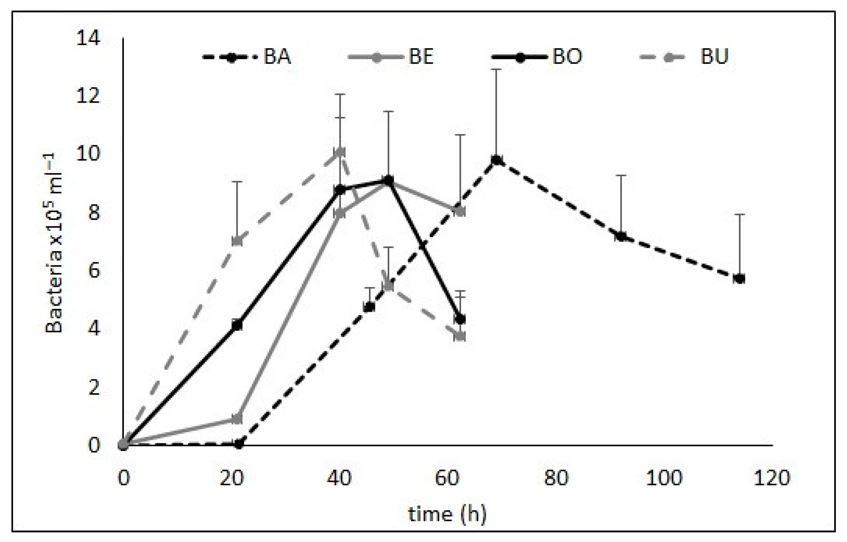
Figure 1. Growth of free-living bacteria in the microcosms inoculated with bacteria attached to microplastics. Error bars: minimum–maximum values for BO, BE, and BU and standard deviation for BA. BA: plastic bag, BO: polyethylene bottle, BE: acrylic beads, BU: cigarette butts.
Diversity 15 00115 g001
Figure 2. Heat map of relative OTU abundance in particle-attached (PA) and free-living (FL) communities. Particles refer to BA microplastics. Only OTUs that were abundant (≥1%) in the PA and/or FL communities are shown.
Diversity 15 00115 g002
Table 1. Abundant (≥0.1%) free-living OTUs released in the BA microcosms, associated species, and indicative functions of the major groups or specific members of the community.
GroupOTU Closest RelativesFunctions
Cytophaga–Flavobacteria clusterOTU12Arcicella sp.Degradation of high-molecular-weight organic matter (e.g., cellulose, chitin, and pectin) [39].
OTU563Flavobacterium succinicans
OTU7338Flavobacterium resistens
OTU19uncultured Flavobacterium
OTU53Emticicia sp.
OTU463Chryseobacterium indoltheticum
OTU88Flavobacterium aquatile
AlphaproteobacteriaOTU50Brevundimonas sp.Degradation of aromatic compounds and environmental pollutants (e.g., chlorophenol) [45]. Emerging as global opportunistic pathogens [46].
OTU484Sphingopyxis chilensis
BetaproteobacteriaOTU16Acidovorax sp.Degradation of xenobiotics and environmental pollutants (e.g., phenol), terephthalate assimilation [42,43], close associations with bloom-forming cyanobacteria (e.g., Microcystis) and degradation of algal-derived metabolites (e.g., microcystins and glycolate) [38]. Emergent opportunistic healthcare-associated pathogens with antibiotic resistance [44].
OTU7238Acidovorax sp.
OTU43Delftia tsuruhatensis
OTU120Comamonas jiangduensis
OTU1516uncultured Acidovorax
GammaproteobacteriaOTU1Acinetobacter johnsoniiDegradation of phenanthrene and other aromatic compounds and xenobiotics. Opportunistic pathogens with synergistic relationships with algae and pathogens with antibiotic resistance [35,38].
OTU9Acinetobacter piperi
OTU26Pseudomonas migulae
Disclaimer/Publisher’s Note: The statements, opinions and data contained in all publications are solely those of the individual author(s) and contributor(s) and not of MDPI and/or the editor(s). MDPI and/or the editor(s) disclaim responsibility for any injury to people or property resulting from any ideas, methods, instructions or products referred to in the content.