Article

Eight New Records of Siphonophores (Cnidaria: Hydrozoa) in Korean Waters

Department of Life Science, College of Natural Sciences, Hanyang University, Seoul 04763, Korea
*
Author to whom correspondence should be addressed.
Diversity 2022, 14(6), 494; https://doi.org/10.3390/d14060494
Submission received: 4 June 2022 / Revised: 16 June 2022 / Accepted: 16 June 2022 / Published: 17 June 2022
(This article belongs to the Collection Collection of Experts’ Researches on Aquatic Life (CEREAL))

Abstract

:
Taxonomical research on siphonophores is lacking despite their high biodiversity. We collected siphonophores in Jeju and the southern coasts of Korea and conducted morphological redescriptions using multi-focus stacked digital images. As a result, we revealed eight unrecorded siphonophores (Abyla haeckeli, Ceratocymba leuckartii, Bassia bassensis, Dimophyes arctica, Lensia subtilis, Lensia subtiloides, Muggiaea atlantica, and Nanomia bijuga). Six genera, excluding Muggiaea, were reported for the first time in Korean waters, and subfamily Abylinae, to which Abyla haeckeli and Ceratocymba leuckartii belong, was also first recorded in Korea. The distributions and habitats of these species were summarized, with most of the species having cosmopolitan distributions but variations in individual size depending on environmental conditions. Phylogenetic analysis based on mtCOI was conducted for each family. Finally, we updated the species list of siphonophores in Korean waters to include 3 suborders, 5 families, 3 subfamilies, 14 genera, and 21 species. Our findings constitute baseline data for further research on Korean siphonophores.

1. Introduction

Siphonophores are gelatinous zooplankton characterized by a unique colonial life [1]. About 190 species are known worldwide [2], and it is estimated that there are many more in the pelagic environment [3]. However, most studies on gelatinous zooplankton focus on Scyphozoa, or true jellyfish, blooming, toxicity, and removal, while taxonomic research on siphonophores has not reflected their high biodiversity [4,5]. Although some ecological papers include siphonophore records, there are few data available to confirm morphological features. The main reason for this deficiency in data is difficulty in classification due to the fragility of the gelatinous body and polymorphism. In addition, the situation is even worse because there is a lack of morphological experts of this taxonomic group. Thus, in recent years, species identification, reconstructions of life cycles, and clarifications of phylogeny based on molecular tools have been used to complement morphological analyses [3,6].
The order Siphonophorae Eschscholtz, 1829, is traditionally divided into three suborders: (1) Calycophorae Leuckart, 1854—no pneumatophore, but one or two nectophores; (2) Cystonectae Haeckel, 1887—pneumatophore at the apical end; (3) Physonectae Haeckel, 1888—pneumatophore and nectosome [7,8].
Abylidae L. Agassiz, 1862, is divided into two subfamilies (Abylinae L. Agassiz, 1862; Abylopsinae Totton, 1954) and nine species belonging to five genera. The main difference between the subfamilies is that Abylinae has a rectangular apical facet and Abylopsinae has a non-apical facet [9]. Usually, it includes two polygonal nectophores, and the anterior nectophore is smaller than the posterior. It is the most highly derived siphonophore group [10].
Diphyidae Quoy and Gaimard, 1827, the largest group among the siphonophores [11], is divided into three subfamilies (Diphyinae Quoy and Gaimard, 1827; Giliinae Pugh and Pagès, 1995; Sulculeolariinae Totton, 1954), has 45 species belonging to 8 genera, and shows the highest diversity in the Calycophorae [12]. In the polygastric phase, diphyids have one or two dissimilar nectophores. Anterior nectophores are usually bullet-shaped and contain major morphological features such as somatocysts, ridges, and the hydroecium. Therefore, most morphological records of Diphyidae highlight the anterior nectophore.
Agalmatidae Brandt, 1834, is a physonect subject to many taxonomic controversies [9]. Genera Marrus Totton, 1954, Frillagalma Daniel, 1966, Lychnagalma Haeckel, 1888, and others were excluded and are now referred to the Agalmatidae sensu stricto [6,13], which consists of three long-stem genera (Agalma Eschscholtz, 1825, Halistemma Huxley, 1859, Nanomia A. Agassiz, 1865) and two short-stem genera (Athorybia Eschscholtz, 1829 and Melophysa Haeckel, 1888). Nectophores are arranged in two rows in the stem, with the shape of the nectophore and ridge patterns being critical morphological features.
In this study, eight unrecorded siphonophores (belong to Abylidae, Diphyidae, and Agalmatidae) were collected from Jeju (an island in the southern part of Korea) and southern coasts of Korea. We performed morphological redescriptions using multi-focus stacked digital images, and the distributions and environmental conditions were summarized. In addition, we confirmed phylogenies and genetic distances in each family using molecular analyses based on the mitochondrial cytochrome c oxidase subunit I (mtCOI) sequences. Finally, we confirmed the entire siphonophore record in Korea and established the species list.

2. Materials and Methods

2.1. Sample Collection

Samples were collected from the Jeju and southern coasts of Korea during the R/V Dongbaek (April 2018) and R/V Eardo (June 2021) cruises (Figure 1). We used a plankton net (mesh size: 200 µm, Ø: 60 cm) towed vertically from the bottom to the surface at each station. The mouth of the net was equipped with a flowmeter (Hydro-Bios, Altenholz, Germany) to determine the volume of water filtered during each tow. The temperature and salinity of the surface layer of seawater were recorded using a CTD (CastAway-CTD, Sontek, San Diego, CA, USA) at each station (Table 1). After towing, the samples were immediately split into 1/2 aliquots using a Folsom plankton splitter. Each aliquot was fixed in 5% neutralized formalin solution (stored at room temperature) and 99% ethanol (stored at 4 °C). Specimens were sorted from the samples using Live Insect Forceps (26029-10, Fine Science Tools Inc., Foster City, CA, USA) under a stereomicroscope (Olympus SZX7, Tokyo, Japan). All materials were deposited in the Marine Biodiversity Institute of Korea (MABIK) and invertebrate collection of the National Institute of Biological Resources (NIBR), Korea.

2.2. Morphological Analysis

Siphonophore specimens were observed and identified under a stereomicroscope using descriptions, illustrations, and terminology drawn from the literature [9,10,11,12,14]. Photographs of specimens were collected at various focal points using a digital camera (Olympus PEN Lite E-PL3, Tokyo, Japan) connected to the stereomicroscope with sidelights on the dark field. We stacked multi-focus images using Helicon Focus 7 software (version: 7.5.1, Helicon Soft, Kharkiv, Ukraine). Then, we cropped and moved objects from the photographs to a black background with a scale bar using Adobe Photoshop CS6 (version: 13.0 × 64, Adobe, San Jose, CA, USA).

2.3. DNA Extraction, Amplification, and Sequencing

For DNA extraction, we sorted specimens from 99% ethanol plankton samples and transferred them to distilled water for three hours to wash and remove ethanol. The LaboPass™ Genomic Isolation-Tissue miniprep kit (Cosmogenetech Co., Seoul, Korea) was used to extract genomic DNA following the manufacturer’s protocols. We amplified mtCOI (about 650–850 bp) via polymerase chain reaction (PCR) using the AccuPower® PCR PreMix (Bioneer Co., Daejeon, Korea) and performed thermal cycling using a TaKaRa Thermal Cycler Dice Touch TP350 (Takara Bio Inc., Kusatsu, Japan). PCR products consisted of 5 μL premix, 15 μL distilled water, 3 μL DNA template, and 1 μL each of the forward and reverse primers, for a 25 μL total reaction volume per tube. We used modified jgLCO1490 and jgHCO2198 primers for gene amplification with the following thermo-cycling conditions: initial 5 min at 95 °C, followed by 42 cycles of 20 sec at 95 °C, 1 min at 48 °C, and 1 min at 72 °C, and ending with a 5 min at 72 °C referenced and transformed from Geller et al. [15]. We also used COF and COR primers with the following thermo-cycling conditions: 5 cycles of 50 s at 94 °C, 50 s at 45 °C, and 2 min at 70 °C, followed by 30 cycles of 50 s at 94 °C, 50 s at 50 °C, and 2 min at 68 °C 37 [16]. Amplifications were confirmed by electrophoresis on a 1% agarose gel (AGAROSE I™, Amresco Inc., Solon, OH, USA; LaboPass™ Buffer 50X TAE, Cosmogenetech Co., Seoul, Korea) with Staining STAR (Dynebio Inc., Seongnam, Korea) for 20 min at 100 V with a 100 bp DNA ladder (Bioneer Co., Daejeon, Korea). PCR products were purified using a LaboPass™ PCR Purification Kit (Cosmogenetech Co., Seoul, Korea) following the manufacturer’s protocols. Purified PCR products were sent to Bionics Inc. (Seoul, Korea) for DNA sequencing. For sequencing, an ABI automatic capillary sequencer was used with the same primer sets as used for amplification. The quality of each obtained sequence was evaluated, and low-resolution peaks were checked by comparing forward and reverse strands using Finch TV software (ver. 1.4.0) (Geospiza Inc., Denver, CO, USA). BLAST [17] search confirmed the obtained sequences as siphonophores without contaminants. Sequence information from this study was deposited in the GenBank database (ON532017–ON532018, ON532083–ON532086, ON539931, ON540133–ON540143, ON553405).

2.4. Sequence Analysis

For molecular analyses, we used three datasets (Abylidae: 31, Diphyidae: 116, Agalmatidae: 56) consisting of mtCOI sequences obtained from this study and from GenBank (Table S1). The datasets were aligned using the Clustal W algorithm [18] with default parameters embedded in Molecular Evolutionary Genetics Analysis 11 software (MEGA11, version: 11.0.11) [19]. The genetic distances between intra-genus sequences were calculated using the Kimura 2-parameter (K2P) model [20] by MEGA11 software. Phylogenetic trees were constructed using Bayesian Inference (BI) and Maximum Likelihood (ML) approaches. Gap calibration was conducted using FastGap (version: 1.2) [21,22]. The BI analyses were performed using MrBayes software (version: 3.2.6) [23] with parameters based on the Akaike Information Criterion (AIC) [24,25] calculated by jModelTest software (version: 2.1.7) [26,27]. Markov Chain Monte Carlo (MCMC) runs were conducted with the following parameters: ngen = 10,000,000, nchains = 4, samplefreq = 100, savebrlens = yes, and printfreq = 1000. The BI trees were constructed using the “sump” command with burnin = 250 to summarize the parameters and the “sumt” command with burnin = 250 to summarize the tree. ML analyses and best-fit model selection (Abylidae: GTR + F + G4, Diphyidae: GTR + F + I + G4, Agalmatidae: GTR + F + G4) were performed using IQTree web server [28] with 1000 bootstrapping replicates for phylogenetic tree reconstruction [29]. All trees were visualized using FigTree (version: 1.4.2).

3. Results

Class Hydrozoa Owen, 1843.
Subclass Hydroidolina Collins, 2000.
Order Siphonophorae Eschscholtz, 1829.
Suborder Calycophorae Leuckart, 1854.
Family Abylidae L. Agassiz, 1862.
Subfamily Abylinae L. Agassiz, 1862.
Genus Abyla Quoy and Gaimard, 1827.

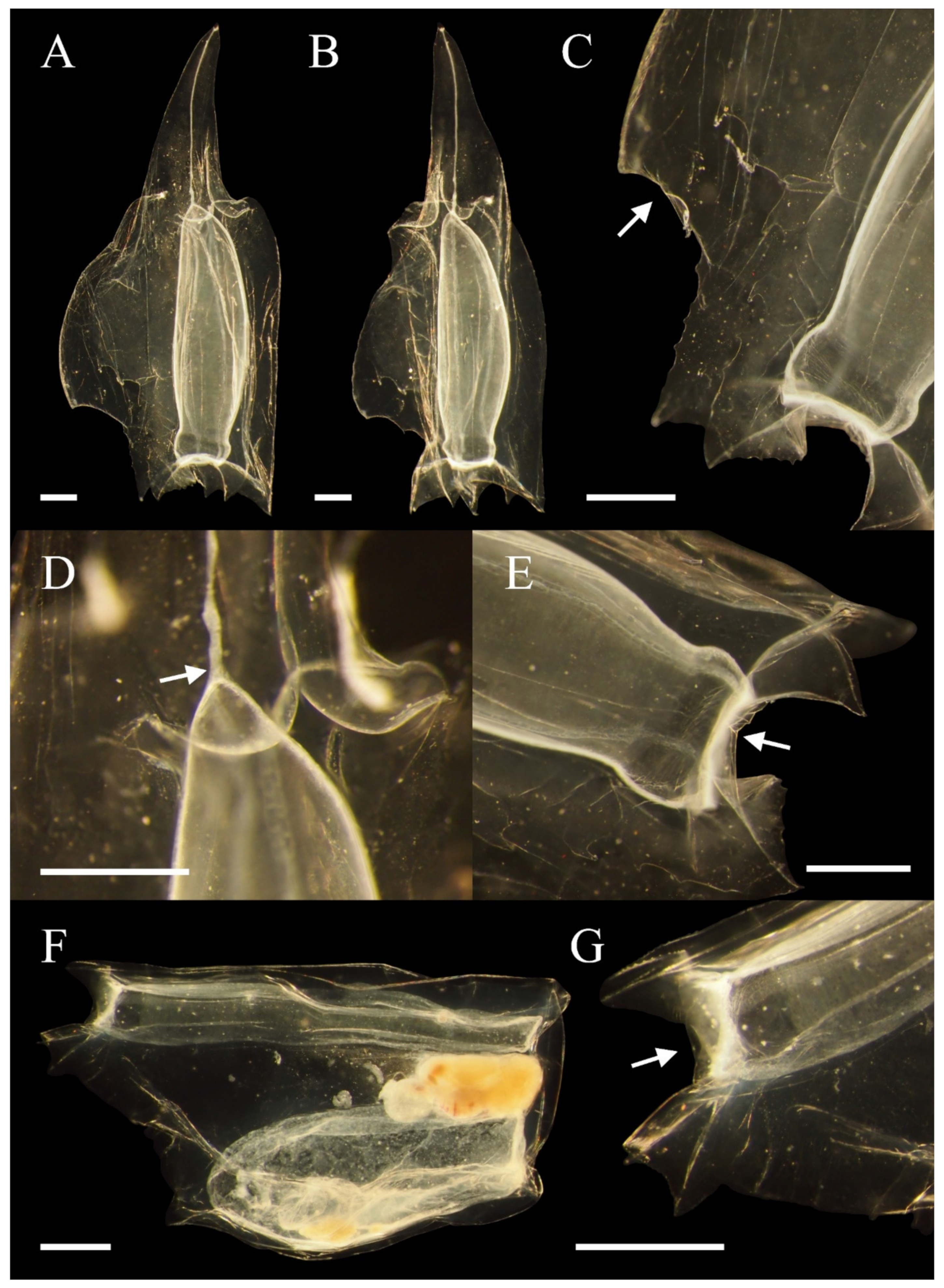
3.1. Abyla haeckeli (Lens and van Riemsdijk, 1908)

3.1.1. Synonymy

Abyla ingeborgae Sears, 1953: [30], p. 39, 42.
Amphiroa dispar Bedot, 1896: [31], p. 373, pl. 12, figs. 5, 6.
Abyla haeckeli Lens and van Riemsdijk, 1908: [32], p. 32, pl. 5, figs. 39–41; [9], p. 210, text-fig. 143, pl. 36, figs. 6, 8; [10], p. 151–152.

3.1.2. Material Examined

One posterior nectophore (NIBRIV0000896907), D7, 26 April 2018, collected by Nayeon Park and Eunha Choi was examined.

3.1.3. Descriptions

See Figure 2A–E. The posterior nectophore is about 13 mm in length. The tapering apical apophysis is long, extended, and includes an elongated pedicular canal. The basal end of the pedicular canal meets the apex of the nectosac. There are four ridges, with the two ventral ridges forming the hydroecial wing, which have many hook-shaped serrate. The right and left ventral hydroecial wings are extended widely with a comb between them. Only two or three teeth are on the comb. The ventral margin is thick. Ostial teeth are conspicuous and have fine serrations. The lateral teeth around the ostium are closer to the dorsal tooth than to the ventral teeth. The right lateral tooth and left lateral tooth are similar in size. The dorsal tooth is large and dominant. The dorsal facet is flat and much narrower than the ventral facet. There is a curvature in the connection of the apical apophysis.

3.1.4. Remarks

Abyla is the genus with the most morphological variation among the siphonophores, currently including three species, with type species Abyla trigona Quoy and Gaimard, 1827. Abyla currently includes the former ‘Ceratocymba’ and ‘Trigona’. In Abyla, the ventro-lateral facet of the anterior nectophore is divided by a horizontal ridge.
Most Abyla posterior nectophores have five or more combs, but Abyla haeckeli has only two or three. According to Sears [30], little information is available regarding the morphology of young posterior nectophores. However, there are two or three combs, as in the adult specimens we observed. In several other references such as Totton [9,10], the descriptions of comb and serrate are confused. However, it is correct that the comb is inside between the right and left hydroecial wings, and the serrate is on the ridge. The description of Abyla ingeborgae Sears, 1953, by Totton [9] said “in haeckeli right tooth is the smaller”. In our specimen, the left and right tooth were not significantly different. According to Totton [10], specimens from Atlantic stations were larger than the specimens observed in the present study. The anterior nectophore was not observed in this study.

3.1.5. Distribution

The distribution is as follows: Pacific (North Pacific; tropico-equatorial Pacific; Japan; Peru; California regions [33]; South China Sea [34]), Atlantic (Southwest Atlantic [35]), Mediterranean (eastern Mediterranean [36]), and Indian (Red Sea [36]; Laccadive Sea [37]). It is known to live in deeper waters than the shallow coast and is found not only in warm layers, but also in cold waters of about 8–12 °C.
Genus Ceratocymba Chun, 1888.

3.2. Ceratocymba leuckartii (Huxley, 1859)

3.2.1. Synonymy

Abyla leuckartii Huxley, 1859: [38], p. 49, pl. 36, figs. 2–5, 7; text-figs. 139, 141B,D.
Ceratocymba leuckartii (Huxley, 1859): [32], p. 34–35, figs. 41–45; [9], p. 205; figs. 139, 141, pl. 36, figs. 2–5, 7; [30], p. 67; [39], p. 215–216, pl. 8, figs. 8–11; [40], p. 19, fig. 2.

3.2.2. Material Examined

One anterior nectophore (NIBRIV0000896909), D7, 26 April 2018, collected by Nayeon Park and Eunha Choi, was examined.

3.2.3. Descriptions

See Figure 2F,G. The anterior nectophore is about 5–7 mm in length, is a polygon consisting of seven facets, and is flat on the lateral view. The apical facet is not divided by another ridge and is a single facet that is flat and hexagonal and does not have a peak. Both the ventral and dorsal facets are long and narrow. However, the ventral facet is narrower and fusiform, while the dorsal facet is longer and rectangular. The lateral ridges are close to the ventral facet. The basal ends of lateral ridges meet the lateral teeth at the entrance of the hydroecium. Due to the oblique lateral ridges, the dorso-lateral and ventro-lateral facets are incomplete and unevenly divided. The dorso-lateral facet is about two–three times wider than the ventro-lateral facet. The dorsal teeth are more conspicuous than the lateral teeth and have small serrates. The somatocyst, hydroecium, and the apex of nectosac are located at the same level close to the nectophore apex. The somatocyst is pear-shaped with a thick mid portion and makes up most of the body. The hydroecium widens away from the apical facet. The nectosac is long and cylindrical.

3.2.4. Remarks

Genus Ceratocymba currently includes three species. The type species is Ceratocymba sagittata (Quoy and Gaimard, 1827). Unlike the previous genus Abyla, the posterior nectophore is narrow, and the wing is not extended. However, in this study, we were unable to compare the posterior nectophores of these two genera.
According to Pagès [41], many features such as the height of the apex and cylindrical thickness are described as similar in the somatocyst, hydroecium, and nectosac. However, the somatocyst was thickest, and made up most of our specimens. The ventral facet is described as a fusiform, but it cannot be defined as completely fusiform because there are horizontal ridges in the apical direction and the lateral ridge is located much closer to the ventral facet than in other species in the same genus. The posterior nectophore was not observed in this study.

3.2.5. Distribution

The distribution is as follows: Pacific (western Pacific; Solomon Island; Easter Island; Tuamotu Archipelago; Hawaii; California [33]; South China Sea [34]), Atlantic ([35]; Benguela Current [41]), Antarctic (Peninsula [42]). This species lives in waters of various temperatures from tropical regions to Antarctic [40] and has a cosmopolitan distribution. Although they are mainly epipelagic, some populations have also been found in bathypelagic environments.
Subfamily Abylopsinae Totton, 1954.
Genus Bassia Agassiz, 1862.

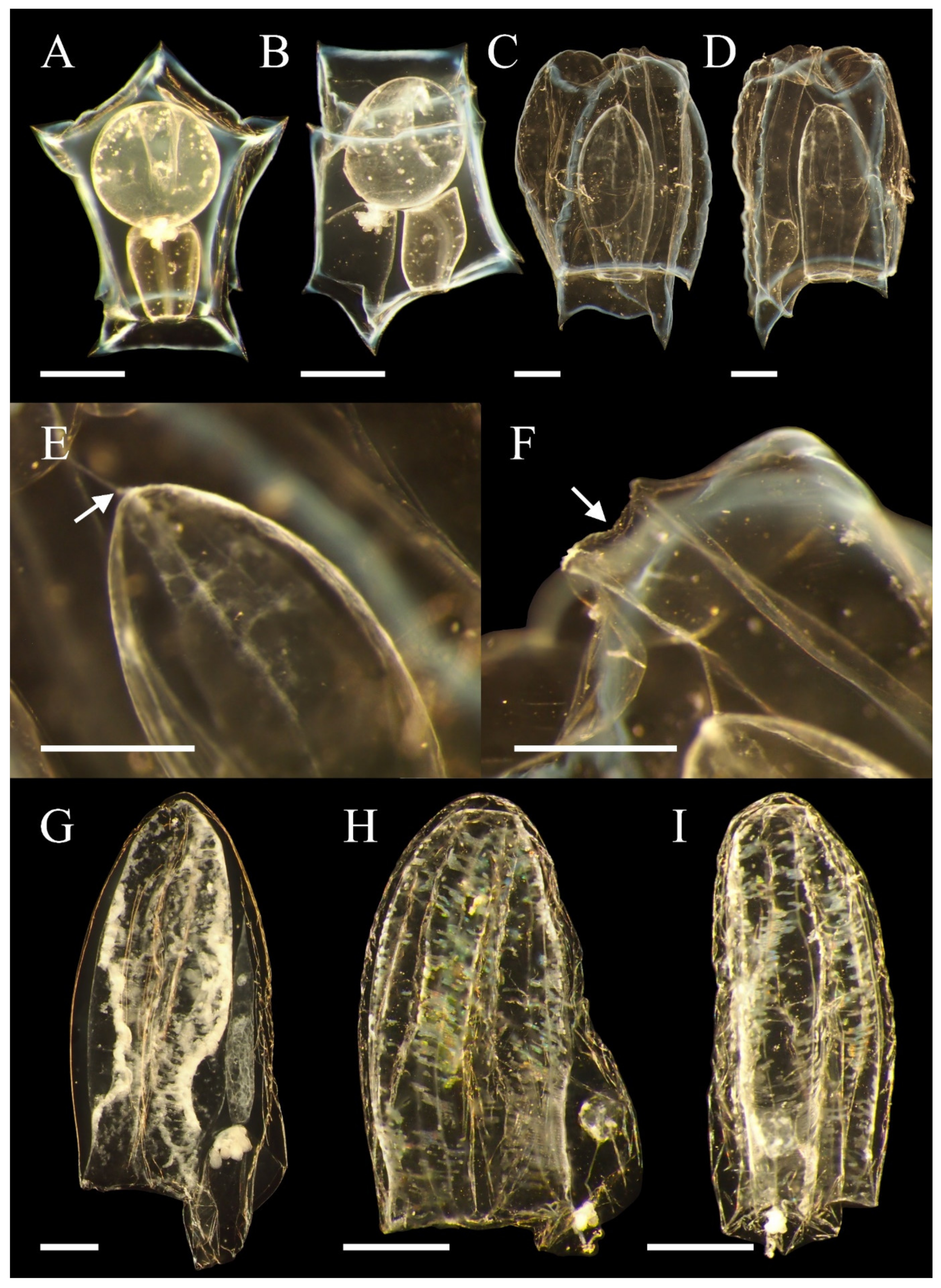
3.3. Bassia bassensis (Quoy and Gaimard, 1833)

3.3.1. Synonymy

Diphyes bassensis Quoy and Gaimard, 1833: [43], p. 91, pl. 4, figs. 18–20.
Abyla bassensis Huxley, 1859: [38], p. 45.
Bassia bassensis (Quoy and Gaimard, 1833): [44], p. 229–231, pl. 12, fig. 8, pl. 14, fig. 9; [1], p. 70–72, fig. 11; [9], pl. 40, fig. 5; [11], p. 134, fig. 60; [30], p. 94–98, figs. 2F and 28B–C; [41], figs. 52A–C; [45], p. 339–340; fig. 18; [46], fig. 59.

3.3.2. Material Examined

One posterior nectophore (NIBRIV0000896908); one anterior nectophore, and one posterior nectophore (MABIKCN00081182), D8, 26 April 2018, collected by Nayeon Park and Eunha Choi, were examined.

3.3.3. Descriptions

See Figure 3A–F. The anterior nectophore is about 2–4 mm in length, and is a polyhedron consisting of a total of seven facets, of which the ventral and dorsal facets form a conspicuous pentagon. There are no median ridges in the ventral or dorsal facets. The two apical lateral facets meet toward the apical end and form a central apical ridge. The apical-lateral and baso-lateral facets are divided by the lateral ridge, and the apical-lateral facet is half as broad as the baso-lateral facet. Ridges are opaque, blue-white tinged, and conspicuous. The spherical somatocyst is large enough to account for more than half of the nectophore. The somatocyst is not an apical projection and is not elongated along the ventral facet. The apexes of the nectosac and hydroecium lie below the somatocyst and are located at the same level as the basal part of the somatocyst. The apex of the nectosac reaches about halfway to the nectophore and does not expand between the somatocyst and dorsal facet. However, it faces the dorsal facet rather than the ventral facet and is located closer. In the nectosac, the short thick lateral radial canal passes through and reaches the ostial. The lateral radial canal forms a gradual curve. The hydroecium is similar in length and size to the nectosac and reaches the middle of the entire nectophore. The hydroecium is deep, but the basal part does not protrude into the basal facet. The hydroecium narrows toward the apex and widens toward the basal end. The vertical end of the hydroecium wing is pointed and has fine serrations.
The posterior nectophore is about 5–8 mm in length, is longer than the anterior nectophore, and the width is similar to or wider than the anterior nectophore. In addition, the posterior nectophore is softer than the anterior nectophore. There are four longitudinal ridges that form a square column. Ridges are opaque, blue-white tinged, and conspicuous. However, the ridges are winding, not straight. The basal part of each ridge has small serrations. There are two teeth in the basal part of the ventral facet, with the right tooth larger than the left. The two teeth are connected in an oblique curve. The inner flaps of the ventral wings are connected to the inside of the hydroecium, which is hook-shaped. There are small serrations in the basal ridges of the hydroecium. There is a columnar passage running in the apical direction from the hydroecium and reaches the apex of the nectophore. The columnar passage protrudes outward and is connected to part of the anterior nectophore. A pedicular canal descends from the passage and reaches the apex of the nectosac.

3.3.4. Remarks

Genus Bassia includes only Bassia bassensis. Anterior nectophores have many similarities to Genus Abylopsis, but the somatocyst is not fusiform to the apex.
The apex levels of nectosac and hydroecium differed between specimens, and in most cases, the apex of the nectosac is slightly higher. Kirkpatrick and Pugh [11] noted that the dorsal ridge was absent, but it was clearly observed in our specimens. In cases of posterior nectophores, there are many similarities to Abylopsis eschscholtzii (Huxley, 1859), so it is easy to be confused. However, it can be distinguished by its unique and conspicuously-colored ridges in Bassia. Totton [9] mentioned that the ostial end is twisted, but after considering his figures and our specimens, the ostial part is straight, and non-twisted in form. The four longitudinal ridges are winding, which might have occurred during fixation. Mills et al. [46] described an anterior nectophore of 7 mm and a posterior nectophore up to 15 mm, but our adult specimens were smaller.

3.3.5. Distribution

The distribution is as follows: Pacific ([46]; Japan [33]; South China Sea [34]; Hawaii; Easter Islands [33]), Atlantic ([35,46]; Ireland [46]; Benguela Current [41]), Mediterranean [36,46], and Indian ([46]; Red Sea [36]; Laccadive Sea [37]). It is mainly found in tropical, subtropical, and shallow waters [46]. However, some records have mentioned that it lives not only in the shallow, but also in the deep sea [37]. In China, it has been recorded more in winter than in summer [34].
Family Diphyidae Quoy and Gaimard, 1827.
Subfamily Diphyinae Quoy and Gaimard, 1827.
Genus Dimophyes Moser, 1925.

3.4. Dimophyes arctica (Chun, 1897)

3.4.1. Synonymy

Diphyes arctica Chun, 1897: [47], p. 19, pl. 1, figs. 1–5 and 9.
Eudoxia arctica Chun, 1897: [47], p. 19.
Diphyes borealis Chun, 1897: [47], p. 99.
Dimophyes arctica (Chun, 1897): [48], p. 76; [7], p. 488, figs. 3.67, 3.79; [9], p. 184, pl. 33, figs. 1–2, 7, text-fig. 122; [11], p. 106, fig. 47; [12], figs. 52–53; [41], p. 93, fig. 37; [45], p. 363; [46], pl. 60E; [49], fig. 2, Table 1.

3.4.2. Material Examined

The following materials were examined: four anterior nectophores (NIBRIV0000896890), E1, 15 June 2021; two anterior nectophores (NIBRIV0000896892), E2; five anterior nectophores (MABIKCN00081183), E6, 17 June 2021; three anterior nectophores (NIBRIV0000896891), E4; two anterior nectophores (NIBRIV0000896893), E7, 18 June 2021, collected by Nayeon Park.

3.4.3. Descriptions

See Figure 3G. The anterior nectophore is long and bullet-shaped with a blunt apex, about 7–10 mm in length, with no longitudinal ridges. There are longitudinal folds (furrows) on the lateral surfaces instead of ridges. The depth of the fold is variable, both shallow and deep. There is no serrate. The blunt tip of the nectosac faces toward the apex. The surface of the nectosac has a horizontal pattern. The nectosac appears transparent or opaque depending on the individual and makes up 9/10 or more of the nectophore. The somatocyst is fusiform and narrows toward the apex. The ratio of somatocyst to total anterior nectophore is about 1/2–3/4. The somatocyst stalk is short and the somatocyst has oil droplets. The hydroecium is medium in depth and rounded, with a rounded basal ridge. The hydroecium is about 1/5 of the length of the nectophore. The upper ridges of the hydroecium and the basal part of the somatocyst are located above the ostial. Ostium accounts for about 1/2–2/3 of the width of the nectophore. There are no ostial teeth, and the mouth plate is undivided. The mouth plate is about 1/3 of the length of the somatocyst and is almost triangular.

3.4.4. Remarks

Genus Dimophyes includes only Dimophyes arctica. The absence of a longitudinal ridge is the main feature, but Muggiaea bargmannae Totton, 1954, and some Lensia species also lack a longitudinal ridge, and the difference between these species lies in whether the mouth plate is divided [12].
According to Kirkpatrick and Pugh [11], the anterior nectophore is up to 15 mm in length. However, our specimens were relatively small. The depth of the fold and transparency of the nectosac are different for each specimen, which is thought to be an effect of fixation. The internal pedicular canal had not been well observed, but this was also considered an artifact of fixation. In previous records, various expressions for somatocyst shapes are used, such as spindle-shaped, inverted carrot, etc. In Mapstone [12], Dimophyes arctica is divided into two types: wide (rounded apex, wide nectophore) and narrow (pointed apex, narrow nectophore), but we observed only the wide type in this study. This is interpreted as a functional difference [50], and there are also suggestions that the difference is an artifact of fixation [12]. The posterior nectophore was not observed in this study, but according to the literature it is reduced.

3.4.5. Distribution

The distribution is as follows: Pacific (Northeast Pacific; Americas [36,46]; Canadian waters [12]; Bellingshausen Sea; British North Sea [9]; California; South China Sea [33]; Sagami Bay [51]), Atlantic ([35]; southern Greenland; Iceland [46]; Caribbean [36]; Korsfjord [52]), Indian [33], Arctic ([9]; Chinese National Arctic [53]), and Antarctic ([53]; Weddel Sea [9]). According to [10], Dimophyes arctica inhabits cold waters, mainly found at 200–600 m or below 900 m, but our specimens were obtained within 100 m of warm water, unlike the previously described environmental conditions. Therefore, it is a cosmopolitan species that appears in all waters worldwide, including the Arctic and Antarctic. Although it is also found in tropical regions with high temperatures, Arctic and Antarctic specimens are more abundant and larger [33].
Genus Lensia Totton 1932.

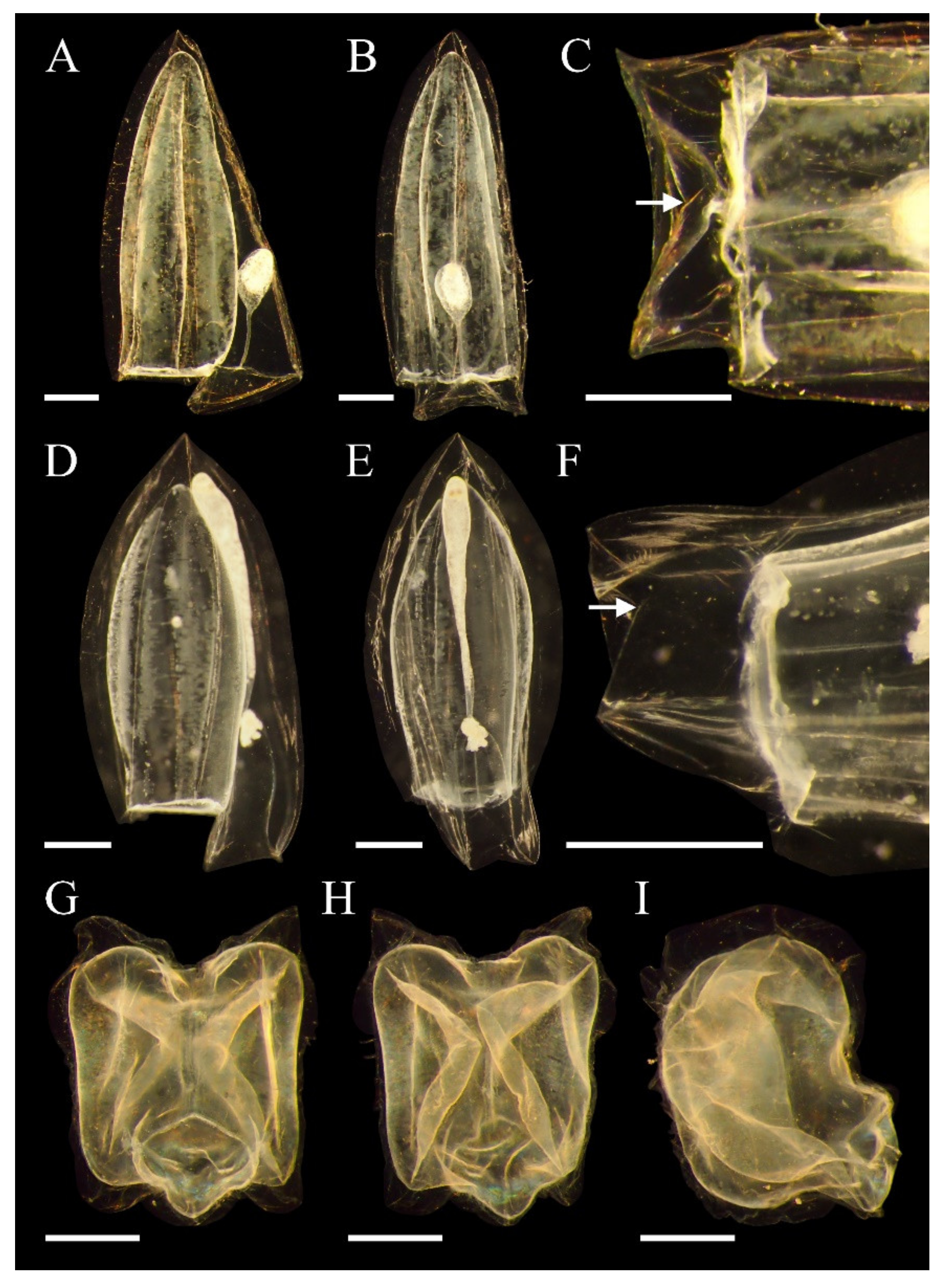
3.5. Lensia subtilis (Chun, 1886)

3.5.1. Synonymy

Ersaea elongata Will, 1844: [54], p. 1–86.
Diphyes subtilis Chun, 1886: [55], p. 681, figs. 3–7.
Lensia subtilis (Chun, 1886): [10], p. 114; [7], p. 490, figs. 3.99, 3.109; [9], p. 168, text-figs. 104–105; [11], p. 102, fig. 44; [41], p. 91, fig. 35; [42], p. 40–41, fig. 62; [44], p. 343–344; [45], p. 367–368, fig. 34.

3.5.2. Material Examined

One anterior nectophore (NIBRIV0000882819), D8, 26 April 2018, collected by Nayeon Park and Eunha Choi, was examined.

3.5.3. Descriptions

See Figure 3H,I. The anterior nectophore is about 4–5 mm in length. There is a bullet-shaped anterior nectophore with a blunt apex, with no longitudinal ridges. There are four inward-facing longitudinal folds on the lateral surfaces instead of ridges that divide the surface of the nectophore as they contract. There is no serrate. The blunt tip of the nectosac faces the apex. The surface of the nectosac is patterned horizontally. The nectosac occupies a height of about 9/10 or more of the nectophore. The somatocyst is a small sphere and has a long stalk and oil droplets. The ratio of somatocyst to total anterior nectophore is about 1/2–1/4. The hydroecium is shallow in depth, round, and wide, inclined toward the ventral facet, so that the upper ridge of the hydroecium is above the ostium. The basal ridge of the hydroecium is rounded. The ostium accounts for more than 3/5 of the width of the nectophore. There are no ostial teeth, and the mouth plate is divided into small sizes. Each mouth plate is rounded. The basal facet of the mouth plate is inclined toward the ventral facet.

3.5.4. Remarks

Genus Lensia includes 26 species and is the most morphologically diverse among the diphyids. The anterior nectophore generally has a pentagonal cross-section, but depending on the species, might have 3, 5, 7, 15, or more longitudinal ridges. Some species do not have ridges. The arrangement of ridges varies. Lensia is smaller in size and has a more fragile body than other diphyids.
Lensia subtilis exhibits broad variation in size ([9]: up to 11 mm; [11]: up to 10 mm; [14]: 2.56 mm; [41]: up to 3.2 mm). Our specimen was about 5 mm in length. Nishiyama [14] mentioned that the hydroecium is below the ostial level, but in our specimens, the hydroecium started at the ostial level and angled obliquely to the rear. Finally, the apex of the hydroecium was above the ostial level. Excluding the shape of the somatocyst, our specimen was similar to the anterior nectophore of Lensia meteori (Leloup, 1934). It was also similar to Dimophyes arctica in that there is a fold instead of ridge, but the fold of this species was much more pronounced and similar to a ridge. The identification key in Kirkpatrick and Pugh [11] mentioned that Lensia subtilis has five non-crested ridges, and a pair of lateral ridges often is not clearly observed, but p. 102 states that there are four longitudinal folds, as confirmed in our specimens and those described in previous studies. The posterior nectophore was not observed in this study.

3.5.5. Distribution

The distribution is as follows: Pacific ([56,57]; Japan; Hawaii; California [33]; South China Sea [34]); Atlantic ([35,56,57]; British; Celtic Sea [11]; Brazil; Santa Catarina [14]; Benguela Current [41]), Mediterranean [11,33,36], and Indian ([56,57]; East African coast [10]). It mainly lives in warm and shallow tropical water [14].

3.6. Lensia subtiloides (Lens and van Riemsdijk, 1908)

3.6.1. Synonymy

Diphyes subtiloides Lens and van Riemsdijk, 1908: [32], p. 46, pl. 7, figs. 59–61.
Lensia subtiloides (Lens and van Riemsdijk, 1908): [45], p. 364, figs. 31–32; [7], p. 491, figs. 3.100, 3.110; [9], text-figs. 95–96, 97B; [41], p. 92, fig. 36; [57], p. 130–132, figs. 10J–N.

3.6.2. Material Examined

The following material was examined: one anterior nectophore (NIBRIV0000882820), D4; one anterior nectophore (NIBRIV0000882821); one anterior nectophore (MABIKCN00081184), D5, 27 April 2018; one anterior nectophore (NIBRIV0000882822), D7; one anterior nectophore (NIBRIV0000882823), D8; one anterior nectophore (NIBRIV0000896894), D10, 26 April 2018, collected by Nayeon Park and Eunha Choi.

3.6.3. Descriptions

See Figure 4A–C. The anterior nectophore is about 7 mm in length, bullet-shaped, and has a pointed apex. Five complete longitudinal ridges meet at the apex. The narrow rounded tip of nectosac faces the apex. The surface of the nectosac is patterned horizontally. The nectosac occupies about 9/10 or more of the nectophore. The somatocyst is oblique oval or club-shaped, with a long stalk. The ratio of somatocyst to total anterior nectophore is about 1/2–1/3. The somatocyst has oil droplets. The hydroecium is shallow in depth, rounded, and wide. The hydroecium starts at the ostial level and descends almost straight to the ventral facet. Therefore, the hydroecium and the ostial level are at the same level. The basal ridge of the hydroecium is rounded. The ostium accounts for more than 1/2–2/3 of the width of the nectophore. There are no ostial teeth, and the mouth plate is divided into small parts of equal size. The length of the mouth plate is about 1/3 of the somatocyst. Each mouth plate is rounded. The two mouth plate wings slightly overlap toward the ventral facet.

3.6.4. Remarks

Lensia subtiloides is the type species of the genus Lensia.
Our specimens were larger than those described in previous records ([9]: up to 5 mm; [14]: 3.96 mm; [41]: 3.7 mm). Although crested ridges are mentioned in previous descriptions, but are not clear as a result of actual observation, they might be artifacts of fixation. In Totton [9], the base of the somatocyst is described as above the ostial, but in our specimens it is almost the same or finely above. In Nishiyama [14], the somatocyst is about 1/10 of the height of the nectophore and slightly bent. Our specimens were much longer, about 1/2–1/3, and straighter. It is possible that Nishiyama [14] describes juvenile individuals. The somatocyst was similar to that of Chelophyes contorta (Lens and van Riemsdijk 1908) but was not skewed left or right. There was a difference in the number of ridges and the somatocyst shape from the Lensia subtilis mentioned earlier. The posterior nectophore was not observed in this study.

3.6.5. Distribution

The distribution is as follows: Pacific ([10,56,57]; Americas [14]; Hawaii; California; San Diego; Japan [33]; Thailand [33]; South China Sea [33,34]), Atlantic ([14,35,56,57]; British; Celtic Sea [11]; Brazil; Santa Catarina [14]; Benguela Current [41]), Mediterranean [11,33,36], and Indian (Red Sea [10]). It mainly lives in warm waters and surface layers [10].
Genus Muggiaea Bush, 1851.

3.7. Muggiaea atlantica (Cunningham, 1892)

3.7.1. Synonymy

Muggiaea atlantica Cunningham, 1892: [58], p. 212–215, figs. 1–2; [11], p. 104, fig. 46; [12], p. 491, figs. 3.74, 3.85; [41], p. 93–95, fig. 38; [44], p. 187, pl. 7, fig. 1, pl. 9, figs. 7–8; [59], p. 254, fig. 69.

3.7.2. Material Examined

The following material was examined: three anterior nectophores (NIBRIV0000882809), D2; three anterior nectophores (NIBRIV0000882815), D14, 23 April 2018; three anterior nectophores (NIBRIV0000896897), D7; three anterior nectophores (NIBRIV0000882812); four anterior nectophores (MABIKCN00081185), D8; three anterior nectophores (NIBRIV0000882813), D9; four anterior nectophores (NIBRIV0000896898), D10; three anterior nectophores (NIBRIV0000882814), D11, 26 April 2018; three anterior nectophores (NIBRIV0000882808), D3; three anterior nectophores (NIBRIV0000896899), D4; three anterior nectophores (NIBRIV0000882810), D5; three anterior nectophores (NIBRIV0000882811), D6, 27 April 2018, collected by Nayeon Park and Eunha Choi.

3.7.3. Descriptions

See Figure 4D,F. The anterior nectophore is about 5–7 mm in length. The bullet-shaped anterior nectophore has a pointed apex. Five complete longitudinal ridges meet at the apex. The ridge has a small serrate. The anterior nectophore has a pentagonal cross-section. The rounded tip of the nectosac faces the apex. The surface of the nectosac is patterned horizontally. The nectosac occupies about 7/10 of the nectophore. The somatocyst is long filiform, and the apical end is thickly inflated. The somatocyst reaches the apex of the nectosac and is about 4/5 of the anterior nectophore. The somatocyst is closely attached to the ventral facet of the nectosac and has oil droplets. The hydroecium is deep and rounded and reaches 1/3 of the nectosac. Therefore, the upper ridge of the hydroecium and the basal part of the somatocyst are located above the ostial. The basal ridge of the hydroecium is flat. The ostium accounts for about 1/2 of the width of the nectophore. There are no ostial teeth, and the mouth plate is divided into two small parts of unequal size. The left mouth plate wing is more prominent than the right. The length of the mouth plate is about 1/4 of the somatocyst. Each mouth plate is triangular and has serrations. The two mouth plate wings are slightly overlapped and face the ventral facet.

3.7.4. Remarks

Genus Muggiaea currently includes four species. The type species is Muggiaea kochi (Will 1844). The lengths of hydroecium and somatocyst in Muggiaea are important morphological features.
Although it is equivalent in size and shape to Muggiaea kochi, it has a different ratio of somatocyst to height of the hydroecium. In Mapstone [12], it is described as laterally flat and less flat than other diphyids because it is a balanced pentagon. Muggiaea atlantica has wide variation in size ([12]: up to 11 mm; [41]: 4.5 mm; [58]: 3–7 mm). In Kirkpatrick and Pugh [11], the somatocyst is described as long and thin. However, some of our specimens were thick. Although it is mentioned as having strong crested ridges in Kirkpatrick and Pugh [11], it is not clear as a result of actual observation, and this might be an artifact of fixation. Depending on the specimens, the somatocyst exceeds the apex of the nectosac or is just below. Unlike Mills et al. [46], we did not observe any individual with a hydroecium reaching 1/2 of nectophore. The posterior nectophore was not developed.

3.7.5. Distribution

The distribution is as follows: Pacific ([11]; Northeast Pacific [44]; San Diego; California; Peru [33]; South China Sea [34]; Sagami Bay [51]; Canadian waters [12]), Atlantic ([11,33,35]; Benguela Current [41]), Mediterranean [11,33,36], and Indian ([11,33]; Red Sea [36]). Many records locate it within 100 m [12] and in high water temperatures [9]. According to Pagès [41], Muggiaea atlantica is more abundant in the Atlantic and Mediterranean than the Pacific and Indian Oceans.
Suborder Physonectae Haeckel, 1888.
Family Agalmatidae Brandt, 1834.
Genus Nanomia A. Agassiz, 1865.

3.8. Nanomia bijuga (Delle Chiaje, 1844)

3.8.1. Synonymy

Physsophora bijuga Delle Chiaje, 1844: [60], pl. 181, figs. 3–6.
Anthemodes canariensis Haeckel, 1869: [61], p. 18, figs. 3–10.
Halistemma pictum Metschnikoff 1874: [62], p. 306, pl. 2, figs. 1–11.
Halistemma tergestinum Claus, 1878: [63], p. 1, pl. 1, figs. 1–11, pl. 2, figs. 1–10, 12–15, pl. 4.
Agalmopsis fragile Fewkes, 1882: [64], p. 844.
Anthemodes moseri Agassiz and Mayer, 1902: [65], p. 167, pl. 12, figs, 49–57.
Nanomia bijuga (Delle Chiaje, 1844): [9], p. 68, figs. 32–36; [7], p. 483, figs. 3.14, 3.28; [66], p. 212, figs. 125A–D; [67], p. 747, fig. 5.

3.8.2. Material Examined

The following material was examined: two anterior nectophores (NIBRIV0000896896), D14, 23 April 2018; two anterior nectophores (NIBRIV0000882817), D8; two anterior nectophores (NIBRIV0000882818); one anterior nectophore (MABIKCN00081186), D9; one anterior nectophore (NIBRIV0000896895), D10, 26 April 2018; two anterior nectophores (NIBRIV0000882816), D6, 27 April 2018, collected by Nayeon Park and Eunha Choi.

3.8.3. Descriptions

See Figure 4G–I. The nectophore is about 2–4 mm in length and rectangular in axial or abaxial, lateral views. The nectophore is flatted along the proximal-distal side. The upper corners are ear-shaped in lateral view. The nectosac is opaque, pale brown, and fills the nectophore. The nectosac surface has a scale pattern. Axial wings are small, triangular, and folded in the proximal direction. The thrust block is small, thin, and incomplete, and located above the end of the axial wing. There are incomplete upper lateral, complete lateral, vertical lateral, and lower lateral ridges on the surface of the nectophore. Upper lateral ridges occur at the end of each axial wing, run to the nectophore apex, and then obliquely to the midline of the upper surface of the nectophore. The upper lateral ridge pairs are incomplete at both distal ends. Lateral ridges occur in upper lateral ridges and run to the ostium of the nectophore. Vertical lateral ridges occur at the upper lateral ridge near the nectophore apex and descend to the ostial level and meet the lower lateral ridges. Lower lateral ridges occur from an upper lateral ridge at the end of the axial wing and run to the ostium. In the proximal part, the lower lateral ridges meet the upper lateral ridges in the axial wing. The ostial regions are wider than the mouth plate, accounting for about 1/2 of the width of the nectophore. The mouth plate is short and undivided. There is a thick velum inside the ostial. Radial canals occur from the internal pedicular canal and extend along the surfaces of the nectosac. The upper radial canal runs superiorly and passes through the upper nectosac. The lower radial canal passes through the lower nectosac surface and enters the ostial ring canal. Lateral radial canals on the left and right sides are looped to the nectosac.

3.8.4. Remarks

Genus Nanomia includes two species, and the type species is Nanomia cara Agassiz, 1865. Nanomia is an agalmid siphonophore in which male and female gonophores are alternately attached on the siphosome. The most distinct difference from other agalmatids is possession of a partial involucrum on the unicornuate tentillum. They have small bright pigments. They form linear colonies and have nectophores with many ridges. In addition, the nectosac fills most of the nectophores, unlike other agalmatids. ‘Nanomia’ is the generic name of the species referred to as Nanomia cara in the past, while ‘Bijuga’ was referred to as Stephanomia Lesueur and Petit, 1807 [12].
Nanomia bijuga is easily confused with Nanomia cara but can be identified by the size of the axial wing, the proximal-distal inward fold, and the degree of flatness. If the entire colony is secured, Nanomia bijuga can be distinguished by its much smaller size. In general, all ridges except the upper lateral ridges are complete, but some specimens are difficult to clearly confirm. We did not observe the mantle canal, its ascending or descending branches, or the external pedicular canal. This might be an artifact of fixation. Only traces of the internal pedicular canals could be observed in some individuals, so we infer the location where the radial canals occurred. In general, the internal pedicular canal in agalmatids is short and originates from the external pedicular canal of the proximal nectosac surface. In Mapstone [12], the nectophore is 2–6 mm in length. We did not observe specimens of about 6 mm. In Totton [9], vertical lateral ridges are shown to meet lower lateral ridges at the mid-height of the nectophore, but were down to the ostium level in the specimens we observed.

3.8.5. Distribution

The distribution is as follows: Pacific ([7,9,46]; Northeast Pacific [9]; Sagami Bay [68]; Canadian waters [12]), Atlantic ([7,9,46]; North Atlantic [69]; Benguela Current [41]; British waters [10]), Mediterranean [9,46], and Indian [9,46]. It mainly lives in warm waters.

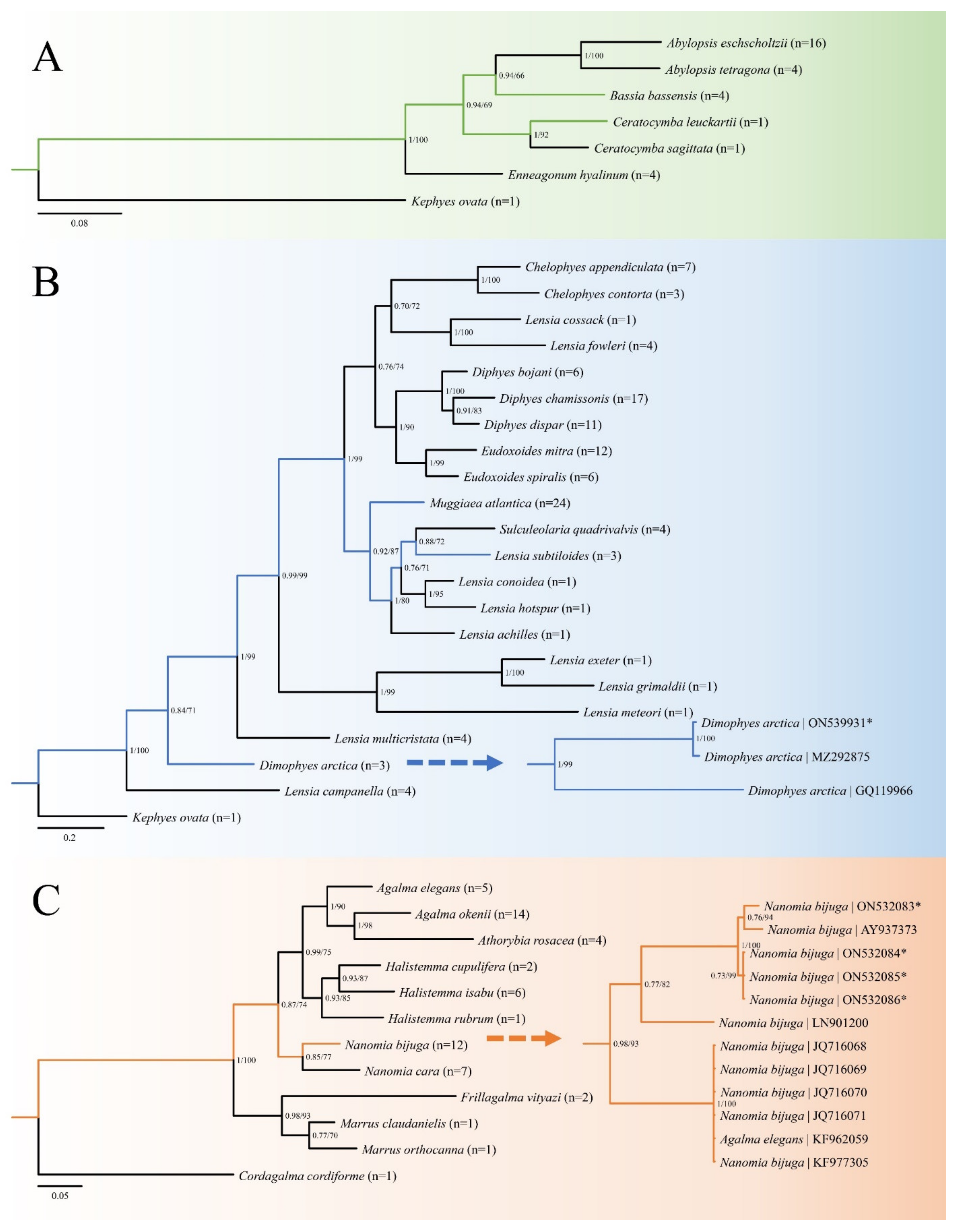
3.9. Molecular Phylogenetic Analysis and Genetic Diversity

A total of 19 new mtCOI sequences for five siphonophore species were obtained in this study (Table S1). The sequences were limited to some ethanol specimens, not formalin-only specimens. We recovered the phylogeny of three families within siphonophores based on newly acquired sequences and GenBank data. Phylogenetic trees were constructed using two algorithms (BI and ML) and showed identical topologies with high support values (Figure 5). The K2P genetic distances (Tables S2–S6) in each intra-genus analysis (Ceratocymba, Dimophyes, Lensia, Muggiaea, Nanomia) were calculated to check the differences between our sequences and GenBank data.
In Abylidae, all genera formed a monophyletic group (Figure 5A). However, based on the subfamily, the genera belonging to Abylopsinae (Abylopsis, Bassia, Enneagonum) were polyphyletic. Bassia bassensis was a sister clade to the genus Abylopsis (BI = 0.94; ML = 66), followed by Ceratocymba (BI = 0.94; ML = 69). Ceratocymba leuckartii differed by 0.104 in K2P genetic distance compared to Ceratocymba sagittata in the same genus (Table S2).
In Diphyidae, all genera except Lensia formed a monophyletic group (Figure 5B), while Lensia was polyphyletic throughout the Diphyidae. Dimophyes arctica was located far from most diphyids (BI = 0.84; ML = 71) and branched into two groups (BI = 1; ML = 99). This observation was confirmed using K2P genetic distances, with the difference from GQ119966 ranging from 0.342–0.363 (Table S3). Lensia subtiloides formed a sister relationship with Sulculeolaria quadrivalvis (BI = 0.88; ML = 72), and no other Lensia, and was then combined with other Lensia (Lensia conoidea, Lensia hotspur, Lensia achilles) (BI = 0.76; ML = 71). Lensia subtiloides had few differences in the same species (0.005–0.007), but distances ranged from 0.207–0.613 vs. other species in the same genus (Table S4). Muggiaea atlantica formed the sister group to the SulculeolariaLensia branch (BI = 0.92; ML = 87). In Muggiaea atlantica, there was little difference within species (0.000–0.006) (Table S5).
In Agalmatidae, all genera except Agalma formed a monophyletic group (Figure 5C). Agalma was paraphyletic. Nanomia combined with the AthorybiaAgalmaHalistemma branch (BI = 0.87; ML = 74). In this study, we additionally confirmed the positions of Halistemma cupulifera, Marrus, and Frillagalma. Nanomia bijuga branched into at least three groups (BI = 0.98, 0.77, 0.73; ML = 93, 82, 99). K2P genetic distances ranged from 0.000–0.193 within Nanomia bijuga (Table S6). This was similar to distances between Nanomia cara and Nanomia bijuga (0.169–0.191).

4. Discussion

4.1. Morphological Analysis and Distribution

We conducted morphological redescriptions of eight species previously unrecorded in Korean waters that were collected from Jeju and southern coasts of Korea. We include the morphological discussions for each species in the Remarks sections. Distributions and habitats were also summarized.
Most of the habitat features of siphonophores are based on type localities, but the actual distributions are cosmopolitan regardless of temperature and salinity. In particular, species such as Dimophyes arctica range from the polar regions to the equator. However, in some cases, there was significant size variation depending on temperature [33], which supports the observations of Bouillon and Boero [70], who previously mentioned that the size of gelatinous zooplankton is greatly affected by habitat. Abyla haeckeli, Bassia bassensis, and Dimophyes arctica specimens observed in this study were much smaller in size than previous records. Abyla haeckeli was often found in waters deeper than the coast [33] and in cold seas of 8–12 °C. Bassia bassensis was more abundant in winter than summer [34]. Dimophyes arctica mainly inhabits cold water [10] but was also found in tropical regions with high water temperatures, but individuals from the Arctic and Antarctic were more abundant and larger [33]. However, the collection sites in this study were much warmer than in previous studies, with a temperature range of 13.3–23.2 °C, and the individuals we collected were much smaller in size, perhaps due to these environmental conditions. That being said, the temperatures presented in this research are limited to the surface layer and should be noted as they are different from the depth of water from which our specimens were collected. Nevertheless, since the temperature of the surface layer is an important indicator of the characteristics of the sea, these relationships need to be considered.
In ecological studies of adjacent countries in Pacific waters, such as China and Japan, siphonophores tended to be recorded in the species lists without morphological descriptions. Therefore, more species were expected to exist in Korean waters, and we pursued additional investigations. In addition, although not covered in this study, oceanographic settings and siphonophore assemblage composition and abundance need to be investigated in further studies. These approaches will lead to a broad understanding of Siphonophores.

4.2. Molecular Analysis

Polyphyly of Abylopsinae suggests that the presence or absence of the apical facet dividing subfamilies Abylopsinae and Abylinae is not a useful phylogenetic feature (Figure 5A). According to Zheng et al. [71], the average intra-genus distance between mtCOI is 0.166 in Ceratocymba, which was considered to be a corresponding figure (Table S2).
In Figure 5B, the overall locations were similar to the Diphyidae phylogeny suggested in Park et al. [49], but there were changes, such as the location of Lensia cossack, as more species were included. Regarding the divided two groups of Dimophyes arctica, we considered the possibility of regional DNA variation because the locality of GQ119966 was the North Atlantic, while those of ON539931 and MZ292875 were the Northwest Pacific. However, even considering this, it was a much greater difference than intraspecies variation and was thought to represent either a cryptic species or misidentification of Atlantic specimens, while the distances between the Northwest Pacific samples were 0.013, which was a level of variation appropriate within the same species (Figure 5B, Table S3). In Lensia, the difference within the same genus was much greater than the 0.166 (Table S4) suggested in Zheng et al. [71]. The phylogenetic results for Lensia were ambiguous. Lensia is the most diverse genus within the Diphyidae. Therefore, it might be necessary to redefine the genus. The K2P distances of Muggiaea atlantica verified with our specimens are correctly identified (Table S5).
Figure 5C presents the same result as in the Agalmatidae phylogenetic tree suggested by Park et al. [72]. In the K2P distances of Nanomia (Table S6), although the localities varied (Northwest Pacific, North Atlantic, etc.), the group was not divided based on geography. It was also a much more significant difference than intraspecific variation, as in the case of Dimophyes arctica, and was thought to represent a cryptic species or misidentification. KF962059 (Agalma elegans (Sars, 1846)) was included in the Nanomia bijuga group, which was thought to represent misidentification.

4.3. Siphonophores in Korean Waters

Park and Song described the first siphonophores in Korean waters in 2004 [73]. Since then, only 8 genera and 13 species remained on the definitive list until 2020 [74]. This number is far less than 10% of the entire siphonophore (about 190 species [2]). The reason for this is not just that a small number of siphonophores live in Korean waters. Considering the cosmopolitan of siphonophores mentioned previously, it might be that they have been continuously introduced in Korean waters from the past through geographical movement. However, due to the difficulty of taxonomical research and the lack of experts, many of them may have been omitted.
Seven species except Muggiaea atlantica were also first reported at the genus level. In particular, subfamily Abylinae, which includes Abyla haeckeli and Ceratocymba leuckartii, was also first reported at the subfamily level. As a result, in the present study, the species list of siphonophores was updated to include 3 suborders, 5 families, 3 subfamilies, 14 genera, and 21 species (Table 2). These results serve as important basic data for future research in siphonophores and are expected to contribute to securing national biological resources.

Supplementary Materials

The following supporting information can be downloaded at https://www.mdpi.com/article/10.3390/d14060494/s1, Table S1: DNA sequences used in the phylogenetic analyses of this study; Table S2: K2P distances of mtCOI sequences between genus Ceratocymba from Genbank and this study; Table S3: K2P distances of mtCOI sequences between genus Dimophyes from Genbank and this study; Table S4: K2P distances of mtCOI sequences between genus Lensia from Genbank and this study; Table S5: K2P distances of mtCOI sequences between genus Muggiaea from Genbank and this study; Table S6: K2P distances of mtCOI sequences between genus Nanomia from Genbank and this study.

Author Contributions

All authors contributed to the study conception and design. Sample and data collection, N.P. and W.L.; data analysis, N.P.; writing—original draft preparation, N.P.; writing—review and editing, W.L. All authors have read and agreed to the published version of the manuscript.

Funding

This study was supported by following research grants: National Institute of Biological Resources (NIBR), funded by the Ministry of Environment (MOE) of the Republic of Korea (NIBR202203105; NIBR202231206); Marine Biodiversity Institute of Korea (MABIK, 2022M01100); Korea Institute of Ocean Science and Technology (KIOST, PE9998C); Korea Polar Research Institute (KOPRI, PE21900).

Institutional Review Board Statement

Not applicable.

Data Availability Statement

The voucher specimens of the species examined in the present study were deposited in the National Institute of Biological Resources (NIBR) and National Marine Biodiversity Institute of Korea (MABIK). The datasets generated and/or analyzed during the current study are available in Genbank and this published article (and its Supplementary Materials).

Acknowledgments

We would like to express sincere thanks to the members of the Biodiversity Laboratory, Hanyang University, and to the captains and crew of the R/V Dongbaek and Eardo for their help during the collecting process. In addition, we are grateful to Andrey A. Prudkovsky for advice on the taxonomical approach.

Conflicts of Interest

The authors declare no conflict of interest. The funders had no role in the design of the study; in the collection, analyses, or interpretation of data; in the writing of the manuscript; or in the decision to publish the results.

References

  1. Pugh, P. The vertical distribution of the siphonophores collected during the SOND cruise, 1965. J. Mar. Biol. Assoc. U. K. 1974, 54, 25–90. [Google Scholar] [CrossRef]
  2. Palma, S.; Retamal, M.C.; Silva, N.; Silva, C. Horizontal and vertical distributions of siphonophores in relation to oceanographic conditions in Chilean Patagonian fjords. Sci. Mar. 2014, 78, 339–351. [Google Scholar] [CrossRef]
  3. Munro, C.; Siebert, S.; Zapata, F.; Howison, M.; Damian-Serrano, A.; Church, S.H.; Goetz, F.E.; Pugh, P.R.; Haddock, S.H.; Dunn, C.W. Improved phylogenetic resolution within Siphonophora (Cnidaria) with implications for trait evolution. Mol. Phylogenet. Evol. 2018, 127, 823–833. [Google Scholar] [CrossRef] [PubMed]
  4. Chuan, C.H.; Venmathi Maran, B.A.; Yap, T.K.; Cheong, K.C.; Syed Hussein, M.A.; Saleh, E. New Records of Cubozoan and Scyphozoan Jellyfish from Sabah Waters, Malaysia. Diversity 2021, 13, 420. [Google Scholar] [CrossRef]
  5. Marambio, M.; Canepa, A.; Lòpez, L.; Gauci, A.A.; Gueroun, S.K.; Zampardi, S.; Boero, F.; Yahia, O.K.-D.; Yahia, M.N.D.; Fuentes, V. Unfolding jellyfish bloom dynamics along the Mediterranean basin by transnational citizen science initiatives. Diversity 2021, 13, 274. [Google Scholar] [CrossRef]
  6. Dunn, C.W.; Pugh, P.R.; Haddock, S.H. Molecular phylogenetics of the Siphonophora (Cnidaria), with implications for the evolution of functional specialization. Syst. Biol. 2005, 54, 916–935. [Google Scholar] [CrossRef] [Green Version]
  7. Pugh, P. (Ed.) Siphonophorae. South Atlantic Zooplankton; Backhuys Publishers: Leiden, The Netherlands, 1999; Volume 1, pp. 467–511. [Google Scholar]
  8. Mapstone, G.M. Global diversity and review of Siphonophorae (Cnidaria: Hydrozoa). PLoS ONE 2014, 9, e87737. [Google Scholar] [CrossRef]
  9. Totton, A.K.; Bargmann, H.E. A Synopsis of the Siphonophora; British Museum (Natural History): London, UK, 1965. [Google Scholar]
  10. Totton, A.K. Siphonophora of the Indian Ocean together with systematic and biological notes on related specimens from other oceans. Discov. Rep. 1954, 27, 1–162. [Google Scholar]
  11. Kirkpatrick, P.; Pugh, P. Siphonophores and Velellids: Keys and Notes for the Identification of the Species; Brill Archive: Leiden, The Netherlands, 1984; Volume 29. [Google Scholar]
  12. Mapstone, G.M. Siphonophora (Cnidaria, Hydrozoa) of Canadian Pacific Waters; NRC Research Press: Ottawa, ON, Canada, 2009. [Google Scholar]
  13. Pugh, P. The taxonomic status of the genus Moseria (Siphonophora, Physonectae). Zootaxa 2006, 1343, 1–42. [Google Scholar] [CrossRef]
  14. Nishiyama, E.Y.; Araujo, E.M.; Oliveira, O.M. Species of Lensia (Cnidaria: Hydrozoa: Siphonophorae) from southeastern Brazilian waters. Zoologia 2016, 33, e20160030. [Google Scholar] [CrossRef] [Green Version]
  15. Geller, J.; Meyer, C.; Parker, M.; Hawk, H. Redesign of PCR primers for mitochondrial cytochrome c oxidase subunit I for marine invertebrates and application in all-taxa biotic surveys. Mol. Ecol. Resour. 2013, 13, 851–861. [Google Scholar] [CrossRef]
  16. Schuchert, P. DNA barcoding of some Pandeidae species (Cnidaria, Hydrozoa, Anthoathecata). Rev. Suisse Zool. 2018, 125, 101–127. [Google Scholar]
  17. Altschul, S.F.; Gish, W.; Miller, W.; Myers, E.W.; Lipman, D.J. Basic local alignment search tool. J. Mol. Biol. 1990, 215, 403–410. [Google Scholar] [CrossRef]
  18. Thompson, J.D.; Higgins, D.G.; Gibson, T.J. CLUSTAL W: Improving the sensitivity of progressive multiple sequence alignment through sequence weighting, position-specific gap penalties and weight matrix choice. Nucleic Acids Res. 1994, 22, 4673–4680. [Google Scholar] [CrossRef] [Green Version]
  19. Tamura, K.; Stecher, G.; Kumar, S. MEGA11: Molecular evolutionary genetics analysis version 11. Mol. Biol. Evol. 2021, 38, 3022–3027. [Google Scholar] [CrossRef]
  20. Kimura, M. A simple method for estimating evolutionary rates of base substitutions through comparative studies of nucleotide sequences. J. Mol. Evol. 1980, 16, 111–120. [Google Scholar] [CrossRef]
  21. Simmons, M.P.; Ochoterena, H. Gaps as characters in sequence-based phylogenetic analyses. Syst. Biol. 2000, 49, 369–381. [Google Scholar] [CrossRef] [Green Version]
  22. Young, N.D.; Healy, J. GapCoder automates the use of indel characters in phylogenetic analysis. BMC Bioinform. 2003, 4, 6. [Google Scholar] [CrossRef] [Green Version]
  23. Ronquist, F.; Teslenko, M.; Van Der Mark, P.; Ayres, D.L.; Darling, A.; Höhna, S.; Larget, B.; Liu, L.; Suchard, M.A.; Huelsenbeck, J.P. MrBayes 3.2: Efficient Bayesian phylogenetic inference and model choice across a large model space. Syst. Biol. 2012, 61, 539–542. [Google Scholar] [CrossRef] [Green Version]
  24. Akaike, H. A new look at the statistical model identification. IEEE Trans. Autom. Control. 1974, 19, 716–723. [Google Scholar] [CrossRef]
  25. Yamaoka, K.; Nakagawa, T.; Uno, T. Application of Akaike’s information criterion (AIC) in the evaluation of linear pharmacokinetic equations. J. Pharmacokinet. Biopharm. 1978, 6, 165–175. [Google Scholar] [CrossRef] [PubMed]
  26. Darriba, D.; Taboada, G.L.; Doallo, R.; Posada, D. jModelTest 2: More models, new heuristics and parallel computing. Nat. Methods 2012, 9, 772. [Google Scholar] [CrossRef] [PubMed] [Green Version]
  27. Guindon, S.; Gascuel, O. A simple, fast and accurate method to estimate large phylogenies by maximum-likelihood. Syst. Biol. 2003, 52, 696–704. [Google Scholar] [CrossRef] [Green Version]
  28. Trifinopoulos, J.; Nguyen, L.-T.; von Haeseler, A.; Minh, B.Q. W-IQ-TREE: A fast online phylogenetic tool for maximum likelihood analysis. Nucleic Acids Res. 2016, 44, W232–W235. [Google Scholar] [CrossRef] [PubMed] [Green Version]
  29. Felsenstein, J. Confidence limits on phylogenies: An approach using the bootstrap. Evolution 1985, 39, 783–791. [Google Scholar] [CrossRef] [PubMed]
  30. Sears, M. Notes on Siphonophores: A Revision of the Abylinae; Museum of Comparative Zoology: Cambridge, MA, USA, 1953. [Google Scholar]
  31. Bedot, M. Les Siphonophores de la Baie d’Amboine: Étude Suivie d’une Revision de la Famille des Agalmidae. Rev. Suisse Zool. 1896, 3, 367–414. [Google Scholar] [CrossRef]
  32. Lens, A.D.; van Riemsdijk, M.T.A.A. The Siphonophora of the Siboga Expedition; Late EJ Brill: Leiden, The Netherlands, 1908; Volume 9. [Google Scholar]
  33. Alvarino, A. Siphonophores of the Pacific with a Review of the World Distribution; University of California Press: Berkeley, CA, USA, 1971. [Google Scholar]
  34. Li, K.Z.; Yin, J.Q.; Huang, L.M.; Song, X.Y. Comparison of siphonophore distributions during the southwest and northeast monsoons on the northwest continental shelf of the South China Sea. J. Plankton Res. 2012, 34, 636–641. [Google Scholar] [CrossRef]
  35. Schiariti, A.; Dutto, M.S.; Morandini, A.C.; Nagata, R.M.; Pereyra, D.Y.; Tapia, F.A.P.; Briz, L.D.; Genzano, G. An overview of the Medusozoa from the Southwestern Atlantic. In Plankton Ecology of the Southwestern Atlantic; Springer: Berlin/Heidelberg, Germany, 2018; pp. 413–449. [Google Scholar]
  36. Alvariño, A. Distribution of siphonophores in the regions adjacent to the Suez and Panama Canals. Fish. Bull. 1974, 72, 527–546. [Google Scholar]
  37. Rengarajan, K. Distribution of siphonophores along the west coast of India and the Laccadive Sea. J. Mar. Biol. Assoc. India 1975, 17, 56–72. [Google Scholar]
  38. Huxley, T.H. The Oceanic Hydrozoa; A Description of the Calycophoridae and Physophoridae Observed during the Voyage of HMS “Rattlesnake” in the Years 1846–1850; Edward Bowditch: London, UK, 1859. [Google Scholar]
  39. Daniel, R.; Daniel, A. On the siphonophores of the Bay of Bengal. I. Madras Coast. J. Mar. Biol. Assoc. India 1963, 5, 185–220. [Google Scholar]
  40. Casanova, B. Spatial and structural evolution on the euphausiid populations from the Antartic to the gulf of Aden. Investig. Pesq. 1980, 44, 377–394. [Google Scholar]
  41. Pagès, F.; Gili, J.-M.; Bouillon, J. Medusae (Hydrozoa, Scyphozoa, Cubozoa) of the Benguela Current (southeastern Atlantic). Sci. Mar. 1992, 56, 1–64. [Google Scholar]
  42. Alvariño, A. Antarctic Siphonophores from Plankton Samples of the United States Antarctic Research Program: Eltanin Cruises for Spring, Summer, Fall, and Winter (Cruises 3–5, 8–23, 25–28, 30, 35, and 38); American Geophysical Union: Washington, DC, USA, 1990; Volume 49. [Google Scholar]
  43. Quoy, J.; Gaimard, J. Zoophytes. Voyage de l’Astrolabe. Zoologie 1833, 4, 1–390. [Google Scholar]
  44. Bigelow, H.B. The Siphonophorae; Cambridge Museum: Cambridge, UK, 1911; Volume 38. [Google Scholar]
  45. Totton, A.K. Siphonophora; British Museum: London, UK, 1932. [Google Scholar]
  46. Mills, C.E.; Marques, A.C.; Migotto, A.E.; Calder, D.R.; Hand, C.; Rees, J.; Haddock, S.; Dunn, C.; Pugh, P. Hydrozoa: Polyps, hydromedusae, and siphonophora. In The Light and Smith Manual: Intertidal Invertebrates from Central California to Oregon; Carlton, J.T., Ed.; University of California Press: Berkeley, CA, USA, 2007; pp. 118–167. [Google Scholar]
  47. Chun, C. Die Siphonophoren der Plankton-Expedition; Lipsius & Tischer: Kiel, Germany, 1897. [Google Scholar]
  48. Bigelow, H.B. Medusae and Siphonophorae Collected by the US Fisheries Steamer “Albatross” in the Northwestern Pacific, 1906; US Government Printing Office: Washington, DC, USA, 1913; Volume 44.
  49. Park, N.; Yeom, J.; Jeong, R.; Lee, W. Novel attempt at discrimination of a bullet-shaped siphonophore (Family Diphyidae) using matrix-assisted laser desorption/ionization time of flight mass spectrometry (MALDI-ToF MS). Sci. Rep. 2021, 11, 19077. [Google Scholar] [CrossRef]
  50. Stepanjants, S.; Dianov, M. The computer approach to the study of the morphological and biological peculiarities of siphonophora Dimophyes arctica (Chun, 1897). Data bases and computer graphics in zoological investigations. Tr. Zool. Inst. Akad. Nauk. SSSR 1997, 269, 154–165. [Google Scholar]
  51. Grossmann, M.M.; Lindsay, D.J. Diversity and distribution of the Siphonophora (Cnidaria) in Sagami Bay, Japan, and their association with tropical and subarctic water masses. J. Oceanogr. 2013, 69, 395–411. [Google Scholar] [CrossRef]
  52. Hosia, A.; Båmstedt, U. Seasonal abundance and vertical distribution of siphonophores in western Norwegian fjords. J. Plankton Res. 2008, 30, 951–962. [Google Scholar] [CrossRef]
  53. Gao, S. The vertical distribution of the Medusae, Pteropoda, Heteropoda and Thaliacea in the East China Sea. Stud. Mar. Sin 1990, 31, 83–92. [Google Scholar]
  54. Will, J.F. Horae Tergestinae Oder Beschreibung und Anatomie der im Herbste 1843 bei Triest Beobachteten Akalephen; Voss: Hamburg, Germany, 1844. [Google Scholar]
  55. Chun, C. Über Bau und Entwickelung der Siphonophoren. Sitz. Königlich Preuss. Akad. Wiss. 1886, 1886, 681–688. [Google Scholar]
  56. Margulis, R.Y. Distribution of Siphonophores of the genus Lensia (suborder Calycophorae) in the Atlantic. Okeanologiya 1971, 11, 80–84. [Google Scholar]
  57. Daniel, R. Siphonophora from the Indian Ocean; Zoological Survey of India: Kolkata, India, 1974; Volume 15. [Google Scholar]
  58. Cunningham, J. On a species of siphonophore observed at Plymouth. J. Mar. Biol. Assoc. U. K. 1892, 2, 212–215. [Google Scholar] [CrossRef] [Green Version]
  59. Daniel, R. Fauna of India and the Adjacent Countries. Coelenterata: Hydrozoa, Siphonophora; Zoological Survey of India: Kolkata, India, 1985. [Google Scholar]
  60. Delle Chiaje, S. Animali Senza Vertebre del Regno di Napoli. Descrizione e Notomia Degli Animali Invertebrati Della Sicilia Citeriore Osservati Vivi Negli Anni 1822–1830; C. Batelli: Naples, Italy, 1844; Volume 8. [Google Scholar]
  61. Haeckel, E. Ueber Arbeitstheilung in Natur-und Menschenleben: Vortrag; Charisius: Berlin, Germany, 1869. [Google Scholar]
  62. Metschnikoff, E. Studien über die Entwicklung der Medusen und Siphonophoren. Z. Wiss. Zool. 1874, 24, 15–83. [Google Scholar]
  63. Claus, C. Über Halistemma tergestinum n. sp. Nebst Bemerkungen über den Feinern Bau der Physophoriden; Alfred Hölder: Vienna, Austria, 1878. [Google Scholar]
  64. Fewkes, J.W. The Siphonophores. Am. Nat. 1883, 17, 833–845. [Google Scholar] [CrossRef]
  65. Agassiz, A.; Murray, J. Reports on the Scientific Results of the Expedition to the Tropical Pacific, in Charge of Alexander Agassiz, by the US Fish Commission Steamer “Albatross” from August, 1899, to March, 1900, Commander Jefferson F. Moser, USN, Commanding: With Remarks on the Deep-sea Deposits. Preliminary Report and List of Stations. I; Museum of Comparative Zoology: Cambridge, MA, USA, 1902. [Google Scholar]
  66. Bouillon, J.; Medel, M.D.; Pagès, F.; Gili, J.M.; Boero, F.; Gravili, C. Fauna of the Mediterranean hydrozoa. Sci. Mar. 2004, 68, 5–438. [Google Scholar] [CrossRef] [Green Version]
  67. Dunn, C.W.; Wagner, G.P. The evolution of colony-level development in the Siphonophora (Cnidaria: Hydrozoa). Dev. Genes Evol. 2006, 216, 743–754. [Google Scholar] [CrossRef]
  68. Lindsay, D.J. A checklist of midwater cnidarians and ctenophores from Sagami Bay–species sampled during submersible surveys from 1993–2004. Bull. Plankton Soc. Jpn. 2006, 53, 104–110. [Google Scholar]
  69. Pugh, P.R. The distribution of siphonophores in a transect across the North Atlantic Ocean at 32 N. J. Exp. Mar. Biol. Ecol. 1975, 20, 77–97. [Google Scholar] [CrossRef]
  70. Bouillon, J.; Boero, F. The hydrozoa: A new classification in the ligth of old knowledge. Thalass. Salentina 2000, 24, 3–45. [Google Scholar]
  71. Zheng, L.; He, J.; Lin, Y.; Cao, W.; Zhang, W. 16S rRNA is a better choice than COI for DNA barcoding hydrozoans in the coastal waters of China. Acta Oceanol. Sin. 2014, 33, 55–76. [Google Scholar] [CrossRef]
  72. Park, N.; Prudkovsky, A.A.; Lee, W. Integrated taxonomy for halistemma species from the northwest pacific ocean. Water 2020, 12, 3283. [Google Scholar] [CrossRef]
  73. Park, J.H.; Song, J.I. Two new records of hydromedusae (Cnidaria: Hydrozoa) in Korea. Anim. Syst. Evol. Divers. 2004, 20, 31–37. [Google Scholar]
  74. Park, N.; Lee, W. Four new records of family Diphyidae (Hydrozoa: Siphonophorae) in Korean waters. J. Species Res. 2020, 9, 131–146. [Google Scholar]
  75. Park, J.H.; Won, J.H. Two New Records of Shiphonophores (Hydrozoa: Siphonophora) in Korea. Anim. Syst. Evol. Divers. 2005, 21, 201–205. [Google Scholar]
  76. Park, J.H. Five new records of hydromedusae (Cnidaria: Hydrozoa) in Korea. Anim. Syst. Evol. Divers. 2007, 23, 175–181. [Google Scholar] [CrossRef] [Green Version]
  77. Park, J.H.; Won, J.H. Three new records of marine Hydromedusae (Cnidaria: Hydrozoa) in Korea. Anim. Syst. Evol. Divers. 2004, 20, 179–184. [Google Scholar]
  78. Park, J.H. Two New Records of Siphonophores (Cnidaria: Hydrozoa: Siphonophora) in Korean Waters. Anim. Syst. Evol. Divers. 2010, 26, 67–70. [Google Scholar] [CrossRef] [Green Version]
Figure 1. Map of collecting stations.
Diversity 14 00494 g001
Figure 2. (AE) Abyla haeckeli posterior nectophore. (A) Lateral view; (B) dorsal view; (C) hydroecial wing (arrowed); (D) pedicular canal and apex of nectosac (arrowed); (E) ostium (arrowed); (F,G) Ceratocymba leuckartii anterior nectophore; (F) lateral view; (G) ostium (arrowed). Scale = 1 mm.
Diversity 14 00494 g002
Figure 3. (A,B) Bassia bassensis anterior nectophore. (A) Ventral view; (B) lateral view; (CF) Bassia bassensis posterior nectophore; (C) dorsal view; (D) lateral view; (E) pedicular canal and apex of nectosac (arrowed); (F) columnar passage (arrowed); (G) Dimophyes arctica anterior nectophore; (H,I) Lensia subtilis anterior nectophore; (H) lateral view; (I) ventral view. Scale = 1 mm.
Diversity 14 00494 g003
Figure 4. (AC) Lensia subtiloides anterior nectophore; (A) lateral view; (B) ventral view; (C) mouth plate (arrowed); (DF) Muggiaea atlantica anterior nectophore; (D) lateral view; (E) ventral view; (F) mouth plate (arrowed); (GI) Nanomia bijuga nectophore; (G) upper view; (E) lower view; (F) lateral view. Scale = 1 mm.
Diversity 14 00494 g004
Figure 5. Molecular phylogenetic tree of three families based on mtCOI. (A) Abylidae; (B) Diphyidae; (C) Agalmatidae. All positions containing gaps and missing data were eliminated. The first number along the branches represents BI, the second number represents ML bootstrap values; n: indicates the number of sequences in this analysis. * New acquired sequences.
Diversity 14 00494 g005
Table 1. Sample collection information with CTD data of surface layer.
StationLatitude (N)Longitude (E)DateDepth (m)Temperature (°C)Salinity
D234°27′28.3″127°52′53.0″23 April 20184213.334.1
D334°13′12.6″127°53′5.8″27 April 20186514.234.2
D433°57′25.7″127°53′17.6″27 April 20189016.534.4
D533°41′17.9″127°53′22.7″27 April 201810717.934.5
D633°25′27.7″127°53′45.6″27 April 201811118.034.5
D733°18′43.7″127°21′52.9″26 April 201813318.434.3
D833°36′20.6″127°21′27.4″26 April 20189817.934.5
D933°51′33.0″127°14′43.8″26 April 20188216.834.4
D1033°37′25.9″126°46′34.0″26 April 201813015.834.3
D1133°50′21.2″126°46′43.8″26 April 20189515.934.4
D1434°7′40.7″127°8′47.8″23 April 20184013.834.2
E134°21′0.0″128°11′60.0″15 June 20216020.033.2
E234°17′60.0″128°3′0.0″17 June 20216520.032.8
E433°57′0.0″128°0′0.0″18 June 20218021.632.6
E633°51′5.0″127°42′1.0″17 June 20218021.332.9
E733°32′60.0″127°47′60.0″18 June 202110523.232.3
Table 2. Species list of siphonophores in Korean waters.
SuborderFamilySubfamilyGenusSpeciesReferences
CalycophoraeAbylidaeAbylinaeAbylaAbyla haeckeli (Lens and van Riemsdijk, 1908)This study
CeratocymbaCeratocymba leuckartii (Huxley, 1859)This study
AbylopsinaeAbylopsisAbylopsis eschscholtzii (Huxley, 1859)[75]
Abylopsis tetragona (Otto, 1823)[76]
BassiaBassia bassensis (Quoy and Gaimard, 1833)This study
DiphyidaeDiphyinaeChelophyesChelophyes appendiculata (Eschscholtz, 1829)[74]
Chelophyes contorta (Lens and van Riemsdijk, 1908)[74]
DimophyesDimophyes arctica (Chun, 1897)This study
DiphyesDiphyes bojani (Eschscholtz, 1825)[77]
Diphyes chamissonis (Huxley, 1859)[75]
Diphyes dispar (Chamisso and Eysenhardt, 1821)[76]
EudoxoidesEudoxoides mitra (Huxley, 1859)[74]
Eudoxoides spiralis (Bigelow, 1911)[74]
LensiaLensia subtilis (Chun, 1886)This study
Lensia subtiloides (Lens and van Riemsdijk, 1908)This study
MuggiaeaMuggiaea atlantica (Cunningham, 1892)This study
Muggiaea bargmannae (Totton, 1954)[77]
CystonectaePhysaliidae PhysaliaPhysalia physalis (Linnaeus, 1758)[73]
Rhizophysidae BathyphysaBathyphysa conifera (Studer, 1878)[78]
PhysonectaeAgalmatidae AgalmaAgalma okenii (Eschscholtz, 1825)[78]
NanomiaNanomia bijuga (Delle Chiaje, 1844)This study
3531421
Publisher’s Note: MDPI stays neutral with regard to jurisdictional claims in published maps and institutional affiliations.

