Article

Postglacial Expansion Routes and Mitochondrial Genetic Diversification of the Freshwater Pearl Mussel in Europe and North America

by
Ilya V. Vikhrev
1,*,
Evgenii P. Ieshko
2,
Alexander V. Kondakov
1,
Nikolai S. Mugue
3,
Galina V. Bovykina
1,
Denis A. Efremov
2,
Andrei G. Bulakhov
3,
Alena A. Tomilova
1,
Olesya A. Yunitsyna
1 and
Ivan N. Bolotov
1
1
N.P. Laverov Federal Center for Integrated Arctic Research, The Ural Branch of Russian Academy of Sciences, 163000 Arkhangelsk, Russia
2
Institute of Biology, Karelian Research Centre, Russian Academy of Sciences, 185910 Petrozavodsk, Russia
3
Russian Federal Research Institute of Fisheries and Oceanography, 107140 Moscow, Russia
*
Author to whom correspondence should be addressed.
Diversity 2022, 14(6), 477; https://doi.org/10.3390/d14060477
Submission received: 10 May 2022 / Revised: 10 June 2022 / Accepted: 10 June 2022 / Published: 13 June 2022
(This article belongs to the Special Issue Aquatic Biodiversity: Evolution, Taxonomy and Conservation)

Abstract

:
The freshwater pearl mussel Margaritifera margaritifera is a unionid species distributed across Northwestern Russia, Fennoscandia, Western and Southwestern Europe, and the Atlantic Coast of North America. In this study, we reconstructed the post-glacial expansion routes of this species based on FST genetic distances and the fact that M. margaritifera distribution is directly connected with salmonid expansion. The freshwater-pearl-mussel populations from North America and Northeastern Europe were the closest groups, judging by FST distances, supporting the concept of the North Atlantic Salmo salar colonization of the Barents and White Sea basins. We also documented that unique haplotypes in the populations of the Baltic and White Sea basins may have originated in isolated glacial refugia in Eastern and Northeastern Europe. The Iberian clade was the most distant group of populations, which is consistent with the previously observed role of the Iberian Peninsula as a glacial refugium. The high genetic diversity in the populations of Northern and Eastern Karelia was facilitated by migrants utilizing complex periglacial hydrological networks and by admixture in the contact zone where the migration flows met. We confirm that this region should be considered as a major center of genetic diversity within the European part of the species’ range.

1. Introduction

The freshwater pearl mussel Margaritifera margariitfera (L., 1758) is one of the most remarkable species of freshwater mussels in the Northern Hemisphere, and once served as a source of pearl production. At the same time, it is endangered throughout its range, which extends from Northwestern Russia through Fennoscandia and Western and Southwestern Europe to the Atlantic Coast of North America. Inhabiting a narrow ecological niche in fast-flowing, low-mineral streams and rivers, the freshwater pearl mussel is sensitive to habitat degradation due to dam construction, riverbed transformations, agriculture and forestry within watersheds, and climate change [1,2,3,4]. Another significant threat is the degradation of the Atlantic salmon Salmo salar (L., 1758) and brown trout Salmo trutta (L., 1758) populations, which are utilized by glochidia, parasitic larvae of M. margaritifera, as hosts after release from the parental organism [5,6]. Glochidia metamorphose to juvenile mussels on the gills of Salmo salar and Salmo trutta and spread along the watercourse during this parasitic stage.
The association with certain host-fish species may shape the population genetic structure of the freshwater pearl mussel. The results of microsatellite-based studies showed clear divergence in a freshwater pearl mussel population utilizing different hosts in Norway [7], but not in Ireland [8]. Host specificity provides a link to the variable genetic structure of the species, as mussel populations limited to Atlantic salmon show higher genetic diversity and weaker differentiation than those limited to brown trout as the host [9]. Another source of the host-dependent genetic structuring is the life-cycle biology of the host fish. Regardless of the host species, the genetic differentiation is lower among mussel populations sustained by sea migration than by resident hosts, while the genetic diversity in such populations is higher [10].
The highest genetic diversity of the freshwater pearl mussel, as estimated by microsatellite markers, was observed in the northern part of the European range [11,12] and resembled the results obtained from North American populations. The small genetic differentiation among the sampling locations throughout Northeastern North America suggested a single genetic population as the most probable result [13]. The same result was obtained in studies of several M. margaritifera populations from Massachusetts when using the single-nucleotide polymorphism approach [14].
In contrast with the northern populations, high genetic differentiation but low genetic diversity of freshwater pearl mussel populations was discovered in Central Europe [12,15]. The freshwater pearl mussel populations in this region appear to be more genetically similar within the same drainage system than between drainage systems [15]. Even lower genetic diversity was observed in populations from the southern edge of the species’ range, i.e., the Iberian Peninsula [16,17], despite the fact that the region was an important freshwater refugium during the Last Glacial Maximum.
Fast-evolving molecular markers, such as microsatellites, and the SNP approach are effective tools for reconstructing the recent demographic history of populations and are obligatory for conservation management [14,18]. However, studies of more ancient demographic processes require molecular markers with a slower nucleotide substitution rate, which can reflect more ancient patterns of the population structure. Fossil-calibrated phylogeny returned extremely slow nucleotide substitution rates for the family Margaritiferidae [19]. The application of the mtDNA data for reconstructions of the species’ phylogeography in the Holocene allowed to detect freshwater glacial refugia in Southern Europe using the mitochondrial cytochrome c oxidase subunit I (COI) gene polymorphism between the lineages of Anodonta anatina (see [20,21]) and the ancient mtDNA ND1 region sequencing of S. salar [22].
Studies that attempt to infer population genetic patterns using mitochondrial DNA, particularly the COI marker, are rather limited. Machordom et al. [23] studied geographically isolated population groups of the freshwater pearl mussel from Iberia, Ireland, and Northern Europe and identified two mitochondrial lineages in the north, from the western Atlantic coast through Ireland to the Kola Peninsula, and in the south, from Ireland to the Iberian Peninsula. However, the M. margaritifera population in the Vuokkinjoki River in Karelia contains representatives of both lineages and is characterized by considerable genetic variation and high haplotype richness [24]. The mitochondrial DNA studies of M. margaritifera, mentioned above, showed that the COI-based phylogeographic reconstructions may highlight hidden patterns of the post-glacial diversification of M. margaritifera.
In our study, we estimate M. margaritifera postglacial expansion routes and diversification patterns based on a broad-scale dataset of the COI sequences covering almost the entire species distribution, with special attention to the northeastern part of the species range. The objectives of this study are to (i) determine the genetic structure of M. margaritifera populations using available COI sequences generated from newly collected samples and those obtained from NCBI GenBank; (ii) compare the species’ genetic diversity at the global and regional scales; and (iii) reconstruct the post-glacial expansion routes of M. margaritifera from putative glacial refugia throughout the species’ range.

2. Materials and Methods

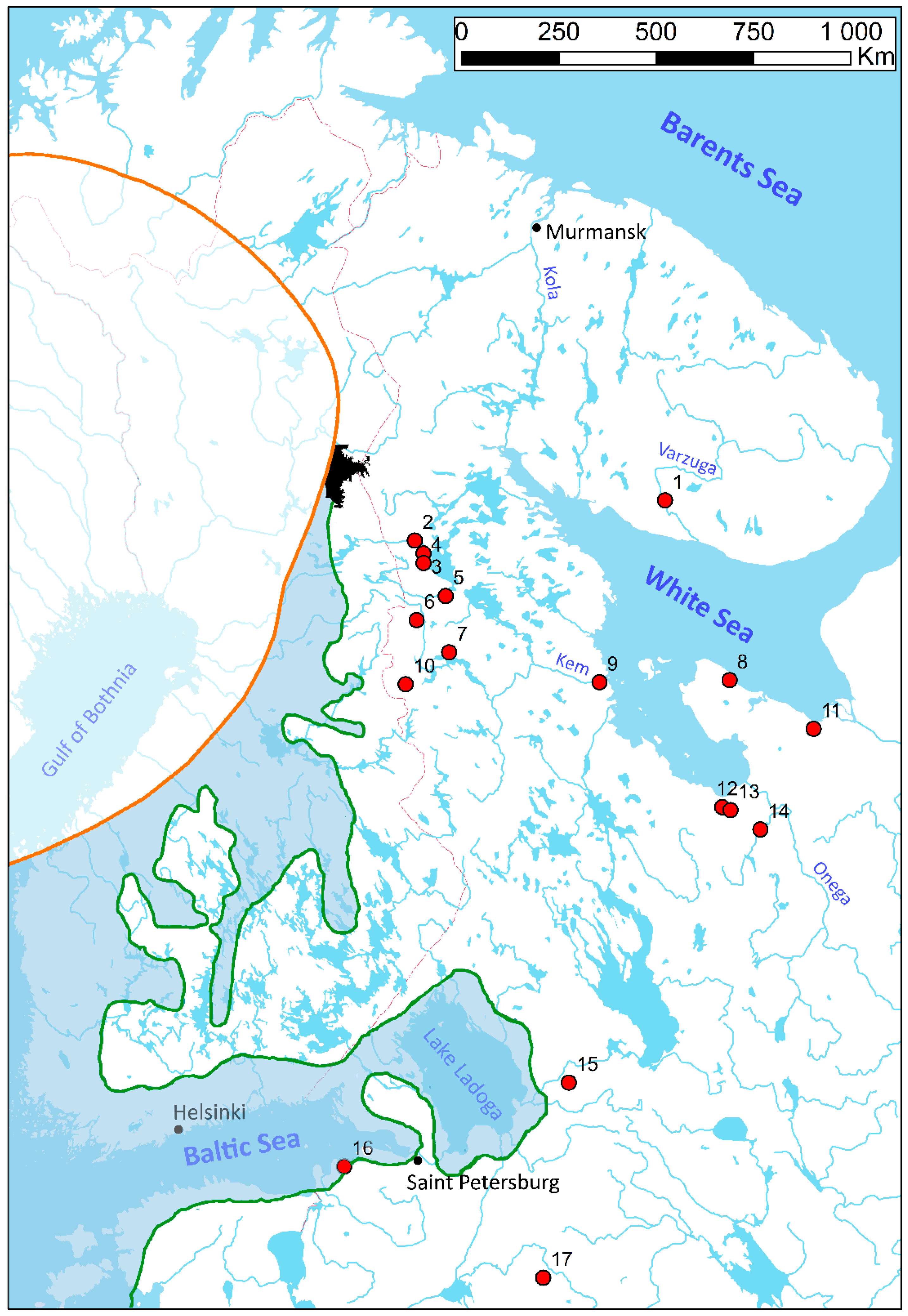
2.1. Sample Collection, DNA Extraction, PCR, and Sequencing

The main body of the material was collected through a series of field surveys on rivers and streams of the White Sea and the Baltic Sea drainages in Europe (Table S1). Soft-tissue samples of M. margaritifera were snipped from the mussel’s foot by a non-lethal procedure [25,26] and immediately stored in 96% ethanol. After snipping, all mussels were carefully returned to the habitat. Soft-tissue samples were deposited in the collection of the Russian Museum of Biodiversity Hotspots (RMBH) of the N. Laverov Federal Center for Integrated Arctic Research of the Ural Branch of the Russian Academy of Sciences (FCIArctic) and in the collection of the Karelian Research Centre of the Russian Academy of Sciences.
Total genomic DNA was extracted from the tissue snips using the NucleoSpin tissue kit (Macherey-Nagel GmbH and Co., KG, Düren, Germany), following the manufacturer’s protocol. For molecular analyses, we obtained sequences of the COI gene. The sequences were amplified and sequenced using the primer pair, LCO1490 and HCO2198 [27]. The PCR mix contained approximately 100 ng of total cellular DNA, 10 pmol of each primer, 200 μmol of each dNTP, 2.5 μL of PCR buffer (with 10 × 2 mmol MgCl2), 0.8 units of Taq DNA polymerase (SibEnzyme Ltd., Novosibirsk city, Russia), and H2O, which totaled a final volume of 25 μL. Thermocycling included one cycle at 95 °C (4 min), followed by 28–32 cycles of 95 °C (50 s), 48–50 °C (50 s), and 72 °C (50 s), and a final extension at 72 °C (5 min). Forward and reverse sequencing were performed on an automatic sequencer (ABI PRISM3730, Applied Biosystems, Waltham, MA, USA) using the ABI PRISM BigDye Terminator v.3.1 reagent kit [28]. The resulting COI gene sequences were checked manually using BioEdit v. 7.2.5 [29]. In addition, 99 COI sequences of M. margaritifera were obtained from NCBI GenBank (Table S1). The sequences were aligned using the MUSCLE algorithm implemented in MEGAX [30]. For the subsequent analyses, each COI sequence of the aligned datasets was trimmed, leaving a 628-base-pair fragment.

2.2. Genetic Diversity, Genetic Differentiation, and Demographic History

The dataset under investigation covers most of the M. margaritifera range and was structured at the global (Figure 1) and regional (Figure 2) scales. At the global scale, we analyzed five subsets corresponding to certain groups of populations. The White Sea (WS; n = 317), Baltic Sea (BS; n = 99) and West–North Atlantic (WNA; n = 10) groups were delineated by the corresponding sea drainage. The exclusion was the Juojoki River, which flows to the northern rim of the Gulf of Bothnia in Sweden, but in our analysis, we included it in the WS group based on our assumption that rivers of northern Finland and Sweden were likely connected with the White Sea through a system of periglacial lakes and rivers (see the Discussion below). The Suomujoki River in Finland and the Zapadnaya Litsa River on the Kola Peninsula belong to the Barents Sea basin, but we included them in the WS group of populations based on the evidence that the Kola Strait was one the main migration corridors between the White and Barents Seas during the Scandinavian Ice Sheet retreat [31]. Iberian (IBER; n = 20) and British (BRIT; n = 11) population groups were determined based on previous phylogenetic studies of freshwater mussels, indicating that two COI lineages of M. margaritifera are distributed in Europe [23] and the existence of an Iberian glacial refugium of freshwater biota [20,21].
At the regional scale, we selected rivers where samples of sufficient size (n ≥ 7 in our dataset) were collected and considered them as populations in subsequent analysis. All these populations belong to the WS and BS groups of populations. The same subset was used to investigate the differentiation between salmon- and trout-associated freshwater pearl-mussel populations (Table S2). The Juojoki population was not included in this analysis, since the only unique haplotypes from this river basin are available in the GenBank.
Genetic diversity was estimated through haplotype diversity (Hd) and nucleotide diversity (Pi) calculations. Genetic differentiation was estimated through calculation of pairwise FST’s distances by the method of Tajima and Nei, and inter- and intrapopulation genetic variability was estimated by AMOVA. To detect deviation from mutational-drift equilibrium in the studied groups and populations, we calculated Fu’s F and Tajima’s D neutrality tests. Ramos-Onsins and Rozas’s R2 neutrality test was calculated in R-studio using pegas package [32], which is the most powerful test for detecting the deviation in small samples [33]. In the case of significance, in at least one neutrality test, we examined the frequency distributions of pairwise mismatch between sequences (MMD). The observed mismatch distribution was compared with that obtained under models of spatial expansion and population expansion for the evidence of model fit by calculating the sum of squared deviations (SSD) of the observed data relative to the model and Harpending’s raggedness statistic (HRag). Genetic diversity indices, FSTs distances, AMOVA, neutrality tests, and MMD were calculated using Arlequin v. 3.5.1.2 [34] all with 10,000 permutations.
To estimate the phylogeographic structure of haplotypes obtained from groups of populations, we applied a median joining network approach using Network v. 5.0 with default settings [35].
The Mantel test was performed to detect the geographic structure of population genetic differentiation based on matrices of geographical distances and FST distances. Principal component analysis (PCA) was performed to reduce the haplotype frequencies matrix to principal components (PCs) to estimate the contribution of certain haplotypes to the genetic differentiation of M. margaritifera populations in dependence on two factors “basin” and “host”. To test the hypothesis of host-fish and basin-induced genetic differences between populations of M. margaritifera from the WS and BS groups, the general linear models (GLM) were used. We used the PCs scores plotted against the variable “host” (1—Salmo salar, and 2—S. trutta) as a covariate and the group of populations as a factor with two levels. Statistical calculations were performed in PAST v4.03 [36].

3. Results

3.1. Genetic Diversity

In total, 32 haplotypes were identified from the studied dataset of the M. margaritifera sequences. The population groups share a relatively high level of haplotype diversity, with the mean Hd value varying from 0.35 ± 0.12 in the IBER group of populations to 0.89 ± 0.075 in the WNA group of populations (Table 1). At the same time, all the groups have an extremely low level of nucleotide diversity, with the mean π value not exceeding 0.004 ± 0.002 in the WNA and BRIT groups. Twenty of the observed haplotypes belong to the BS and WS groups of populations. At the regional scale, the haplotype diversity varies from 0 in the Peypia River, where one haplotype occurs, to 0.88 ± 0.03 in the Kem River. The nucleotide diversity also returned low values, with the maximum π value of 0.004 ± 0.002 in the Varzuga, Vuokkijoki, Solza, and Nimenga rivers.

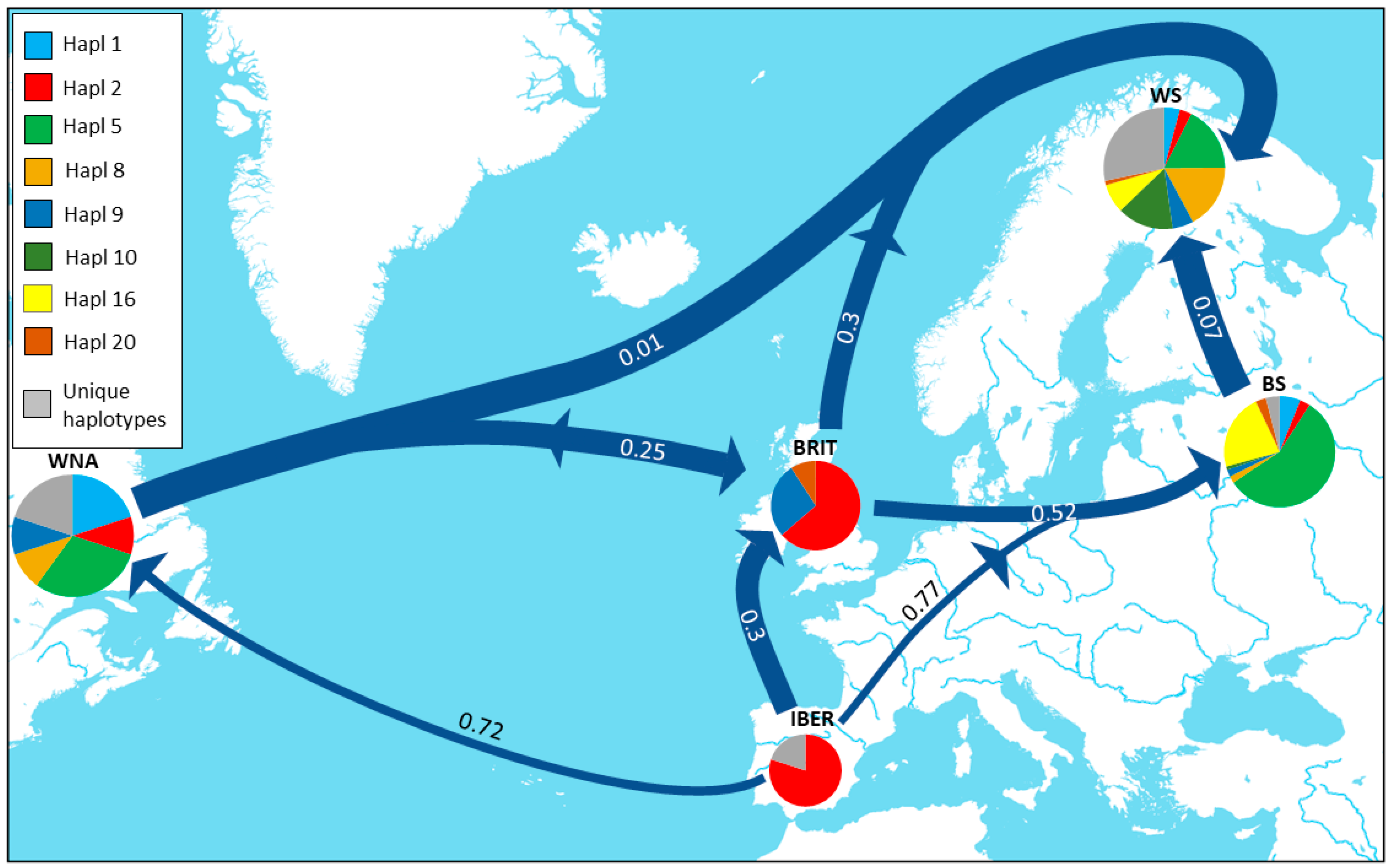
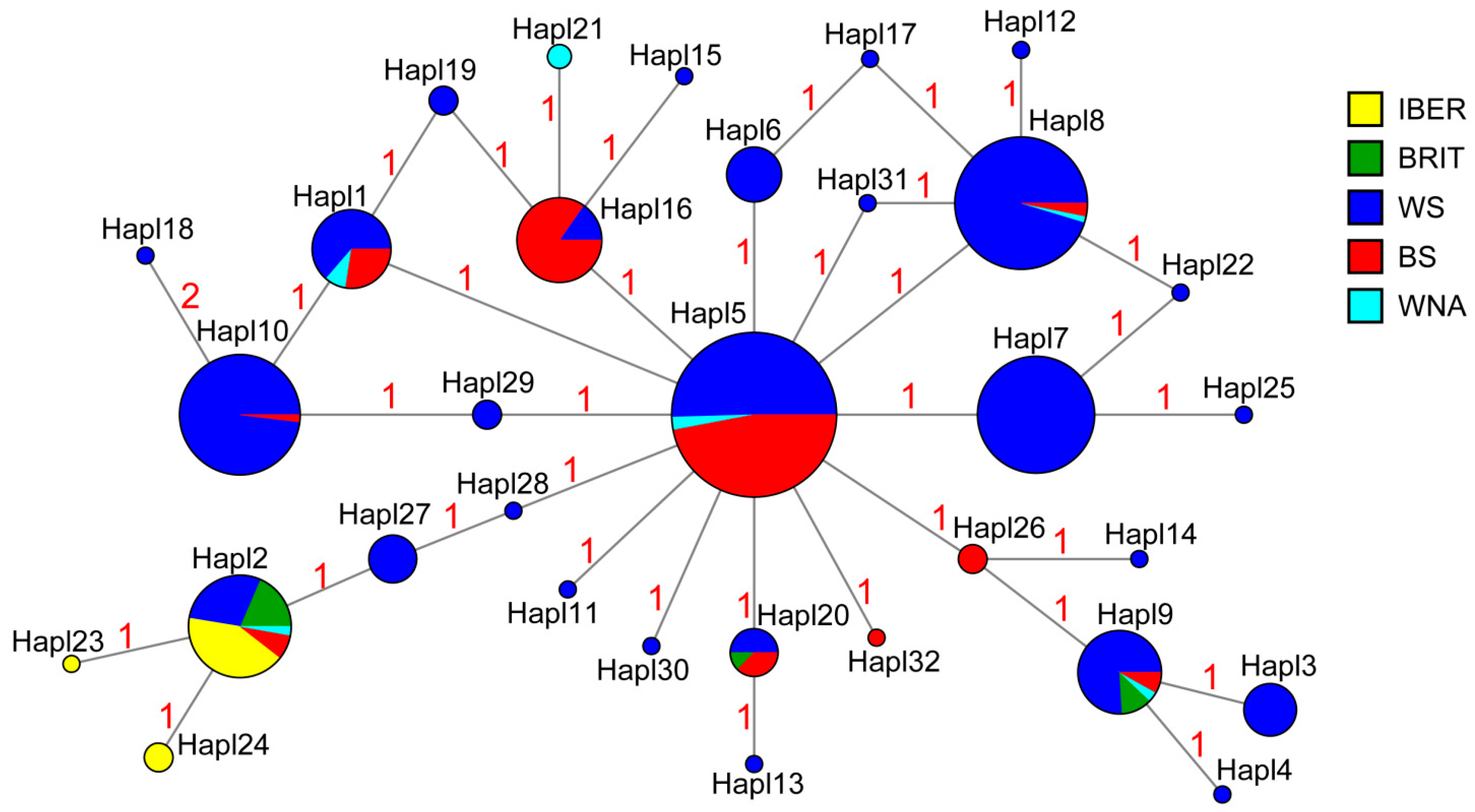
3.2. Genetic Differentiation and Genetic Structure

The AMOVA showed low genetic differentiation irrespective of the data structure tested (Table 2). The contribution of the intrapopulation variability to the general variability was almost four times higher than the contribution of the interpopulation variability (61.64% vs. 14.32%), and more than twice as high as the contribution of the variability between the groups of populations (24.23%). We also tested the molecular differentiation between the groups of populations delineated by the corresponding fish host (S. salar or S. trutta). Only the interpopulation variability was found to be significant, with 20.64% of variance, while the intrapopulation variability was almost four times higher (79.81%), but not significant. Interestingly, between the groups of populations delineated by the corresponding fish-host, we found genetic similarity in the COI gene (−0.45%). The results of the AMOVA, showing low but significant interpopulation molecular differences, suggested an unclear population structure in the studied dataset.
The molecular distances were estimated by Tajima and Nei’s method, which returned insignificant values for the WNA-BS and WNA-WS pairs (Table 3). The most distant group of populations in terms of molecular distances is IBER, which is relatively close to the BRIT group (FST’s = 0.31) and distant from the rest. It is noteworthy that IBER is more closely connected to WS (FST’s = 0.55) than to BS or WNA (FST’s = 0.77 and 0.72, respectively). The molecular distances revealed relatively close connections between the Northeast European and North American populations in the past, while the populations from Iberia were barely connected to the others or lost these connections much earlier.
The matrix of the haplotype frequencies (Table S3) observed in the populations was reduced to principal components, and two PCA were obtained. For the first PCA, the host fish (S. salar or S. trutta) was used as an independent variable and, for the second PCA, the corresponding group (WS or BS) was such a variable. Prior to analyzing the PCA outputs, we ran a GLM analysis to check whether there was significant dependence of the PC distribution on the “host” or “basin”.
The results of the GLMs revealed that the PC1 and PC2 scores differ significantly between the populations from the White Sea and the Baltic Sea basins (Table 4). This is consistent with the PCA results, according to which PC1 and PC2 significantly contribute to the overall variability (Table 5). The loadings of certain haplotypes in PC1 and PC2 show that Hapl5, prevailing in the Peypia and Khorinka rivers, and Hapl16, which is mostly observed in the Yanega River, are associated with the BS group, while Hapl7, Hapl8, and Hapl10 are associated with the WS group (Tables S2 and S4, Figure S1). The GLMs also revealed that the PC6 scores differ significantly between populations associated with different hosts, but that the contribution of PC6 to the overall dispersion is insignificant, with a value of 2.6% (Table 5).
The median-joining haplotype network supports the AMOVA results (Figure 3). The network’s structure is almost star-like, and indicating the presence of the most abundant haplotype, which is mostly shared between the WS and BS groups of populations, and much less with the WNA group. Twenty-two haplotypes belong exclusively to a certain group of populations and eighteen of them are from the WS group. Almost all the haplotypes have a neighbor in one mutational step; the only exception is haplotype H18, from the Juojoki River, which stands in two mutational steps from its neighboring haplotype, H10. We found that six haplotypes are present in the WNA group, but only one is unique, and the rest are shared with the European populations. The haplotype network has five alternative connection loops between haplotypes from Northern Europe and North America, which suggests repeat and reverse mutations as a consequence of multiple migration events. The haplotypes H23, H24, and H2 observed in the Iberian populations are clustered together in the network, forming the least diversified group of populations.

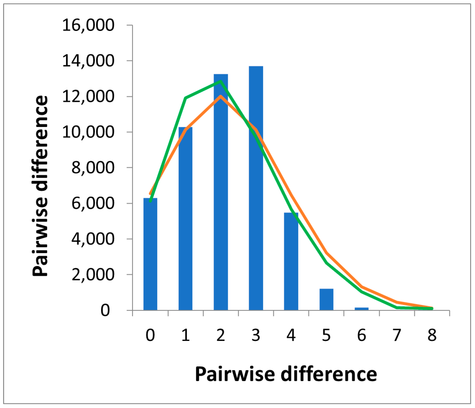
3.3. Demographic Trends

Deviation from the mutation-drift equilibrium was detected only for the WS group of populations (Fu’s Fs = −12.66; p < 0.02) (Table 1). Usually, negative values on the Fu’s Fs test indicate the passage of a population through a bottleneck or a recent population expansion event. The observed mismatch distribution for the WS resembles a bell curve (Figure 4). This kind of unimodal distribution shows a genetic signal associated with a sudden population expansion; however, the verification tests differed significantly from the modeled distribution for a spatial expansion model and did not match the expectation of a sudden population expansion, rejecting both demographic models (Table S5). We did not use mismatch distribution computations to test demographic scenarios in the BS, WNA, BRIT, and IBER groups of populations, or in any of the populations at the regional scale, because the neutrality tests did not indicate deviation from the mutational-drift equilibrium.

4. Discussion

We sequenced COI gene fragments of M. margaritifera from populations inhabiting watercourses in the northeastern margin of the species range to clarify its post-glacial demographic history in the region and to estimate the genetic diversity and population differentiation patterns. The weak phylogenetic signal in our dataset prevented us from inferring the demographic trends and processes in the studied populations. However, combining newly sequenced fragments with the data available from NCBI GenBank, we suggest post-glacial routes of M. margaritifera expansion from putative glacial refugia through the species range.
The level of haplotype diversity observed in our global dataset varies among the designated groups of populations. The lowest level was observed in the Iberian group of populations. This finding could be an artefact of the low sample volume (n = 20), but a group of populations from the opposite side of the Atlantic with even lower sample volume (n = 10) shows a twice-higher haplotype diversity, which is comparable to the observations from the Baltic Sea and the White Sea basins. At the global scale, our results are consistent with the previous observations inferred from microsatellites, showing a latitudinal gradient in genetic diversity, with the lowest levels in the Iberian and the highest in the Scandinavian populations [11]. A reduced genetic drift due to a longer reproductive life span and higher population densities in the northern portion of the species’ range, together with the better conditions of the populations due to the lower anthropogenic impact in the area, were suggested as the possible causes of an inverse latitudinal trend in the spatial distribution of the genetic diversity [11,12]. Our COI-based data are unable to reflect such relatively recent changes in genetic diversity corresponding to adaptations to northern environments, and our data also have a limited power to detect the footprints of the anthropogenic disturbances in the genotype. Reflecting more ancient genetic processes, our dataset may indicate that the populations from the Iberian Peninsula are older and lost their COI diversity due to a lack of new immigrants, while the freshwater pearl-mussel populations in the northern areas are younger and host immigrants from western and southern areas, and possibly from glacial refugia in Eastern Europe (see below). This speculative suggestion may explain the inconsistency in the observed latitudinal gradient of the genetic diversity, based on the theoretical expectation that the genetic diversity in post-glacial populations should decrease with distance from glacial refugia [40]. The high level of haplotype diversity, together with the extremely low level of nucleotide diversity, indicates a high number of closely related haplotypes. This pattern is specific for young populations derived from parental populations that have recently expanded from refugia after the Last Glacial Maximum [41,42,43].
At the regional scale, we did not observe a significant correlation between the haplotype diversity and the latitudinal gradient (Pearson rs = 0.42, p > 0.05). Nevertheless, the most diverse populations, such as Kem, Vozhma, Kozha, and Varzuga, are from the north and the monotonous population in the Peypia River is from the south, although there are a couple of populations from the northern part of the range with the same level of diversity as that in the southern populations. The Nuris and Tuhka, which are tributaries of the Kumskoe Reservoir, share a relatively low level of haplotype diversity (0.3), whereas the other tributary of the same reservoir, the Tavajoki, features a higher haplotype diversity value (0.5). At first glance, this difference could be explained by the disproportion in the sample size, which was less than 10 for the Nuris and Tuhka, respectively, and 20 for the Tavajoki. Conversely, the lower level of genetic diversity may indicate continuing population decline [8]. Unfavorable environmental conditions can lead to increasing mortality in populations. The freshwater-pearl-mussel population from the Tuhka River is located in the Pyaozerski settlement and is thus exposed to heavy anthropogenic pressure. The mean levels of Cd and Pb observed in the mussels’ soft tissues were 0.31 mg/kg and 0.12 mg/kg, respectively (E.P. Ieshko, personal observation). By contrast, the concentrations of the mentioned elements in the freshwater pearl mussels’ soft tissues collected from the populations of the Paanajarvi National Park were much lower (Cd: 0.10–0.06 mg/kg; and Pb: 0.05–0.07 mg/kg, E.P. Ieshko, personal observation). The potentially high mortality of the freshwater pearl mussels in the Nuris River may be connected with spring floods. The mean channel slope of the river is approximately 5.5 m/km (E.P. Ieshko, personal observation). Seasonal floods may seriously disturb freshwater-pearl-mussel beds in such a steep channel.
Another peculiar observation regarding these tributaries of the Kumskoe Reservoir was the absence of haplotypes common in populations of the Tavajoki and Tuhka rivers (Table S3). Moreover, one of the two haplotypes observed in the Tukha population is unique, and the other, Hapl27, is only shared with the Kem population. The genetic uniqueness of the Tuhka population could be related to the anthropogenic impact discussed above, as well as to the population differentiation in proglacial waterbodies. Our data prevented us from drawing an unambiguous conclusion, but they did suggest the necessity of investigating the Tukha population using advanced molecular techniques.
The zero diversity in the Peypia population is consistent with the neutral model, which links the population’s genetic diversity with its effective population size and the gene’s mutation rate [44]. In the Peypia River, we observed a small population living in a small, isolated stream [45]. Examples of the same pattern of genetic diversity have been reported for bivalves [46], lizards [47], and platypus [48]. The effect of geographic isolation could also be multiplied by hermaphroditism, which occurs in M. margaritifera populations in extreme conditions [49,50]. Moreover, the Peypia population is subject to a number of anthropogenic effects from the neighboring city of Saint-Petersburg [45]. Small, isolated populations lose rare alleles through genetic drift; such reductions in genetic diversity can make species more vulnerable to extinction, because greater diversity increases adaptability and long-term population persistence [51,52].
Our COI-based analyses returned low genetic differentiation and unclear population structure in the analyzed dataset both at the global and the regional scales. The AMOVA results returned much higher variability intrapopulation than both interpopulation and even intergroup, independently of the group designation, i.e., sea basins or associated host-fish. However, the low but significant differences between the populations and between the groups prevented us from drawing a conclusion as to the lack of structure among the river drainages, as was observed previously in North-European and North American populations [11,13], based on microsatellites with a faster mutational rate than that in the COI gene.
The lack of differentiation between the populations from North America and the White Sea and Baltic Sea drainages and the low differentiation between other pairs of population groups point to relatively recent and multiple connections between different parts of the species range. In the case of slowly evolving species, such as M. margaritfera, the COI-based genetic structure reflects the haplotype ordering that took place before the Last Glacial Maximum or at early stages of post-glacial dispersion. The mean mutational rate of the COI gene in the family Margaritiferidae is slow (0.216% per site per million years in [19]), and it is comparable with that in the Unionidae (0.265% per site per million years in [53] and 0.150% per site per million years in [54]). It not enough time has passed since the Last Glacial Maximum for the clear diversification of most of the populations or groups of populations of M. margaritifera to occur. On the other hand, 23 unique haplotypes were observed in the median-joining COI haplotype network (Figure 3).
Two alternative hypotheses can be suggested to explain the presence of unique haplotypes in the populations of the White and Baltic Sea basins. The first is that the haplotypes differentiated after the Last Glacial Maximum, when the populations were isolated geographically or via association with a certain host-fish species. Considering the mean mutational rate of the COI gene in the family Margaritiferidae, this assumption is questionable, unless we concede that the evolution rate in the studied gene region has accelerated since the Last Glacial Maximum. The second hypothesis is that the unique haplotypes in the modern populations came from populations that existed in ice-dammed lakes and related freshwater basins during the glaciation events. Several lakes extended along the glacial margin in Eastern Europe, as documented in the literature, i.e., Lake Komi, the White Sea Basin Lake, and the Baltic Lake [55,56]. We observed unique haplotypes in the BS and WS regions, which could indicate that periglacial lakes, such as the White Sea Basin Lake and the Baltic Sea Lake, were probably not directly connected. Within the WS region, unique haplotypes were observed in the Juojoki, Tuhka, Uhta, and Vozhma Rivers. Remarkably, the last three populations belong to the upper part of the Kem River basin, but none of the unique haplotypes were found in the main channel of the Kem River. The difficulties involved in the reconstruction of the pre- and postglacial river network rearrangements, resulting in a complicated picture of the haplotype distribution, prevented us from drawing robust conclusions about the source of this genetic differentiation.
In their study, Machordom et al. [23] identified different mitochondrial lineages in the north (River Dereen) and the south (River Nore). According to their data, the northern lineage extends northwards from Ireland to the Kola Peninsula, and the southern lineage extends from Ireland to the south. Our expanded dataset does not support a strict geographical division between these lineages, since the “southern” haplotype (Hapl2) was found in several rivers from the northern part of the species’ range. Despite the generally unclear genetic structure in our dataset, we observed that the Iberian group of populations is less diverse and the most distinct in terms of FST distances. We speculate that the distinctiveness of the Iberian populations is further evidence of the existence of the Iberian freshwater glacial refugium, which played an important role in the evolutionary history of freshwater mussels [20,21] and fishes [22,57]. The M. margaritifera distribution to North America may have started from the Iberian refugium, adjacent river basins in Southern France, and the British Isles (Figure 1). The significant genetic distance between the Iberian and North American populations may indicate that this path of M. margaritifera expansion from the refugium discontinued much earlier than the northern path from Iberia to the British Isles. Alternatively, the exchange of migrants between the IBER and WNA regions was initially weak, and the penetration to the British Isles was the main direction of the spread from the refugium (Figure 1). However, the relatively genetic proximity between WNA and BRIT groups of populations indicates that the exchanges of immigrants between the North American and British populations played an important role in freshwater-pearl-mussel distribution across the ocean.
Freshwater-pearl-mussel distribution is directly connected with the transport of larvae on the gills of host fish, i.e., S. salar and S. trutta, as primary hosts. Fishes play an important role not only in freshwater pearl-mussel spread within the river basin, but also as genetic data point out, between neighboring river basins [10]. Glochidial transport across sea waters has been confirmed experimentally [58] and seems to be the only way for M. margaritifera to spread through the entire modern range.
The closest groups in our dataset, judging by the FST distances, are the populations from the river basins of Northern Europe and North America (Table 3). The difference between them is insignificant, which supports the theory of the restocking of North European rivers by Salmo salar from North America [31]. The second insignificant FST value was between the WNA and BS groups, which appears to be peculiar because the difference between the WS and BS is very small, but significant. We can assume that the spread of freshwater pearl mussels to the Baltic Sea basin from the west contributed significantly to the colonization of the region and may indicate the western path of the salmonid penetration of the Baltic Sea basin (Figure 1). At the same time, the southern part of the Baltic Sea drainage may have acted as a glacial refugia itself. Both the finding of unique haplotypes in the BS region and the paleontological record of freshwater pearl mussels from Middle-Weichselian deposits in North-Western Lithuania [59] indicate that M. margaritifera populations existed in the peri- and proglacial water bodies of the Baltic Ice Lake basin, to which they migrated from the area of the present-day Black Sea, together with brown trout [60]. The unique haplotypes discovered in the Khorinka and Ihala Rivers may be the remnants of the genetic diversity of freshwater-pearl-mussel populations of the refugium.
We did not find COI genetic differentiation between the populations of M. margaritifera utilizing Salmo salar or S. trutta as hosts. This pattern is consistent with the results obtained from freshwater pearl mussels in Ireland based on microsatellites [8], but contradicts the results of microsatellite-based studies in Norway, where a clear diversification between these two groups was shown [7]. However, the high genetic diversity and low genetic differentiation observed through our dataset resemble the genetic structure found in recent freshwater pearl-mussel populations utilizing sea-migrating hosts [10]. In the case of post-glacial freshwater pearl-mussel colonization history, the distribution associated with anadromous hosts has resulted in a diverse but poorly differentiated population genetic structure.
The insignificant genetic difference and high rate of gene flow between the North American and White Sea M. margaritifera populations support the hypothesis that the White Sea populations of Atlantic salmon were re-colonizers from a glacial refugium located somewhere in the Western Atlantic Ocean [31]. Common haplotype-sharing between the White Sea and the Baltic Sea freshwater pearl-mussel populations and secondary contact loops between them in the haplotype network highlight that Baltic Sea migrants might have participated in the recolonization of the White Sea. This pattern is supported by the post-glacial colonization history of Atlantic salmon [31,61] and brown trout [62].
There is a discussion over whether phylogeographic connections existed between salmon stocks in the Baltic Sea and the White Sea [31,61] or did not [63]. The phylogenetic data inferred from the COI sequences of the freshwater pearl mussels support the first hypothesis. The most abundant haplotype in the north-eastern part of the species range, Hapl5, is spreading from the population in the Khorinka River, Novgorod Oblast, northward, to the population of the Mutkajoki River in Northern Karelia. The direction of the gene flow can hardly be validated using our data; however, we can assume that a two-directional colonization, from the south and from the north, took place. The retreat of the Scandinavian Ice Sheet after the Last Glacial Maximum caused the emergence of multiple freshwater bodies connected to and disconnected from each other from the Late Weichselian onward. The ice-dammed Salla Lake, which was initially connected to the White Sea via the Kutsajoki River and then drained to the ancient Baltic Sea (during the Ancylus Lake stage) approximately 10,500 years ago [39,64], is an example of the temporal dynamic of sea-basin divides (Figure 2).
Complex hydrological networks along the glacier margin could facilitate gene drift and admixture between populations from freshwater basins, which subsequently separate. The ice-flow directions in Northern Fennoscandia during the Late Weichselian indicate that the water bodies in the region previously had connections with the Arctic Ocean and the White Sea ([65]: Figure 26), and possibly with the paleo-strait between the mainland and “Kola Island”. These connections represented one of the paths of North American S. salar pioneers penetration [31], and existed 10,500—10,200 years ago ([66]: Figure 3). Paleogeographic reconstructions of the recession of the ice margin reveal that approximately 11,000—10,500 years ago, the ice sheet retreated from Karelia in the north-westward direction, which allowed the ice-dammed Ancylus Lake to extend northward up to the Polar Circle (Figure 2) ([38]: Figure 3; [37]: Figure 9). The Ancylus Lake water’s penetration following the deglaciation of Central Finland opened the way for immigrants from the Baltic Sea, which contributed to high genetic diversity in the region. Direct connections between the Baltic Sea and White Sea faunas were most probably terminated after the Ancylus Lake regressed and turned into the Littorina Sea approximately 9800 years ago [38].
The complex history of the colonization of the Baltic, White, and Barents Sea basins by Atlantic salmon and brown trout reflects the gradual retreat of the Scandinavian Ice Sheet after the Last Glacial Maximum and can be considered as a basic scheme of freshwater-pearl-mussel expansion through the species range. Our data confirmed close connections between the freshwater biota of Northern Europe and the eastern coast of North America. Haplotype 5 dominates in the BS group, but it is also widely represented in the WS group (Figure 4 and Table S3). Given that it is especially abundant in the populations of the Kem River basin and the Mutkajoki River, the observation of this haplotype highlighted these freshwater basins as contact zones where migration flows met following the glacier retreat, as was shown for Central Sweden ([11] and references herein).
The COI-based data show a rather ancient picture of M. margaritifera genetic diversification and diversity but, despite the slow mutational substitution rate, may also trace populations in unfavorable conditions. The genetic diversity serves as an indicator in this case. The populations with low diversity, such as those from the Nuris and Tukha rivers and, especially, from the Peypia River, which shows zero diversity, require conservation and, possibly, restoration efforts. The northern populations, with their exceptional COI genetic diversity, are also a high conservation priority. Postglacially colonized areas, from Lapland [12] to Karelia, are remarkable sites of genetic diversity within the European range of the species. However, the weak phylogenetic signal in the studied populations prevents the detection of actual demographic processes and cannot infer actual population structure. These data are crucial for the conservation management of the species [8,13]. Therefore, population-genetic studies of M. margaritfera populations from the north-eastern margin of the range based on molecular markers with faster evolutionary rates, such as microsatellites, or on those covering a larger portion of the genome, such as a single-nucleotide polymorphism (SNP), are highly necessary.

Supplementary Materials

The following supporting information can be downloaded at: https://www.mdpi.com/article/10.3390/d14060477/s1. Figure S1. Principal-component scores for the first two PC axes obtained by using PCA on matrix of haplotype frequencies in Margaritifera margaritifera populations. The green dots indicate pearl-mussel populations belonging to the White Sea group (WS), and the pink dots indicate pearl-mussel populations belonging to the Baltic Sea group (BS). Biplot vectors indicate relative loadings of certain haplotypes in PCs. Convex hull is shown for each species. Table S1. List of sequences used in this study, including the location, the haplotype numbers, and NCBI GenBank accession numbers and references. Table S2. Distribution of Margariitfera margaritifera populations between factors for PCA and GLM analysis, and references with information about host fish. Table S3. Matrix of COI haplotype frequencies in populations of Margaritifera margaritifera. Table S4. Loading values of haplotypes to PC1 and PC2 obtained using matrix of haplotype frequencies in Margaritifera margaritifera populations. PCA under the “basin” factor. Table S5. Mismatch distribution verification tests for spatial population expansion and size population expansion assumptions for the WS group of Margaritifera margaritifera populations. Statistically significant values are marked in bold (p < 0.05). Refs [67,68,69,70,71,72,73,74,75,76,77,78] are cited in Supplement Material.

Author Contributions

I.V.V., E.P.I., A.V.K. and I.N.B. developed the concept of this study. I.V.V., E.P.I., A.V.K., I.N.B. and D.A.E. collected the samples. A.V.K., N.S.M., G.V.B., A.G.B., A.A.T. and O.A.Y. designed and carried out the molecular analyses. A.V.K. performed the phylogenetic modeling. I.V.V. created the maps and performed the population genetics and statistical data analyses. I.V.V. wrote the paper, with input from E.P.I. and I.N.B. All authors have read and agreed to the published version of the manuscript.

Funding

The study was funded by the Russian Science Foundation, project number 21-74-10130 (including fieldworks in Arkhangelsk, Leningrad and Novgorod oblasts and molecular analyses). The phylogenetic modeling, population genetic analysis, mapping, and manuscript preparation were funded by the Ministry of Science and Higher Education of the Russian Federation, project number FUUW-2022-0056. The fieldwork in Karelia was funded by ENI-CBC Kolarctic project, SALMUS, KO1017 ‘Salmonid Fish and Freshwater Pearl Mussel—Ecosystem Services and Biodiversity in the Green Belt of Fennoscandia’ (EI and DE), under the state-ordered projects number 122032100130-3 (EI) and 0218-2019-0081 (DE).

Data Availability Statement

Not Applicable.

Acknowledgments

We are grateful to those who assisted us in the collection of the samples for the molecular-genetic analyses: Mikhail B. Kabakov, Svetlana E. Sokolova, Valentina S. Artamonova, Yulia V. Bespalaya, Olga V. Aksenova, Andrey S. Aksenov, Mikhail Yu. Gofarov, and Gennady A. Dvoryankin. We would also like to express our sincere gratitude to Alexander A. Makhrov and Natalia A. Zubrii, who not only assisted us in field sampling, but also provided valuable advice regarding the methodology and concept of the manuscript. Special thanks are due to Olga S. Kislova, who proofread our manuscript and suggested changes to the English grammar and style, which made the text read more smoothly. We are deeply grateful to three anonymous reviewers, whose comments helped us to significantly improve the manuscript.

Conflicts of Interest

The authors declare no conflict of interest.

References

  1. Geist, J. Strategies for the Conservation of Endangered Freshwater Pearl Mussels (Margaritifera margaritifera L.): A Synthesis of Conservation Genetics and Ecology. Hydrobiologia 2010, 644, 69–88. [Google Scholar] [CrossRef] [Green Version]
  2. Moorkens, E.A. Addressing the Conservation and Rehabilitation of Margaritifera margaritifera (L.) Populations in the Republic of Ireland within the Framework of the Habitats and Species Directive. J. Conchol. 2010, 40, 339–350. [Google Scholar]
  3. Makhrov, A.; Bespalaya, J.; Bolotov, I.; Vikhrev, I.; Gofarov, M.; Alekseeva, Y.; Zotin, A. Historical Geography of Pearl Harvesting and Current Status of Populations of Freshwater Pearl Mussel Margaritifera margaritifera (L.) in the Western Part of Northern European Russia. Hydrobiologia 2014, 735, 149–159. [Google Scholar] [CrossRef]
  4. Bolotov, I.N.; Makhrov, A.A.; Gofarov, M.Y.; Aksenova, O.V.; Aspholm, P.E.; Bespalaya, Y.V.; Kabakov, M.B.; Kolosova, Y.S.; Kondakov, A.V.; Ofenböck, T.; et al. Climate Warming as a Possible Trigger of Keystone Mussel Population Decline in Oligotrophic Rivers at the Continental Scale. Sci. Rep. 2018, 8, 35. [Google Scholar] [CrossRef] [PubMed]
  5. Young, M.; Williams, J. The Status and Conservation of the Freshwater Pearl Mussel Margaritifera margaritifera Linn. in Great Britain. Biol. Conserv. 1983, 25, 35–52. [Google Scholar] [CrossRef]
  6. Young, M.R.; Cosgrove, P.J.; Hastie, L. The Extent of, and Causes for, the Decline of a Highly Threatened Naiad: Margaritifera margaritifera. In Cology and Evolutionary Biology of the Freshwater Mussels Unionoidea; Bauer, G., Wachtler, K., Eds.; Springer: Berlin, Germany, 2001; p. 337357. [Google Scholar]
  7. Karlsson, S.; Larsen, B.M.; Larsen, M.; Hindar, K. Host-Dependent Genetic Variation in Freshwater Pearl Mussel (Margaritifera margaritifera L.). Hydrobiologia 2014, 735, 179–190. [Google Scholar] [CrossRef]
  8. Geist, J.; Moorkens, E.; Killeen, I.; Feind, S.; Stoeckle, B.C.; Connor, Á.O.; Kuehn, R. Genetic Structure of Irish Freshwater Pearl Mussels (Margaritifera margaritifera and Margaritifera durrovensis): Validity of Subspecies, Roles of Host Fish, and Conservation Implications. Aquat. Conserv. Mar. Freshw. Ecosyst. 2018, 28, 923–933. [Google Scholar] [CrossRef]
  9. Wacker, S.; Larsen, B.M.; Jakobsen, P.; Karlsson, S. High Levels of Multiple Paternity in a Spermcast Mating Freshwater Mussel. Int. J. Bus. Innov. Res. 2018, 17, 8126–8134. [Google Scholar] [CrossRef] [Green Version]
  10. Österling, M.; Lopes-Lima, M.; Froufe, E.; Hadzihalilovic, A.H.; Arvidsson, B. The Genetic Diversity and Differentiation of Mussels with Complex Life Cycles and Relations to Host Fish Migratory Traits and Densities. Sci. Rep. 2020, 10, 17435. [Google Scholar] [CrossRef]
  11. Geist, J.; Söderberg, H.; Karlberg, A.; Kuehn, R. Drainage-Independent Genetic Structure and High Genetic Diversity of Endangered Freshwater Pearl Mussels (Margaritifera margaritifera) in Northern Europe. Conserv. Genet. 2010, 11, 1339–1350. [Google Scholar] [CrossRef]
  12. Geist, J.; Kuehn, R. Host-Parasite Interactions in Oligotrophic Stream Ecosystems: The Roles of Life-History Strategy and Ecological Niche. Mol. Ecol. 2008, 17, 997–1008. [Google Scholar] [CrossRef] [PubMed]
  13. Zanatta, D.T.; Stoeckle, B.C.; Inoue, K.; Paquet, A.; Martel, A.L.; Kuehn, R.; Geist, J. High Genetic Diversity and Low Differentiation in North American Margaritifera margaritifera (Bivalvia: Unionida: Margaritiferidae). Biol. J. Linn. Soc. 2018, 123, 850–863. [Google Scholar] [CrossRef]
  14. Farrington, S.J.; King, R.W.; Baker, J.A.; Gibbons, J.G. Population Genetics of Freshwater Pearl Mussel (Margaritifera margaritifera) in Central Massachusetts and Implications for Conservation. Aquat. Conserv. Mar. Freshw. Ecosyst. 2020, 30, 1945–1958. [Google Scholar] [CrossRef]
  15. Geist, J.; Kuehn, R. Genetic Diversity and Differentiation of Central European Freshwater Pearl Mussel (Margaritifera margaritifera L.) Populations: Implications for Conservation and Management. Mol. Ecol. 2005, 14, 425–439. [Google Scholar] [CrossRef]
  16. Bouza, C.; Castro, J.; Martínez, P.; Amaro, R.; Fernández, C.; Ondina, P.; Outeiro, A.; San Miguel, E. Threatened Freshwater Pearl Mussel Margaritifera margaritifera L. in NW Spain: Low and Very Structured Genetic Variation in Southern Peripheral Populations Assessed Using Microsatellite Markers. Conserv. Genet. 2007, 8, 937–948. [Google Scholar] [CrossRef]
  17. Stoeckle, B.C.; Araujo, R.; Geist, J.; Kuehn, R.; Toledo, C.; Machordom, A. Strong Genetic Differentiation and Low Genetic Diversity of the Freshwater Pearl Mussel (Margaritifera margaritifera L.) in the Southwestern European Distribution Range. Conserv. Genet. 2017, 18, 147–157. [Google Scholar] [CrossRef] [Green Version]
  18. Taeubert, J.-E.; Geist, J. The Relationship between the Freshwater Pearl Mussel (Margaritifera margaritifera) and Its Hosts. Biol. Bull. 2017, 44, 67–73. [Google Scholar] [CrossRef]
  19. Bolotov, I.N.; Vikhrev, I.V.; Bespalaya, Y.V.; Gofarov, M.Y.; Kondakov, A.V.; Konopleva, E.S.; Bolotov, N.N.; Lyubas, A.A. Multi-Locus Fossil-Calibrated Phylogeny, Biogeography and a Subgeneric Revision of the Margaritiferidae (Mollusca: Bivalvia: Unionoida). Mol. Phylogenet. Evol. 2016, 103, 104–121. [Google Scholar] [CrossRef]
  20. Froufe, E.; Sobral, C.; Teixeira, A.; Sousa, R.; Varandas, S.; Aldridge, D.C.; Lopes-Lima, M. Genetic Diversity of the Pan-European Freshwater Mussel Anodonta anatina (Bivalvia: Unionoida) Based on CO1: New Phylogenetic Insights and Implications for Conservation. Aquat. Conserv. Mar. Freshw. Ecosyst. 2014, 24, 561–574. [Google Scholar] [CrossRef] [Green Version]
  21. Tomilova, A.A.; Lyubas, A.A.; Kondakov, A.V.; Vikhrev, I.V.; Gofarov, M.Y.; Kolosova, Y.S.; Vinarski, M.V.; Palatov, D.M.; Bolotov, I.N. Evidence for Plio-Pleistocene Duck Mussel Refugia in the Azov Sea River Basins. Diversity 2020, 12, 118. [Google Scholar] [CrossRef] [Green Version]
  22. Consuegra, S.; De Leániz, C.G.; Serdio, A.; González Morales, M.; Straus, L.G.; Knox, D.; Verspoor, E. Mitochondrial DNA Variation in Pleistocene and Modern Atlantic Salmon from the Iberian Glacial Refugium. Mol. Ecol. 2002, 11, 2037–2048. [Google Scholar] [CrossRef] [PubMed]
  23. Machordom, A.; Araujo, R.; Erpenbeck, D.; Ramos, M.-Á. Phylogeography and Conservation Genetics of Endangered European Margaritiferidae (Bivalvia: Unionoidea). Biol. J. Linn. Soc. 2003, 78, 235–252. [Google Scholar] [CrossRef] [Green Version]
  24. Välilä, S.J.; Knott, K.E.; Ieshko, E.P.; Veselov, A.E.; Taskinen, J.K. Variation in the COI Gene of the Freshwater Pearl Mussel Margaritifera margaritifera from River Vuokkijoki 1. Biol. Bull. 2017, 44, 92–98. [Google Scholar] [CrossRef]
  25. Karlsson, S.; Larsen, B.M.; Eriksen, L.; Hagen, M. Four Methods of Nondestructive DNA Sampling from Freshwater Pearl Mussels Margaritifera margaritifera L. (Bivalvia:Unionoida). Freshw. Sci. 2013, 32, 525–530. [Google Scholar] [CrossRef]
  26. Berg, D.J.; Haag, W.R.; Guttman, S.I.; Sickel, J.B. Mantle Biopsy: A Technique for Nondestructive Tissue-Sampling of Freshwater Mussels. J. N. Am. Benthol. Soc. 1995, 14, 577–581. [Google Scholar] [CrossRef] [Green Version]
  27. Folmer, O.; Black, M.; Hoeh, W.; Lutz, R.; Vrijenhoek, R. DNA Primers for Amplification of Mitochondrial Cytochrome c Oxidase Subunit I from Diverse Metazoan Invertebrates. Mol. Mar. Biol. Biotechnol. 1994, 3, 294–299. [Google Scholar] [PubMed]
  28. Bolotov, I.N.; Kondakov, A.V.; Vikhrev, I.V.; Aksenova, O.V.; Bespalaya, Y.V.; Gofarov, M.Y.; Kolosova, Y.S.; Konopleva, E.S.; Spitsyn, V.M.; Tanmuangpak, K.; et al. Ancient River Inference Explains Exceptional Oriental Freshwater Mussel Radiations. Sci. Rep. 2017, 7, 2135. [Google Scholar] [CrossRef] [Green Version]
  29. Hall, T.A. BioEdit: A User-Friendly Biological Sequence Alignment Editor and Analysis Program for Windows 95/98/NT. Nucleic Acids Symp. Ser. 1999, 41, 95–98. [Google Scholar]
  30. Kumar, S.; Stecher, G.; Li, M.; Knyaz, C.; Tamura, K. MEGA X: Molecular Evolutionary Genetics Analysis across Computing Platforms. Mol. Biol. Evol. 2018, 35, 1547–1549. [Google Scholar] [CrossRef]
  31. Makhrov, A.A.; Verspoor, E.; Artamonova, V.S.; O’Sullivan, M. Atlantic Salmon Colonization of the Russian Arctic Coast: Pioneers from North America. J. Fish Biol. 2005, 67, 68–79. [Google Scholar] [CrossRef]
  32. Paradis, E. Pegas: An R Package for Population Genetics with an Integrated–Modular Approach. Bioinformatics 2010, 26, 419–420. [Google Scholar] [CrossRef] [PubMed] [Green Version]
  33. Ramos-Onsins, S.E.; Rozas, J. Statistical Properties of New Neutrality Tests Against Population Growth. Mol. Biol. Evol. 2002, 19, 2092–2100. [Google Scholar] [CrossRef] [Green Version]
  34. Excoffier, L.; Lischer, H.E.L. Arlequin Suite Ver 3.5: A New Series of Programs to Perform Population Genetics Analyses under Linux and Windows. Mol. Ecol. Resour. 2010, 10, 564–567. [Google Scholar] [CrossRef] [PubMed]
  35. Bandelt, H.J.; Forster, P.; Röhl, A. Median-Joining Networks for Inferring Intraspecific Phylogenies. Mol. Biol. Evol. 1999, 16, 37–48. [Google Scholar] [CrossRef] [PubMed]
  36. Hammer, D.A.T.; Ryan, P.D.; Hammer, Ø.; Harper, D.A.T. Past: Paleontological Statistics Software Package for Education and Data Analysis. Palaeontol. Electron. 2001, 4, 178. [Google Scholar]
  37. Stroeven, A.P.; Hättestrand, C.; Kleman, J.; Heyman, J.; Fabel, D.; Fredin, O.; Goodfellow, B.W.; Harbor, J.M.; Jansen, J.D.; Olsen, L.; et al. Deglaciation of Fennoscandia. Quat. Sci. Rev. 2016, 147, 91–121. [Google Scholar] [CrossRef] [Green Version]
  38. Andrén, T.; Björck, S.; Andrén, E.; Conley, D.; Zillén, L.; Anjar, J. The Development of the Baltic Sea Basin During the Last 130 Ka. In The Baltic Sea Basin; Harff, J., Björck, S., Hoth, P., Eds.; Springer: Berlin/Heidelberg, Germany, 2011; pp. 75–97. ISBN 978-3-642-17219-9. [Google Scholar]
  39. Johansson, P. The Deglaciation in the Eastern Part of the Weichselian Ice Divide in Finnish Lapland; Geological Snrvey of Finland: Rovaniemi, Finland, 1995. [Google Scholar]
  40. Hewitt, G.M. Some Genetic Consequences of Ice Ages, and Their Role in Divergence and Speciation. Biol. J. Linn. Soc. 1996, 58, 247–276. [Google Scholar] [CrossRef]
  41. Bernatchez, L.; Wilson, C.C. Comparative Phylogeography of Nearctic and Palearctic Fishes. Mol. Ecol. 1998, 7, 431–452. [Google Scholar] [CrossRef] [Green Version]
  42. Berendzen, P.B.; Simons, A.M.; Wood, R.M. Phylogeography of the Northern Hogsucker, Hypentelium Nigricans (Teleostei: Cypriniformes): Genetic Evidence for the Existence of the Ancient Teays River. J. Biogeogr. 2003, 30, 1139–1152. [Google Scholar] [CrossRef]
  43. Inoue, K.; Monroe, E.M.; Elderkin, C.L.; Berg, D.J. Phylogeographic and Population Genetic Analyses Reveal Pleistocene Isolation Followed by High Gene Flow in a Wide Ranging, but Endangered, Freshwater Mussel. Heredity 2014, 112, 282–290. [Google Scholar] [CrossRef] [Green Version]
  44. Kimura, M. The Neutral Theory of Molecular Evolution; Cambridge University Press: Cambridge, UK, 1983; ISBN 9780521231091. [Google Scholar]
  45. Popov, I.Y.; Ostrovsky, A.N. Survival and Extinction of the Southern Populations of Freshwater Pearl Mussel Margaritifera margaritifera in Russia (Leningradskaya and Novgorodskaya Oblast). Hydrobiologia 2014, 735, 161–177. [Google Scholar] [CrossRef]
  46. Walters, A.D.; Taynor, K.N.; Berg, D.J. Genetic Diversity in the Threatened Freshwater Mussel Lampsilis Powellii. Freshw. Mollusk Biol. Conserv. 2021, 24, 26–33. [Google Scholar] [CrossRef]
  47. Hague, M.T.J.; Routman, E.J. Does Population Size Affect Genetic Diversity? A Test with Sympatric Lizard Species. Heredity 2016, 116, 92–98. [Google Scholar] [CrossRef] [Green Version]
  48. Furlan, E.; Stoklosa, J.; Griffiths, J.; Gust, N.; Ellis, R.; Huggins, R.M.; Weeks, A.R. Small Population Size and Extremely Low Levels of Genetic Diversity in Island Populations of the Platypus, Ornithorhynchus anatinus. Ecol. Evol. 2012, 2, 844. [Google Scholar] [CrossRef] [PubMed]
  49. Bauer, G. Reproductive Strategy of the Freshwater. J. Anim. Ecol. 1987, 56, 691–704. [Google Scholar] [CrossRef]
  50. Grande, C.; Araujo, R.; Ramos, M.A. The Gonads of Margaritifera auricularia (Spengler, 1793) and M. Margaritifera (Linnaeus, 1758) (Bivalvia: Unionoidea). J. Molluscan Stud. 2001, 67, 27–35. [Google Scholar] [CrossRef] [Green Version]
  51. Reed, D.H.; Frankham, R. Correlation between Fitness and Genetic Diversity. Conserv. Biol. 2003, 17, 230–237. [Google Scholar] [CrossRef]
  52. Hoffman, J.R.; Willoughby, J.R.; Swanson, B.J.; Pangle, K.L.; Zanatta, D.T. Detection of Barriers to Dispersal Is Masked by Long Lifespans and Large Population Sizes. Ecol. Evol. 2017, 7, 9613–9623. [Google Scholar] [CrossRef]
  53. Froufe, E.; Duarte, D.V.; Teixeira, A.; Sousa, R.; Varandas, S.; Ghamizi, M.; Zieritz, A.; Lopes-Lima, M. Who Lives Where? Molecular and Morphometric Analyses Clarify Which Unio Species (Unionida, Mollusca) Inhabit the Southwestern Palearctic. Org. Divers. Evol. 2016, 43500, 597–611. [Google Scholar] [CrossRef] [Green Version]
  54. Zieritz, A.; Froufe, E.; Bolotov, I.; Gonçalves, D.V.; Aldridge, D.C.; Bogan, A.E.; Gan, H.M.; Gomes-Dos-Santos, A.; Sousa, R.; Teixeira, A.; et al. Mitogenomic Phylogeny and Fossil-Calibrated Mutation Rates for All F- and M-Type MtDNA Genes of the Largest Freshwater Mussel Family, the Unionidae (Bivalvia). Zool. J. Linn. Soc. 2021, 193, 1088–1107. [Google Scholar] [CrossRef]
  55. Mangerud, J.; Jakobsson, M.; Alexanderson, H.; Astakhov, V.; Clarke, G.K.C.; Henriksen, M.; Hjort, C.; Krinner, G.; Lunkka, J.P.; Möller, P.; et al. Ice-Dammed Lakes and Rerouting of the Drainage of Northern Eurasia during the Last Glaciation. Quat. Sci. Rev. 2004, 23, 1313–1332. [Google Scholar] [CrossRef]
  56. Krinner, G.; Mangerud, J.; Jakobsson, M.; Crucifix, M.; Ritz, C.; Svendsen, J.I. Enhanced Ice Sheet Growth in Eurasia Owing to Adjacent Ice-Dammed Lakes. Nature 2004, 427, 429–432. [Google Scholar] [CrossRef] [PubMed]
  57. Bernatchez, L. The Evolutionary History of Brown Trout (Salmo trutta L.) Inferred from Phylogeographic, Nested Clade, and Mismatch Analyses of Mitochondrial DNA Variation. Evolution 2001, 55, 351–379. [Google Scholar] [CrossRef] [PubMed]
  58. Treasurer, J.W.; Turnbull, T. The Pathology and Seawater Performance of Farmed Atlantic Salmon Infected with Glochidia of Margaritifera margaritifera. J. Fish Biol. 2000, 57, 858–866. [Google Scholar] [CrossRef]
  59. Satkūnas, J.; Grigienė, A.; Jusienė, A.; Damušytė, A.; Mažeika, J. Pleni-weichselian Sequences in the Venta River Valley and Vicinities (North-Western Lithuania), Exemplified by the Purviai Outcrop. In Proceedings of the Quaternary of Western Lithuania: From the Pleistocene Glaciations to the Evolution of the Baltic Sea: The INQUA Peribaltic Group Field Symposium, Plateliai, Lithuania, 27 May–2 June 2007; Guobytė, R., Stančikaitė, M., Eds.; Lithuanian Geological Survey, Institute of Geology and Geography: Vilnius, Lithuania, 2007; pp. 71–72. [Google Scholar]
  60. Artamonova, V.S.; Bolotov, I.N.; Vinarski, M.V.; Makhrov, A.A. Fresh-and Brackish-Water Cold-Tolerant Species of Southern Europe: Migrants from the Paratethys That Colonized the Arctic. Water 2021, 13, 1161. [Google Scholar] [CrossRef]
  61. Kazakov, R.V.; Titov, S.F. Geographical Patterns in the Population Genetics of Atlantic Salmon, Salmo salar L., on U.S.S.R. Territory, as Evidence for Colonization Routes. J. Fish Biol. 1991, 39, 1–6. [Google Scholar] [CrossRef]
  62. Cortey, M.; Vera, M.; Pla, C.; GarcÍa-MarÍn, J.L. Northern and Southern Expansions of Atlantic Brown Trout (Salmo trutta) Populations during the Pleistocene. Biol. J. Linn. Soc. 2009, 97, 904–917. [Google Scholar] [CrossRef] [Green Version]
  63. Zueva, K.J. Evolutionary Genomics of Adaptation in Atlantic Salmon From Northern Europe. Ph.D. Thesis, University of Turku, Turku, Finland, 2021. [Google Scholar]
  64. Johansson, P. Late Weichselian Deglaciation in Finnish Lapland. Appl. Quat. Res. Cent. Part Glaciat. Terrain 2007, 2007, 47–54. [Google Scholar]
  65. Johansson, P.; Lunkka, J.P.; Sarala, P. Late Pleistocene Glacigenic Deposits from the Central Part of the Scandinavian Ice Sheet to Younger Dryas End Moraine Zone. In Proceedings of the INQUA Peribaltic Working Group Meeting and Excursion in Finland, Kilpisjärvi, Finland, 12–17 June 2011; Johansson, P., Lunkka, J.-P., Sarala, P., Eds.; Geological Survey of Finland: Rovaniemi, Finland, 2011. ISBN 9789522171634. [Google Scholar]
  66. Elina, G.A.; Lukashov, A.D.; Yurkovskaya, T.K. Late Glacial and Holocene Time in the East Fennoscandia (Palaeovegetation and Palaeogeography); Karelian Research Centre: Petrozavodsk, Russia, 2000. [Google Scholar]
  67. Ziuganov, V.; Kaliuzhin, S.; Beletsky, V.; Popkovitch, E. The Pearl Mussel-Salmon Community in the Varzuga River, Northwest Russia: Problems of Environmental Impacts. In Ecology and Evolution of the freshwater mussels Unionoida; Springer: Berlin/Heidelberg, Germany, 2001; Volume 145, pp. 359–366. [Google Scholar] [CrossRef]
  68. Ieshko, E.P.; Geist, J.; Murzina, S.A.; Veselov, A.E.; Lebedeva, D.I.; Ziuganov, V.V. The Characteristics of the Infection of Juvenile Atlantic Salmon with Glochidia of the Freshwater Pearl Mussel in Rivers of Northwest Russia. Knowl. Manag. Aquat. Ecosyst. 2016, 6, 10. [Google Scholar] [CrossRef] [Green Version]
  69. Popov, I. In the Search of the Lost Pearl; Springer International Publishing: Cham, Switzerland, 2021. [Google Scholar]
  70. Popov, I.Y. Overfishing in the Baltic Sea basin in Russia, its impact on the pearl mussel, and possibilities for the conservation of riverine ecosystems in conditions of high anthropogenic pressure. Biol. Bull. 2017, 44, 39–44. [Google Scholar] [CrossRef]
  71. Araujo, R.; Schneider, S.; Roe, K.J.; Erpenbeck, D.; Machordom, A. The origin and phylogeny of Margaritiferidae (Bivalvia, Unionoida): A synthesis of molecular and fossil data. Zool. Scr. 2016, 46, 289–307. [Google Scholar] [CrossRef]
  72. Gomes-Dos-Santos, A.; Lopes-Lima, M.; Castro, L.F.C.; Froufe, E. Molluscan genomics: The road so far and the way forward. Hydrobiologia 2019, 847, 1705–1726. [Google Scholar] [CrossRef]
  73. Huff, S.W.; Campbell, D.; Gustafson, D.L.; Lydeard, C.; Altaba, C.R.; Giribet, G. Investigations into the Phylogenetic Relationships of Freshwater Pearl Mussels (Bivalvia: Margaritiferidae) Based on Molecular Data: Implications for Their Taxonomy and Biogeography. J. Molluscan Stud. 2004, 70, 379–388. [Google Scholar] [CrossRef] [Green Version]
  74. Källersjö, M.; Von Proschwitz, T.; Lundberg, S.; Eldenäs, P.; Erséus, C. Evaluation of ITS RDNA as a Complement to Mitochondrial Gene Sequences for Phylogenetic Studies in Freshwater Mussels: An Example Using Unionidae from North-Western Europe. Zool. Scr. 2005, 34, 415–424. [Google Scholar] [CrossRef]
  75. LeBlanc, F.; Steeves, R.; Belliveau, V.; Akaishi, F.; Gagné, N. Detecting the brook floater, a freshwater mussel species at risk, using environmental DNA. Aquat. Conserv. Mar. Freshw. Ecosyst. 2021, 31, 1233–1244. [Google Scholar] [CrossRef]
  76. Sharma, P.P.; Zardus, J.D.; Boyle, E.E.; González, V.L.; Jennings, R.; McIntyre, E.; Wheeler, W.C.; Etter, R.J.; Giribet, G. Into the deep: A phylogenetic approach to the bivalve subclass Protobranchia. Mol. Phylogenet. Evol. 2013, 69, 188–204. [Google Scholar] [CrossRef]
  77. Vikhrev, I.V.; Bolotov, I.N.; Altun, A.; Gofarov, M.; Dvoryankin, G.A.; Kondakov, A.; Ozcan, T.; Ozcan, G. The revenant: Rediscovery of Margaritifera homsensis from Orontes drainage with remarks on its taxonomic status and conservation (Bivalvia: Margaritiferidae). Syst. Biodivers. 2017, 16, 69–80. [Google Scholar] [CrossRef]
  78. Whelan, N.V.; Geneva, A.J.; Graf, D.L. Molecular phylogenetic analysis of tropical freshwater mussels (Mollusca: Bivalvia: Unionoida) resolves the position of Coelatura and supports a monophyletic Unionidae. Mol. Phylogenet. Evol. 2011, 61, 504–514. [Google Scholar] [CrossRef]
Figure 1. Map showing the distribution of COI haplotypes among Margaritifera margaritifera groups of populations and putative colonization patterns of the species. WNA—West–North Atlantic (n = 10), IBER—Iberia (n = 20), BRIT—Britain (n = 20), BS—Baltic Sea (n = 99), WS—White Sea (n = 317). For details, see Table S1. Values within or near arrows indicate FST’s distances. Arrow widths inversely correspond to FST’s values. The map was created using ESRI ArcGIS 10 software (Environmental Systems Research Institute, Redlands, CA, USA); the topographic base of the map was created with Natural Earth Free Vector and Raster Map Data (https://www.naturalearthdata.com, accessed on 15 March 2022).
Diversity 14 00477 g001
Figure 2. Map of sampling locations of the Margaritifera margaritifera populations used for analysis at the regional scale with paleogeographic reconstructions showing: orange line—the glacier margin position at 10.5 Kyr BP ([37]: Figure 9), green line—the Ancylus Lake during the maximum transgression at 10.5 Kyr BP ([38]: Figure 4.7), black filling—area occupied by the Salla Ice Lake immediately before the rapid lowering of the water level and draining to the Ancylus Lake ([39]: Figure 36). Sampling location numbers correspond to the population numbers in Table 1. The map was created using ESRI ArcGIS 10 software (Environmental Systems Research Institute, Redlands, CA, USA); the topographic base of the map was created with Natural Earth Free Vector and Raster Map Data (https://www.naturalearthdata.com, accessed on 9 May 2022); the graphics were drawn using Adobe Photoshop 2022 (Adobe Inc., San Jose, CA, USA).
Diversity 14 00477 g002
Figure 3. Median-joining haplotype network of the COI sequences of Margaritifera margaritifera dataset (n = 457), delineating five groups of populations. The red numbers near branches indicate the numbers of nucleotide substitutions between haplotypes. Size of circles corresponds to the number of available sequences for each haplotype (smallest circle = 1 sequence). The list of sequences is presented in Table S1.
Diversity 14 00477 g003
Figure 4. Diagram of the mismatch distribution (MMD) of pairwise differences of the COI sequences of Margaritifera margaritifera from the WS group of populations. Simulated MMD under expectations of spatial expansion is marked by the orange line, and that under sudden population expansion is marked by the green line.
Diversity 14 00477 g004
Table 1. Summary of molecular diversity indices estimated from the COI sequencing data of Margaritifera margaritifera: sample size (n), number of haplotypes (NH), haplotype diversity (Hd), and nucleotide diversity (π) with standard-deviation values; results of deviation from mutational-drift equilibrium by different tests, statistically significant values are in bold (p < 0.05 for Tajima’s D and R2, and p < 0.02 for Fu’ Fs). Population numbers correspond to the sampling location numbers in Figure 2.
NoPopulationnNHHd ± SDπ ± SDRamos and Onsis’s R2-TestFu’s FS-TestTajima’s D-Test
R2p-ValueFSp-ValueDp-Value
1Varzuga840.82 ±0.100.004 ± 0.0020.210.780.790.6800.810.810
2Mutkajoki2330.54 ±0.100.002 ± 0.0020.170.772.210.8700.750.770
3Nuris720.29 ±0.200.001 ± 0.0010.350.70−0.090.240−1.000.230
4Tavajoki2020.52 ±0.040.001 ± 0.0010.261.001.460.6901.530.960
5Tuhka820.25 ±0.180.0004 ± 0.0010.330.74−0.180.200−1.050.220
6Vozhma1570.86 ± 0.060.003 ± 0.0020.120.30−2.000.080−0.380.400
7Ukhta1450.7 ± 0.100.002 ± 0.0010.160.62−0.830.330−1.020.160
8Lopshenga2840.65 ± 0.060.003 ± 0.0020.20.951.650.8001.510.920
9Kem30100.88 ± 0.030.003 ± 0.0020.110.43−2.800.080−0.450.360
10Vuokkijoki22100.83 ± 0.060.004 ± 0.0020.130.47−3.380.027−0.730.250
11Solza3080.82 ± 0.050.004 ± 0.0020.130.70−0.140.5270.180.620
12Maloshuika3050.7 ± 0.070.003 ± 0.0020.190.890.470.6201.260.900
13Nimenga3080.8 ± 0.040.004 ± 0.0020.110.47−0.830.350−0.300.400
14Kozha1560.86 ± 0.050.002 ± 0.0020.140.47−1.530.1200.010.600
15Yanega3050.54 ± 0.100.002 ± 0.0010.10.36−0.050.470−0.510.380
16Peypia30100n/a n/a n/a
17Khorinka3030.35 ± 0.1030.001 ± 0.0010.090.18−0.500.200−0.500.330
Population group
BS99100.62 ± 0.050.002 ± 0.0010.050.07−5.260.027−1.410.060
WNA1060.89 ± 0.070.004 ± 0.0020.130.18−1.510.117−1.260.110
WS317270.88 ± 0.010.003 ± 0.0020.050.12−12.660.002−1.010.160
BRIT1130.56 ± 0.130.004 ± 0.0030.210.832.870.9301.030.860
IBER2030.35 ± 0.120.001 ± 0.0010.130.47−0.770.200−0.810.235
Table 2. Analysis of molecular variance (AMOVA) measured among populations of Margaritifera margaritifera, delineated by the corresponding sea basin, and among populations from the north-eastern part of the range related to different fish hosts (see text for details).
Structure TestedSource of Variation% VarianceFixation Indexp
BasinAmong groups24.230.190<0.001
Among populations within groups14.320.390<0.001
Within populations61.460.240<0.001
HostAmong groups−0.450.210<0.001
Among populations within groups20.640.200<0.001
Within populations79.81−0.0060.48
Table 3. Mean genetic divergences (Tajima and Nei FST distances, %, statistically significant values with p < 0.05 are in bold) for the COI dataset between groups of Margaritifera margaritifera populations.
Group of PopulationsBRITBSIBERWNAWS
BRIT00.518290.306920.253870.30222
BS 00.765730.021650.06705
IBER 00.721990.55286
WNA 00.00520
WS 0
Table 4. Results of general linear models (GLMs) of the principal components (PCs) obtained from the COI haplotype frequencies in Margaritifera margaritifera populations from the White and Baltic Sea basins, utilizing Salmo salar or S. trutta as host-fish. Only models with statistically significant differences (p < 0.05, marked in bold) are presented.
Response VariableSourced.fSSFp
PC1Host---n.s.
Basin1317.7625.2820.04
Error14842.306
PC2Host---n.s.
Basin1116.6154.9180.04
Error14331.983
PC6Host121.1576.4390.02
Basin---n.s.
Error1446.001
Table 5. Results of the principal component analysis (PCA) of the haplotype frequencies matrix in Margaritifera margaritifera populations under the factor “Basin”: eigenvalues and percentages of contribution to the overall variability.
PCEigenvalue% Variance
184.662451.3110
228.811217.4610
318.829111.4120
415.34439.2995
57.29024.4183
64.22212.5588
72.05811.2473
81.34010.8122
90.92640.5615
100.64880.3932
110.37590.2278
120.28490.1726
130.13830.0838
140.05460.0331
150.01380.0084
160.00010
Publisher’s Note: MDPI stays neutral with regard to jurisdictional claims in published maps and institutional affiliations.

