Article

Two New Amanita Species in Section Amanita from Thailand

1
Department of Biology, Faculty of Science, Chiang Mai University, Chiang Mai 50200, Thailand
2
Doctor of Philosophy Program in Applied Microbiology (International Program), Faculty of Science, Chiang Mai University, Chiang Mai 50200, Thailand
3
Center for Informational Biology, School of Life Science and Technology, University of Electronic Science and Technology of China, Chengdu 611731, China
4
Research Center of Microbial Diversity and Sustainable Utilization, Chiang Mai University, Chiang Mai 50200, Thailand
5
Academy of Science, The Royal Society of Thailand, Bangkok 10300, Thailand
*
Author to whom correspondence should be addressed.
Diversity 2022, 14(2), 101; https://doi.org/10.3390/d14020101
Submission received: 27 December 2021 / Revised: 27 January 2022 / Accepted: 28 January 2022 / Published: 30 January 2022
(This article belongs to the Special Issue The Hidden Fungal Diversity in Asia)

Abstract

:
Based on a survey of macro-fungi in northern and northeastern Thailand, nine samples collected in 2020 are identified as Amanita and introduced here as two new species, Amanita kalasinensis and A. ravicrocina. Typical macro- and microscopical characteristics indicate that both of these two species belong to Amanita section Amanita, but differ from other currently known species. Amanita kalasinensis is characterized by having a greyish yellow pileus covering with a conical to granuliform, yellowish white volval remnant; the presence of clamps; and a broadly ellipsoid to ellipsoid basidiospore. Amanita ravicrocina is characterized by having a brown to greyish orange pileus covering with a patchy, white volval remnant; a collar-like volval remnant on the stipe; and a subglobose to broadly ellipsoid basidiospore. Multi-gene phylogenetic analysis of partial nuclear rDNA internal transcribed spacer region (ITS), partial nuclear rDNA large subunit region (nrLSU), RNA polymerase II second largest subunit (RPB2), partial translation elongation factor 1-alpha (TEF1-α), and beta-tubulin gene (TUB) also revealed that positions of A. kalasinensis and A. ravicrocina are well-supported within A. section Amanita, but form distinct lineages and do not show any close relationship with any species. The detailed morphological features, line-drawing illustration, and comparison with morphological similar taxa are provided.

1. Introduction

Amanita Pers. is a widespread basidiomycetous genus comprising more than 600 species all over the world [1,2,3,4,5]. According to recent studies [3,4,5], this genus was proposed to be divided into three subgenera and eleven sections [subgenus Amanita Pers., containing: section Amanita Pers., section Amarrendiae (Bougher & T. Lebel) Zhu L. Yang, Y.Y. Cui, Q. Cai & L.P. Tang, section Caesareae Singer ex Singer and section Vaginatae (Fr.) Quél; subgenus Amanitina (E. J. Gilbert) E. J. Gilbert, containing: section Amidella (J. E. Gilbert) Konrad & Maubl., section Arenariae Zhu L. Yang, Y.Y. Cui & Q. Cai, section Phalloideae (Fr.) Quél., section Roanokenses Singer ex Singer, section Strobiliformes Singer ex Q. Cai, Zhu L. Yang & Y.Y. Cui and section Validae (Fr.) Quél., and subgenus Lepidella Beauseigneur, containing section Lepidella Corner & Bas only]. Species in Amanita section Amanita are characterized by having agaricoid basidioma, persistent volval remnants on the pileus, striate pileal margins, truncate lamellulae, basal bulb, and inamyloid basidiospores [1,2,3,4]. To date, there are thirteen species from Amanita sect. Amanita, namely, A. aff. mira Corner & Bas, A. altipes Zhu L. Yang, M. Weiß & Oberw., A. concentrica T. Oda, C. Tanaka & Tsuda, A. digitosa Boonprat. & Parnmen, A. melleialba Zhu L. Yang, Qing Cai & Yang Y. Cui, A. obsita Corner & Bas, A. orientigemmata Zhu L. Yang & Yoshim. Doi, A. rubrovolvata S. Imai, A. siamensis Sanmee, Zhu L. Yang, P. Lumyong & Lumyong, A. sinensis Zhu L. Yang, A. subglobosa Zhu L. Yang, A. submelleialba Yuan S. Liu & S. Lumyong, and A. sychnopyramis f. subannulata Hongo, reported in Thailand [4,6,7,8,9,10,11,12].
During the period of macrofungal investigation in the rainy season of 2020, nine interesting specimens were collected from deciduous forests dominated by Dipterocarpus and Shorea species in northern and northeastern Thailand (Chiang Rai, Kalasin, and Sakon Nakhon Provinces, Thailand). Morphological examination and molecular analyses indicated that these collections herein reported represent two species new to science.

2. Materials and Methods

2.1. Morphological Study

The following information was recorded at the collecting sites: geographic coordinates, forest type, substrate type, and field photographs. Small pieces of tissue from the cap and/or stipe were taken and dried by silica gel to prepare for the molecular material [13], and the remaining specimens were dried at 35–45 °C for at least twelve hours to prepare for the morphological material and later were deposited at the Herbarium of Biology Department (CMUB) and the Herbarium of Sustainable Development of Biological Resources (SDBR), Faculty of Science, Chiang Mai University, Thailand.
Macroscopic characters were described based on field notes and field images. Color codes and names were recorded according to Kornerup and Wanscher [14]. Marginal striations on the pileus were expressed as a proportion of the ratio of striation length to the radius of the pileus (nR). Microscopic features were observed from dried specimens mounted in distilled water, 5% aqueous KOH (w/v), 1% Congo red (w/v), and Melzer’s reagent under a Leica DM500 microscope to depict all tissues [2,3]. Sections of the pileipellis were cut radial-perpendicularly, and halfway between the center and margin of the pileus, and sections of the stipitipellis were taken from the center of the stipe, along the middle part along the longitudinal axis. For the description of basidiospores, the term [n/m/p] represents that n basidiospores were measured from m basidiomata of p collections. Dimensions for basidiospores are given as (a–) b–c (–d), in which ‘b–c’ represents a minimum of 90% of the measured and extreme values ‘a’ and ‘d’ are given in parentheses whenever necessary. Q denotes the ratio of length divided by width of the basidiospore in the side view, Qm denotes the average Q of n measured basidiospores, and SD is their standard deviation. The results are presented as Q = Qm ± SD. Basidiomata size and spores shape are defined according to Bas [15].

2.2. DNA Extraction, PCR Amplification, and Sequencing

Methods of DNA extraction, PCR amplification, and sequencing protocols were conducted based on previous studies [4,12,16]. Five primer pairs ITS1F/ITS4 [17,18], LR0R/LR5 [19], EF1-983F/EF1-1567R [20], Am-6 F/Am-7 R, and Am-β-tubulin F/Am-β-tubulin R [21] were used to amplify ITS, nrLSU, TEF1-α, RPB2, and TUB, respectively. Sequences generated in this study were subjected to BLASTn (http://www.ncbi.nlm.nih.gov (accessed on 15 December 2021)) analysis and submitted to GenBank.

2.3. Phylogenetic Analyses

Detailed information about the sequences retrieved from GenBank and the sequences newly generated in this study was analyzed (Table 1). Sequences of five gene regions were aligned with MAFFT v.7 [22] using the G-INS-i iterative refinement algorithm, and then checked visually and manually optimized using BioEdit v.7.0.9 [23]. Gblocks v. 0.91b [24] was used to check and exclude the ambiguously aligned regions for ITS, based on two options “Allow smaller final blocks” and “Allow gap positions within the final blocks”. Maximum likelihood analyses were carried out for each single gene dataset using the same settings used for concatenated analysis to test potential conflicts among the five genes. Sequence Matrix v.100.0 was applied to combine the five gene fragments for further phylogenetic analysis and the concatenated dataset was deposited in TreeBASE under the number of 29155. Phylogenetic tree inference was performed using both Bayesian inference (BI) and maximum likelihood (ML), as detailed in Dissanayake et al. [25]. The best-fit model of nucleotide substitution was determined for each single gene dataset using MrModeltest v. 2.3 [26] following the default parameters.
The ML analysis was performed at the CIPRES web portal [27] using RAxML v.8.2.12 as part of the “RAxML-HPC BlackBox” tool [28] with default settings, except the “Estimate proportion of invariable sites (GTRGAMMA+I)” was set to be “yes” for both single-gene and combined gene analyses. Phylogenetic inference was first performed on each single-gene alignment, and as there was no evident conflict (with ML bootstrap support ≥75%), then multiple-gene alignments and trees were built. The Bayesian analysis was performed using MrBayes v.3.1.2 [29]. Posterior probabilities [30] were determined by Markov chain Monte Carlo sampling (MCMC) [31] in MrBayes v.3.1.2. Six simultaneous Markov chains were run from random trees for 1 million generations and trees were sampled every 100th generation (the critical value for the topological convergence diagnostic is 0.01). The first 25% of trees were discarded and the remaining trees were used for calculating posterior probabilities in the majority rule consensus tree. The phylogenetic tree was visualized with FigTree v.1.4.4 [32].

3. Results

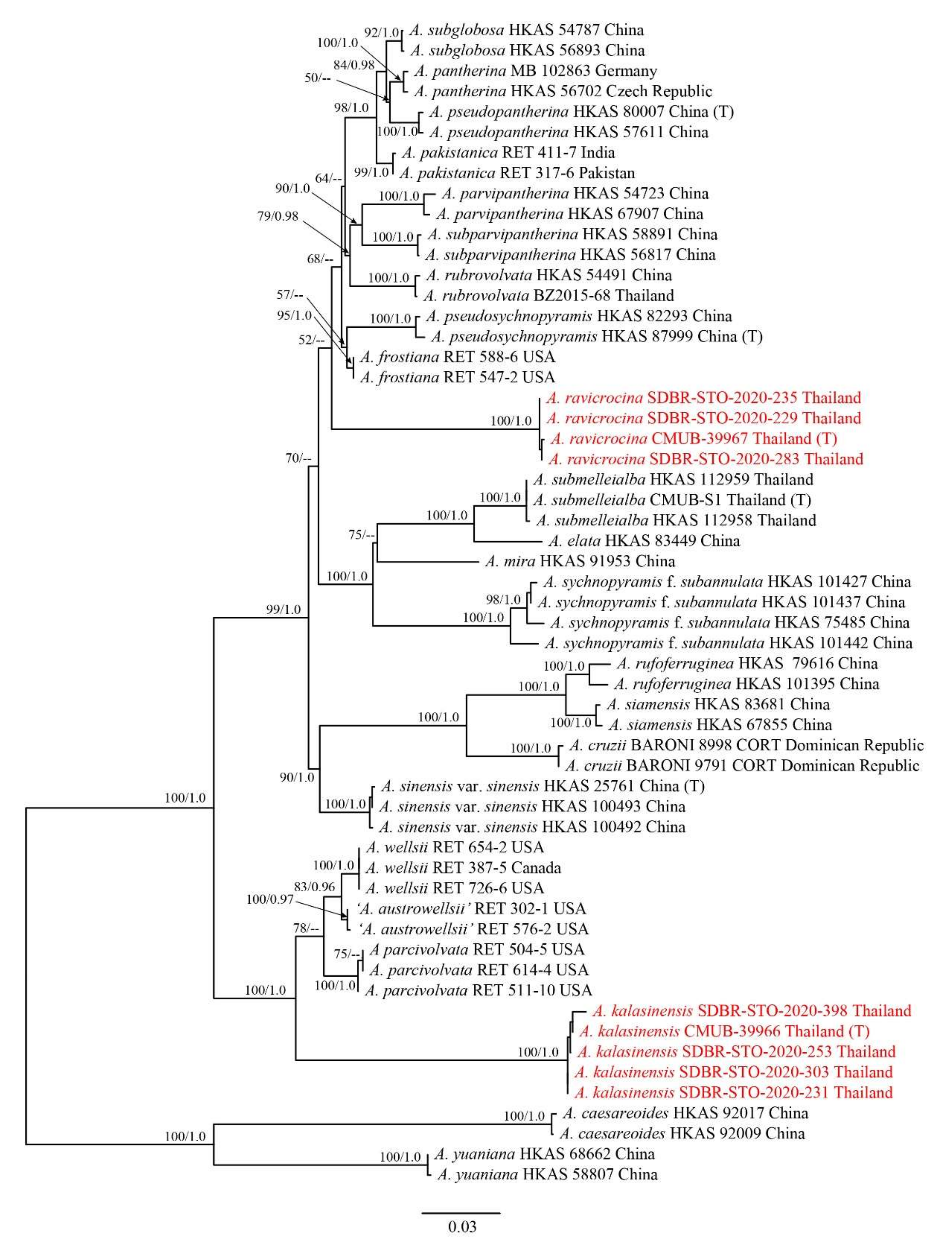
3.1. Phylogenetic Analyses
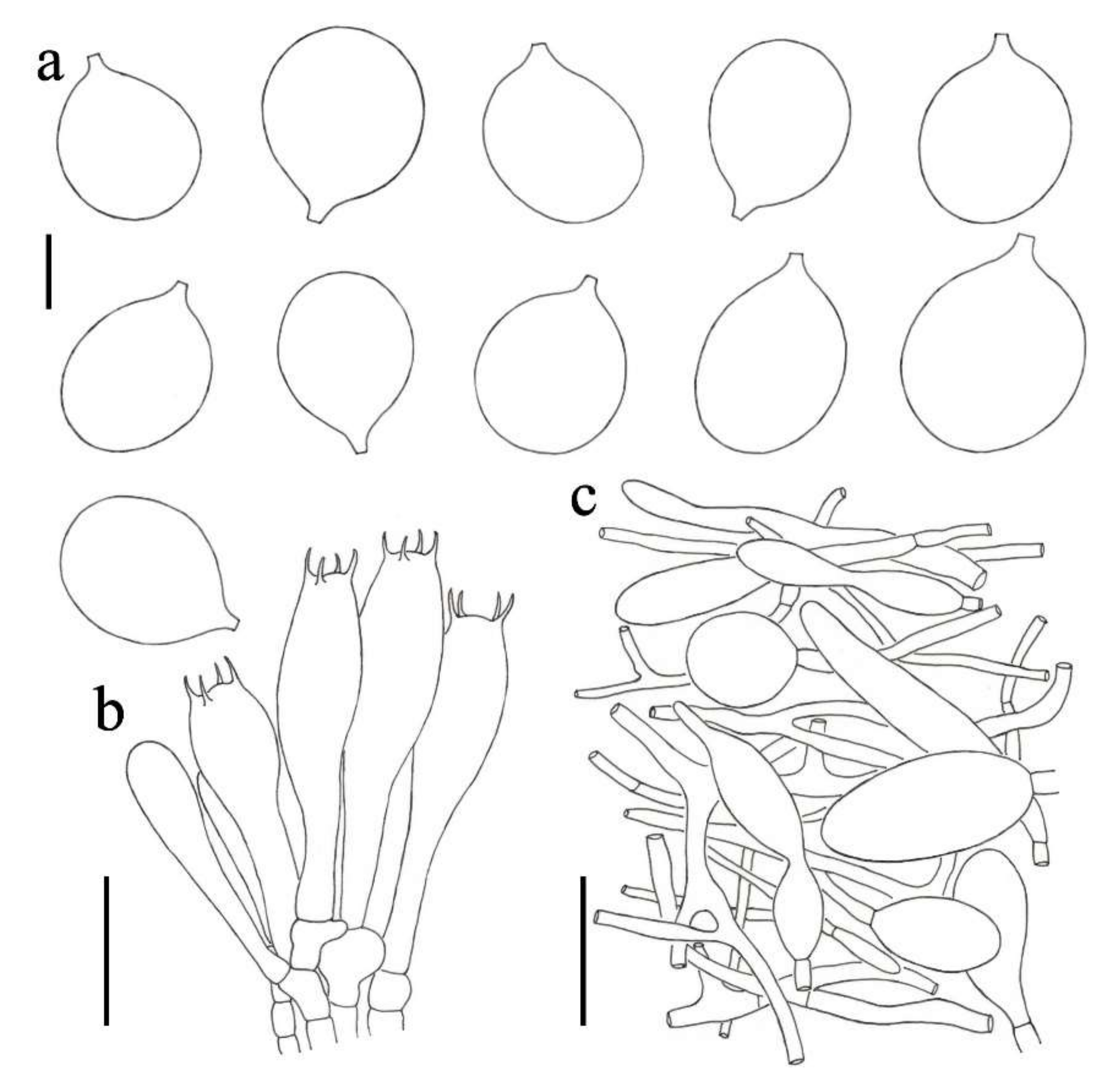
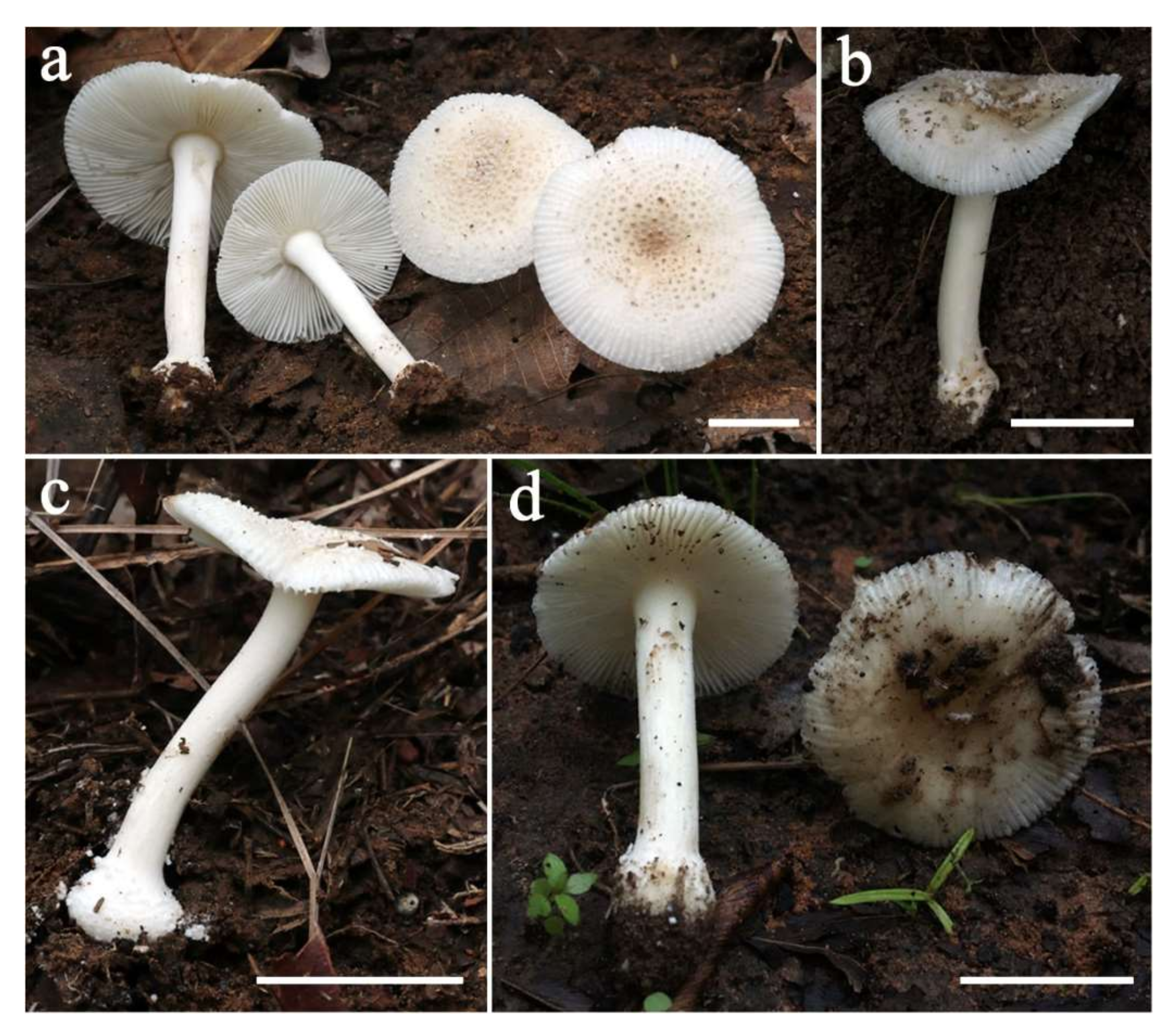
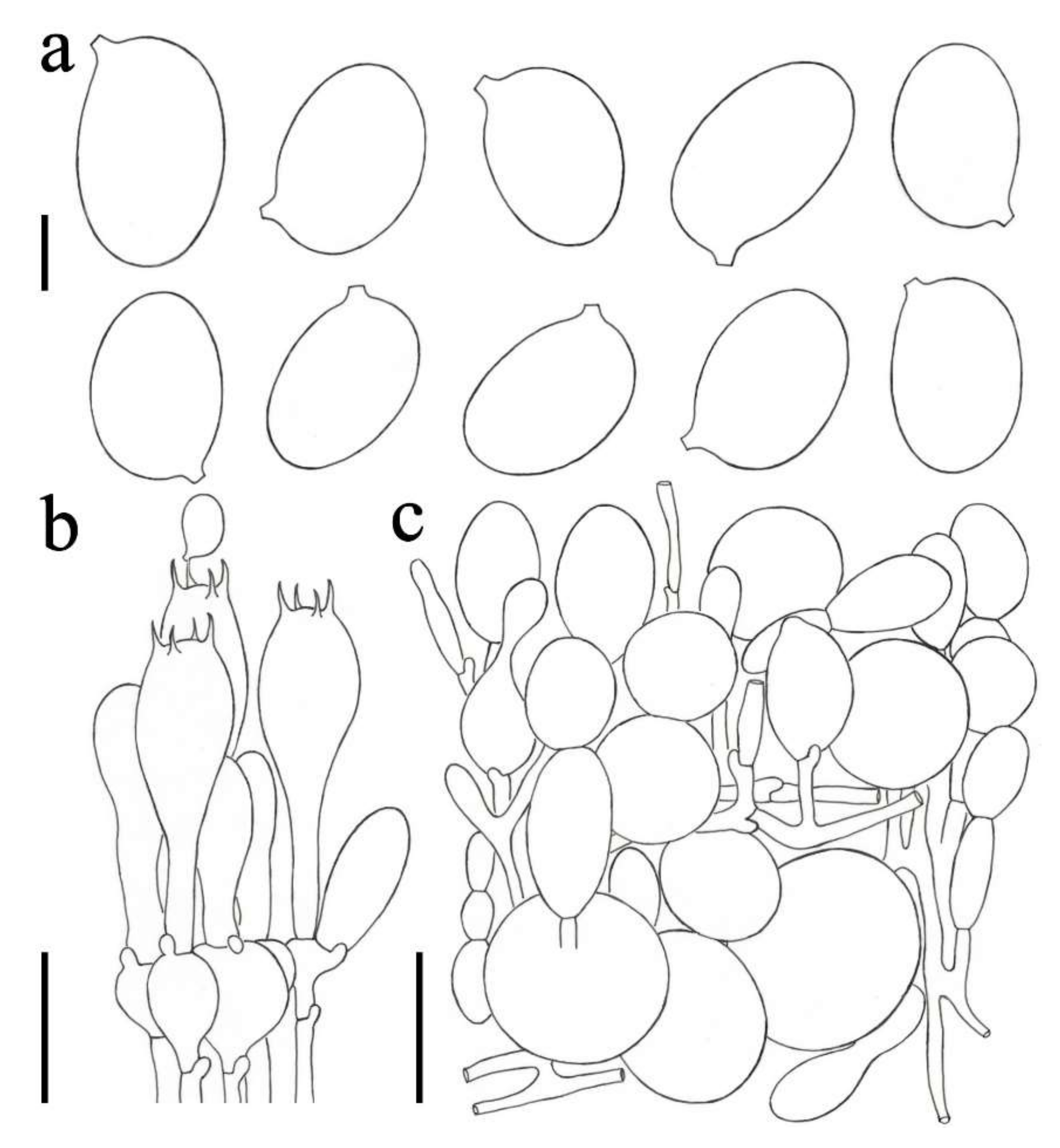
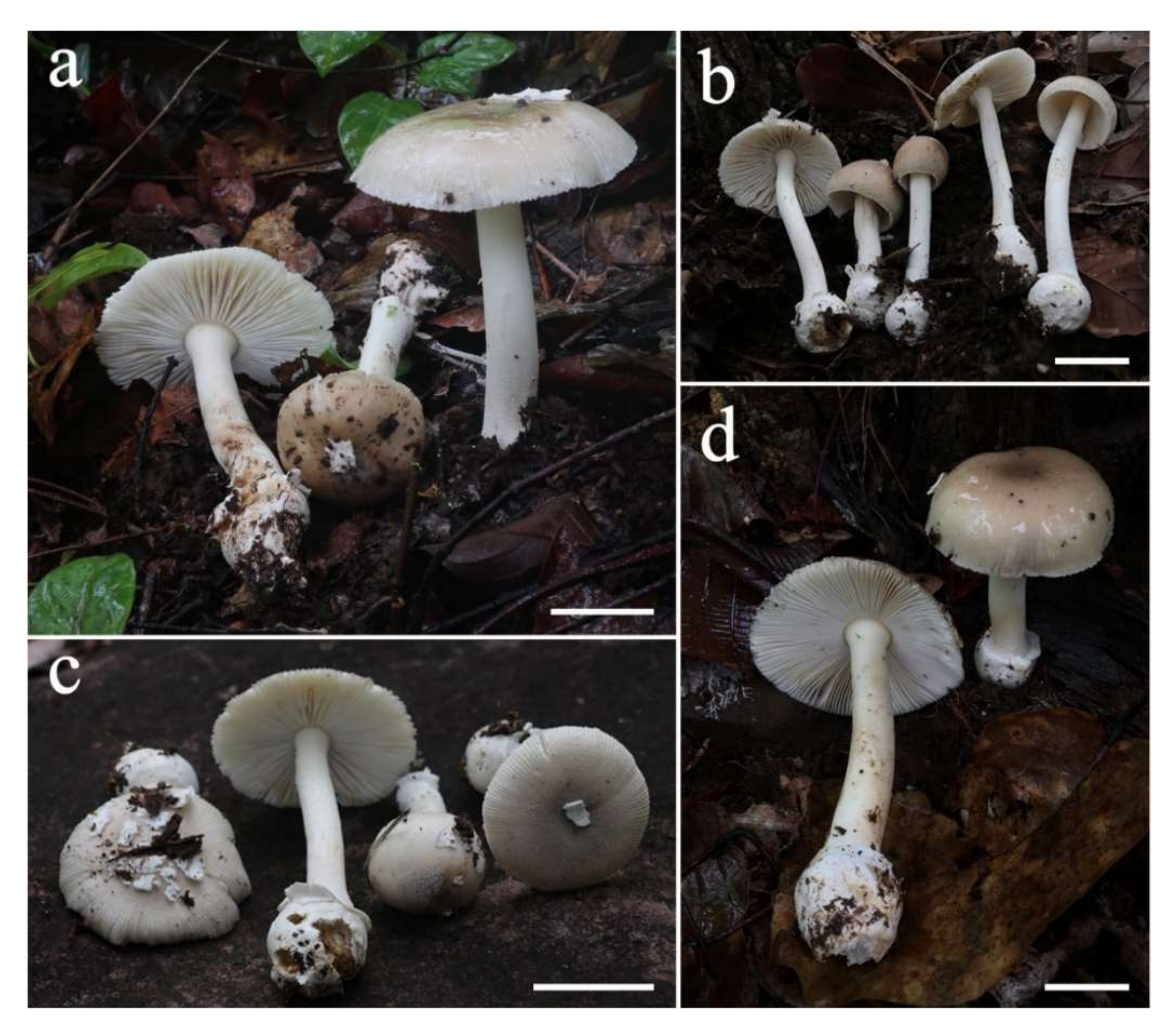
The best-fit models for the five genes were as follows: general time reversible + proportion of invariable sites + gamma distribution (GTR + I + G) for ITS, nrLSU, and TEF1-α; Hasegawa-Kishino-Yano (HKY) + I + G for RPB2; and Kimura 2-parameter (K80) + G for TUB. The concatenated dataset was partitioned into five parts by sequence region. The model HKY + I + G and K80 + G could not be implemented in RAxML, thus the GTR + I + G model, which included all parameters of the selected model, was used instead.
The multi-gene dataset comprised 212 sequences, including 33 newly generated and 179 retrieved from GenBank. Amanita yuaniana (HKAS58807), A. yuaniana (HKAS68662), A. caesareoides (HKAS92009), and A. caesareoides (HKAS92017) from Amanita section Caesarea were set as the outgroup taxa. The concatenated dataset comprised 2769 positions (ITS: 1–445; nrLSU: 446–1343; RPB2: 1344–2014; TEF1-α: 2015–2576; and TUB: 2577–2803) after alignment, including the gaps.
Bayesian and RAxML analysis of the combined dataset resulted in phylogenetic reconstructions with largely similar topologies, thus the result of maximum likelihood (RAxML) tree is shown in Figure 1. In our phylogenetic results, five collections representing Amanita kalasinensis and four collections representing A. ravicrocina, respectively, formed a monophyletic lineage from other extant species with credible support values, which could be recognized as two new species.

3.2. Taxonomy

Amanita kalasinensis Yuan S. Liu & S. Lumyong, sp. nov. (Figure 2 and Figure 3).
Mycobank number: 842342.
Holotype: Thailand, Kalasin Province: Kham Muang District, Na Bon, 16°53′05″ N, 103°41′49″ E, alt. 239 m, 12 August 2020, Yuan S. Liu, STO-2020-233 (CMUB-39966).
Etymology: The specific epithet ‘kalasinensis’ refers to the Kalasin province of Thailand, where the holotype was collected.
Description: Basidioma small to medium-sized. Pileus 3.0–5.7 cm diam., convex, plano-convex to applanate, slightly depressed at center, white (1A1) to greyish yellow (4B3–5), often darker at center and becoming paler towards margin; volval remnants on pileus conical, pyramidal to granuliform, 1–2 mm dia., white (1A1) to yellowish white (4A2), densely arranged on the disk; margin striate (ca. 0.3–0.5), non-appendiculate; context white, unchanging. Lamellae free, crowded, white (1A1); lamellulae truncate, plentiful. Stipe 3.5–8.0 cm long × 0.6–0.9 cm diam., subcylindrical and slightly tapering upward, with apex slightly expanded, white (1A1) to yellowish white (4A2), sometimes with greyish yellow (4B3–4) tinge, covered with white (1A1) fibrils, often becoming floccose near basal bulb; context white (1A1), stuffed; basal bulb globose to subglobose, 0.9–1.4 cm diam., white (1A1); volval remnants on stipe base granuliform to floccos, white (1A1) to light yellow (4B4–5). Annulus absent. Odor not recorded.
Lamellar trama bilateral. Mediostratum 15–20 μm wide, composed of abundant clavate to cylindrical inflated cells (54–145 × 13–30 μm); filamentous hyphae abundant, 4–12 μm wide; vascular hyphae scarce. Lateral stratum composed of abundant ellipsoid to clavate inflated cells (32–66 × 13–37 μm), diverging at an angle of ca. 30° to 45° to mediostratum; filamentous hyphae abundant to very abundant, 3–10 μm wide. Subhymenium 20–30 μm thick, with 2–3 layers of globose, ellipsoid, or irregular inflated cells, 6–24 × 6–18 μm. Basidia (Figure 3b) 37–52 × 12–16 μm, clavate, four-spored; sterigmata 4–6 μm long; basal septa clamped. Basidiospores (Figure 3a) [100/3/3] (8.5–) 9.0–11.5 (–13.0) × (6.0–) 7.0–8.5 (–10.0) μm, avl X avw = 10.3 × 7.4 μm, Q = (1.19–) 1.25–1.53 (–1.67) μm, Qm = 1.40 ± 0.09, broadly ellipsoid to ellipsoid, inamyloid, colorless, thin-walled, smooth; apiculus short but wide, width up to 1.5 μm. Lamellar edge appearing as a sterile strip, composed of very abundant to nearly dominant globose, subglobose, ellipsoid, or irregular inflated cells (11–26 × 8–25 μm), single and terminal or in chains of 2–3, thin-walled, colorless; filamentous hyphae scattered, 3–6 μm wide, irregularly arranged or parallel to lamellar edge. Pileipellis 130–205 μm thick, composed of radial, thin-walled, colorless, filamentous hyphae 3–17 μm wide; vascular hyphae scarce. Volval remnants on pileus (Figure 3c) composed of vertically to subvertically arranged elements: filamentous hyphae fairly abundant to abundant, 2–8 μm wide, colorless, thin-walled, branching, anastomosing; inflated cells abundant, globose, subglobose, to ellipsoid, sometimes irregular, 23–65 × 15–55 μm, colorless, thin-walled, terminal or in chains of 2–3; vascular hyphae scarce. Volval remnants on stipe base is semblable with the structure of volval remnants on pileus, composed of irregularly arranged elements: filamentous hyphae very abundant to nearly dominant, 2–11 (–15) μm wide, colorless, thin-walled, branching, anastomosing; inflated cells fairly abundant, globose, subglobose to ellipsoid, sometimes irregular, 12–88 × 10–55 μm, colorless, thin-walled; vascular hyphae scarce. Stipe trama composed of longitudinally arranged, clavate terminal cells, 90–265 × 20–48 μm; filamentous hyphae abundant, 2–16 μm wide; vascular hyphae scarce. Clamps present in all parts of basidioma.
Habitat: Solitary to scattered on soil in tropical deciduous forests dominant by Dipterocarpus and Shorea. Basidioma occurs in the rainy season during May to October.
Distribution: Currently known from northern and northeastern Thailand.
Additional collections examined: Thailand, Kalasin Province: Kham Muang District, Na Bon, 16°53′05″ N, 103°41′49″ E, alt. 239 m, 12 August 2020, Yuan S. Liu, STO-2020-231 (SDBR-STO-2020-231); Kalasin Province: Somdet District, Mahachai, 16°48′38″ N, 103°46′13″ E, alt. 197 m, 14 August 2020, Yuan S. Liu, STO-2020-253 (SDBR-STO-2020-253); Sakon Nakhon Province: Kut Bak District, Na Mong, 17°06′04″ N, 103°54′32″ E, alt. 208 m, 15 August 2020, Yuan S. Liu, STO-2020-303 (SDBR-STO-2020-303); Chiang Mai Province: Mueang Chiang Mai District, Suthep, 18°48′10″ N, 98°57′02″ E, alt. 343 m, 26 August 2020, Yuan S. Liu, STO-2020-398 (SDBR-STO-2020-398).
Notes: Amanita kalasinensis is similar to several species that have a light yellow tinge pileus covered by pyramidal to granuliform, white volva remnants, such as A. parvipantherina Zhu L. Yang, M. Weiß & Oberw., A. sychnophyramis f. subannulata Hongo, and A. sychnophyramis Corner & Bas f. sychnopyramis. However, both A. parvipantherina [33,34] and A. sychnophyramis f. subannulata [1,2,3,35] differ from A. kalasinensis by having a white or brownish annulus on its stipe, as well as a darker brownish pileus. Amanita sychnophyramis f. sychnophyramis, occurring in Singapore, China, and Malaysia [3,36,37], is discerned from A. kalasinensis by having a larger and darker brownish pileus. Furthermore, compared with the former species, which is short of clamp and has a globose to subglobose basidiospore (6.5–8.5 × 6–8 μm, Q = 1.01–1.11, Qm = 1.06 ± 0.03), A. kalasinensis has obvious clamps and a broadly ellipsoid to ellipsoid basidiospore (9.0–11.5 × 7.0–8.5 μm, Q = 1.25–1.53 μm, Qm = 1.40 ± 0.09).
Amanita ravicrocina Yuan S. Liu & S. Lumyong sp. nov. (Figure 4 and Figure 5).
Mycobank number: 842343.
Holotype: Thailand. Sakon Nakhon Province: Phu Phan District, Khok Phu, 17°00′09″ N, 103°57′34″ E, alt. 291 m, 15 August 2020, Yuan S. Liu, STO-2020-282 (CMUB-39967).
Etymology: ravicrocina, from ravus = greyish, and crocinus = orange, refers to its greyish orange pileus.
Description: Basidioma small to medium-sized. Pileus 3.0–8.5 cm diam., convex to plano-convex, often slightly depressed at center, brown (5E5), greyish orange (5B4–5) to orange white (5A2), often darker at center and becoming paler towards margin; volval remnants on pileus often persistent as large, thick, white (1A1) patches slightly attached on pileus; margin striate (ca. 0.3–0.5), non-appendiculate; context white, unchanging. Lamellae free, crowded, white (1A1); lamellulae truncate. Stipe 5.6–13.1 cm long × 0.8–1.4 cm diam., slender, subcylindrical and slightly tapering upward, with apex slightly expanded, white (1A1), covered with white (1A1) fibrils, becoming floccose near basal bulb; context white (1A1), fistulose; basal bulb globose to subglobose, 1.8–2.9 cm diam., white (1A1); volval remnants on stipe base formed a collar-like or shortly limbate volva on limit between stipe and basal bulb, white (1A1). Annulus absent. Odor not recorded.
Lamellar trama bilateral. Mediostratum 15–30 μm wide, composed of abundant clavate to cylindrical inflated cells (25–160 × 12–35 μm); filamentous hyphae abundant, 2–7 μm wide; vascular hyphae scarce. Lateral stratum composed of abundant ellipsoid to clavate inflated cells (50–155 × 7–18 μm), diverging at an angle of ca. 30° to 45° to mediostratum; filamentous hyphae abundant, 2–8 μm wide. Subhymenium 20–30 μm thick, with 2–3 layers of subglobose, pyriform, or irregular cells, 8–19 × 5–10 μm. Basidia (Figure 5b) 38–52 × 10–14 μm, clavate, four-spored; sterigmata 3–6 μm long; basal septa lacking clamps. Basidiospores (Figure 5a) [100/5/4] (7.0–) 8.0–9.5 (–10.5) × (6.0–) 7.0–8.5 (–9.0) μm, avl X avw = 8.7 × 7.5 μm, Q = (1.00–) 1.06–1.29 (–1.36) μm, Qm = 1.16 ± 0.08, subglobose to broadly ellipsoid, inamyloid, colorless, thin-walled, smooth; apiculus small. Lamellar edge appearing as a sterile strip, composed of abundant subglobose to ellipsoid inflated cells (8–25 × 6–12 μm), single and terminal or in chains of 2–3, thin-walled, colorless; filamentous hyphae fairly abundant, 2–8 μm wide, irregularly arranged or parallel to lamellar edge. Pileipellis 60–100 μm thick, composed of radial, thin-walled, colorless, filamentous hyphae 2–10 μm wide; vascular hyphae scarce. Volval remnants on pileus (Figure 5c) composed of irregularly arranged elements: filamentous hyphae abundant to very abundant, 2–10 μm wide, colorless, thin-walled, branching, anastomosing; inflated cells abundant, globose, subglobose, fusiform to ellipsoid, sometimes irregular, 20–90 × 14–46 μm, colorless, thin-walled, terminal or in chains of 2–4; vascular hyphae scarce. Volval remnants on stipe base is semblable with the structure of volval remnants on pileus, composed of irregularly arranged elements: filamentous hyphae very abundant to nearly dominant, 2–9 (–12) μm wide, colorless, thin-walled, branching, anastomosing; inflated cells fairly abundant to abundant, globose, subglobose, ellipsoid to clavate, sometimes irregular, 18–90 × 11–35 μm, colorless, thin-walled; vascular hyphae scarce. Stipe trama composed of longitudinally arranged, clavate terminal cells, 75–235 × 18–36 μm; filamentous hyphae abundant, 3–11 μm wide; vascular hyphae scarce. Clamps absent in all parts of basidioma.
Habitat: Solitary to scattered on soil in tropical deciduous forests dominant by Dipterocarpus and Shorea. Basidioma occurs in the rainy season during May to October.
Distribution: Currently known from northeastern Thailand.
Additional collections examined: Thailand, Kalasin Province: Kham Muang District, Na Bon, 16°53′05″ N, 103°41′49″ E, alt. 239 m, 12 August 2020, Yuan S. Liu, STO-2020-229 (SDBR-STO-2020-229); Yuan S. Liu, STO-2020-235 (SDBR-STO-2020-235); Sakon Nakhon Province: Phu Phan District, Khok Phu, 17°00′09″ N, 103°57′34″ E, alt. 291 m, 15 August 2020, Yuan S. Liu, STO-2020-283 (SDBR-STO-2020-283).
Notes: Amanita ravicrocina has a collar-like volva remnant on the limit between stipe and inflated basal bulb, which is not a common characteristic in A. section Amanita. Coupled with a brown tone pileus surface, A. ravicrocina could be easily singled out from other species in A. section Amanita, except A. ibotengutake T. Oda, C. Tanaka & Tsuda, A. pseudopantherina Zhu L. Yang ex Yang-Yang Cui, Qing Cai & Zhu L. Yang, and A. subglobosa Zhu L. Yang. Nevertheless, the latter three taxa are distinguished from A. ravicrocina by having the membranous annulus and pyramidal to subconical volva remnants on pileus [1,4,38]. In addition, A. parvipantherina [4,33] also possesses close likeness with A. ravicrocina. Both of them have small to medium-sized basidiomata and a light grey to brown pileus surface. However, A. parvipantherina has a white to brownish annulus, and its volva remnants on pileus are verrucose to pyramidal, while A. ravicrocina has a short annulus and appears as patchy volva remnants on pileus.

4. Discussion

As mentioned above, thirteen species in section Amanita have been described in Thailand. Although most of these species are based on both of morphologic and phylogenetic evidences, four species, namely, A. aff. mira, A. obsita, A. siamensis, and A. subglobosa, have only morphologic data [6], which need to be supplemented with more molecular data to confirm their taxonomic status. Our study, along with other previous studies, allowed to discover the high diversity of Amanita species in Thailand [6,7,8,9,10,11,12], indicating that more taxa remain to be discovered and documented.
In our multi-gene phylogenetic analyses, both Amanita kalasinensis and A. ravicrocina formed a well-supported (BS = 100%, PP = 1.0) monotypic clade. However, the position of A. ravicrocina is not stable. Despite that many potential causes could lead to these unstable topologies, the primary reason might be that A. ravicrocina has significant differences from any other existing Amanita species in each single gene (the highest similarity and its query cover of initial BLAST searches results in GenBank for ITS and nrLSU of A. ravicrocina samples were from following: A. frostiana—RET 588-6 (KP313583) with 89.23% similarity and 99% query cover for ITS, and A. altipes—BZ2013-42 (MH716040) with 97.58% similarity and 100% query cover for nrLSU).
The erratic positions of Amanita ravicrocina and its distinct sequences provide indirect evidence that there are probably some new taxa related to A. ravicrocina that remain to be discovered. Therefore, further exploration of Amanita diversity is critical, which could reveal more members in this section.

Author Contributions

Conceptualization, Y.S.L. and S.L.; methodology, Y.S.L.; formal analysis, Y.S.L.; resources, Y.S.L.; data curation, Y.S.L.; writing—original draft preparation, Y.S.L.; writing—review and editing, J.L., J.K. and S.L.; supervision, J.L. and S.L.; project administration, S.L.; funding acquisition, S.L. All authors have read and agreed to the published version of the manuscript.

Funding

This study was supported by TA&RA Scholarship, graduate school, Chiang Mai University, and partially supported by Chiang Mai University, Thailand.

Institutional Review Board Statement

Not applicable.

Data Availability Statement

The DNA sequences data obtained from this study were deposited in GenBank under accession numbers: ITS (OM040561, OM040562, OM040563, OM040564, OM040565, OM040566, OM040567, OM040568, OM040569), nrLSU (OM040552, OM040553, OM040554, OM040555, OM040556, OM040557, OM040558, OM040559, OM040560), RPB2 (OM066913, OM066914, OM066915, OM066916, OM066917, OM066918), TEF1-α (OM066919, OM066920, OM066921, OM066922, OM066923, OM066924), and TUB (OM066925, OM066926, OM066927).

Acknowledgments

We are very grateful to Zhu-Liang Yang for his guidance in microscopic features observation and drawing. We also appreciate Jean Evans I. Codjia for his valuable suggestions.

Conflicts of Interest

The authors declare no conflict of interest.

References

  1. Yang, Z.L. Die Amanita-Arten von Südwestchina. Bibl. Mycol. 1997, 170, 1–240. [Google Scholar]
  2. Yang, Z.L. Amanitaceae. Flora Fungorum Sinicorum 27; Science Press: Beijing, China, 2005; pp. 1–258. [Google Scholar]
  3. Yang, Z.L. Atlas of the Chinese Species of Amanitaceae; Science Press: Beijing, China, 2015; pp. 1–213. [Google Scholar]
  4. Cui, Y.Y.; Cai, Q.; Tang, L.P.; Liu, J.W.; Yang, Z.L. The family Amanitaceae: Molecular phylogeny, higher-rank taxonomy and the species in China. Fungal Divers. 2018, 91, 5–230. [Google Scholar] [CrossRef]
  5. Amanitaceae org. Available online: http://www.amanitaceae.org/?Genus%20Amanita (accessed on 5 December 2021).
  6. Sanmee, R.; Tulloss, R.E.; Lumyong, P.; Dell, B.; Lumyong, S. Studies on Amanita (Basidiomycetes: Amanitaceae) in Northern Thailand. Fungal Divers. 2008, 3, 97–123. [Google Scholar]
  7. Li, G.J.; Hyde, K.D.; Zhao, R.L.; Hongsanan, S.; Abdel-Aziz, F.A.; AbdelWahab, M.A.; Alvarado, P.; Alves-Silva, G.; Ammirati, S.F.; Ariyawansa, H.A.; et al. Fungal diversity notes 253–366: Taxonomic and phylogenetic contributions to fungal taxa. Fungal Divers. 2016, 78, 1–237. [Google Scholar] [CrossRef]
  8. Thongbai, B.; Tulloss, R.E.; Miller, S.L.; Hyde, K.D.; Chen, J.; Zhao, R.L.; Raspé, O. A new species and four new records of Amanita (Amanitaceae; Basidiomycota) from Northern Thailand. Phytotaxa 2016, 286, 211–231. [Google Scholar] [CrossRef] [Green Version]
  9. Thongbai, B.; Miller, S.L.; Stadler, M.; Stadler, M.; Wittstein, K.; Hyde, K.D.; Lumyong, S.; Raspé, O. Study of three interesting Amanita species from Thailand: Morphology, multiple-gene phylogeny and toxin analysis. PLoS ONE 2017, 12, e0182131. [Google Scholar] [CrossRef] [PubMed] [Green Version]
  10. Thongbai, B.; Hyde, K.D.; Lumyong, S.; Raspé, O. High undescribed diversity of Amanita section Vaginatae in northern Thailand. Mycosphere 2018, 9, 462–494. [Google Scholar] [CrossRef]
  11. Phookamsak, R.; Hyde, K.D.; Jeewon, R.; Bhat, D.J.; Jones, E.B.G.; Maharachchikumbura, S.S.N.; Raspe, O.; Karunarathna, S.C.; Wanasinghe, D.N.; Hongsanan, S.; et al. Fungal diversity notes 929–1035: Taxonomic and phylogenetic contributions on genera and species of fungi. Fungal Divers. 2019, 95, 1–273. [Google Scholar] [CrossRef] [Green Version]
  12. Liu, Y.S.; Liu, J.K.; Mortimer, P.E.; Lumyong, S.L. Amanita submelleialba sp. nov. in section Amanita from northern Thailand. Phytotaxa 2021, 513, 129–140. [Google Scholar] [CrossRef]
  13. Zhang, L.F.; Yang, Z.L. Recommendation of several methods for preserving the materials of macro fungi for molecular biological research. J. Fungal Res. 2004, 2, 60–61. [Google Scholar]
  14. Kornerup, A.; Wanscher, J.H. Methuen Handbook of Colour, 3rd ed.; Eyre Methuen: London, UK, 1978; pp. 1–243. [Google Scholar]
  15. Bas, C. Morphology and subdivision of Amanita and a monograph of its section Lepidella. Persoonia 1969, 5, 285–579. [Google Scholar]
  16. Cai, Q.; Cui, Y.Y.; Yang, Z.L. Lethal Amanita species in China. Mycologia 2016, 108, 993–1009. [Google Scholar] [CrossRef] [Green Version]
  17. White, T.J.; Bruns, T.; Lee, S.; Taylor, J.W. Amplification and direct sequencing of fungal ribosomal RNA genes for phylogenetics. In PCR Protocols: A Guide to Methods and Applications; Innis, M.A., Gelfand, D.H., Sninsky, J.J., White, T.J., Eds.; Academic Press: San Diego, CA, USA, 1990; Volume 38, pp. 315–322. [Google Scholar]
  18. Gardes, M.; Bruns, T.D. ITS primers with enhanced specificity for basidiomycetes application to the identification of mycorrhizae and rusts. Mol. Ecol. 1993, 2, 113–118. [Google Scholar] [CrossRef] [PubMed]
  19. Vilgalys, R.; Hester, M. Rapid genetic identification and mapping of enzymatically amplified ribosomal DNA from several Cryptococcus species. J. Bacteriol. 1990, 172, 4238–4246. [Google Scholar] [CrossRef] [Green Version]
  20. Rehner, S.A.; Buckley, E. A Beauveria phylogeny inferred from nuclear ITS and EF1-α sequences: Evidence for cryptic diversification and links to Cordyceps teleomorphs. Mycologia 2005, 97, 84–98. [Google Scholar] [CrossRef] [PubMed]
  21. Cai, Q.; Tulloss, R.E.; Tang, L.P.; Tolgor, B.; Zhang, P.; Chen, Z.H.; Yang, Z.L. Multi-locus phylogeny of lethal amanitas: Implications for species diversity and historical biogeography. BMC Evol. Biol. 2014, 14, 143. [Google Scholar] [CrossRef] [Green Version]
  22. Katoh, K.; Standley, D.M. MAFFT multiple sequence alignment software version 7: Improvements in performance and usability. Mol. Biol. Evol. 2013, 30, 772–780. [Google Scholar] [CrossRef] [Green Version]
  23. Hall, T.A. BioEdit: A user-friendly biological sequence alignment editor and analysis program for Windows 95/98/NT. Nucleic Acids Symp. Ser. 1999, 41, 95–98. [Google Scholar]
  24. Castresana, J. Selection of conserved blocks from multiple alignments for their use in phylogenetic analysis. Mol. Biol. Evol. 2000, 17, 540–552. [Google Scholar] [CrossRef] [Green Version]
  25. Dissanayake, A.J.; Bhunjun, C.S.; Maharachchikumbura, S.S.N.; Liu, J.K. Applied aspects of methods to infer phylogenetic relationships amongst fungi. Mycosphere 2020, 11, 2652–2676. [Google Scholar] [CrossRef]
  26. Nylander, J.A.A. MrModeltest v2. Program Distributed by the Author; Evolutionary Biology Centre, Uppsala University: Uppsala, Sweden, 2004. [Google Scholar]
  27. Miller, M.A.; Pfeiffer, W.; Schwartz, T. Creating the CIPRES Science Gateway for Inference of Large Phylogenetic Trees. In Proceedings of the Gateway Computing Environments Workshop, New Orleans, LA, USA, 14 November 2010; pp. 1–8. [Google Scholar]
  28. Stamatakis, A. RAxML Version 8: A tool for Phylogenetic Analysis and Post-Analysis of Large Phylogenies. Bioinformatics 2014, 30, 1312–1313. [Google Scholar] [CrossRef] [PubMed]
  29. Ronquist, F.; Huelsenbeck, J.P. MrBayes3: Bayesian phylogenetic inference under mixed models. Bioinformatics 2003, 19, 1572–1574. [Google Scholar] [CrossRef] [PubMed] [Green Version]
  30. Rannala, B.; Yang, Z. Probability distribution of molecular evolutionary trees: A new method of phylogenetic inference. J. Mol. Evol. 1996, 43, 304–311. [Google Scholar] [CrossRef] [PubMed]
  31. Larget, B.; Simon, D.L. Markov chain Monte Carlo algorithms for the Bayesian analysis of phylogenetic trees. Mol. Biol. Evol. 1999, 16, 750–759. [Google Scholar] [CrossRef]
  32. FigTree v1.4.4. Available online: http://tree.bio.ed.ac.uk/software/figtree/ (accessed on 25 November 2021).
  33. Yang, Z.L.; Weiß, M.; Oberwinkler, F. New species of Amanita from the Eastern Himalaya and adjacent regions. Mycologia 2004, 96, 636–646. [Google Scholar] [CrossRef] [PubMed]
  34. Bhatt, R.P.; Mehmood, T.; Uniyal, P.; Singh, U. Six new records of genus Amanita (Amanitaceae) from Uttarakhand, India. Curr. Res. Environ. Appl. Mycol. 2017, 7, 161–182. [Google Scholar] [CrossRef]
  35. Hongo, T. Notulae Mycologicae (10). Mem. Shiga Univ. 1971, 21, 62–68. [Google Scholar]
  36. Corner, E.J.H.; Bas, C. The genus Amanita in Singapore and Malaya. Persoonia 1962, 2, 241–304. [Google Scholar]
  37. Lee, S.S. A Field Guide to the Larger Fungi of FRIM; Forest Research Institute Malaysia: Kuala Lumpur, Malaysia, 2017; pp. 1–174. [Google Scholar]
  38. Oda, T.; Yamazaki, T.; Tanaka, C.; Terashita, T.; Taniguchi, N.; Tsuda, M. Amanita ibotengutake sp. nov., a poisonous fungus from Japan. Mycol. Prog. 2002, 1, 355–365. [Google Scholar] [CrossRef]
Figure 1. RAxML tree based on a combined of ITS + nrLSU + RPB2 + TEF1-α + TUB dataset. Bootstrap values (BS) for ML ≥ 50% and posterior probabilities (PP) for BI ≥ 0.95 are placed above or below the branches, respectively. Newly generated sequences are indicated in red and sequences from type material are marked with (T). The tree is rooted with Amanita yuaniana (HKAS58807 and HKAS68662) and A. caesareoides (HKAS92009 and HKAS92017).
Diversity 14 00101 g001
Figure 2. Different collections of Amanita kalasinensis shown in the field. (a) CMUB-39966 (holotype). (b) SDBR-STO-2020-231. (c) SDBR-STO-2020-303. (d) SDBR-STO-2020-398. Scale bars: (ad) 2 cm.
Diversity 14 00101 g002
Figure 3. Amanita kalasinensis (CMUB-39966, holotype). (a) Basidiospores. (b) Hymenium and subhymenium. (c) Longitudinal section of volval remnants on pileus. Bars: (a) 4 μm; (b) 20 μm; (c) 40 μm.
Diversity 14 00101 g003
Figure 4. Different collections of Amanita ravicrocina shown in the field. (a) CMUB-39967 (holotype). (b) SDBR-STO-2020-235. (c) SDBR-STO-2020-229. (d) SDBR-STO-2020-283. Scale bars: (ad) 3 cm.
Diversity 14 00101 g004
Figure 5. Amanita ravicrocina (CMUB-39967, holotype). (a) Basidiospores. (b) Hymenium and subhymenium. (c) Longitudinal section of volval remnants on pileus. Bars: (a) 4 μm; (b) 20 μm; (c) 40 μm.
Diversity 14 00101 g005
Table 1. Species names, voucher numbers, countries, and their respective GenBank accession numbers of the taxa used in this study.
Species NameVoucherCountryGenBank Accession Number
ITSnrLSURPB2TEF1-αTUB
‘Amanita austrowellsii’RET 302-1USAMN963578MN963578
‘A. austrowellsii’RET 576-2USAMN963579MN963579
A. cruziiTBARONI 8998 (CORT)Dominican RepublicKC855222KC855222MH508750MH485478
A. cruziiBARONI 9791 (CORT)Dominican RepublicKC855223KC855223MH508751
A. elataHKAS 83449ChinaMH508334MH486486MH485965MH508763MH485488
A. frostianaRET 547-2USAKP313581
A. frostianaRET 588-6USAKP313583
A. kalasinensisSDBR-STO-2020-231ThailandOM040561OM040552
A. kalasinensisTCMUB-39966ThailandOM040562OM040553OM066913OM066919OM066925
A. kalasinensisSDBR-STO-2020-253ThailandOM040563OM040554OM066914OM066920
A. kalasinensisSDBR-STO-2020-303ThailandOM040564OM040555
A. kalasinensisSDBR-STO-2020-398ThailandOM040565OM040556
A. miraHKAS 91953ChinaMH508437MH486646MH486097
A. pakistanicaRET 317-6PakistanKX365198KX365199
A. pakistanicaRET 411-7IndiaMG991745MG991814
A. pantherinaHKAS 56702Czech RepublicMH508487KR824782KR824789KR824825MH485670
A. pantherinaMB-102863GermanyMH508488MH486743MH486167MH508976MH485671
A. parcivolvataRET 504-5USAKP313586
A. parcivolvataRET 511-10USAKP313584
A. parcivolvataRET 614-4USAMN963585MN963584
A. parvipantherinaHKAS 54723ChinaMH508495KR824780KR824802KR824807MH485676
A. parvipantherinaaHKAS 67907ChinaMH508498KR824781KR824803KR824808MH485679
A. pseudopantherina THKAS 80007ChinaMH508514MH486777MH486191MH509004MH485698
A. pseudopantherinaHKAS 57611ChinaMH508511MH486774MH486188MH485695
A. pseudosychnopyramisHKAS 82293ChinaMH508529MH486790MH486204MH485712
A. pseudosychnopyramis THKAS 87999ChinaMH508530KR824778KR824790KR824824MH485713
A. rubrovolvataBZ2015-68ThailandKY747465KY747477KY656882KY656863
A. rubrovolvataHKAS 54491ChinaJN943178JN941153JQ031116KR824823
A. rufoferrugineaHKAS 101395ChinaMH508578MH486839MH486249MH485753
A. rufoferrugineaHKAS 79616ChinaMH508579MH486842MH486252MH485756
A. siamensisHKAS 67855ChinaMH508592MH486864MH486271MH485773
A. siamensisHKAS 83681ChinaMH508593MH486866MH486273MH485774
A. sinensis var. sinensis THKAS 25761ChinaAB096059AF024474AB095864
A. sinensis var. sinensisHKAS 100492ChinaMH508594MH486867MH486274MH485775
A. sinensis var. sinensisHKAS 100493ChinaMH508595MH486868MH486275MH485776
A. subglobosaHKAS 54787ChinaMH508618MH486900MH486301MH509121MH485801
A. subglobosaHKAS 56893ChinaJN943176JN941157JQ031120KR824826MH485802
A. submelleialba TCMUB-S1ThailandMZ045688MZ045693MZ048619MZ048624MZ048629
A. submelleialbaHKAS 112958ThailandMZ045685MZ045690MZ048616MZ048621MZ048626
A. submelleialbaHKAS 112959ThailandMZ045686MZ045691MZ048617MZ048622MZ048627
A. subparvipantherinaHKAS 56817ChinaJN943171JN941160JQ031114KR824815MH485818
A. subparvipantherinaHKAS 58891ChinaMH508628MH486918MH486316MH509136MH485819
A. sychnopyramis f. subannulataHKAS 101427ChinaMH508631MH486922MH509139MH485824
A. sychnopyramis f. subannulataHKAS 101437ChinaMH508632MH486923MH486319MH509140MH485825
A. sychnopyramis f. subannulataHKAS 101442ChinaMH508633MH486925MH486321MH509142MH485826
A. sychnopyramis f. subannulataHKAS 75485ChinaMH508634MH486926MH509143MH485827
A. wellsiiRET 387-5CanadaKU248115OK285332
A. wellsiiRET 654-2USAOK299151OK299151
A. wellsiiRET 726-6USAOK299169OK299169
A. ravicrocinaSDBR-STO-2020-229ThailandOM040566OM040557OM066915OM066921OM066926
A. ravicrocinaSDBR-STO-2020-235ThailandOM040567OM040558OM066916OM066922OM066927
A. ravicrocinaTCMUB-39967ThailandOM040568OM040559OM066917OM066923
A. ravicrocinaSDBR-STO-2020-283ThailandOM040569OM040560OM066918OM066924
Outgroup
A. caesareoidesHKAS 92009ChinaMH508285MH486421MH485901MH508708
A. caesareoidesHKAS 92017ChinaMH508286MH486422MH485902MH508709
A. yuanianaHKAS 58807ChinaMH508653MH486954MH486347MH509174MH485852
A. yuanianaHKAS 68662ChinaMH508654MH486957MH486350MH509177MH485854
Newly generated sequences in this study are in black bold, while holotypes are marked with “T”.
Publisher’s Note: MDPI stays neutral with regard to jurisdictional claims in published maps and institutional affiliations.

