Article

New Strain of Cyphellophora olivacea Exhibits Striking Tolerance to Sodium Bicarbonate

Department of Genetics and Applied Microbiology, Faculty of Science and Technology, University of Debrecen, 4032 Debrecen, Hungary
*
Author to whom correspondence should be addressed.
Diversity 2022, 14(12), 1023; https://doi.org/10.3390/d14121023
Submission received: 17 October 2022 / Revised: 15 November 2022 / Accepted: 21 November 2022 / Published: 24 November 2022
(This article belongs to the Topic Fungal Diversity)

Abstract

:
The cyanobacterium strain Synechococcus cedrorum SAG 88.79 stock culture has fungal contamination stated by the Sammlung von Algenkulturen der Universität Göttingen itself. In this recent work, this particular fungal strain was isolated, identified, and morphologically characterised. The fungal strain AGSC12 belongs to the species Cyphellophora olivacea, with respect to the sequence similarity, phylogeny, and morphology of the strain. Colony morphology and growth capability were examined on SMA, EMMA, PDA, MEA, YEA, and YPA plates. Growth of the colonies was the most successful on YPA plates, followed by PDA and MEA containing plates. Surprisingly, the AGSC12 strain showed extreme tolerance to NaHCO3, albeit it, is is considered a general fungistatic compound. Moreover, positive association between the AGSC12 and SAG 88.79 strains was revealed, as the SAG 88.79 strain always attained higher cell density in co-cultures with the fungus than in mono-cultures. Besides, a taxonomic note on the SAG 88.79 strain itself was also stated.

1. Introduction

The genus Cyanobacterium (family: Cyanobacteriaceae, order: Chroococcales) originally proposed by Rippka and Cohen-Bazire in 1983 comprises more than 13 species [1,2,3,4]. The type species of this genus is Cyanobacterium stanieri Rippka and Cohen-Bazire, a free-living unicellular photosynthetic bacterium [1]. Its homotypic synonym is Synechococcus cedrorum Sauvageau according to the NCBI Taxonomy [5,6]. The strain S. cedrorum SAG 88.79 was originally isolated in Chad by M. Lefevre in 1963. This strain was originally described as Synechococcus on the basis of morphology, then transferred to the genus Cyanothece, and later to Cyanobacterium, based on the results of more thorough morphological examinations, the type of cell division, and the ultrastructural patterns [7,8]. Phylogenetic analyses of the 16S rDNA and ITS rDNA sequences suggested that the strains C. stanieri PCC 7202 and S. cedrorum SAG 88.79 are identical, but there were some observable differences in the carotenoid compositions between the two strains [3].
It is broadly known that algae and cyanobacteria exhibit a wide range of relationships with fungi, from mutualistic relations to parasitic characteristics. Symbiotic relationships reach their highest levels in the form of lichens or mycophycobiosis [9,10,11]. However, free living cells also could be beneficial to each other, even if they do not have strong physical or metabolic interdependences [12,13].
The SAG 88.79 culture, which is maintained in SAG (The Culture Collection of Algae at Goettingen University), has fungal contaminants (https://sagdb.uni-goettingen.de/detailedList.php?str_number=88.79, accessed on 15 October 2022). Though the origin of the fungal contamination is not known, its presence is fascinating on its own. It is widely known that NaHCO3 and Na2CO3 both have a strong statin effect on most fungi, as the compounds are able to inhibit fungal growth even at relatively low concentrations [14,15,16,17,18,19,20]. In contrast, the Spirulina medium (which is used for the maintenance of the SAG 88.79 strain) contains approximately 160 mmol NaHCO3 and 38 mmol Na2CO3, whose amounts are more than enough to hinder fungal growth. It would be interesting to know whether the fungal companion is less sensitive to NaHCO3 and Na2CO3, or whether the metabolism of the cyanobacterial strain makes fungal propagation possible in that hostile medium.
We strongly believe it would be beneficial to the scientific community if we identified the fungal companion. Thus, the aim of this study was to isolate and identify that certain fungal strain and to provide taxonomical and morphological characterisation. We assessed the sensitivity of the strain to different compounds and compared it to other fungal strains. Furthermore, we established the nature of the relationship between the cyanobacterial and fungal strains.

2. Materials and Methods

2.1. Strains and Culturing Media

The strains used in this study are listed in Table 1. The cyanobacterial strain was maintained and cultivated in Spirulina and ES medium (in liquid or on slant agar) at room temperature (25 °C) with atmospheric CO2 under natural light. Fungal strains were maintained in PDA medium at room temperature.
Recipes of the Spirulina and ES media are available at the website of SAG (https://www.uni-goettingen.de/en/186449.html, accessed on 15 October 2022) [21]. YEL, YEA, YPL, YPA, SML, and SMA were described by Sipiczki and Ferenczy [22], while EMML and EMMA minimal media were made according to Mitchinson [23]. PDA and MEA were purchased from VWR (VWR, Debrecen, Hungary) [24,25].
PDA plates containing NaHCO3 and NaCl were prepared as follows: a twofold concentration of PDA medium was sterilised in an autoclave and mixed with sterile NaHCO3 or NaCl aqueous solution after cooling to 35–40 °C. NaHCO3 containing media were prepared in two ways: NaHCO3 version I was sterilised by autoclaving, NaHCO3 version II was filter sterilised (VWR, Debrecen, Hungary). The reason for that is the heat instability of the NaHCO3. According to PubChem, NaHCO3 begins to decompose around 50 °C, releasing CO2, H2O and Na2CO3. As the recipe of the Spirulina medium does not mention filter sterilisation in the case of the NaHCO3, both NaHCO3 solutions were tested. Consequently, the effect of Na2CO3 tested only indirectly.

2.2. Isolation of the Unknown Fungal Strain

The unknown fungal strain was isolated from the culture broth of S. cedrorum by transferring 10 µL of liquid drops to Petri dishes (VWR, Debrecen, Hungary) containing EMMA medium. The Petri dishes were stored at room temperature. The obtained fungal isolate was transferred to PDA medium and was maintained at room temperature.

2.3. Morphological Observations and Microscopy

Fungal colony morphologies and growth ability were investigated on YEA, YPA, SMA, EMMA, MEA, and PDA containing plates using quadruplicate samples in the case of each medium. Fungal hyphae were observed with the sandwich method under an Olympus BX-40 light microscope (Olympus, Tokyo, Japan) and were photographed with an Olympus DP-70 camera (Olympus, Tokyo, Japan) [26,27]. Aerial mycelia were investigated under a Carl Zeiss Jena stereo microscope (Carl Zeiss Microscopy GmbH, Jena, Germany).

2.4. Tolerance Tests

The NaHCO3 and NaCl tolerance of the fungal strains were tested with the drop plate method using PDA medium. A loopful amount of fungal net and conidia were washed into 1 mL of sterile water and mixed thoroughly, then droplets of 20 µL volume from the supernatants were placed onto the surface of the PDA plates. The plates were incubated at ambient temperature, and the growths of the fungal colonies were checked and measured occasionally. Since the growth rates of the fungal strains listed in Table 1 are substantially different, a custom scoring system, ranging from 0 to 3, was used for the evaluation of fungal growth. Unit 3 indicates normal growth in comparison to the untreated control, whereas unit 2 represents slower growth and/or altered phenotypes (e.g., less pigmented colonies). Unit 1 denotes retarded growth, which generally means small and unusual phenotypes of the fungal colonies. Consequently, unit 0 means no fungal growth at all. Categorisations were made by statistical significance or thorough investigation by eye.

2.5. Interaction Tests

S. cedrorum and C. olivacea were separated by consecutive dilutions then mono- and co-cultivated in liquid media and on agar plates. Solid Spirulina media were flooded with either a cell mixture or sequentially (first with cyanobacteria, then with fungi), and the growth of the cultures was monitored by eye and photographed on occasion.
At the same time, exponentially growing cyanobacteria were inoculated to fresh Spirulina medium with or without the fungal strain. In this case, approximately 7 × 7 mm samples from the fungal net growing in liquid media (YEL or YPL) were cut with a sterile scalpel (VWR, Debrecen, Hungary). The remaining liquid from the surface of the hyphal net was dried with a sterile filter paper (VWR, Debrecen, Hungary) to avoid adding extra nutrients to the fresh media. Then, the hyphal net was added to the cyanobacterial culture with sterile forceps (VWR, Debrecen, Hungary). The optical density (OD) at λ = 730 nm was measured using an UV-1601 Shimadzu spectrophotometer (Shimadzu Europa GmbH, Duisburg, Germany) to monitor the cyanobacterial growth. Individually cultivated cyanobacteria were used as a control. We performed the experiments with 2 replicates twice.

2.6. DNA Analysis Methods

Genomic DNA was isolated from exponential-phase cyanobacterium cultures grown in 50 mL of Spirulina medium for 7 days with the glass bead method [28]. DNA isolation from fungal hyphae was performed as follows: the fungal strain was grown in a 300 mL Erlenmeyer flask (VWR, Debrecen, Hungary) in 100 mL YEL medium at room temperature for 10 days. Then, the hyphal batch was ground to powder under liquid nitrogen with a pestle in a mortar. Genomic DNA was isolated with the GenEluteTM Plant Genomic DNA Miniprep Kit (Sigma-Aldrich Hungary Ltd., Budapest, Hungary), according to the manufacturer’s protocol.
The isolated genomic DNAs were used as templates for the PCR reactions. Bacterial 23S rDNA was amplified with the primers 23SU1 and 23SU2 in the case of S. cedrorum [29]. PCR was conducted under the following cycling conditions: one cycle at 95 °C for 2 min; 35 cycles of 95 °C for 30 s, 55 °C for 60 s, and 72 °C for 2 min. These cycles were followed by a final extension step at 72 °C for 10 min.
The fungal ITS1-5.8S-ITS2 region was amplified with the primer pair of ITS1-ITS4 in the case of the fungal strain [30]. PCR cycling conditions were the following: one cycle at 95 °C for 2 min; 30 cycles of 95 °C for 60 s, 55 °C for 60 s, and 72 °C for 90 s, followed by a final extension step at 72 °C for 10 min. DreamTaq polymerase KIT (Life Technologies Hungary Ltd., Budapest, Hungary) was used in both cases.
To check the efficiency of the DNA amplifications, PCR products were subjected to electrophoresis through 1.0% agarose gels in 1xTBE buffer. Gels were stained with ethidium-bromide. Qualities and sizes of the bands were checked by comparing them to 1 kb DNA ladder (Thermo Scientific SM0332, Life Technologies Hungary Ltd., Budapest, Hungary) under UV-transilluminator (UVP Bio-Doc-It Imaging System, Ultra-Violet Products Ltd., Cambridge, UK).
Quality-checked PCR products were purified with the ethanol precipitation method [31]. Concentrations of the purified DNAs were measured with a Qubit fluorometer (Life Technologies Hungary Ltd., Budapest, Hungary). Sanger sequencing reactions were performed by GeneArt or Microsynth AG using the primer pairs mentioned earlier.

2.7. Bioinformatics

The DNA sequences resulting from the Sanger sequencing were curated manually by creating pairwise alignments with a Needleman-Wunsch algorithm available at the website of EMBL-EBI (https://www.ebi.ac.uk/Tools/psa/emboss_needle/, accessed on 15 October 2022). The refined consensus sequences were used further. BLASTn searches were performed at the website of NCBI (https://blast.ncbi.nlm.nih.gov/Blast.cgi?LINK_LOC=blasthome&PAGE_TYPE=BlastSearch&PROGRAM=blastn, accessed on 15 October 2022) using megablast with standard parameters either in the standard nucleotide collection database or in the rRNA/ITS database of fungi type and reference material. Fungal ITS sequence was also checked in the UNITE database (https://unite.ut.ee/analysis.php, accessed on 15 October 2022) [32].
The concerning DNA sequences extracted from the database of NCBI are listed in Table S2 [33,34,35,36,37,38,39,40,41,42,43,44,45,46,47,48,49,50,51,52]. Phylogenetic trees were created on the website of Phylogeny.fr (http://www.phylogeny.fr/, accessed on 15 October 2022) [53]. DNA sequences were aligned with MUSCLE (full mode, maximum iteration: 16) (http://www.phylogeny.fr/one_task.cgi?task_type=muscle, accessed on 15 October 2022) [54], and the ambiguous regions were removed with GBLOCKS v0.91b (http://www.phylogeny.fr/one_task.cgi?task_type=gblocks, accessed on 15 October 2022) [55]. For the phylogeny, BioNJ and PhyML algorithms were used. In the case of BioNJ (http://www.phylogeny.fr/one_task.cgi?task_type=bionj, accessed on 15 October 2022), the K2P substitution model was applied [56]. The PhyML v3.0 algorithm (http://www.phylogeny.fr/one_task.cgi?task_type=phyml, accessed on 15 October 2022) was used with the HKY85 substitution model [57]. The number of substitution rate categories was adjusted to 4. Gamma distribution parameters, proportions of invariable sites, and transition/transversion ratio were all estimated. Branch supports were estimated from bootstrap analysis (100 replicates—BioNJ) and the approximate likelihood ratio test (aLRT—PhyML). The trees were displayed with FigTree v1.4.2 (http://tree.bio.ed.ac.uk/software/figtree/, accessed on 15 October 2022).

2.8. Statistical Analyses

Normal distribution of the data was tested by the Shapiro-Wilk test. In the case of normally distributed data, one-way ANOVA and repeated-measures ANOVA tests were performed, followed by Tukey’s pairwise as post hoc tests. In other cases, the Kruskal-Wallis test was used, followed by Dunn’s post hoc test with Bonferroni correction. P values were considered significant below the alpha level of 0.05. All statistical analyses were performed in PAST v.4.03 software [58].

2.9. Data Availability

The isolated fungal strain AGSC12 is available upon request. The sequences generated during the current study are available in the GenBank repository with the following accession numbers: MW363512 and MT860449.

3. Results

3.1. Taxonomic Note on the SAG 88.79 Strain

The analyses of the 16S rDNA and ITS rDNA sequences suggested that the strains C. stanieri PCC 7202 and S. cedrorum SAG 88.79 were identical, but minor differences could be observed in the carotenoid compositions between the two strains [3]. We amplified the 23S rDNA sequence of the SAG 88.79 strain and sequenced it from both directions, then we performed a similarity search on the website of NCBI with the 769 nucleotide length consensus sequence (GenBank accession: MW363512). According to our findings, the most similar sequence was the 23S rDNA of the C. stanieri PCC 7202 strain with a 99.22% sequence identity.

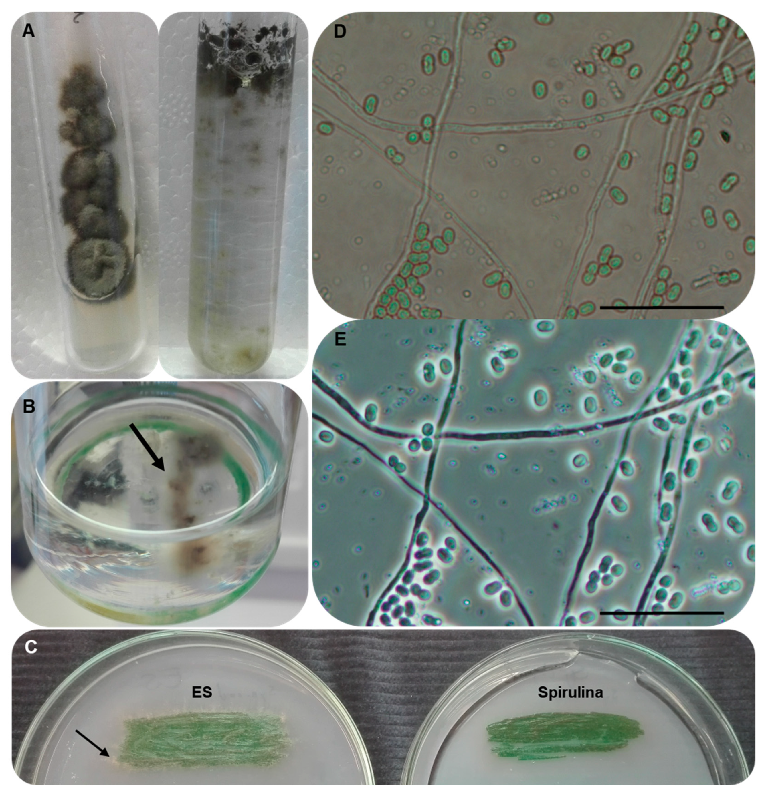
3.2. Isolation of the Unknown Fungal Strain

As was mentioned earlier, the culture collection SAG itself stated that the SAG 88.79 strain has a fungal contaminant. At the very first cultivation of the cyanobacterium strain upon arrival, no fungal strain could be spotted by the naked eye. However, when we transferred 10 µL of liquid drops from the original cyanobacterial culture broth to different media (EMMA and YEA) suitable for fungi, a single greyish mould-like colony appeared. We scraped away some fungal material with a sterile loop from the surface of the colony and streaked it onto Petri dishes containing YEA medium to obtain single colonies. Single colonies were then transferred to fresh PDA media and maintained at room temperature. As the single colonies were identical in their morphology, we presumed that the isolates belonged to the same strain. We gave the name AGSC12 to the new strain (Figure 1A).
After a long cultivation period, we observed that the fungal strain appeared in both the liquid and solid cultures of the cyanobacterial strain, as well (Figure 1B,C). A stock culture of the SAG 88.79 strain has been maintained on Spirulina slant agar in our lab for years, and the fungal strain AGSC12 is still observable among the cyanobacterial cells (Figure 1D,E).

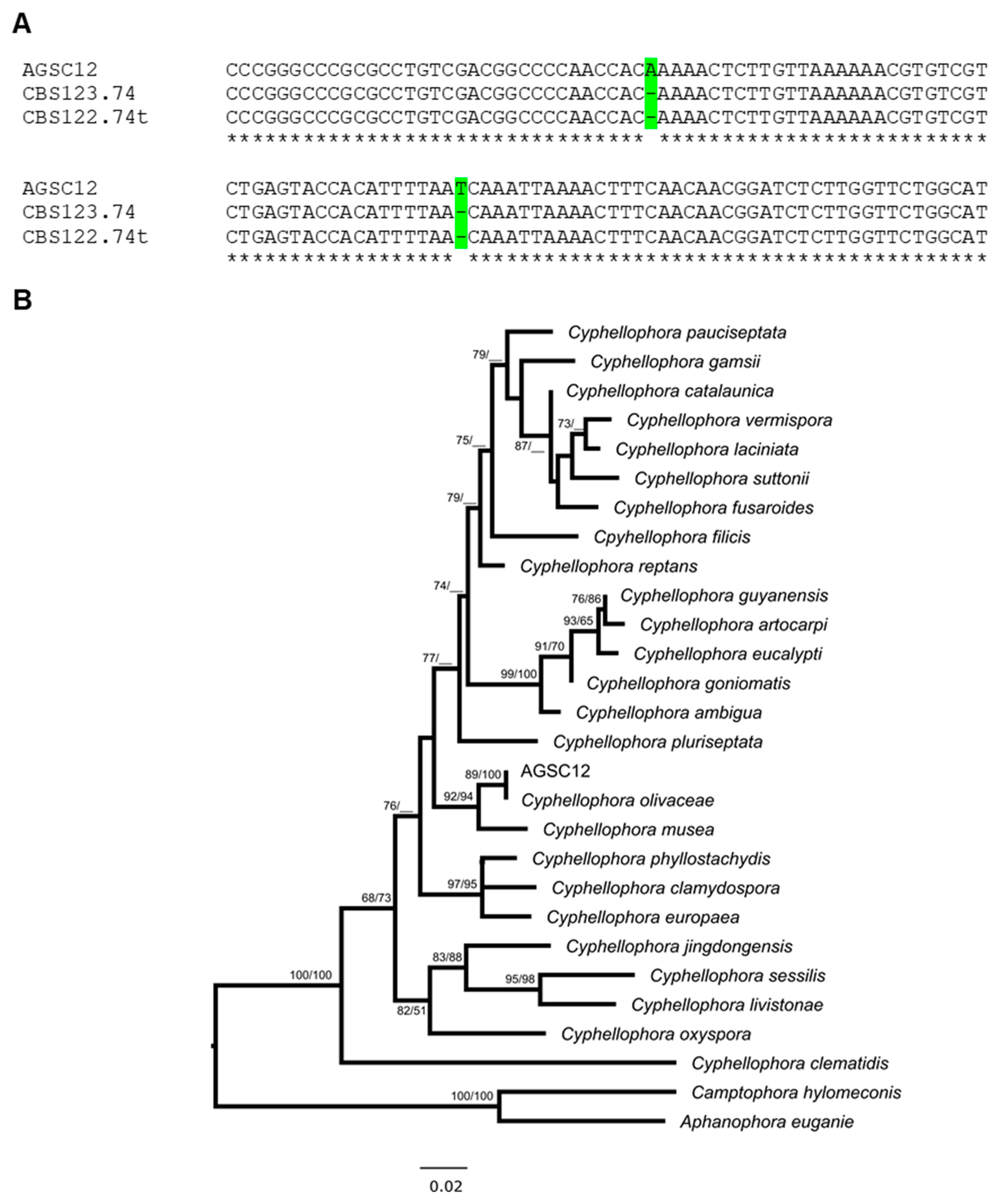
3.3. Molecular Identification of the AGSC12 Strain

For taxonomic identification, the ITS1-5.8S-ITS2 region of the new strain was amplified and sequenced from both directions. After that, a consensus sequence of 612 nucleotides (GenBank accession: MT860449) was subjected to similarity searches at the websites of NCBI and UNITE. We searched for type/reference strains whose sequences were not different from the AGSC12 sequence by more than three substitutions or indels. The Cyphellophora olivacea CBS 122.74 type strain proved to be the most similar to our isolate, as they shared a 99.64% sequence identity. The next best search hit was C. pluriseptata CBS 286.85, with a 91.13% pairwise identity. Accordingly, the inclusion of additional barcode sequences was not necessary. Based on multiple alignments of the isolate AGSC12, C. olivacea CBS 122.74 and CBS 123.74, we assigned our strain to the species Cyphellophora olivacea (Figure 2A).

3.4. Phylogenetic Analyses of the AGSC12 Strain and Its Relatives

For further conviction on the taxonomic position of the strain AGSC12, we searched for all the publicly available Cyphellophora sequences of type strains that were assigned to a species. According to the MycoBank database, there are 35 identified species, which belong to the genus Cyphellophora to date (https://www.mycobank.org/, accessed on 6 October 2022). We managed to find most of the ITS sequences of ex-type strains in that genus. C. indica and C. taiwanensis neither have cultures nor have any sequences available, so we were not able to include them in any phylogenetic analyses [33]. C. eugeniae and C. hylomeconis were transferred to new genera, as Aphanophora eugeniae and Camptophora hylomeconis [34]. According to Crous et al., C. vermispora is identical to C. aestival [59]. Finally, we used the sequences of 27 ex-type strains to build phylogenetic trees (Supplementary Table S1). We created character-based (PhyML) and distance-based (BioNJ) phylogenetic trees using 401 well-aligned sites (broadly 44% of the concerning ITS sequences) (Figure 2B and Supplementary Figure S1). Although the two trees exhibited some minor discrepancies in the topologies, our strain AGSC12 was situated in a common branch with C. olivacea at both of the trees. Thus, we were convinced that AGSC12 was likely a new strain of C. olivacea.

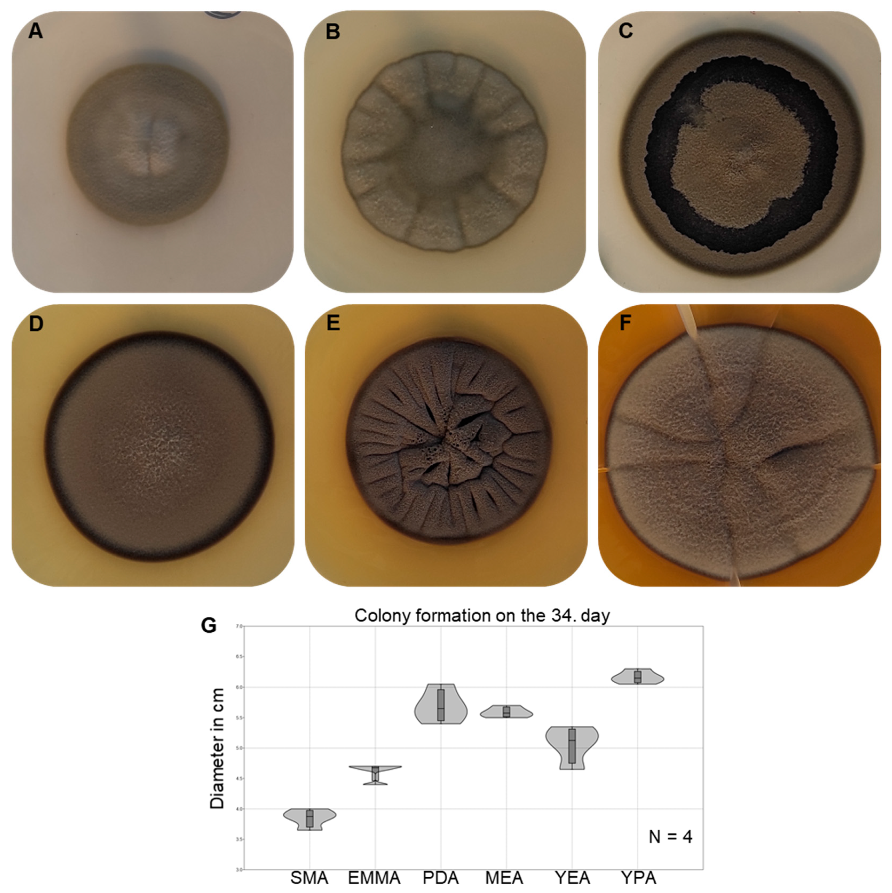
3.5. Morphological Characterisation and Growth Ability of the Strain AGSC12

Species: Cyphellophora olivacea (W. Gams), Réblová & Unter [34].
Basionym. Phialophora olivacea W. Gams [49].
Teleomorph: unknown.
Material examined: fungal contaminant of the cyanobacterial strain SAG 88.79 obtained from the culture collection of Sammlung von Algenkulturen der Universität Göttingen.
Colony morphology and growth capability were examined on SMA, EMMA, PDA, MEA, YEA, and YPA plates (Figure 3A–F). Young colonies are greyish with a pale olivaceous margin. The surface is covered with aerial mycelia. Colonies attain 12–19 mm in diameter after nine days of cultivation at room temperature, depending on the type of media. Mature colonies exhibit the same look as the young colonies but have darker colours and more definitive margins.
Since the strain AGSC12 does not belong to a new species, only a short description is provided here. A thorough description can be seen in [34]. Young vegetative hyphae are smooth, thin, rarely septate, and hyaline, with an approximate width of 1–2 µm. Intercalary branching of the vegetative hyphae is observable. Old hyphae are a bit melanised, and they are sometimes widened and bulky (Supplementary Figure S2A). Phialides are slightly pigmented and are formed terminally, laterally, or intercalary (Supplementary Figure S2B). Conidia are nonseptate at maturity with a shape from clavate to fusiform but mainly ovoidal (Supplementary Figure S2C,D).
The strain AGSC12 can grow in both aerobic and anaerobic conditions in liquid media, but it prefers the aerobic condition. On the plates of complete media, colony expansion proved to be the most successful on YPA (one-way ANOVA test, P = 2.162 × 10⁻¹¹), followed by PDA and MEA (Figure 3G). The SMA minimal medium was the least preferred by the strain (Figure 3G).

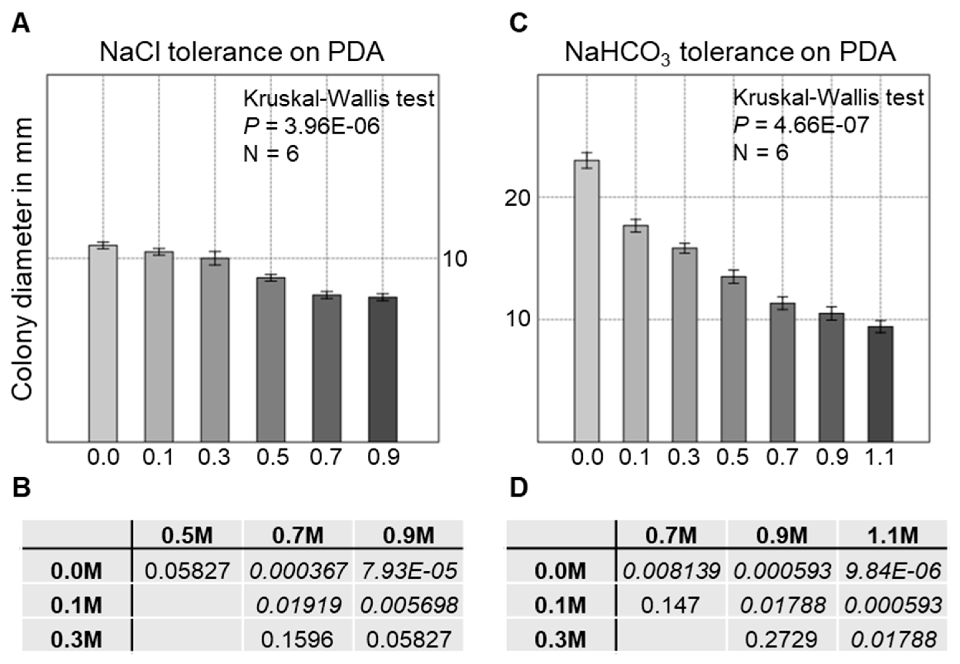
3.6. Salt Tolerance of the Strain AGSC12 Compared to Other Fungal Strains

As the S. cedrorum strain is maintained in Spirulina medium, which contains a notable amount of NaHCO3, we wanted to test the salt tolerance of the fungal companion compared to other fungi. First, we performed a general NaCl tolerance test on PDA plates to exclude potentially sensitive strains. All the used fungal strains were able to grow successfully even at 0.9 M NaCl concentration (Figure 4A and Table 2). F. graminearum responded to the emerging concentration of NaCl with less pigment production (Figure 4A). However, it seemed that the strain AGSC12 showed a weak but definite sensitivity to NaCl as the strain produced significantly smaller colonies (Kruskal-Wallis test, P = 3.96 × 10⁻⁶) (Figure 5A).
In contrast to that, on NaHCO3 version I and version II containing media, colony formation of A. niger and P. expansum was completely inhibited above 0.1 M and 0.2 M concentrations, respectively (Table 2 and Supplementary Figures S3 and S5). Although F. graminearum was able to initiate colony formation even at 0.7 M concentrations, their colonies showed pigmentless and altered phenotypes compared to the untreated controls (Table 2 and Supplementary Figures S3, S5 and S6). The colony morphology seemed normal only at the 0.1 M concentration (Supplementary Figures S3 and S5). Strikingly, C. olivacea AGSC12 was able to produce viable colonies, even at ~1.1 M concentrations, but the colonies were smaller in diameter (Kruskal-Wallis test, P = 4.66 × 10⁻⁷) (Figure 4B and Figure 5B, Table 2 and Supplementary Figures S4–S6). However, the AGSC12 strain did not show any substantial differences in the colony forming ability on the two NaHCO3-containing media but had a darker colour on the version II medium (Figure 4B). According to the stereo microscopic examinations, the aerial hyphae of C. olivacea AGSC12 turned melanised from hyaline and bulky from thin in response to the elevated NaHCO3 concentrations.

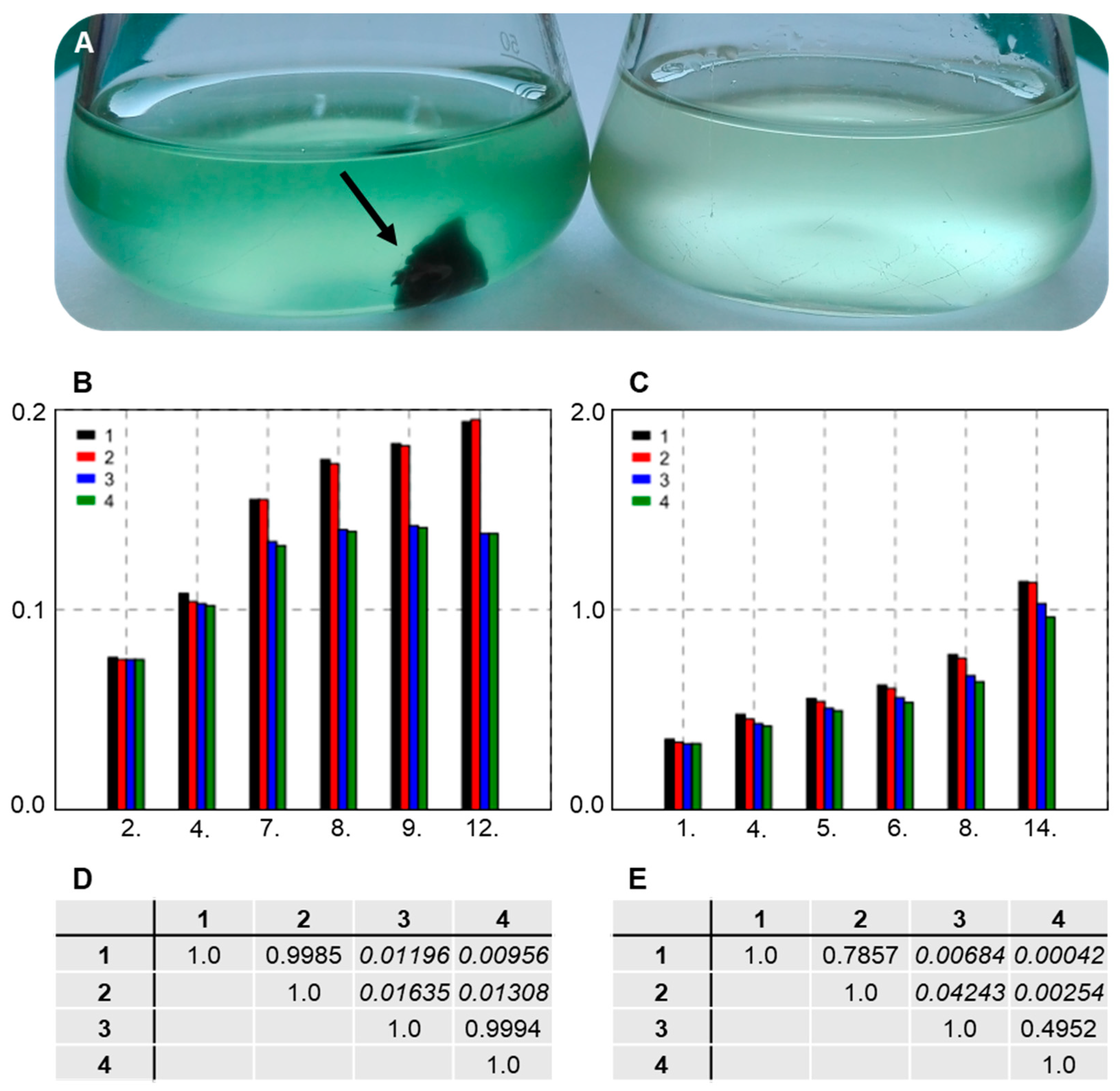
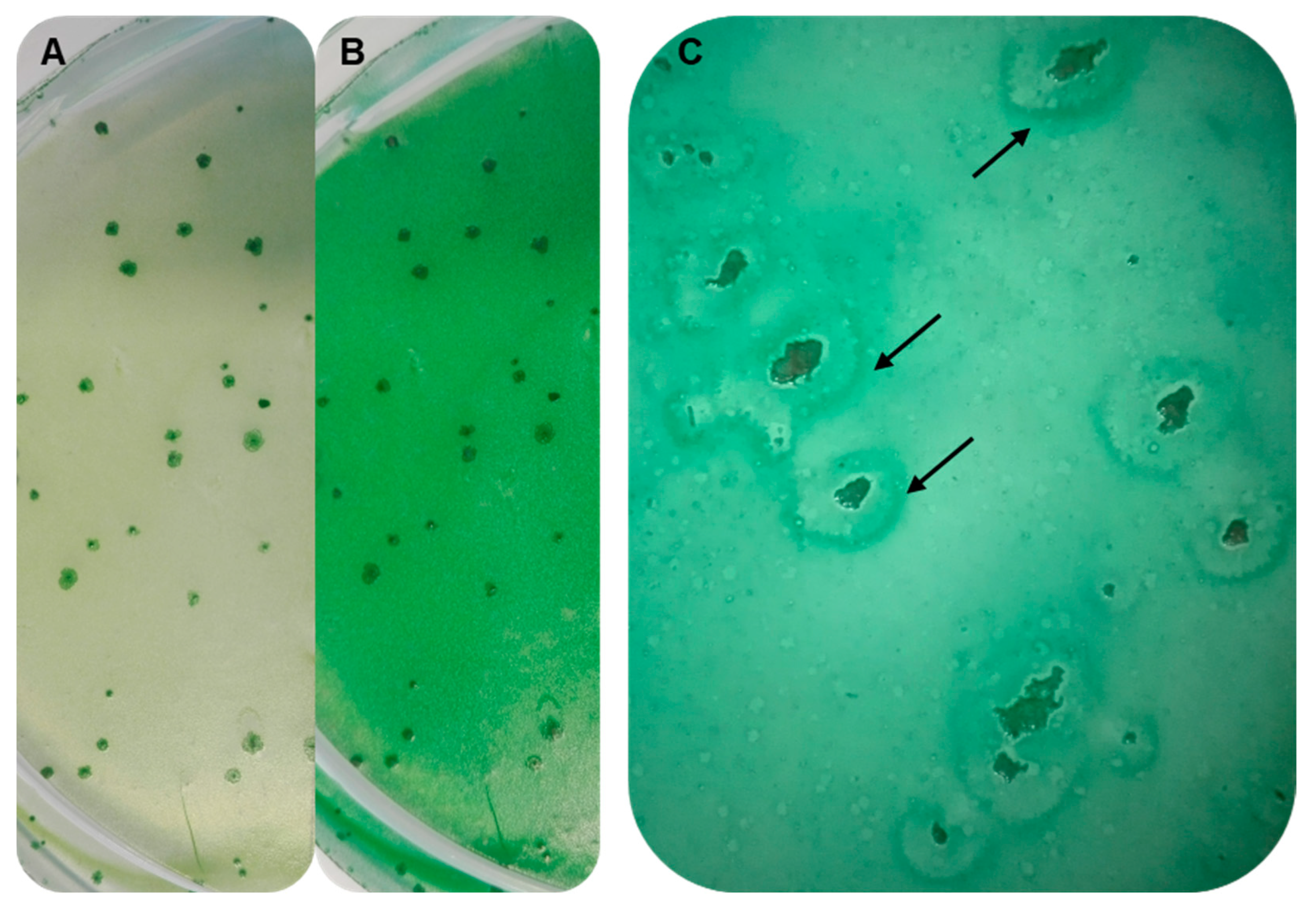
3.7. Establishing Interaction between the Strains SAG 88.79 and AGSC12

Since we had no information about the interactions (if any) between the strains AGSC12 and SAG 88.79, we wanted to examine the possibility of that. Thus, we separated the two strains with serial dilutions. After this, we obtained fungal-free cyanobacterial cultures, and we cultivated them in mono- and co-cultures in Spirulina medium. As the AGSC12 strain was not able to increase its biomass substantially in the Spirulina medium, afterwards we concentrated only on the growth of the SAG 88.79 strain.
We inoculated an equal amount of cyanobacterial cells into liquid Spirulina medium with or without the AGSC12 strain. We monitored the changes in the cell density with an OD measurement (λ = 730 nm). We performed the experiments at two different times with 2–2 samples (initial OD = 0.03 and 0.3). In both experiments, co-cultures gained significantly higher OD per unit time than mono-cultures (repeated-measures ANOVA, P = 0.001427 and P = 0.000232, respectively) (Figure 6A–E).
On the plates of Spirulina medium, we used two different approaches. At one point, we flooded the plates with a mixture of the SAG 88.79 and AGSC12 strains. At another time, we flooded the plates with the fungus-free cyanobacterial strain first, let them dry, then flooded them with fungus-containing sterile water. We observed that the cyanobacterial strain became denser around the fungal strain in both cases (Figure 7A–C).

4. Discussion

Here, we provided a brief report on the fungal contaminant of the S. cedrorum (C. stanieri) SAG 88.79 strain, besides a taxonomic note on the cyanobacterium itself.
The 23S rDNA sequence of the SAG 88.79 strain was almost identical with the C. stanieri PCC 7202 strain. Thus, our results are in accordance with Komárek et al. [2] and with Moro et al. [3] that the strain SAG 88.79 could be a strain of C. stanieri, rather than a strain of S. cedrorum. However, it is important to note that the original S. cedrorum strain described by Sauvageau is not available for comparison to gain more evidence, so the exact taxonomic evaluation of the strain is cumbersome.
The fungal strain AGSC12 isolated from the culture broth of the SAG 88.79 strain belongs to the Cyphellophora olivacea species according to the sequence, phylogenetic, and morphological analyses. The genus Cyphellophora (G.A. de Vries emend. Réblová & Unter) consists of mould-like species from widespread ecological niches. Some of the species can be hazardous to health: for example, the type species of the genus C. laciniata, along with C. europaea and C. pluriseptata were isolated from human skin and nails [41,48,50]. Other species, such as C. guyanensis, C. oxyspora, C. sessilis, C. jingdongensis and C. olivacea were isolated from plant materials or non-biological substrates [43,47,49,60]. The genus is continuously expanding with the introduction of new species and with the occasional transfer from other genera [59,61,62].
Cyphellophora olivacea (W. Gams) and Réblová & Unter (Basionym. Phialophora olivacea W. Gams) ex-type strain CBS 122.74 was originally isolated from wallpaper in Kiel-Kitzeberg (Germany) in 1966 [34,49]. Other strains originated from various substrates, such as stalactites, dead petioles of Chamaerops humilis, surface of Inonotus obliquus, or the surface of treated pine poles and washing machine soap dispensers [49,63,64]. The variety of occurrences of this fungus predicts that the species has a wide tolerance range for environmental factors. Although we have not tested the AGSC12 strain in many different circumstances, we showed that the strain has a striking tolerance for the NaHCO3 on its own. In general, NaHCO3 is considered as a harmless compound, but it could be dangerously harmful to aquatic life (from planktonic organisms to fishes and amphibians) [65]. Besides, this compound has a strong statin effect on most fungi even at low concentrations [14,15,16,17,18,19,20]. For instance, Penicillium digitatum is completely inhibited by 14.1 mmol of NaHCO3, while Botrytis cinerea could be inhibited by 50 mmol [15,20]. Letscher-Bru et al. tested the effect of NaHCO3 on 70 different yeasts, moulds, and dermatophytes, and they found that a concentration of 10 g/L NaHCO3 inhibited the growth of 80% of all the fungal isolates [19]. According to our findings, the isolated strain AGSC12 can colonize even a medium containing 1.1 M (~92.4 g) NaHCO3. Consequently, this strain may be a good candidate in cooperation with certain cyanobacteria and algae for the bioremediation of NaHCO3-containing wastewater (e.g., natural coalbed water and used thermal water).
Although the species inhabits various niches, to our best knowledge, this is the first report of a strain co-existing with cyanobacteria. However, it is unclear whether SAG 88.79 was already accompanied by the fungal strain at its isolation in 1963 or if its culture became contaminated by the fungus afterwards, during its maintenance in the culture collection. According to Göttlich et al., the fungal flora of groundwater-derived public drinking water in certain regions of Germany was dominated by Phialophora species [66]. We should bear in mind that the genus Cyphellophora was proposed first in 2013 by Réblová et al. and that C. olivacea was considered as Phialophora olivacea earlier [34]. Thus, there is a chance that the AGSC12 strain appeared in the culture broth of the SAG 88.79 strain during its maintenance. Nevertheless, it seemed that the presence of the fungal strain was beneficial to the cyanobacterium, as we observed that the SAG 88.79 strain always attained a higher cell density in co-cultures with the fungus than in mono-cultures.
To reveal the exact mode of interaction between the strains is outside the scope of the current study, but we are planning to further investigate this phenomenon. There is emerging knowledge to prove that “artificial” symbiosis can be beneficial to the participant species even if the strains do not have common evolutionary history [67,68].

Supplementary Materials

The following supporting information can be downloaded at: https://www.mdpi.com/article/10.3390/d14121023/s1, Figure S1: Comparison of different phylogenetic tress; Figure S2: Microscopic examination of the fungal strain AGSC12; Figure S3: NaHCO3 (II) tolerance of the comparator species; Figure S4: NaHCO3 (II) tolerance of the AGSC12 strain; Figure S5: NaHCO3 (I) tolerance of the examined fungal species; Figure S6: NaHCO3 (I) tolerance of the FGSC 9075 and AGSC12 strains; Table S1: List of species used for the phylogeny.

Author Contributions

Conceptualization: L.A.-S. and M.S.; Methodology: L.A.-S., H.C. and M.S.; Validation: L.A.-S.; Formal analysis: L.A.-S. and M.S.; Investigation: L.A.-S. and L.A.P.; Resources: I.M. and M.S.; Writing—original draft preparation: L.A.-S.; Writing—review and editing: L.A.-S., L.A.P., H.C., I.M. and M.S.; Visualization: L.A.-S.; Supervision. M.S.; Funding acquisition: M.S. and I.M. All authors have read and agreed to the published version of the manuscript.

Funding

This research was founded by grant K-124417 provided by the National Research, Development and Innovation Office of Hungary and GINOP-2.2.1-15-2016-00029, co-financed by the European Union and the State of Hungary. The study received further funding from the project 2019-1.1.1-PIACI-KFI-2019-00228. Lajos Acs-Szabo received a scholarship from the EFOP-3.6.1-16-2016-00022 “Debrecen Venture Catapult Program”, provided by the European Union and the European Social Fund. Laszlo Attila Papp was supported by FIK 20428-3/2018 and the Thematic Excellence Programme (TKP2020-IKA-04) of the Ministry for Innovation and Technology in Hungary projects.

Institutional Review Board Statement

Not applicable.

Data Availability Statement

All data generated or analysed during this study are included in this published article (and its Supplementary files). The sequences generated during the current study are available in the GenBank repository with the following accession numbers: MW363512 and MT860449.

Acknowledgments

The authors thank Anita Kovacs-Bordan and Ilona Lakatos for skilful technical assistance.

Conflicts of Interest

The authors declare no conflict of interest.

References

  1. Rippka, R.; Cohen-Bazire, G. The Cyanobacteriales: A legitimate order based on the type strain Cyanobacterium stanieri? Ann. Microbiol. 1983, 134, 21–36. [Google Scholar] [CrossRef] [PubMed]
  2. Komárek, I.; Cepák, V.; Kaštovský, J.; Sulek, J. What are the cyanobacterial genera Cyanothece and Cyanobacterium? Contribution to the combined molecular and phenotype taxonomic evaluation of cyanobacterial diversity. Algol. Stud. 2004, 113, 1–36. [Google Scholar] [CrossRef]
  3. Moro, I.; Rascio, N.; La Rocca, N.; Di Bella, M.; Andreoli, C. Cyanobacterium aponinum, a new Cyanoprokaryote from the microbial mat of Euganean Thermal Springs (Padua, Italy). Algol. Stud. 2007, 123, 1–15. [Google Scholar] [CrossRef]
  4. Komárek, J.; Kaštovský, J.; Mareš, J.; Johansen, J.R. Taxonomic classification of cyanoprokaryotes (cyanobacterial genera) 2014, using a polyphasic approach. Preslia 2014, 86, 295–335. [Google Scholar]
  5. Sauvageau, C. Sur les algues d’eau douce recoltées en Algérie pendant le session de la Societé Botanique en 1892. Bulletin de la Société Botanique de France 1892, 39, civ–cxxviii. [Google Scholar] [CrossRef]
  6. NCBI Taxonomy Browser: Cyanobacterium stanieri PCC 7202. Available online: https://www.ncbi.nlm.nih.gov/Taxonomy/Browser/wwwtax.cgi?mode=Info&id=292563&lvl=3&lin=f&keep=1&srchmode=1&unlock (accessed on 15 October 2022).
  7. Komárek, J. Taxonomic review of the genera Synechocystis SAUV, 1892, Synechococcus NÄG, 1849 and Cyanothece gen. nov. (Cyanophyceae). Arch. Für Protistenkd. 1976, 118, 119–179. [Google Scholar]
  8. Komárek, J.; Kopecký, J.; Cepák, V. Generic characters of the simplest cyanoprokaryotes Cyanobium, Cyanobacterium and Synechococcus. Cryptogam. Algol. 1999, 20, 209–222. [Google Scholar] [CrossRef]
  9. Honegger, R. Functional aspects of the lichen symbiosis. Annu. Rev. Plant Physiol. Plant Mol. Biol. 1991, 42, 553–578. [Google Scholar] [CrossRef]
  10. Singh, G.; Dal Grande, F.; Divakar, P.K.; Otte, J.; Crespo, A.; Schmitt, I. Fungal-algal association patterns in lichen symbiosis linked to macroclimate. New Phytol. 2017, 214, 317–329. [Google Scholar] [CrossRef]
  11. Grimm, M.; Grube, M.; Schiefelbein, U.; Zühlke, D.; Bernhardt, J.; Riedel, K. The Lichens’ Microbiota, Still a Mystery? Front. Microbiol. 2021, 12, 623839. [Google Scholar] [CrossRef]
  12. Gimmler, H. Mutualistic Relationships Between Algae and Fungi (Excluding Lichens). In Progress in Botany; Esser, K., Lüttge, U., Kadereit, J.W., Beyschlag, W., Eds.; Springer: Berlin/Heidelberg, Germany, 2001; Volume 62, pp. 194–214. [Google Scholar] [CrossRef]
  13. Calcott, M.J.; Ackerley, D.F.; Knight, A.; Keyzers, R.A.; Owen, J.G. Secondary metabolism in the lichen symbiosis. Chem. Soc. Rev. 2018, 47, 1730–1760. [Google Scholar] [CrossRef]
  14. Corral, L.G.; Post, L.S.; Montville, T.J. Antimicrobial activity of sodium bicarbonate. A research note. J. Food Sci. 1988, 53, 981–982. [Google Scholar] [CrossRef]
  15. Smilanick, J.L.; Margosan, D.A.; Mlikota, F.; Usall, J.; Michael, I.F. Control of Citrus Green Mold by Carbonate and Bicarbonate Salts and the Influence of Commercial Postharvest Practices on Their Efficacy. Plant Dis. 1999, 83, 139–145. [Google Scholar] [CrossRef] [PubMed] [Green Version]
  16. Droby, S.; Wisniewski, M.; Ghaouth, A.E.; Wilson, C. Influence of food additives on the control of postharvest rots of apple and peach and efficacy of the yeast-based biocontrol product aspire. Postharvest Biol. Technol. 2003, 27, 127–135. [Google Scholar] [CrossRef]
  17. Gamagae, S.U.; Sivakumar, D.; Wijeratnam, R.S.W.; Wijesundera, R.L.C. Use of sodium bicarbonate and Candida oleophila to control anthracnose in papaya during storage. Crop Prot. 2003, 22, 775–779. [Google Scholar] [CrossRef]
  18. Zamani, M.; Sharifi Tehrani, A.; Ali Abadi, A.A. Evaluation of antifungal activity of carbonate and bicarbonate salts alone or in combination with biocontrol agents in control of citrus green mold. Commun. Agric. Appl. Biol. Sci. 2007, 72, 773–777. [Google Scholar] [PubMed]
  19. Letscher-Bru, V.; Obszynski, C.M.; Samsoen, M.; Sabou, M.; Waller, J.; Candolfi, E. Antifungal activity of sodium bicarbonate against fungal agents causing superficial infections. Mycopathologia 2013, 175, 153–158. [Google Scholar] [CrossRef] [PubMed]
  20. Türkkan, M.; Özcan, M.; Erper, I. Antifungal effect of carbonate and bicarbonate salts against Botrytis cinerea, the casual agent of grey mould of kiwifruit. Akademik Ziraat Dergisi 2017, 6, 107–114. [Google Scholar] [CrossRef] [Green Version]
  21. Aiba, S.; Ogawa, T. Assessment of growth yield of a blue-green alga: Spirulina platensis, in axenic and continuous culture. J. Gen. Microbiol. 1977, 102, 179–182. [Google Scholar] [CrossRef] [Green Version]
  22. Sipiczki, M.; Ferenczy, L. Protoplast fusion of Schizosaccharomyces pombe auxotrophic mutants of identical mating-type. Mol. Gen. Genet. 1977, 151, 77–81. [Google Scholar] [CrossRef]
  23. Mitchison, J.M. Physiological and cytological methods for Schizosaccharomyces pombe. Methods Cell Biol. 1970, 4, 131–165. [Google Scholar] [CrossRef]
  24. Beever, R.E.; Bollard, E.G. The Nature of the Stimulation of Fungal Growth by Potato Extract. J. Gen. Microbiol. 1970, 60, 273–279. [Google Scholar] [CrossRef] [Green Version]
  25. Thom, C.; Church, M.B. The Aspergilli; Williams & Wilkins, Co.: Baltimore, MD, USA, 1926; p. 272. [Google Scholar]
  26. Sipiczki, M.; Takeo, K.; Grallert, A. Growth polarity transitions in a dimorphic fission yeast. Microbiology 1998, 144, 3475–3485. [Google Scholar] [CrossRef] [PubMed] [Green Version]
  27. Balazs, A.; Batta, G.; Miklos, I.; Acs-Szabo, L.; Vazquez de Aldana, C.R.; Sipiczki, M. Conserved regulators of the cell separation process in Schizosaccharomyces. Fungal Genet. Biol. 2012, 49, 235–249. [Google Scholar] [CrossRef] [PubMed] [Green Version]
  28. Hoffman, C.S.; Winston, F. A ten-minute DNA preparation from yeast efficiently releases autonomous plasmids for transformation of Escherichia coli. Gene 1987, 57, 267–272. [Google Scholar] [CrossRef] [PubMed]
  29. del Campo, E.M.; del Hoyo, A.; Royo, C.; Casano, L.M.; Alvarez, R.; Barreno, E. A single primer pair gives a specific ortholog amplicon in a wide range of Cyanobacteria and plastid-bearing organisms: Applicability in inventory of reference material from collections and phylogenetic analysis. Mol. Phylogenet. Evol. 2010, 57, 1323–1328. [Google Scholar] [CrossRef] [PubMed]
  30. White, T.J.; Bruns, T.; Lee, S.; Taylor, J. Amplification and direct sequencing of fungal ribosomal RNA genes for phylogenetics. PCR Protoc. A Guide Methods Appl. 1990, 18, 315–322. [Google Scholar] [CrossRef]
  31. Sambrook, J.; Russell, D.W. Standard ethanol precipitation of DNA in microcentrifuge tubes. CSH. Protoc. 2006, 2006, pdb.prot4456. [Google Scholar] [CrossRef]
  32. Nilsson, R.H.; Larsson, K.H.; Taylor, A.; Bengtsson-Palme, J.; Jeppesen, T.S.; Schigel, D.; Kennedy, P.; Picard, K.; Glöckner, F.O.; Tedersoo, L.; et al. The UNITE database for molecular identification of fungi: Handling dark taxa and parallel taxonomic classifications. Nucleic Acids Res. 2019, 47, D259–D264. [Google Scholar] [CrossRef]
  33. Feng, P.; Lu, Q.; Najafzadeh, M.J.; Ende, A.H.; Sun, J.; Li, R.; Xi, L.; Vicente, V.; Lai, W.; Lu, C.; et al. Cyphellophora and its relatives in Phialophora: Biodiversity and possible role in human infection. Fungal Divers. 2012, 65, 17–45. [Google Scholar] [CrossRef] [Green Version]
  34. Réblová, M.; Untereiner, W.A.; Réblová, K. Novel evolutionary lineages revealed in the Chaetothyriales (Fungi) based on multigene phylogenetic analyses and comparison of ITS secondary structure. PLoS ONE 2013, 8, e63547. [Google Scholar] [CrossRef] [PubMed]
  35. Gao, L.; Ma, Y.; Zhao, W.; Wei, Z.; Gleason, M.L.; Chen, H.; Hao, L.; Sun, G.; Zhang, R. Three New Species of Cyphellophora (Chaetothyriales) Associated with Sooty Blotch and Flyspeck. PLoS ONE 2015, 10, e0136857. [Google Scholar] [CrossRef] [PubMed]
  36. Crous, P.W.; Wingfield, M.J.; Guarro, J.; Cheewangkoon, R.; van der Bank, M.; Swart, W.J.; Stchigel, A.M.; Cano-Lira, J.F.; Roux, J.; Madrid, H.; et al. Fungal Planet description sheets: 154213. Persoonia 2013, 31, 188–296. [Google Scholar] [CrossRef] [PubMed]
  37. Madrid, H.; Hernández-Restrepo, M.; Gené, J.; Cano, J.; Guarro, J.; Silva, V. New and interesting chaetothyrialean fungi from Spain. Mycol. Prog. 2016, 15, 1179–1201. [Google Scholar] [CrossRef] [Green Version]
  38. Crous, P.W.; Schumacher, R.K.; Akulov, A.; Thangavel, R.; Hernández-Restrepo, M.; Carnegie, A.; Cheewangkoon, R.; Wingfield, M.; Summerell, B.; Quaedvlieg, W.; et al. New and Interesting Fungi. 2. Fungal Syst. Evol. 2019, 3, 57–134. [Google Scholar] [CrossRef]
  39. Cheewangkoon, R.; Groenewald, J.Z.; Summerell, B.A.; Hyde, K.D.; To-Anun, C.; Crous, P.W. Myrtaceae, a cache of fungal biodiversity. Persoonia 2009, 23, 55–85. [Google Scholar] [CrossRef] [Green Version]
  40. Crous, P.W.; Braun, U.; Wingfield, M.J.; Wood, A.R.; Shin, H.D.; Summerell, B.A.; Alfenas, A.C.; Cumagun, C.J.; Groenewald, J.Z. Phylogeny and taxonomy of obscure genera of microfungi. Persoonia 2009, 22, 139–161. [Google Scholar] [CrossRef] [PubMed] [Green Version]
  41. de Hoog, G.S.; Mayser, P.; Haase, G.; Horre, R.; Horrevorts, A.M. A new species, Phialophora europaea, causing superficial infections in humans. Mycoses 2000, 43, 409–416. [Google Scholar] [CrossRef]
  42. Phookamsak, R.; Hyde, K.; Jeewon, R.; Bhat, D.J.; Jones, E.; Maharachchikumbura, S.; Raspé, O.; Karunarathna, S.; Wanasinghe, D.N.; Hongsanan, S.; et al. Fungal diversity notes 929–1035: Taxonomic and phylogenetic contributions on genera and species of fungi. Fungal Divers. 2019, 95, 1–273. [Google Scholar] [CrossRef] [Green Version]
  43. Decock, C.; Delgado-Rodríguez, G.; Buchet, S.; Seng, J.M. A new species and three new combinations in Cyphellophora, with a note on the taxonomic affinities of the genus, and its relation to Kumbhamaya and Pseudomicrodochium. Antonie van Leeuwenhoek 2003, 84, 209–216. [Google Scholar] [CrossRef]
  44. Crous, P.W.; Wingfield, M.J.; Richardson, D.M.; Le Roux, J.J.; Strasberg, D.; Edwards, J.; Roets, F.; Hubka, V.; Taylor, P.W.; Heykoop, M.; et al. Fungal Planet description sheets: 400–468. Persoonia 2016, 36, 316–458. [Google Scholar] [CrossRef] [PubMed]
  45. Crous, P.W.; Wingfield, M.; Lombard, L.; Roets, F.; Swart, W.; Alvarado, P.; Carnegie, A.; Moreno, G.; Luangsa-Ard, J.; Thangavel, R.; et al. Fungal Planet description sheets: 951–1041. Pers.—Mol. Ph. Evol. Fungi 2019, 43, 223–425. [Google Scholar] [CrossRef] [PubMed]
  46. Crous, P.W.; Schubert, K.; Braun, U.; de Hoog, G.S.; Hocking, A.D.; Shin, H.D.; Groenewald, J.Z. Opportunistic, human-pathogenic species in the Herpotrichiellaceae are phenotypically similar to saprobic or phytopathogenic species in the Venturiaceae. Stud. Mycol. 2007, 58, 185–217. [Google Scholar] [CrossRef] [PubMed]
  47. Yang, H.; Hyde, K.; Karunarathna, S.; Deng, C.; Gu, C.; Yang, S.; Zhang, Z. New species of Camptophora and Cyphellophora from China, and first report of sexual morphs for these genera. Phytotaxa 2018, 343, 149–159. [Google Scholar] [CrossRef]
  48. De Vries, G.A. Cyphellophora laciniata nov. gen., nov. sp. and Dactylium fusarioides Fragoso et Ciferri. Mycopathol. Mycol. Appl. 1962, 16, 47–54. [Google Scholar] [CrossRef]
  49. Gams, W.; Holubová-Jechová, V. Chloridium and some other dematiaceous hyphomycetes growing on decaying wood. Stud. Mycol. 1976, 13, 99. [Google Scholar]
  50. de Vries, G.A.; Elders, M.C.; Luykx, M.H. Description of Cyphellophora pluriseptata sp. nov. Antonie van Leeuwenhoek 1986, 52, 141–143. [Google Scholar] [CrossRef]
  51. Matsushima, T. Matsushima Mycological Memoirs; Matsushima Fungus Collection: Kobe, Japan, 1987; Volume 5, pp. 1–100. [Google Scholar]
  52. Walz, A.; de Hoog, G.S. A new species of Cyphellophora. Antonie van Leeuwenhoek 1987, 53, 143–146. [Google Scholar] [CrossRef]
  53. Dereeper, A.; Guignon, V.; Blanc, G.; Audic, S.; Buffet, S.; Chevenet, F.; Dufayard, J.F.; Guindon, S.; Lefort, V.; Lescot, M.; et al. Phylogeny.fr: Robust phylogenetic analysis for the non-specialist. Nucleic Acids Res. 2008, 36, W465–W469. [Google Scholar] [CrossRef]
  54. Edgar, R.C. MUSCLE: Multiple sequence alignment with high accuracy and high throughput. Nucleic Acids Res. 2004, 32, 1792–1797. [Google Scholar] [CrossRef] [Green Version]
  55. Castresana, J. Selection of conserved blocks from multiple alignments for their use in phylogenetic analysis. Mol. Biol. Evol. 2000, 17, 540–552. [Google Scholar] [CrossRef] [PubMed] [Green Version]
  56. Gascuel, O. BIONJ: An Improved Version of the NJ Algorithm Based on a Simple Model of Sequence Data. Mol. Biol. Evol. 1997, 14, 685–695. [Google Scholar] [CrossRef] [PubMed]
  57. Guindon, S.; Gascuel, O. A simple, fast, and accurate algorithm to estimate large phylogenies by maximum likelihood. Syst. Biol. 2003, 52, 696–704. [Google Scholar] [CrossRef] [PubMed] [Green Version]
  58. Hammer, Ø.; Harper, D.A.T.; Ryan, P.D. PAST: Paleontological statistics software package for education and data analysis. Palaeontol. Electron. 2001, 4, 9. [Google Scholar]
  59. Crous, P.W.; Braun, U.; McDonald, B.A.; Lennox, C.L.; Edwards, J.; Mann, R.C.; Zaveri, A.; Linde, C.C.; Dyer, P.S.; Groenewald, J.Z. Redefining genera of cereal pathogens: Oculimacula, Rhynchosporium and Spermospora. Fungal Syst. Evol. 2021, 7, 67–98. [Google Scholar] [CrossRef] [PubMed]
  60. de Hoog, G.S.; Weenink, X.O.; Ende, A.H. Taxonomy of the Phialophora verrucosa complex with the description of two new species. Stud. Mycol. 1999, 43, 107–121. [Google Scholar]
  61. Crous, P.W.; Cowan, D.A.; Maggs-Kölling, G.; Yilmaz, N.; Larsson, E.; Angelini, C.; Brandrud, T.E.; Dearnaley, J.D.W.; Dima, B.; Dovana, F.; et al. Fungal Planet description sheets: 1112–1181. Persoonia 2020, 45, 251–409. [Google Scholar] [CrossRef]
  62. Crous, P.W.; Boers, J.; Holdom, D.G.; Osieck, E.R.; Steinrucken, T.V.; Tan, Y.P.; Vitelli, J.S.; Shivas, R.G.; Barrett, M.; Boxshall, A.G.; et al. Fungal planet description sheets: 1383–1435. Pers.—Mol. Phylogeny Evol. Fungi 2022, 4, 261–371. [Google Scholar] [CrossRef]
  63. Zabel, R.A.; Lombard, F.F.; Wang, C.J.K.; Terracina, F. Fungi associated with decay in treated southern pine utility poles in the eastern United States. Wood Fiber Sci. 1985, 17, 75–91. [Google Scholar]
  64. Isola, D.; Selbmann, L.; de Hoog, G.S.; Fenice, M.; Onofri, S.; Prenafeta-Boldú, F.X.; Zucconi, L. Isolation and screening of black fungi as degraders of volatile aromatic hydrocarbons. Mycopathologia 2013, 175, 369–379. [Google Scholar] [CrossRef]
  65. Harper, D.D.; Farag, A.M.; Skaar, D. Acute toxicity of sodium bicarbonate, a major component of coal bed natural gas produced waters, to 13 aquatic species as defined in the laboratory. Environ. Toxicol. Chem. 2014, 33, 525–531. [Google Scholar] [CrossRef] [PubMed]
  66. Göttlich, E.; Van Der Lubbe, W.; Lange, B.; Fiedler, S.; Melchert, I.; Reifenrath, M.; Flemming, H.C.; De Hoog, S. Fungal flora in groundwater-derived public drinking water. Int. J. Hyg. Environ. Health 2002, 205, 269–279. [Google Scholar] [CrossRef] [PubMed]
  67. Du, Z.Y.; Zienkiewicz, K.; Vande Pol, N.; Ostrom, N.E.; Benning, C.; Bonito, G.M. Algal-fungal symbiosis leads to photosynthetic mycelium. eLife 2019, 8, e47815. [Google Scholar] [CrossRef] [PubMed]
  68. Jiang, L.; Li, T.; Jenkins, J.; Hu, Y.; Brueck, C.L.; Pei, H.; Betenbaugh, M.J. Evidence for a mutualistic relationship between the cyanobacteria Nostoc and fungi Aspergilli in different environments. Appl. Microbiol. Biotechnol. 2020, 104, 6413–6426. [Google Scholar] [CrossRef]
Figure 1. The fungal strain AGSC12 from the culture broth of the cyanobacterium SAG 88.79. (A) The isolated strain AGSC12 on EMMA slant agar and in EMML liquid medium. (B) The presence of the AGSC12 strain in SAG 88.79 culture broth after photobleaching the cyanobacterial cells in Spirulina medium. (C) Fungal hyphae among the cyanobacterial colonies and at the edges of the colonies on ES and Spirulina media containing plates. On ES medium, the fungal growth was stronger, and on Spirulina medium, the fungal presence was indicated by the brownish tone on the top of the cyanobacterial colony. (D,E) Microscopic images of the fungal hyphae among the cyanobacterial cells. (D): Nomarski microscopy. (E): phase-contrast microscopy. Scale bars = 20 µm.
Diversity 14 01023 g001
Figure 2. (A) Multiple sequence alignment of the ITS1-5.8SrDNA-ITS2 regions of the strain AGSC12 and C. olivacea strains CBS122.74 (ex-type strain) and CBS123.74. Only those regions are depicted from the alignment, which exhibited discrepancies (highlighted with green). Otherwise, the alignments were identical. (B) Phylogenetic analyses of the strain AGSC12 and other Cyphellophora species. The numbers at the branches indicate aLRT of Maximum Likelihood (ML) analysis, the other values come from bootstrapping of neighbour joining (NE) analysis (aLRT/bootstrap). Only aLRT and bootstrap values larger than 50 are shown. The current tree layout represents the ML analysis and comparison of ML. NJ can be seen in Supplementary Figure S1. Although some minor differences can be seen at the branching points, the strain AGSC12 clustered to the same branch with the C. olivacea ex-type strain in both the ML and NJ analyses.
Diversity 14 01023 g002
Figure 3. Morphology and growth capability of the C. olivacea strain AGSC12. (AF) Colony formation of SMA, EMMA, PDA, MEA, YEA, and YPA plates in the order shown. Photos were taken after 34 days of cultivation. (G) Distribution of the measured diameters of the fungal colonies growing on different media at the 34th day. Violin plots show the kernel density for each sample. The box plots in the violin plots indicate the 25–75 percent quartiles. Horizontal lines within the boxes show the medians of the samples. Minimal and maximal values are depicted by the whiskers. N = sample size. Colony expansion was the most efficient on YPA plates (one-way ANOVA test, P = 2.162 × 10⁻¹¹).
Diversity 14 01023 g003
Figure 4. Colony forming ability of the examined fungal strains on NaCl- and on NaHCO3-containing PDA plates. (A) Tolerance to NaCl. Green mold: Penicillium expansum SZMC 2175; blackish mold: Aspergillus niger CBS 554.65; orange-yellowish mold: Fusarium graminearum FGSC 9075; small grey colonies: Cyphellophora olivacea AGSC12. The NaCl concentrations are the following in the top line from left to right: 0.0 M (control), 0.1 M, 0.3 M and in the bottom line from left to right: 0.5 M, 0.7 M, 0.9 M. All the tested strains were able to produce colonies at the different concentrations. Photos were taken after 7 days of cultivation. (B) The strain C. olivacea AGSC12 tolerance to NaHCO3 I and II contained PDA plates (see methods). This strain showed a striking tolerance to NaHCO3. Photos were taken after 10 days of cultivation. The diameters of the colonies are not to scale; these photos concentrate on the colony morphologies.
Diversity 14 01023 g004
Figure 5. Colony formation of the AGSC12 strain on NaCl- and NaHCO3-containing PDA plates. (A) Colony diameters after 7 days of cultivation at different NaCl concentrations. There were significant differences among the values (Kruskal-Wallis test, P = 3.96 × 10⁻⁶). (B) The data indicate those concentrations when the measured colony diameters showed significant discrepancies according to the Dunn’s post hoc test. The values are the relevant P values. Only those cases are shown where the P values were less than 0.5. Significant values are in italics. (C) Colony diameters at various NaHCO3 (II) concentrations after 14 days of cultivation. There were significant differences among the values (Kruskal-Wallis test, P = 4.66 × 10⁻⁷). (D) Same as (B) for the NaHCO3 concentrations. Whiskers on the bar charts represent standard deviations. N stands for sample size.
Diversity 14 01023 g005
Figure 6. Cultivation of the cyanobacterial strain SAG 88.79 with or without the fungal strain AGSC12. (A) Co-cultures (left) and mono-cultures (right) of the SAG 88.79 strain in liquid Spirulina medium. A black arrow shows the hyphal batch in the medium. (B,C) Bar charts indicate the differences between the co- (sample 1 and 2) and mono-cultures (sample 3 and 4). The y-axles depict the OD values (λ = 730 nm) and the x-axles show the days of sampling. The differences were significant in both cases (repeated-measures ANOVA, P = 0.001427 and P = 0.000232, respectively). (D,E) Tukey’s pairwise post-hoc test demonstrated that all the co-cultures exhibited significantly higher cyanobacterial growth than the mono-cultures did. As in (B) and (C), samples 1 and 2 are for the co-cultures, and samples 3 and 4 are for the mono-cultures. The values in the tables are the corresponding pairwise P-values. Italics show the significant values. The cyanobacterial strain SAG 88.79 attained higher cell density in the presence of the fungal strain AGSC12.
Diversity 14 01023 g006
Figure 7. Cultivation of the cyanobacterial strain SAG 88.79 with or without the fungal strain AGSC12 on agar plates. (A,B) Spirulina plate flooded by the mixture of SAG 88.79 and AGSC12 cells. The cyanobacterial cells became denser near the fungi within a few days of cultivation (A) and retained the advantage over a long cultivation period, too (B). (C) Sequential flooding of the cells. The SAG 88.79 cell-mass was thicker around the fungal colonies as the black arrows indicate. The cyanobacterial strain SAG 88.79 attained higher cell density in the presence of the fungal strain AGSC12 on plates, as well.
Diversity 14 01023 g007
Table 1. List of strains used in this study.
SpeciesStrainOriginMaintaining Medium
Cyanobacterium staniereiSAG 88.79SAGSpirulina
Aspergillus nigerCBS 554.65CBS-KNAWPDA
Penicillium expansumSZMC 2175SZMCPDA
Fusarium graminearumFGSC 9075ATCCPDA
unkown fungusAGSC12This studyPDA
Table 2. Evaluation of the tolerance of the fungal strains to different concentrations of salts (see methods).
SpeciesNaCl Tolerance on PDA
0.0 M0.1 M0.3 M0.5 M0.7 M0.9 M
A. niger333333
P. expansum333333
F. graminearum333333
C. olivacea333322
NaHCO3 I. Tolerance on PDA
0.0 M0.1 M0.3 M0.5 M0.7 M0.9 M1.1 M
A. niger300000ND
P. expansum320000ND
F. graminearum3311100
C. olivacea3333222
NaHCO3 II. Tolerance on PDA
0.0 M0.1 M0.3 M0.5 M0.7 M0.9 M1.1 M
A. niger300000ND
P. expansum320000ND
F. graminearum331110ND
C. olivacea3333222
Publisher’s Note: MDPI stays neutral with regard to jurisdictional claims in published maps and institutional affiliations.

