Review

Detection of Fish Pathogens in Freshwater Aquaculture Using eDNA Methods

1
Department of Aquaculture and Fisheries, University of Arkansas at Pine Bluff, Pine Bluff, AR 71601, USA
2
Department of Continuing Education, College of Menominee Nation, Keshena, WI 54135, USA
3
Department of Plant Science, University of Idaho, Moscow, ID 83844, USA
*
Author to whom correspondence should be addressed.
Diversity 2022, 14(12), 1015; https://doi.org/10.3390/d14121015
Submission received: 21 October 2022 / Revised: 17 November 2022 / Accepted: 18 November 2022 / Published: 22 November 2022
(This article belongs to the Special Issue eDNA for Basic and Applied Sciences)

Abstract

:
Organisms release their nucleic acid in the environment, including the DNA and RNA, which can be used to detect their presence. eDNA/eRNA techniques are being used in different sectors to identify organisms from soil, water, air, and ice. The advancement in technology led to easier detection of different organisms without impacting the environment or the organism itself. These methods are being employed in different areas, including surveillance, history, and conservation. eDNA and eRNA methods are being extensively used in aquaculture and fisheries settings to understand the presence of different fish species and pathogens in water. However, there are some challenges associated with the reliability of results because of the degradation of nucleic acid by several factors. In aquaculture, there are several diseases and parasites detected with these methods. In this review, we discuss different aquaculture diseases and parasites detected with eDNA/eRNA approach and the fate of these nucleic acids when subjected to different water quality and environmental parameters. This review intends to help the researcher with the potential of eDNA/eRNA-based detection of pathogens in aquaculture; this will be useful to predict a potential outbreak before it occurs. Along with that, this paper intends to help people understand several factors that degrade and can hamper the detection of these nucleic acids.

1. Introduction

Environmental DNA (eDNA) analysis is a new scientific technique for identifying species from materials that contain the cellular and extracellular DNA leached off all living organisms. The terminology for eDNA as extracellular DNA is noted by Pietramellara, et al., 2009 [1]. However, researchers are using different terminologies, such as exDNA (extracellular DNA) or cfDNA (cell-free DNA). The idea of obtaining DNA from the environmental sample was first demonstrated in 1986 [2] and called environmental DNA (eDNA). The identification of various eDNA from macro-organisms validated the method as important in a conservation context, and it has been demonstrated in a wide range of ancient and modern habitats, both terrestrial and aquatic [3,4,5,6]. Environmental DNA (eDNA) approaches are becoming more widely used in conservation biology, biodiversity research, and invasion ecology. The most significant benefits of eDNA sampling are the undemanding way of obtaining samples, as the target organism does not need to be isolated. The detection of parasites and diseases in water can also be performed using environmental DNA methods. There is a lot of evidence for the detection of several bacterial species from aquatic environments, including Aeromonas and Flavobacterium.
DNA is leached into the aquatic environment by different means, such as mucosal secretion, bodily fluids, tissues, scales, skin, microbial cells, and cell ruptures. This gives the researcher the potential to isolate DNA from different water sources without impacting the aquatic habitat. eDNA is not only being extracted from water samples, but it is also being extracted from different substrates, including soil, snow, and air as well. The extensive study of eDNA has led historians to identify new species and detect the presence of endangered species. Environmental nucleic acid, including eRNA for the recent infective SARS-CoV-2, has been successfully isolated from hospital air sampling [7]. The advances in diagnostic techniques and instruments are the biggest reasons behind the success of environmental nucleic acid detection. Direct detection in water utilizing eDNA-based approaches eliminates the need to acquire and investigate diseased hosts, reducing disease monitoring efforts and costs dramatically. Eukaryotic micro- and macrobial communities and populations have been effectively detected and monitored using eDNA analysis. The advances in eDNA analysis have resulted in efficient identification and quantification of extracellular nucleic acids in different mediums. DNA metabarcoding, sequencing, quantitative polymerase chain reaction (qPCR), and digital droplet PCR (ddPCR) are different methods being used (Figure 1). This study aims to discuss the potential of eDNA/eRNA in disease surveillance and the fate of extracellular nucleic acids subjected to environmental conditions in the water. Different independent studies are being conducted all over the world to assess the use of the eDNA approach for the surveillance of disease, and our study intend to bring it all together to help fish health researchers, aquaculture farmers, and policymakers.

2. Data Collection

The study was conducted using a systematic literature search using Google Scholar, PubMed, Scopus, NCBI, and Web of Science. The articles were searched only in the English language. Different aquaculture and eDNA-related journals and book chapters were searched, along with the conference proceedings available online. The keywords used to search were ‘eDNA for fish disease’, ‘eDNA degradation’, ‘eDNA in water’, ‘eDNA metabarcoding’, ‘eRNA in fish disease’, ‘Methods of eDNA detection’, ‘Surveillance of fish health using eDNA/eRNA’ and ‘DNA degradation.’ To understand the current knowledge of eDNA use in disease detection, 750 articles were screened and 82 studies were included in this review.

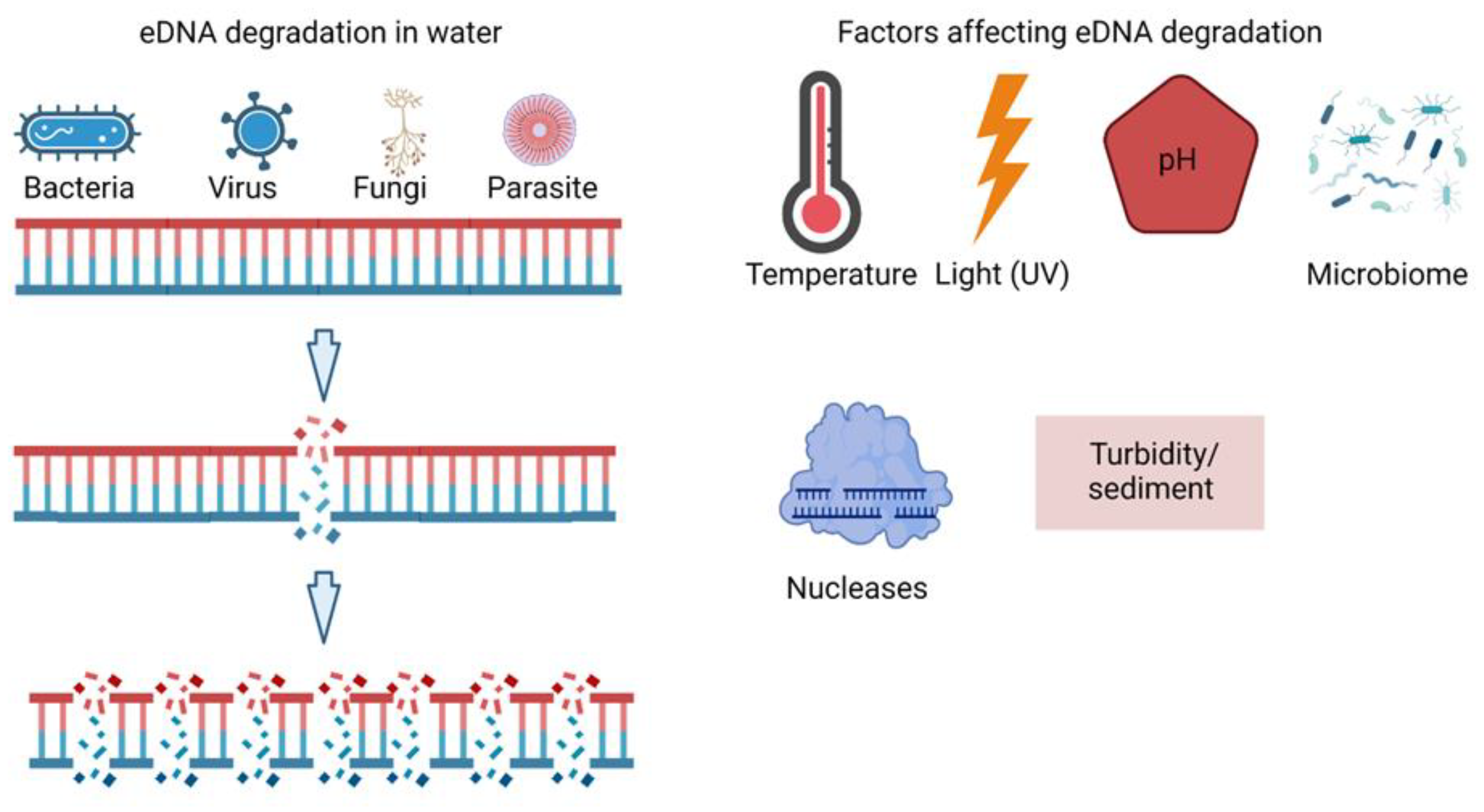
3. eDNA in Fish Disease

3.1. Bacteria

The ability of bacteria, archaea, and fungi cultures to release their genetic material into the extracellular medium, as well as in the context of multicellular microbial communities such as biofilms, has been reported. Bacteria release their DNA in water by different methods, including cell lysis and extrusion. The integrity of DNA released by cell lysis is usually higher, because the exonucleases cannot act quickly to degrade the DNA. Extrusion is used as a survival strategy by certain bacteria such as Deinococcus radiodurans, in which damaged DNA is released and new DNA is synthesized [8]. Many environmental bacteria including Micrococcus, Acinetobacter, Bacillus, Flavobacterium, Azotobacter, Pseudomonas, and Alcaligenes release their genetic material while growing in the media [9,10]. The amount of eDNA found depends on several factors, such as temperature, salinity, turbidity, and vegetation. In freshwater systems, the amount of DNA ranges from 1.74 to 7.77 μg/L [11]. There are many fish bacterial diseases affecting freshwater aquaculture, causing huge economic losses to the farmers. eDNA techniques might help them to predict bacterial load in their farms. Several research studies are being carried out to find an efficient method to detect those pathogens directly from the water samples. In most cases of Flavobacterium columnare infection, a gram-negative bacterium affecting different fish species, is found only externally in the skin, gills, and water samples before being systemic [Table 1]. Early and rigorous F. columnare diagnosis, as well as the implementation of practical preventive measures, are the only credible means of disease control. F. psychrophilum was found in different river water samples in Japan. They found a higher presence of F. psychrophilum during early summer and fall, and the presence of this bacteria depends on the water temperature [12]. In addition to that, F. psychrophilum and Yersinia ruckeri were also detected in salmon recirculatory aquaculture systems (RAS). Similarly, seven distinct species of Aeromonas were confirmed from coastal zones of river basins in Bangladesh. Over the 2-year study period, they also found that the number of bacteria changes with change in temperature using the eDNA method [13]. There are still many bacterial pathogens that are responsible for losses, which are yet to be studied.

3.2. Fungi

Fungi are some of the common fish pathogens in aquaculture settings. The most common fungal disease that affect fish species is saprolegniasis, branchiomycosis, and aspergillosis. Because farmed animals are typically held in high densities and exposed to constant stress and various types of pollutants, the risk of infection and disease spread is higher in fish farms than in wild environments. There are only a few studies on fish fungal disease identification using the eDNA method in aquaculture. Following high mortality outbreaks in the river Loue, for finding S. parasitica in water, a qPCR assay was designed. The pathogen was detected in river water but not in the tap water of surrounding villages [14]. There are other fungi from water identified by this approach that affect amphibians. Batrachochytrium dendrobatidis and B. salamandrivorans, two major fungal diseases of amphibians, were found in water samples in Spain using a qPCR assay [15]. DNA released from fungus cells has received less attention than DNA released from bacterial cells. Although fungi constitute more than 70% of the microbiome in soil, due to the fast rate of DNA degradation in dead fungal cells, the contribution of fungal DNA to the eDNA pool in soil should be insignificant. The fungus can spread to ponds and rivers via rainwater flow and water infiltration. Adequate and efficient methods to detect the presence of these fungal pathogens using eDNA will allow the farmer to predict the fungal disease outbreak, leading to timely management and control strategies.

3.3. Parasites

Environmental DNA (eDNA) sampling methods, in conjunction with different molecular methods, are well suited to quickly detecting the presence of pathogens in different fish farms, which helps the managers with valuable information that can be used to reduce disease threats. Parasites are the most common group of fish pathogens that are being detected easily using eDNA method. Standard fish parasite surveillance entails capturing and euthanizing fish before manually inspecting for the presence of parasites. Using this conventional method is both expensive and time-consuming, and it necessitates the sacrifice of many fish species. eDNA/eRNA fragments of several species in water samples have recently been established as an accurate, low-cost alternative to the traditional monitoring techniques, which require sampling the fish itself. A ddPCR assay was developed to detect eDNA in field samples, demonstrating the utility of eDNA detection in natural water systems for G. salaris [16]. eDNA of Dactylogyrus species was detected in a consignment of ornamental fish water and confirmed by sanger sequencing. Although there are some limitations regarding the use of the eDNA tool as a biosecurity and quarantine method. It detects eDNA from water and not directly from fish, and this might create a false positive even though the fish might not have the targeted parasite; the assay can show positive because of the source water used [17]. Chilodonella abundance was detected at varying levels across the year in the barramundi fish farm monitored in the study [18]. Another assay was developed that can detect low concentrations of parasites in tank water containing goldfish, presumably corresponding to an early stage of disease [19]. As a result, it could be a useful tool for monitoring and controlling ichthyophthiriasis in aquaculture.

3.4. Virus

There are mainly two different forms of viruses that infect fish species, which are DNA viruses and RNA viruses. Several studies have shown that DNA and RNA from viruses can be detected using eDNA or eRNA methods [20,21,22,23,24,25,26]. Since DNA is more stable than RNA, detection of eDNA is more practical and easier than detecting RNA from an environmental source. There are many common forms of virus that are found in freshwater aquaculture, including herpesvirus, viral hemorrhagic septicemia virus (VHSV), infectious hematopoietic necrosis virus (IHNV), golden shiner virus (GSV), channel catfish virus (CCV), red seabream virus, tilapia tilapine virus, and salmon alphavirus.
Table 1. Different fish diseases identified by eDNA/eRNA-based detection systems. The different environments, the samples collected, and the methods used to quantify are listed in the table.
DiseaseEnvironmentMethodReferences
Virus
Cyprinus Herpes Virus (CyHV-3)Lake, Pond, RiverPCR, qPCR[20,21]
Red seabream virusFish Farm
(Seawater)
DNA metabarcoding[22]
Tilapia tilapinevirusPond waterqPCR[23]
Rana VirusLakes, PondsqPCR[24,25]
Salmon aplhavirusSeawaterqPCR, ddPCR[26,27]
Parasite
Gyrodactylus salaris qPCR, ddPCR[16]
Dactylogyrusspp.Shipment waterqPCR [17]
Chilodonella hexastichaPond waterqPCR [18]
Ichthyophthirius multifiliisTank waterqPCR[19]
Myxobolus cerebralisRiver waterqPCR[28]
Ceratonova shastaRiver waterqPCR[29,30]
Parvicapsula minibicornisRiver system qPCR[30]
Tetracapsuloides bryosalmonaeRiver waterqPCR[31,32,33]
Neoparamoeba peruransSea waterqPCR[34]
Schistosoma mansoniTank water, water bodiesqPCR[35]
Fungi
Flavobacterium psychrophilumRiver, RASqPCR, ddPCR[12,36]
Bacteria
Aeromonassp.River, PondqPCR[13,37]
Saprolegnia parasiticaRiver waterqPCR[14]
Yersinia ruckeriRASddPCR[37]

4. eDNA Shedding and Degradation in Water
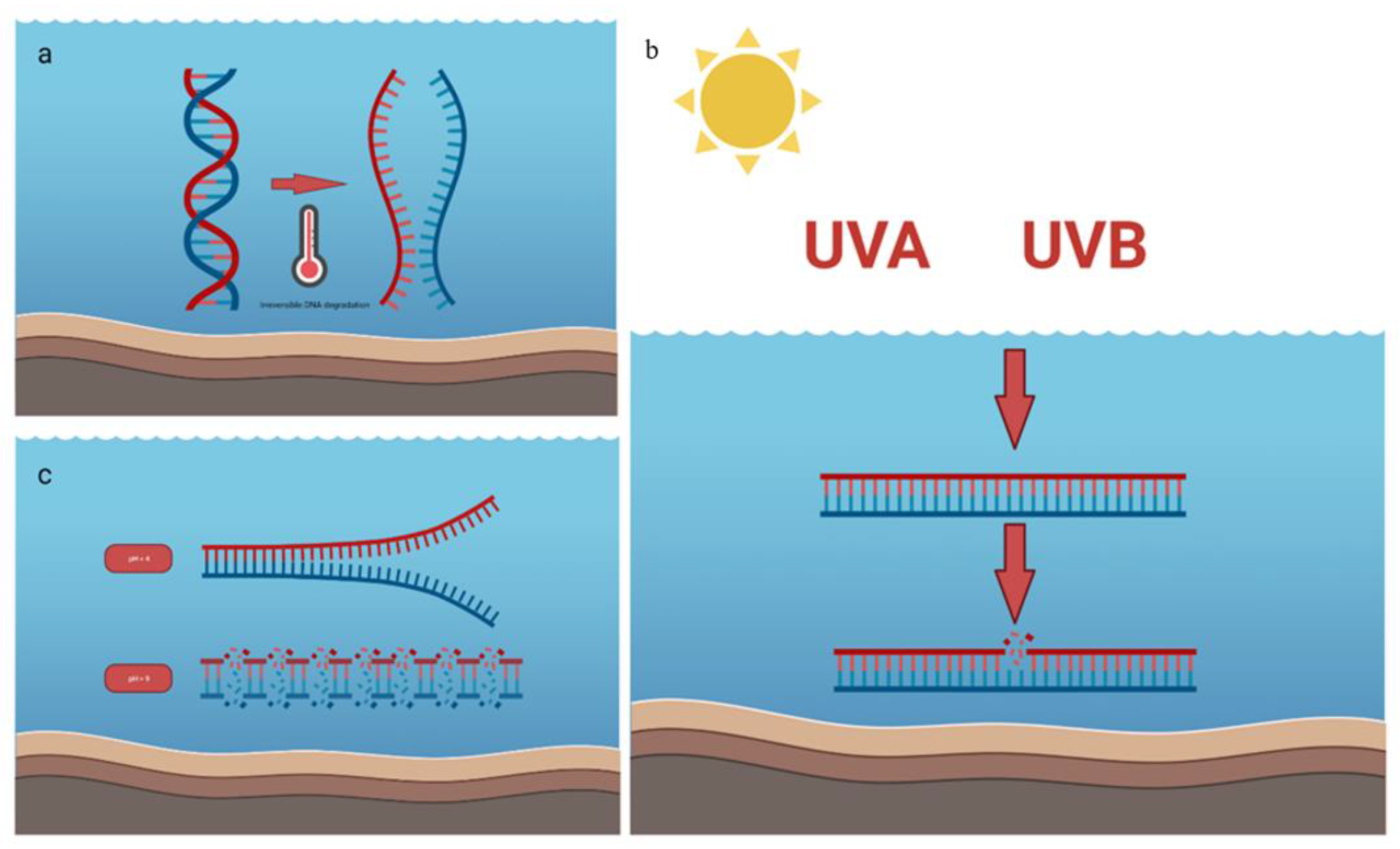
The link between the production and degradation of eDNA/eRNA is crucial for its detection and measurement [38]. When epithelial cells are shed or sloughed off through movement, excretion, and secretion, eDNA/eRNA is released into the environment [39]. Several studies have shown that physiological stress, along with the size and number of individuals, affects the DNA production rate [38,39,40,41]. There is evidence of both intra- and inter-specific heterogeneity in the creation of eDNA/eRNA [Table 2], highlighting a need to better understand the process of eDNA/eRNA generation and degradation in different species and systems [38,41].
A range of factors, including light, temperature, enzymatic activity, and pH impact the breakdown of eDNA (Figure 2). Addressing eDNA detection in an aquatic environment requires a full understanding of these factors and interactions, and the effects they have on eDNA stability. Hence, The factors influencing DNA persistence into three groups: the DNA molecule’s properties, abiotic factors (light, substrates, pH, oxygen, salinity), and biotic factors (microbes and enzymes) [42]. The length, conformations, and whether a DNA molecule is membrane-enclosed or free (also known as “naked”) DNA are all properties of the DNA molecule that affect how quickly DNA breaks down in the environment [43,44,45]. Biotic and abiotic factors affecting DNA degradation in an aquatic environment are discussed below.

4.1. Temperature

DNA deterioration in water occurs more quickly than in soil and sediments, possibly because of increased enzymatic and microbial activity at higher temperatures [46]. According to Matsui, et al., (2001) who investigated the fate of dissolved DNA in a thermally stratified lake, DNA in the warmer epilimnion (upper layer) was destroyed completely in 170 hours, while degrading more slowly in the much cooler hypolimnion (lower layer). In contrast to samples exposed to the full sun for 18 days and those exposed to 20% shade for 18 days, Idaho giant salamander eDNA stored at 4 °C and no light for 18 days contained 2030 and 733 times more eDNA, respectively [46]. Similarly, the experiment carried out by Zulkefil, et al., (2019) found higher degradation of eDNA at 35 °C compared to the control at 5 °C, which was about 60% from the initial concentration [47]. Likewise, the experiment carried out by Tsuji, et al., (2017) to know about the eDNA degradation of two species, ayu sweetfish (Plecoglossus altivelis altivelis) and common carp (Cyprinus carpio), at seven-time points, over a 48-h period, and at three different water temperatures (10 °C, 20 °C, 30 °C), found higher degradation at 30 °C. Some studies have found no effect of temperature on eDNA shedding rate [48,49]. Contrarily, Robson, et al., (2016) discovered that Mozambique tilapia’s DNA shedding rate was dramatically increased by tropical temperatures (23, 29, and 35 °C) (Oreochromis mossambicus) [50]. Inconsistencies in the research findings could point to variations in eDNA production that are species-specific. Higher temperature often leads to degradation by double-strand break of DNA (Figure 3a).

4.2. Ultraviolet light (UV)

Since the stratospheric ozone layer is being destroyed, UV-B radiation can enter the water column and destroy eDNA by rupturing DNA base-pair bonds Figure 3b [51]. Contradictory findings have been found in studies that have investigated how UV affects eDNA degradation. Pilliod, et al. (2014) showed that after eight days of full sun exposure, eDNA was no longer detectable, however, after 11 days in partial shade and after 18 days in complete darkness, eDNA was still detectable in all samples. Given that eDNA decayed exponentially even in the absence of light and that temperature accounted for the bulk of the observed variation in eDNA degradation among samples, the temperature may have a bigger effect on DNA degradation than light [39]. However, the study conducted by Zulkefli et al. (2019) with various levels (20, 50, and 100 µmol m-2s-1) of solar radiation had no observable effect on the degradation rate of eDNA. Interestingly, the same study indicated that UV light, regardless of whether it is UVA or UVB radiation, had no impact on the ability to identify DNA [52,53]. The notable significant differences were seen by co-varying light intensity and temperature (35 °C) at the end of experiment compared to 5 °C treatment [47].
Table 2. Comparison of eDNA decay rates among various types of eDNA, sources, and environmental factors. * Indicates the significant effect on eDNA degradation in the corresponding study. (Table adapted from Zulkefli et al., 2019 [47]).
eDNA TypeSourceEnvironmental FactorDecay Rate, (r) (day−1)Reference
IntracellularCommon carp
Cyprinus carpio
Microbial community *, pH2.52[42]
ExtracellularSediment sample
Cyanobacterium
Anabaena variabilis
Temperature *, microbial activity *, pH, light intensity0.0931–3.2706[47]
ExtracellularSediment and water samplesBased on simplified OECD endurance test0.009–0.133[51]
IntracellularCrustacean
Daphnia magna
pH *, temperature, microbial
activity, total dissolved nitrogen
Water derived 6.552–23.568
Biofilm derived 1.176–17.256
[53]
May fly
Ephemera Danica
Eel
Anguila anguilla
IntracellularAyu sweetfish
Plecoglossus altivelis altivelis
Temperature *, microbial
abundance
0.48–7.2[54]
Common carp
Cyprinus carpio
IntracellularCommon carp
Cyprinus carpio
Temperature *, trophic state *0.35–2.42[55]
IntracellularAmerican bullfrog
Lithobates catesbeianus
UV-B *, temperature *, pH0.243[56]

4.3. pH

DNA hydrolysis is favored in acidic environments [57,58]. In contrast, Stickler, et al., (2015) noted that the increased rate of eDNA degradation at pH 4 was due to interactions with other factors and that the pH level itself had no impact on the degradation. In stream mesocosms with an acid–base gradient, the degradation rate of lotic multispecies eDNA was accelerated to undetectable levels in just two days [53]. Low pH will result in denaturing of the two strands of DNA, while higher pH leads to the degradation of hydrogen bonds and separation of nitrogen bases in DNA (Figure 3c).

4.4. Environmental Parameters

The consistent detection of eDNA in the aquatic environment depends on the flow rate [59,60] of solid materials and dissolved substances in the water column and riverbed [61,62]. It is believed that the properties of sediments (suspended or benthic) might influence eDNA degradation because sediments might adsorb DNA [63], thus reducing the eDNA detection rates. However, eDNA can re-suspend from sediment [64], which can result in false positives or the identification of a species that is no longer present in the environment [65]. Additionally, dissolved materials in the water matrix can affect how quickly DNA is detected, and potentially PCRs, such as humic acids [66]. In this regard, an experiment conducted on environmental conditions on eDNA success in aquatic ecosystems suggested that the presence of sediments is responsible for lower eDNA detection in water samples regardless of flow-through or still-water conditions. This was followed by the delayed release of eDNA in the presence of sediment. Additionally, humic substances had a higher inhibitory effect on eDNA detection, followed by algae and siliceous sediment particles [67]. This study mentioned that application of eDNA methods in field survey conditions strongly depends on site-specific conditions such as water flow conditions, sediment composition, and suspended particles.

5. Discussion

eDNA/eRNA have immense potential for disease risk monitoring, as they can improve our ability to determine the existence, diversity, and quantity of pathogenic organisms. Since traditional pathogen detection techniques frequently entail cultivating or necropsying host tissues, they are equally as resource intensive as those used to detect free-living pathogens. Parasites are often considered less significant in freshwater aquaculture, but they can decrease the final value of the product. In addition to that, there are several cases of co-infection with bacteria and viruses led by parasite infection [67,68,69]. Certain challenges such as accuracy, efficiency, and the fate of eDNA/eRNA are of concern despite the efforts from many researchers around the globe. Sengupta, et al., 2019 used eDNA method for the detection of cercaria, one of the major parasites that affect both aquatic and human species. They used both field-based models and lab-based models to effectively detect the presence of this parasite [70]. However, there are limitations regarding the methods, such as that the rate of decay and the life stage of parasites identified using this technique cannot be determined. Thus, to increase the effectiveness of this method, experiments and research need to be carried out under different conditions. Our review provides comprehensive advances in the detection of freshwater pathogens and parasites using eDNA techniques. In addition to that, our study reviews the effect of different environmental factors on eDNA degradation. The rate of degradation of DNA and RNA is different in the marine environment than in the freshwater system. The half-life of eDNA is found to be in a range of 7 h to 72 h in marine water [71], which is shorter than in the freshwater system [72]. The degradation rate also varies with terrestrial environments and different seasons [73]. Aside from abiotic factors such as oxidation and hydrolysis by depurination, biotic factors such as extracellular DNases produced by heterotrophic microbes are also likely to play a significant role in eDNA persistence dynamics [50,73].

6. Conclusions

eDNA analysis is changing the way we design and implement biodiversity monitoring programs, opening new opportunities for the future. This tool has a high potential for monitoring aquatic biodiversity, including pathogens and parasites. Several high-end sequencing methods are being used to detect the presence and absence of multiple pathogens easily and accurately in freshwater. Detecting pathogens and parasites beforehand is a crucial step in aquaculture. Fish kills due to disease outbreaks are common in aquaculture all around the world, from warm-water to cold-water aquaculture systems. Management can be initiated to prevent the spread of disease and potentially improve the timely treatment of water. This will allow aquaculture farmers to save millions of USD each year, which is lost due to fish kills caused by different diseases around the globe.

Author Contributions

Conceptualization, K.B. and A.K.Y.; writing—original draft preparation, K.B., A.K.Y. and P.J.; writing—review and editing, A.K.Y. All authors have read and agreed to the published version of the manuscript.

Funding

This research received no external funding.

Institutional Review Board Statement

Not applicable.

Conflicts of Interest

The authors declare no conflict of interest.

References

  1. Pietramellara, G.; Ascher, J.; Borgogni, F.; Ceccherini, M.T.; Guerri, G.; Nannipieri, P. Extracellular DNA in Soil and Sediment: Fate and Ecological Relevance. Biol. Fertil. Soils 2009, 45, 219–235. [Google Scholar] [CrossRef]
  2. Ogram, A.; Sayler, G.S.; Barkay, T. The Extraction and Purification of Microbial DNA from Sediments. J. Microbiol. Methods 1987, 7, 57–66. [Google Scholar] [CrossRef]
  3. Haile, J.; Froese, D.; Macphee, R.; Roberts, R.; Arnold, L.; Reyes, A.; Rasmussen, M.; Nielsen, R.; Brook, B.; Robinson, S.; et al. Ancient DNA Reveals Late Survival of Mammoth and Horse in Interior Alaska. Proc. Natl. Acad. Sci. USA 2009, 106, 22352–22357. [Google Scholar] [CrossRef] [Green Version]
  4. Epp, L.S.; Boessenkool, S.; Bellemain, E.P.; Haile, J.; Esposito, A.; Riaz, T.; Erséus, C.; Gusarov, V.I.; Edwards, M.E.; Johnsen, A.; et al. New Environmental Metabarcodes for Analysing Soil DNA: Potential for Studying Past and Present Ecosystems. Mol. Ecol. 2012, 21, 1821–1833. [Google Scholar] [CrossRef]
  5. Bhadury, P.; Austen, M.; Bilton, D.; Lambshead, P.J.; Rogers, A. Molecular Detection of Marine Nematodes from Environmental Samples: Overcoming Eukaryotic Interference. Aquat. Microb. Ecol. 2006, 44, 97–103. [Google Scholar] [CrossRef] [Green Version]
  6. Andersen, K.; Bird, K.L.; Rasmussen, M.; Haile, J.; Breuning-Madsen, H.; Kjaer, K.H.; Orlando, L.; Gilbert, M.T.P.; Willerslev, E. Meta-Barcoding of “dirt” DNA from Soil Reflects Vertebrate Biodiversity. Mol. Ecol. 2012, 21, 1966–1979. [Google Scholar] [CrossRef]
  7. Lednicky, J.A.; Lauzard, M.; Fan, Z.H.; Jutla, A.; Tilly, T.B.; Gangwar, M.; Usmani, M.; Shankar, S.N.; Mohamed, K.; Eiguren-Fernandez, A.; et al. Viable SARS-CoV-2 in the Air of a Hospital Room with COVID-19 Patients. Int. J. Infect. Dis. 2020, 100, 476–482. [Google Scholar] [CrossRef]
  8. Battista, J.R. Against All Odds: The Survival Strategies of Deinococcus Radiodurans. Annu. Rev. Microbiol. 1997, 51, 203–224. [Google Scholar] [CrossRef]
  9. Paget, E.; Simonet, P. On the Track of Natural Transformation in Soil. FEMS Microbiol. Ecol. 1994, 15, 109–117. [Google Scholar] [CrossRef]
  10. Lorenz, M.G.; Wackernagel, W. Bacterial Gene Transfer by Natural Genetic Transformation in the Environment. Microbiol. Rev. 1994, 58, 563–602. [Google Scholar] [CrossRef]
  11. DeFlaun, M.F.; Paul, J.H.; Davis, D. Simplified Method for Dissolved DNA Determination in Aquatic Environments. Appl. Environ. Microbiol. 1986, 52, 654–659. [Google Scholar] [CrossRef] [Green Version]
  12. Tenma, H.; Tsunekawa, K.; Fujiyoshi, R.; Takai, H.; Hirose, M.; Masai, N.; Sumi, K.; Takihana, Y.; Yanagisawa, S.; Tsuchida, K.; et al. Spatiotemporal Distribution of Flavobacterium Psychrophilum and Ayu Plecoglossus Altivelis in Rivers Revealed by Environmental DNA Analysis. Fish. Sci. 2021, 87, 321–330. [Google Scholar] [CrossRef]
  13. Sadique, A.; Neogi, S.B.; Bashar, T.; Sultana, M.; Johura, F.-T.; Islam, S.; Hasan, N.A.; Huq, A.; Colwell, R.R.; Alam, M. Dynamics, Diversity, and Virulence of Aeromonas Spp. in Homestead Pond Water in Coastal Bangladesh. Front. Public Health 2021, 9, 902. [Google Scholar] [CrossRef]
  14. Rocchi, S.; Tisserant, M.; Valot, B.; Laboissière, A.; Frossard, V.; Reboux, G. Quantification of Saprolegnia Parasitica in River Water Using Real-Time Quantitative PCR: From Massive Fish Mortality to Tap Drinking Water. Int. J. Environ. Health Res. 2017, 27, 1–10. [Google Scholar] [CrossRef]
  15. Gonzalez, R.; Curtis, K.; Bivins, A.; Bibby, K.; Weir, M.H.; Yetka, K.; Thompson, H.; Keeling, D.; Mitchell, J.; Gonzalez, D. COVID-19 Surveillance in Southeastern Virginia Using Wastewater-Based Epidemiology. Water Res. 2020, 186, 116296. [Google Scholar] [CrossRef]
  16. Rusch, J.C.; Hansen, H.; Strand, D.A.; Markussen, T.; Hytterød, S.; Vrålstad, T. Catching the Fish with the Worm: A Case Study on EDNA Detection of the Monogenean Parasite Gyrodactylus Salaris and Two of Its Hosts, Atlantic Salmon (Salmo Salar) and Rainbow Trout (Oncorhynchus Mykiss). Parasites Vectors 2018, 11, 333. [Google Scholar] [CrossRef]
  17. Trujillo-González, A.; Edmunds, R.C.; Becker, J.A.; Hutson, K.S. Parasite Detection in the Ornamental Fish Trade Using Environmental DNA. Sci. Rep. 2019, 9, 5173. [Google Scholar] [CrossRef] [Green Version]
  18. Bastos Gomes, G.; Hutson, K.S.; Domingos, J.A.; Chung, C.; Hayward, S.; Miller, T.L.; Jerry, D.R. Use of Environmental DNA (EDNA) and Water Quality Data to Predict Protozoan Parasites Outbreaks in Fish Farms. Aquaculture 2017, 479, 467–473. [Google Scholar] [CrossRef]
  19. Jousson, O.; Pretti, C.; Di Bello, D.; Cognetti-Varriale, A.M. Non-Invasive Detection and Quantification of the Parasitic Ciliate Ichthyophthirius Multifiliis by Real-Time PCR. Dis. Aquat. Organ. 2005, 65, 251–255. [Google Scholar] [CrossRef] [Green Version]
  20. Minamoto, T.; Yamanaka, H.; Takahara, T.; Honjo, M.N.; Kawabata, Z. Surveillance of Fish Species Composition Using Environmental DNA. Limnology 2012, 13, 193–197. [Google Scholar] [CrossRef]
  21. Haramoto, E.; Kitajima, M.; Katayama, H.; Ohgaki, S. Detection of Koi Herpesvirus DNA in River Water in Japan. J. Fish Dis. 2007, 30, 59–61. [Google Scholar] [CrossRef]
  22. Kawato, M.; Yoshida, T.; Miya, M.; Tsuchida, S.; Nagano, Y.; Nomura, M.; Yabuki, A.; Fujiwara, Y.; Fujikura, K. Optimization of Environmental DNA Extraction and Amplification Methods for Metabarcoding of Deep-Sea Fish. MethodsX 2021, 8, 101238. [Google Scholar] [CrossRef]
  23. Taengphu, S.; Kayansamruaj, P.; Kawato, Y.; Delamare-Deboutteville, J.; Mohan, C.; Dong, H.; Senapin, S. Concentration and Quantification of Tilapia Tilapinevirus from Water Using a Simple Iron Flocculation Coupled with Probe-Based RT-QPCR. bioRxiv 2021. [Google Scholar] [CrossRef]
  24. Vilaça, S.T.; Grant, S.A.; Beaty, L.; Brunetti, C.R.; Congram, M.; Murray, D.L.; Wilson, C.C.; Kyle, C.J. Detection of Spatiotemporal Variation in Ranavirus Distribution Using EDNA. Environ. DNA 2020, 2, 210–220. [Google Scholar] [CrossRef]
  25. Miaud, C.; Arnal, V.; Poulain, M.; Valentini, A.; Dejean, T. EDNA Increases the Detectability of Ranavirus Infection in an Alpine Amphibian Population. Viruses 2019, 11, 526. [Google Scholar] [CrossRef] [Green Version]
  26. Weli, S.C.; Bernhardt, L.-V.; Qviller, L.; Myrmel, M.; Lillehaug, A. Development and Evaluation of a Method for Concentration and Detection of Salmonid Alphavirus from Seawater. J. Virol. Methods 2021, 287, 113990. [Google Scholar] [CrossRef]
  27. Bernhardt, L.-V.; Myrmel, M.; Lillehaug, A.; Qviller, L.; Chioma Weli, S. Filtration, Concentration and Detection of Salmonid Alphavirus in Seawater during a Post-Smolt Salmon (Salmo Salar) Cohabitant Challenge. Dis. Aquat. Organ. 2021, 144, 61–73. [Google Scholar] [CrossRef]
  28. Richey, C.A.; Kenelty, K.V.; Van Stone Hopkins, K.; Stevens, B.N.; Martínez-López, B.; Barnum, S.M.; Hallett, S.L.; Atkinson, S.D.; Bartholomew, J.L.; Soto, E. Distribution and Prevalence of Myxobolus Cerebralis in Postfire Areas of Plumas National Forest: Utility of Environmental DNA Sampling. J. Aquat. Anim. Health 2018, 30, 130–143. [Google Scholar] [CrossRef]
  29. Richey, C.A.; Kenelty, K.V.; Hopkins, K.V.S.; Stevens, B.N.; Martínez-López, B.; Hallett, S.L.; Atkinson, S.D.; Bartholomew, J.L.; Soto, E. Validation of Environmental DNA Sampling for Determination of Ceratonova Shasta (Cnidaria: Myxozoa) Distribution in Plumas National Forest, CA. Parasitol. Res. 2020, 119, 859–870. [Google Scholar] [CrossRef]
  30. Hallett, S.L.; Bartholomew, J.L. Application of a Real-Time PCR Assay to Detect and Quantify the Myxozoan Parasite Ceratomyxa Shasta in River Water Samples. Dis. Aquat. Organ. 2006, 71, 109–118. [Google Scholar] [CrossRef]
  31. Carraro, L.; Bertuzzo, E.; Mari, L.; Fontes, I.; Hartikainen, H.; Strepparava, N.; Schmidt-Posthaus, H.; Wahli, T.; Jokela, J.; Gatto, M.; et al. Integrated Field, Laboratory, and Theoretical Study of PKD Spread in a Swiss Prealpine River. Proc. Natl. Acad. Sci. USA 2017, 114, 11992–11997. [Google Scholar] [CrossRef] [Green Version]
  32. Carraro, L.; Hartikainen, H.; Jokela, J.; Bertuzzo, E.; Rinaldo, A. Estimating Species Distribution and Abundance in River Networks Using Environmental DNA. Proc. Natl. Acad. Sci. USA 2018, 115, 11724–11729. [Google Scholar] [CrossRef] [Green Version]
  33. Hutchins, P.R.; Sepulveda, A.J.; Martin, R.M.; Hopper, L.R. A Probe-Based Quantitative PCR Assay for Detecting Tetracapsuloides Bryosalmonae in Fish Tissue and Environmental DNA Water Samples. Conserv. Genet. Resour. 2018, 10, 317–319. [Google Scholar] [CrossRef]
  34. Bridle, A.R.; Crosbie, P.B.B.; Cadoret, K.; Nowak, B.F. Rapid Detection and Quantification of Neoparamoeba Perurans in the Marine Environment. Aquaculture 2010, 309, 56–61. [Google Scholar] [CrossRef]
  35. Alzaylaee, H.; Collins, R.A.; Shechonge, A.; Ngatunga, B.P.; Morgan, E.R.; Genner, M.J. Environmental DNA-Based Xenomonitoring for Determining Schistosoma Presence in Tropical Freshwaters. Parasites Vectors 2020, 13, 63. [Google Scholar] [CrossRef]
  36. Lewin, A.S.; Haugen, T.; Netzer, R.; Tøndervik, A.; Dahle, S.W.; Hageskal, G. Multiplex Droplet Digital PCR Assay for Detection of Flavobacterium Psychrophilum and Yersinia Ruckeri in Norwegian Aquaculture. J. Microbiol. Methods 2020, 177, 106044. [Google Scholar] [CrossRef]
  37. Fong, J.; Cho, H.J.; Park, M.S.; Lim, Y.W. Evaluating Seasonality and Pathogenicity of Aeromonas in Korea Using Environmental DNA. Asian J. Microbiol. Biotechnol. Environ. Sci. 2016, 18, 605–613. [Google Scholar]
  38. Thomsen, P.F.; Kielgast, J.; Iversen, L.L.; Møller, P.R.; Rasmussen, M.; Willerslev, E. Detection of a Diverse Marine Fish Fauna Using Environmental DNA from Seawater Samples. PLoS ONE 2012, 7, e41732. [Google Scholar] [CrossRef]
  39. Pilliod, D.S.; Goldberg, C.S.; Arkle, R.S.; Waits, L.P. Factors Influencing Detection of EDNA from a Stream-Dwelling Amphibian. Mol. Ecol. Resour. 2014, 14, 109–116. [Google Scholar] [CrossRef]
  40. Takahara, T.; Minamoto, T.; Yamanaka, H.; Doi, H.; Kawabata, Z. Estimation of Fish Biomass Using Environmental DNA. PLoS ONE 2012, 7, e35868. [Google Scholar] [CrossRef] [Green Version]
  41. Maruyama, A.; Nakamura, K.; Yamanaka, H.; Kondoh, M.; Minamoto, T. The Release Rate of Environmental DNA from Juvenile and Adult Fish. PLoS ONE 2014, 9, e114639. [Google Scholar] [CrossRef]
  42. Barnes, M.A.; Turner, C.R.; Jerde, C.L.; Renshaw, M.A.; Chadderton, W.L.; Lodge, D.M. Environmental Conditions Influence EDNA Persistence in Aquatic Systems. Environ. Sci. Technol. 2014, 48, 1819–1827. [Google Scholar] [CrossRef]
  43. Romanowski, G.; Lorenz, M.G.; Sayler, G.; Wackernagel, W. Persistence of Free Plasmid DNA in Soil Monitored by Various Methods, Including a Transformation Assay. Appl. Environ. Microbiol. 1992, 58, 3012–3019. [Google Scholar] [CrossRef] [Green Version]
  44. Alvarez, A.J.; Yumet, G.M.; Santiago, C.L.; Toranzos, G.A. Stability of Manipulated Plasmid DNA in Aquatic Environments. Environ. Toxicol. Water Qual. 1996, 11, 129–135. [Google Scholar] [CrossRef]
  45. Zhu, B. Degradation of Plasmid and Plant DNA in Water Microcosms Monitored by Natural Transformation and Real-Time Polymerase Chain Reaction (PCR). Water Res. 2006, 40, 3231–3238. [Google Scholar] [CrossRef]
  46. Matsui, K.; Honjo, M.; Kawabata, Z. Estimation of the Fate of Dissolved DNA in Thermally Stratified Lake Water from the Stability of Exogenous Plasmid DNA. Aquat. Microb. Ecol. 2001, 26, 95–102. [Google Scholar] [CrossRef] [Green Version]
  47. Zulkefli, N.S.; Kim, K.-H.; Hwang, S.-J. Effects of Microbial Activity and Environmental Parameters on the Degradation of Extracellular Environmental DNA from a Eutrophic Lake. Int. J. Environ. Res. Public Health 2019, 16, 3339. [Google Scholar] [CrossRef] [PubMed] [Green Version]
  48. Klymus, K.E.; Richter, C.A.; Chapman, D.C.; Paukert, C. Quantification of EDNA Shedding Rates from Invasive Bighead Carp Hypophthalmichthys Nobilis and Silver Carp Hypophthalmichthys Molitrix. Biol. Conserv. 2015, 183, 77–84. [Google Scholar] [CrossRef]
  49. Takahara, T.; Minamoto, T.; Doi, H. Using Environmental DNA to Estimate the Distribution of an Invasive Fish Species in Ponds. PLoS ONE 2013, 8, e56584. [Google Scholar] [CrossRef] [Green Version]
  50. Robson, H.L.A.; Noble, T.H.; Saunders, R.J.; Robson, S.K.A.; Burrows, D.W.; Jerry, D.R. Fine-Tuning for the Tropics: Application of EDNA Technology for Invasive Fish Detection in Tropical Freshwater Ecosystems. Mol. Ecol. Resour. 2016, 16, 922–932. [Google Scholar] [CrossRef]
  51. Häder, D.-P.; Sinha, R.P. Solar Ultraviolet Radiation-Induced DNA Damage in Aquatic Organisms: Potential Environmental Impact. Mutat. Res. 2005, 571, 221–233. [Google Scholar] [CrossRef] [PubMed]
  52. Mächler, E.; Osathanunkul, M.; Altermatt, F. Shedding Light on EDNA: Neither Natural Levels of UV Radiation nor the Presence of a Filter Feeder Affect EDNA-Based Detection of Aquatic Organisms. PLoS ONE 2018, 13, e0195529. [Google Scholar] [CrossRef] [PubMed]
  53. Seymour, M.; Durance, I.; Cosby, B.J.; Ransom-Jones, E.; Deiner, K.; Ormerod, S.J.; Colbourne, J.K.; Wilgar, G.; Carvalho, G.R.; de Bruyn, M.; et al. Acidity Promotes Degradation of Multi-Species Environmental DNA in Lotic Mesocosms. Commun. Biol. 2018, 1, 4. [Google Scholar] [CrossRef] [Green Version]
  54. Tsuji, S.; Ushio, M.; Sakurai, S.; Minamoto, T.; Yamanaka, H. Water Temperature-Dependent Degradation of Environmental DNA and Its Relation to Bacterial Abundance. PLoS ONE 2017, 12, e0176608. [Google Scholar] [CrossRef]
  55. Eichmiller, J.J.; Best, S.E.; Sorensen, P.W. Effects of Temperature and Trophic State on Degradation of Environmental DNA in Lake Water. Environ. Sci. Technol. 2016, 50, 1859–1867. [Google Scholar] [CrossRef]
  56. Strickler, K.M.; Fremier, A.K.; Goldberg, C.S. Quantifying Effects of UV-B, Temperature, and PH on EDNA Degradation in Aquatic Microcosms. Biol. Conserv. 2015, 183, 85–92. [Google Scholar] [CrossRef]
  57. Lindahl, T. Instability and Decay of the Primary Structure of DNA. Nature 1993, 362, 709–715. [Google Scholar] [CrossRef]
  58. Gates, K.S. An Overview of Chemical Processes That Damage Cellular DNA: Spontaneous Hydrolysis, Alkylation, and Reactions with Radicals. Chem. Res. Toxicol. 2009, 22, 1747–1760. [Google Scholar] [CrossRef] [Green Version]
  59. Ficetola, G.F.; Miaud, C.; Pompanon, F.; Taberlet, P. Species Detection Using Environmental DNA from Water Samples. Biol. Lett. 2008, 4, 423–425. [Google Scholar] [CrossRef] [Green Version]
  60. Nathan, L.R.; Jerde, C.L.; Budny, M.L.; Mahon, A.R. The Use of Environmental DNA in Invasive Species Surveillance of the Great Lakes Commercial Bait Trade. Conserv. Biol. 2015, 29, 430–439. [Google Scholar] [CrossRef]
  61. Rees, H.C.; Maddison, B.C.; Middleditch, D.J.; Patmore, J.R.M.; Gough, K.C. REVIEW: The Detection of Aquatic Animal Species Using Environmental DNA—A Review of EDNA as a Survey Tool in Ecology. J. Appl. Ecol. 2014, 51, 1450–1459. [Google Scholar] [CrossRef]
  62. Shogren, A.J.; Tank, J.L.; Andruszkiewicz, E.; Olds, B.; Mahon, A.R.; Jerde, C.L.; Bolster, D. Controls on EDNA Movement in Streams: Transport, Retention, and Resuspension. Sci. Rep. 2017, 7, 5065. [Google Scholar] [CrossRef] [PubMed] [Green Version]
  63. Stewart, G.J.; Sinigalliano, C.D.; Garko, K.A. Binding of Exogenous DNA to Marine Sediments and the Effect of DNA/Sediment Binding on Natural Transformation of Pseudomonas Stutzeri Strain ZoBell in Sediment Columns. FEMS Microbiol. Lett. 1991, 85, 1–8. [Google Scholar] [CrossRef]
  64. Graf, G.; Rosenberg, R. Bioresuspension and Biodeposition: A Review. J. Mar. Syst. 1997, 11, 269–278. [Google Scholar] [CrossRef]
  65. Stoeckle, B.C.; Beggel, S.; Cerwenka, A.F.; Motivans, E.; Kuehn, R.; Geist, J. A Systematic Approach to Evaluate the Influence of Environmental Conditions on EDNA Detection Success in Aquatic Ecosystems. PLoS ONE 2017, 12, e0189119. [Google Scholar] [CrossRef] [Green Version]
  66. Albers, C.N.; Jensen, A.; Bælum, J.; Jacobsen, C.S. Inhibition of DNA Polymerases Used in Q-PCR by Structurally Different Soil-Derived Humic Substances. Geomicrobiol. J. 2013, 30, 675–681. [Google Scholar] [CrossRef]
  67. Xu, D.-H.; Shoemaker, C.A.; Klesius, P.H. Evaluation of the Link between Gyrodactylosis and Streptococcosis of Nile Tilapia, Oreochromis Niloticus (L.). J. Fish Dis. 2007, 30, 233–238. [Google Scholar] [CrossRef]
  68. Zhang, C.; Li, D.; Chi, C.; Ling, F.; Wang, G. Dactylogyrus Intermedius Parasitism Enhances Flavobacterium Columnare Invasion and Alters Immune-Related Gene Expression in Carassius Auratus. Dis. Aquat. Organ. 2015, 116, 11–21. [Google Scholar] [CrossRef]
  69. Ogut, H.; Cavus, N. A Comparison of Ectoparasite Prevalence and Occurrence of Viral Haemorrhagic Septicemia Virus (VHSV) in Whiting Merlangius Merlangus Euxinus. Revista de Biología Marina y Oceanografía 2014, 49, 91–96. [Google Scholar] [CrossRef] [Green Version]
  70. Sengupta, M.E.; Hellström, M.; Kariuki, H.C.; Olsen, A.; Thomsen, P.F.; Mejer, H.; Willerslev, E.; Mwanje, M.T.; Madsen, H.; Kristensen, T.K.; et al. Environmental DNA for Improved Detection and Environmental Surveillance of Schistosomiasis. Proc. Natl. Acad. Sci. USA 2019, 116, 8931–8940. [Google Scholar] [CrossRef] [Green Version]
  71. Collins, R.A.; Wangensteen, O.S.; O’Gorman, E.J.; Mariani, S.; Sims, D.W.; Genner, M.J. Persistence of Environmental DNA in Marine Systems. Commun. Biol. 2018, 1, 185. [Google Scholar] [CrossRef] [PubMed]
  72. Sassoubre, L.M.; Yamahara, K.M.; Gardner, L.D.; Block, B.A.; Boehm, A.B. Quantification of Environmental DNA (EDNA) Shedding and Decay Rates for Three Marine Fish. Environ. Sci. Technol. 2016, 50, 10456–10464. [Google Scholar] [CrossRef] [PubMed]
  73. Torti, A.; Lever, M.A.; Jørgensen, B.B. Origin, Dynamics, and Implications of Extracellular DNA Pools in Marine Sediments. Mar. Genom. 2015, 24, 185–196. [Google Scholar] [CrossRef] [PubMed]
Figure 1. Different methods of eDNA/eRNA quantification. qPCR, ddPCR, metabarcoding, sequencing, and RT-PCR are generally used to quantify the presence of different organisms from water samples.
Diversity 14 01015 g001
Figure 2. Different factors affecting nucleic acid degradation in the aquatic environment. Nucleic acids are affected by temperature, light, pH, and the environment they are present in.
Diversity 14 01015 g002
Figure 3. Effect of temperature, light and PH on DNA degradation process.
Diversity 14 01015 g003
Publisher’s Note: MDPI stays neutral with regard to jurisdictional claims in published maps and institutional affiliations.

