Article

Transcriptomic Analysis Reveals the Hepatotoxicity of Perfluorooctanoic Acid in Black-Spotted Frogs (Rana nigromaculata)

1
School of Life and Environmental Sciences, Hangzhou Normal University, Hangzhou 311121, China
2
School of Engineering, Hangzhou Normal University, Hangzhou 310018, China
*
Author to whom correspondence should be addressed.
Diversity 2022, 14(11), 971; https://doi.org/10.3390/d14110971
Submission received: 21 October 2022 / Revised: 7 November 2022 / Accepted: 10 November 2022 / Published: 11 November 2022

Abstract

:
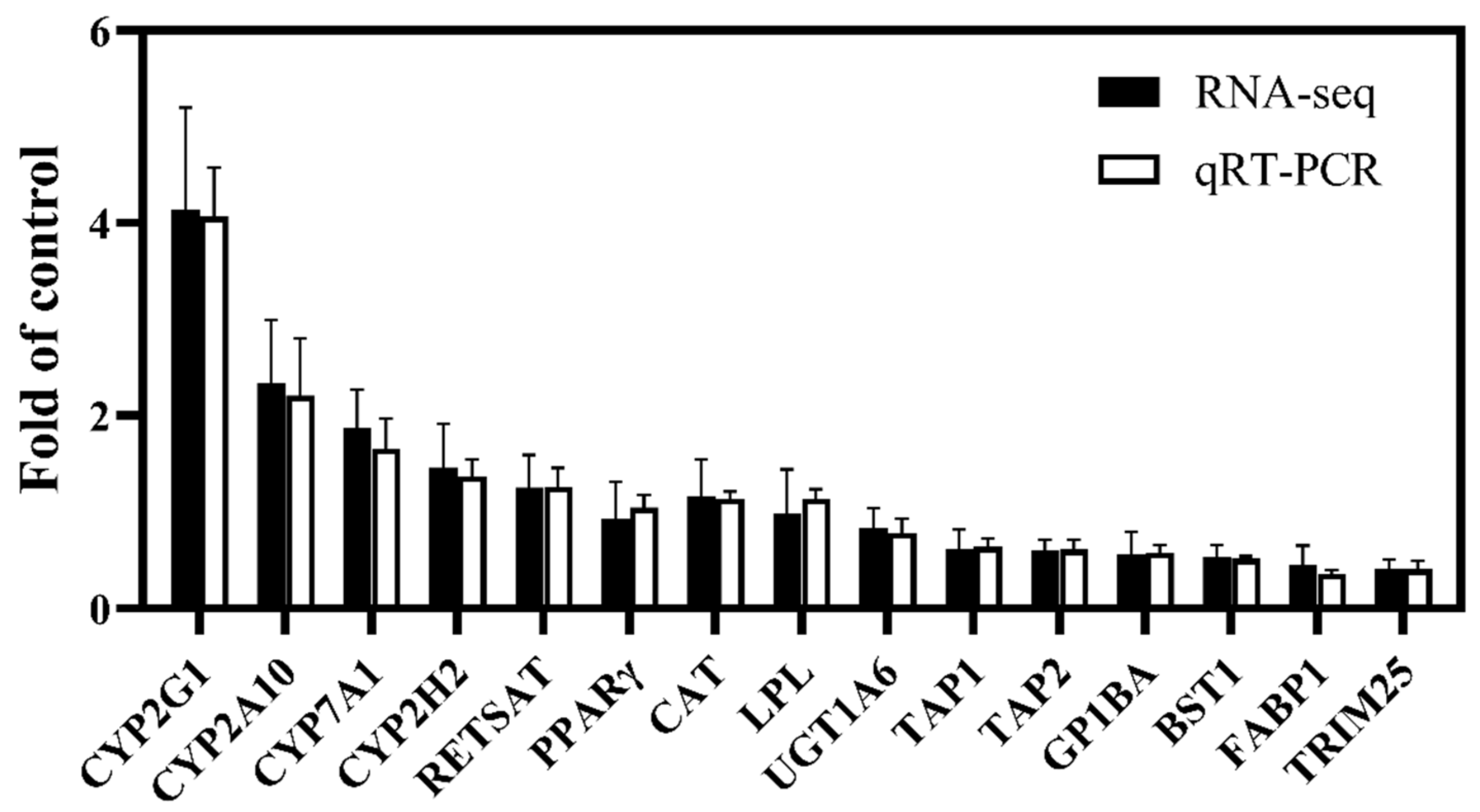
Perfluorooctanoic acid (PFOA) is frequently detected in the environment and accumulates in amphibians such as black-spotted frogs (Rana nigromaculata) with toxic effects; however, the mechanism underlying this toxicity is unclear. In this study, male black-spotted frogs were exposed to 10 μg/L waterborne PFOA for 21 days. Subsequently, the effect of PFOA exposure on gene expression in liver tissue was investigated using transcriptomic techniques. In total, 754 differentially expressed genes (DEGs; 389 up-regulated and 365 down-regulated) were identified. According to Kyoto Encyclopedia of Genes and Genomes enrichment and Gene Ontology functional enrichment analyses, the DEGs were mainly involved in lipid metabolism, endocrine functions, and immunity. Quantitative real-time polymerase chain reaction analysis of 15 selected DEGs revealed a high correlation (R2 = 0.9917) with the transcriptomic results. These results indicated that the PFOA hepatotoxicity in frogs is mediated mainly by lipid metabolism dysregulation, endocrine system disruption, and immunotoxicity. This study provides insights into the hepatotoxic mechanism of PFOA and other perfluorinated compounds in amphibians.

1. Introduction

Perfluorinated compounds (PFCs) are a group of anthropogenic fluorocarbons that have been widely used in consumer and industrial products since the 1950s because of their good thermal stability and high surface activity [1,2,3]. Due to inefficient degradation in the environment combined with their potential for long-distance transfer and bioaccumulation, PFCs are classified as persistent organic pollutants [4,5,6]. Perfluorooctanoic acid (PFOA) is one of the most abundant PFCs in surface water and sediment, atmosphere, wild animals and humans [7,8,9]. With a biological half-life of PFOA of around 3.8 years and the limited potential for discharging or degrading the compound once it enters the organism, PFOA accumulation can cause long-term adverse effects [10]. Therefore, the widespread detection of PFOA in the environment and animals has inevitably raised attention about the environmental health risks of PFOA.
The liver plays a critical role in regulating energy and lipid metabolism as well as the decomposition of toxins, bacteria, blood ammonia, and metabolites, and clearance in bile or urine. The liver is one of the main target organs of PFOA [11]; thus, recent studies have focused on the hepatotoxicity of PFOA in animals. For instance, PFOA exposure led to changes in serum cholesterol accumulation and liver fatty acid composition in mice [12]. In addition, exposure of mice to PFOA in early pregnancy resulted in destructive effects on the uterus and liver as well as exacerbation of the oxidative live damage [13]. In a rat model, 28 days of continuous intragastric administration of PFOA resulted in severe liver injury, accompanied by hepatocyte hypertrophy, cytoplasmic vacuolization, steatosis, and focal hemorrhage [14]. However, the molecular mechanism of PFOA hepatotoxicity remains unclear.
Omics technologies can be used to clarify the molecular mechanism of pollutant toxicity and identify specific biomarkers [15]. Transcriptomics is one of the most widely adopted omics technologies, and is used for qualitative and quantitative analyses of gene expression in cells, tissues, or individuals, and identification of differentially expressed genes (DEGs) as biomarkers [16]. To date, transcriptomic approaches have been widely used to study the toxicological effects of various pollutants such as heavy metals, harmful algal toxins, and fungicides [17]. However, few transcriptome studies on novel pollutants such as PFCs and the underlying mechanism of toxicity have been reported.
Amphibian populations are declining across the world with environmental pollution being one of the important causes. The black-spotted frog (Rana nigromaculata), which represents an important niche in the ecosystem, is classified as a “near threatened” species in the International Union for Conservation of Nature red list of threatened species [18]. The unique aquatic and terrestrial life stages, low mobility, and highly permeable skin render frogs sensitive to environmental pollutants. In our previous study, we showed that the concentration of PFOA in the frog habitat near a fluorochemical industrial area reached 4.1 μg/L, accounting for 70.73% of per- and polyfluoroalkyl substances (PFASs). PFOA in the water inevitably accumulated in the black spotted frog. The level of PFOA in the liver tissue of male black-spotted frogs was 0.56 ng/g [19]. However, the adverse effects of environmentally relevant concentrations of PFOA on frogs and the underlying mechanism remain unclear.
In this study, we investigated the hepatotoxicity of PFOA in amphibians and the underlying molecular mechanism through transcriptomic analysis of the changes in the genome of black-spotted frogs exposed to environmentally relevant concentrations of PFOA for 21 days.

2. Material and Methods

2.1. Reagents

PFOA (CAS No. 335-67-1, >98%) was purchased from Wellington (Wellington Laboratories Inc., Ontario, Canada). Dimethyl sulfoxide (DMSO, >99.9%) used as the PFOA vehicle was obtained from Yeasen (Yeasen Biotechnology Co., Ltd., Shanghai, China). All other reagents applicated were of analytical or chromatographic grade.

2.2. Animals Treatments

Black-spotted frogs were maintained and exposed to PFOA as described previously [20]. Healthy male frogs of about 30 g were exposed to 10 μg/L PFOA or 0.01% DMSO (vehicle) for 21 consecutive days. Due to male frogs having higher body PFAS levels than that of female in our previous field investigation, male frogs were chosen in this study [19]. The exposure level was selected on the basis of the concentration of PFOA (4.12 μg/L) in frog habitats determined in our previous study [19]. Each treatment group consisted of 20 frogs in each of three glass aquariums (n = 3 replicates) containing 2 L of exposure solution. After treatment, frogs were euthanized by pithing, and the liver was removed and stored at −80 °C. All animal experiments were carried out following the humane treatment guidelines established by the Association of Laboratory Animal Sciences.

2.3. Liver Transcriptome Analysis

Total RNA was extracted from the liver tissue (n = 3 replicates) using TRIzol reagent (ComWin Biotech, Beijing, China) following the manufacturer’s instructions. The purity and integrity of the extracted RNA was determined by a Nanodrop2000 (Thermo Fisher Scientific, MIT, USA) and agarose gel electrophoresis, respectively.
The extracted RNA was enriched and purified to isolate the mRNA using polyA tail-specific oligo magnetic beads. Short mRNA fragments were then generated by sonication. Random hexamer primer and reverse transcriptase were used to synthesize the first strand of cDNA, which was the template for the synthesis of the second strand of cDNA. The purified cDNA was used for PCR amplification to enrich the library fragments with a library size of 450 bp. An Agilent 2100 Bioanalyzer (Agilent, CA, USA) was used to determine the total and effective concentrations of the library. Next-generation sequencing technology was then adopted for double-ended sequencing using the Illumina sequencing platform. Cutadapt 1.16 was used to filter low-quality data and obtain clean reads. Data filtering was carried out on the basis of threshold value: at least 10 bp overlap, allowing a base error rate of 20%. Trinity 2.5.1 was used to splice transcription sequences. After splicing, the longest transcript of each gene was extracted as the representative sequence of the gene, which was regard as unigenes. R language 4.1.3 was used to process the data, including differential expression analysis and cluster analysis. DEGs were screened on the basis of threshold value: |log2FoldChange| > 1 and p-value < 0.05. Kyoto Encyclopedia of Genes and Genomes (KEGG) enrichment and Gene Ontology (GO) functional enrichment analyses were carried out on the DEGs, with p-value < 0.05 representing significant enrichment in the corresponding KEGG pathway and GO term and the enrichment factor, p-value, and the number of DEGs representing the degree of enrichment.

2.4. Quantitative Real-Time Polymerase Chain Reaction (qPCR) Analysis

Total RNA extraction in qPCR was performed by the same method as that of transcriptome. Primer sequences for qPCR analysis of target genes are listed in Table S1. Removal of contaminating genomic DNA, reverse transcription of RNA, and qPCR were performed using Hifair® III 1st Strand cDNA Synthesis SuperMix for qPCR (gDNA digester plus) kits and Hieff® qPCR SYBR® Green Master Mix (No Rox) kits (Yeasen Biotechnology Co., Ltd., Shanghai, China). A CFX 384 Touch Real-Time PCR Detection Platform (Bio–Rad, CA, USA) was used to perform qPCR. The PCR protocol was set as follows: the control temperature was 95 °C for 5 min, one cycle, followed by 95 °C denaturation for 10 s, 55 °C annealing for 20 s, and 72 °C for 20 s, a total of 40 cycles. Due to its stable expression after PFOA exposure, GAPDH was selected as the housekeeping gene. Gene expression levels were determined using the 2−ΔΔCT method.

2.5. Statistical Analysis

All data were analyzed with IBM SPSS Statistics v20 (International Business Machines Corporation, NYS, USA). GraphPad Prism 7 (San Diego, CA, USA) was used to create figures. All values are presented as the mean ± standard error of the mean.

3. Results

3.1. Sequencing and Assembly of Transcriptome

As shown in Table 1, clean reads were obtained by deleting low-quality sequencing data from the original sequencing data. More than 33,650,206 filtered sequencing data were obtained in each group, and the proportion of high-quality sequences in the original sequence exceeded 89.36%. Q20 and Q30 were greater than 96.28% and 90.76%%, respectively, and the proportion of GC was 44.17%. A total of 104,143 unigenes were obtained by sequence splicing, with the lengths ranging from 301 to 24,049 bp (mean 979.22 bp; N50 1480 bp). These results indicated that the quality of the transcriptome sequencing data was relatively high and are suitable for use in the next step of the transcriptomic analysis.

3.2. Functional Annotation of Unigenes

The functions of 104,143 assembled unigenes were annotated using the following databases: NCBI Non-redundant Protein Sequences (NR), GO, KEGG, Evolutionary Genetics of Genes: Non-supervised Orthologous Groups (eggNOG), Swiss-Prot Protein Sequence (Swiss-Pro), and Protein Families (Pfam). As shown in Table 2, the number of annotated unigenes in all databases was 10,661, accounting for 10.24% of the total unigenes.

3.3. Differentially Expressed Gene Analysis

A total of 754 DEGs were screened by transcriptome analysis, which consisted of 389 up-regulated genes and 365 down-regulated genes (Figure 1A). As shown in Figure 1B, five biological samples from each of the control and PFOA exposure groups clustered together.

3.4. KEGG Enrichment Analysis

KEGG pathway annotation was carried out to further understand the function of DEGs (Figure 2). The results showed that the DEGs were mainly enriched in steroid hormone biosynthesis, retinol metabolism, arachidonic acid metabolism, folate biosynthesis, linoleic acid metabolism, chemical carcinogenesis, the PPAR signaling pathway, antigen processing and presentation, ovarian steroidogenesis, the serotonergic synapse, and other pathways.

3.5. GO Functional Enrichment Analysis

The DEGs were further classified and analyzed according to the GO database (Figure 3). The results indicated that the DEGs were enriched mainly in monooxygenase activity, oxidoreductase activity, acting on paired donors, with incorporation or reduction in molecular oxygen, the fatty acid metabolic process, steroid hydroxylase activity, the negative regulation of lymphocyte mediated immunity, the monocarboxylic acid metabolic process, the negative regulation of leucocyte-mediated cytotoxicity, the arachidonic acid metabolic process, the negative regulation of leucocyte-mediated immunity, the negative regulation of cell killing, and other pathways.

3.6. Gene Expression Verification

To verify the reliability of transcriptomic data, 15 DEGs identified by transcriptome sequencing were randomly selected for qPCR analysis (Figure 4). The relative expression of each gene under PFOA exposure was basically consistent with the pattern of changes in expression determined by transcriptome sequencing, with a correlation coefficient of R2 = 0.9917, indicating the reliability of the transcriptomic data.

4. Discussion

The mechanism of PFOA toxicity is of great significance as it is ubiquitously detected in the environment, wild animals, and even humans. In this study, transcriptomic analysis revealed 754 DEGs of frog liver in the PFOA group compared with the control group. Further GO term and KEGG enrichment analysis of DEGs revealed that PFOA exposure led mainly to changes in lipid metabolism, endocrine function, and immune-related genes.
The DEGs identified in this study were mainly enriched in lipid metabolism pathways such as arachidonic acid metabolism and linoleic acid metabolism, indicating that PFOA exposure disrupts fatty acid metabolism. Peroxisome-proliferator-activated receptors (PPARs), which are key transcription factors involved in regulating lipid metabolism, are naturally activated by fatty acids and affect the expression of their downstream genes [21]. Due to the structural similarity among fatty acids, PFOA may bind to and activate PPARs, thus modifying the expression of downstream genes and disrupting the lipid homeostasis [22]. PFOA exposure has been reported to induce a stress response in mollusks, accompanied by changes in the PPARγ pathway [23]. Another study of mouse liver transcriptome showed that the DEGs induced by PFOA exposure were mainly enriched in fatty acid metabolism, the PPAR signal pathway, acute inflammatory response, and other pathways [24]. Our previous study indicated that PFOA bind to and activate PPARα, which further disrupted the liver fatty acid composition by activating the PPARα pathway in black-spotted frog [19]. In this study, our transcriptomic analysis showed that components of the PPAR pathway such as the CYP7A1 gene were significantly affected by PFOA exposure, which was consistent with our previous study. CYP7A1 is a critical rate-limiting enzyme that catalyzes the metabolism of cholesterol to bile acid [25]. PFOA exposure led to the significant up-regulation of CYP7A1, thereby promoting the conversion of cholesterol to bile acid. These results indicate that PFOA exposure leads to lipid metabolism dysregulation by disrupting fatty acid metabolism and the PPAR signal pathway as an important molecular mechanism of PFOA hepatotoxicity.
The DEGs identified in this study were also enriched in steroid hormone biosynthesis, ovarian steroidogenesis, retinol metabolism, folate biosynthesis, and other endocrine pathways, suggesting that endocrine disruption is also an important mechanism of PFOA toxicity. A number of studies have shown that PFCs disrupt the endocrine system in mammals [26]. For example, PFOA exposure interfered with the production of primary bile acids, steroids, and unsaturated fatty acids in mice [27]. Another in vitro study indicated that PFOA exposure significantly affected the expression of genes related to glucocorticoid and androgen biosynthesis in rat primary Sertoli cells [28]. In the current study, the expression of endocrine-related genes such as CYP2A10 was significantly altered after exposure to PFOA, indicating that exposure disrupted endocrine function in black-spotted frogs. The CYP2 family is an important member of the cytochrome P450 family of proteins, which participate in the metabolism of endogenous compounds such as steroids and exogenous compounds; thus, its transcription level can indirectly reflect the toxicity of various environmental pollutants [29]. PFOA exposure was reported to significantly enhance the expressions of CYP2B and CYP2B10 genes in mouse liver [30]. Our current study indicated that PFOA exposure significantly up-regulated the expression of CYP2A10, CYP2G1, and CYP2H2, thereby promoting the generation of steroids in the liver and potentially promoting the excretion of PFOA by black-spotted frogs. Collectively, our data indicate that PFOA exposure causes endocrine disruption in the liver of the black-spotted frog, and implicate CYP2 family members as potential biomarkers of PFOA-mediated hepatotoxicity.
The DEGs identified in this study were shown to be clustered in immune disease-related pathways such as allograft rejection, autoimmune thyroid disease, hematopoietic cell lineage, primary immunodeficiency, and graft-versus-host disease. Epidemiological studies have shown that the concentration of PFASs in human serum samples is related to the expression of immune-related biomarkers, which may induce a variety of immune diseases [31]. In this study, PFOA exposure significantly down-regulated the expression of immunity-related genes such as TAP1 and TAP2. TAP family proteins play an important role in antigen processing, which is required to induce immune responses [32]. The decreased expression of TAP1 and TAP2 genes detected in this study indicated that immune function of the black-spotted frog is inhibited. In our previous study, we showed that PFOA resulted in immunosuppression in zebrafish, which aligns with the results of our present study [33]. Overall, these results indicate that PFOA exposure induces immunosuppression by downregulating the expression of immune-related genes, which further damages the immune system and causes immune disease such as primary immunodeficiency. However, the relationship between PFOA stress and immune disease in frogs is still unclear, and further studies are required to confirm the immunotoxic effects of PFOA and clarify the underlying mechanism.

5. Conclusions

We investigated the hepatotoxic effects PFOA by constructing a transcription library from the liver tissue of the black-spotted frog. A total of 754 DEGs were identified after PFOA exposure, of which 389 were up-regulated and 365 were down-regulated. In addition, the reliability of the transcriptomic data was verified by qPCR. KEGG and GO term enrichment analyses further revealed that the DEGs were mainly involved in fatty acid metabolism, steroid hormone biosynthesis, vitamin metabolism, and immunity, indicating that the hepatotoxicity of PFOA in black-spotted frogs is due mainly to dysregulated lipid metabolism, endocrine interference, and immunotoxicity. This study provides a new perspective on PFOA hepatotoxicity and the underlying mechanism in frogs using transcriptomic analysis. In the future, additional omics technologies should be used to systematically explore the biomarkers of PFOA toxicity.

Supplementary Materials

The following are available online at https://www.mdpi.com/article/10.3390/d14110971/s1, Table S1: Target gene primer sequences.

Author Contributions

Sample collection and experiments, H.L., Y.F. and Y.Z. (Yueyue Zheng); data analysis and processing, H.L.; writing—original draft, H.L.; writing—review and editing, Y.H., X.Y., P.G. and Z.L.; project administration, H.Z. and Y.Z. (Yuchi Zhong); funding acquisition, Z.L. All authors have read and agreed to the published version of the manuscript.

Funding

This work was financially supported by the Natural Science Foundation of Zhejiang Province of China (Q21B070028), the National Natural Science Foundation of China (21876037, 32071622), the Talent Support Program of Hangzhou Normal University (4105C5021920445), and the Scientific Research Foundation for Scholars of Hangzhou Normal University (2021QDL063).

Institutional Review Board Statement

Not applicable.

Data Availability Statement

The data are not shared due to restrictions, e.g., privacy and regulation.

Conflicts of Interest

The authors declare no conflict of interest.

References

  1. Wang, Q.; Ruan, Y.; Jin, L.; Lin, H.; Yan, M.; Gu, J.; Yuen, C.N.T.; Leung, K.M.Y.; Lam, P.K.S. Tissue-specific uptake, depuration kinetics, and suspected metabolites of three emerging per- and polyfluoroalkyl substances (PFASs) in marine Medaka. Environ. Sci. Technol. 2022, 56, 6182–6191. [Google Scholar] [CrossRef] [PubMed]
  2. Kotthoff, M.; Müller, J.; Jürling, H.; Schlummer, M.; Fiedler, D. Perfluoroalkyl and polyfluoroalkyl substances in consumer products. Environ. Sci. Pollut. Res. Int. 2015, 22, 14546–14559. [Google Scholar] [CrossRef] [PubMed] [Green Version]
  3. Whitehead, H.D.; Venier, M.; Wu, Y.; Eastman, E.; Urbanik, S.; Diamond, M.L.; Shalin, A.; Schwartz-Narbonne, H.; Bruton, T.A.; Blum, A.; et al. Fluorinated compounds in North American cosmetics. Environ. Sci. Technol. Let. 2021, 8, 538–544. [Google Scholar] [CrossRef]
  4. Suja, F.; Pramanik, B.K.; Zain, S.M. Contamination, bioaccumulation and toxic effects of perfluorinated chemicals (PFCs) in the water environment: A review paper. Water Sci. Technol. 2009, 60, 1533–1544. [Google Scholar] [CrossRef] [PubMed] [Green Version]
  5. Kelly, B.C.; Ikonomou, M.G.; Blair, J.D.; Morin, A.E.; Gobas, F.A.P.C. Food web-specific biomagnification of persistent Organic Pollutants. Science 2007, 317, 236–239. [Google Scholar] [CrossRef]
  6. Fujii, S.; Polprasert, C.; Tanaka, S.; Hong Lien, N.P.; Qiu, Y. New POPs in the water environment: Distribution, bioaccumulation and treatment of perfluorinated compounds—A review paper. J. Water Supply Res. Technol. 2007, 56, 313–326. [Google Scholar] [CrossRef]
  7. Barbarossa, A.; Masetti, R.; Gazzotti, T.; Zama, D.; Astolfi, A.; Veyrand, B.; Pession, A.; Pagliuca, G. Perfluoroalkyl substances in human milk: A first survey in Italy. Environ. Int. 2013, 51, 27–30. [Google Scholar] [CrossRef]
  8. Pan, Y.; Zhang, H.; Cui, Q.; Sheng, N.; Yeung, L.W.Y.; Sun, Y.; Guo, Y.; Dai, J. Worldwide distribution of novel perfluoroether carboxylic and sulfonic acids in surface water. Environ. Sci. Technol. 2018, 52, 7621–7629. [Google Scholar] [CrossRef]
  9. Meng, L.; Lu, Y.; Wang, Y.; Ma, X.; Li, J.; Lv, J.; Wang, Y.; Jiang, G. Occurrence, temporal variation (2010–2018), distribution, and source appointment of per- and polyfluoroalkyl substances (PFAS) in mollusks from the Bohai Sea, China. ACS EST Water 2021, 2, 195–205. [Google Scholar] [CrossRef]
  10. Seals, R.; Bartell, S.M.; Steenland, K. Accumulation and clearance of perfluorooctanoic acid (PFOA) in current and former residents of an exposed community. Environ. Health Perspect. 2011, 119, 119–124. [Google Scholar] [CrossRef]
  11. Guillouzo, A. Liver cell models in in vitro toxicology. Environ. Health Perspect. 1998, 106, 511–532. [Google Scholar] [CrossRef] [Green Version]
  12. Kudo, N.; Yamazaki, T.; Sakamoto, T.; Sunaga, K.; Tsuda, T.; Mitsumoto, A.; Kawashima, Y. Effects of perfluorinated fatty acids with different carbon chain length on fatty acid profiles of hepatic lipids in mice. Biol. Pharm. Bull. 2011, 34, 856–864. [Google Scholar] [CrossRef] [Green Version]
  13. Zhang, Y.; Zhang, L.; Bao, J.; Liu, L.; Wang, X. Perfluorooctanoic acid exposure in early pregnancy induces oxidative stress in mice uterus and liver. Environ. Sci. Pollut. Res. Int. 2021, 28, 66355–66365. [Google Scholar] [CrossRef]
  14. Cui, L.; Zhou, Q.F.; Liao, C.Y.; Fu, J.J.; Jiang, G.B. Studies on the toxicological effects of PFOA and PFOS on rats using histological observation and chemical analysis. Arch. Environ. Contam. Toxicol. 2009, 56, 338–349. [Google Scholar] [CrossRef]
  15. Michel, J.P.; Soto, J.O. Modern approaches into biochemical and molecular biomarkers: Key roles in environmental biotechnology. J. Biotechnol. Biomater. 2016, 6, 216. [Google Scholar] [CrossRef] [Green Version]
  16. Xia, Q.; Li, Z.; Zheng, J.; Zhang, X.; Di, Y.; Ding, J.; Yu, D.; Yan, L.; Shen, L.; Yan, D.; et al. Identification of novel biomarkers for hepatocellular carcinoma using transcriptome analysis. J. Cell. Physiol. 2019, 234, 4851–4863. [Google Scholar] [CrossRef]
  17. Ziegler, P.; Sree, K.S.; Appenroth, K.J. Duckweed biomarkers for identifying toxic water contaminants? Environ. Sci. Pollut. Res. Int. 2019, 26, 14797–14822. [Google Scholar] [CrossRef]
  18. International Union for Conservation of Nature. IUCN Red List of Threatened Species; Version 2020-1; IUCN: Gland, Switzerland, 2020. [Google Scholar]
  19. Lin, H.; Liu, Z.; Yang, H.; Lu, L.; Chen, R.; Zhang, X.; Zhong, Y.; Zhang, H. Per- and polyfluoroalkyl substances (PFASs) impair lipid metabolism in Rana nigromaculata: A field investigation and laboratory study. Environ. Sci. Technol. 2022, 56, 13222–13232. [Google Scholar] [CrossRef]
  20. Lin, H.; Wu, H.; Liu, F.; Yang, H.; Shen, L.; Chen, J.; Zhang, X.; Zhong, Y.; Zhang, H.; Liu, Z. Assessing the hepatotoxicity of PFOA, PFOS, and 6:2 Cl-PFESA in black-spotted frogs (Rana nigromaculata) and elucidating potential association with gut microbiota. Environ. Pollut. 2022, 312, 120029. [Google Scholar] [CrossRef]
  21. Marion-Letellier, R.; Savoye, G.; Ghosh, S. Fatty acids, eicosanoids and PPAR gamma. Eur. J. Pharmacol. 2016, 785, 44–49. [Google Scholar] [CrossRef]
  22. Zhang, L.; Ren, X.M.; Guo, L.H. Structure-based investigation on the interaction of perfluorinated compounds with human liver fatty acid binding protein. Environ. Sci. Technol. 2013, 47, 11293–11301. [Google Scholar] [CrossRef] [PubMed]
  23. Li, F.; Yu, Y.; Guo, M.; Lin, Y.; Jiang, Y.; Qu, M.; Sun, X.; Li, Z.; Zhai, Y.; Tan, Z. Integrated analysis of physiological, transcriptomics and metabolomics provides insights into detoxication disruption of PFOA exposure in Mytilus edulis. Ecotoxicol. Environ. Saf. 2021, 214, 112081. [Google Scholar] [CrossRef]
  24. Sun, S.; Wang, J.; Yao, J.; Guo, H.; Dai, J. Transcriptome analysis of 3D primary mouse liver spheroids shows that long-term exposure to hexafluoropropylene oxide trimer acid disrupts hepatic bile acid metabolism. Sci. Total Environ. 2022, 812, 151509. [Google Scholar] [CrossRef] [PubMed]
  25. Hu, C.; Liu, M.; Wan, T.; Tang, L.; Sun, B.; Zhou, B.; Lam, J.C.W.; Lam, P.K.S.; Chen, L. Disturbances in microbial and metabolic communication across the gut-liver axis induced by a dioxin-like pollutant: An integrated metagenomics and metabolomics analysis. Environ. Sci. Technol. 2021, 55, 529–537. [Google Scholar] [CrossRef] [PubMed]
  26. Guan, R.; Luan, F.; Li, N.; Qiu, Z.; Liu, W.; Cui, Z.; Zhao, C.; Li, X. Identification of molecular initiating events and key events leading to endocrine disrupting effects of PFOA: Integrated molecular dynamic, transcriptomic, and proteomic analyses. Chemosphere 2022, 307, 135881. [Google Scholar] [CrossRef]
  27. Li, R.; Guo, C.; Lin, X.; Chan, T.F.; Su, M.; Zhang, Z.; Lai, K.P. Integrative omics analysis reveals the protective role of vitamin C on perfluorooctanoic acid-induced hepatoxicity. J. Adv. Res. 2022, 35, 279–294. [Google Scholar] [CrossRef]
  28. Wan, H.T.; Lai, K.P.; Wong, C.K.C. Comparative Analysis of PFOS and PFOA Toxicity on Sertoli Cells. Environ. Sci. Technol. 2020, 54, 3465–3475. [Google Scholar] [CrossRef]
  29. Yang, X.; Gong, J.; Zhang, X.; Zhang, W.; Li, D.; Lin, J.; Li, X.; Chai, Y.; Liu, J. The responses of the growth, cytochrome P450 isoenzymes activities and the metabolomics in earthworms to sublethal doses of dichlorvos in soil. Ecotoxicol. Environ. Saf. 2021, 207, 111547. [Google Scholar] [CrossRef]
  30. Cheng, X.; Klaassen, C.D. Perfluorocarboxylic acids induce cytochrome P450 enzymes in mouse liver through activation of PPAR-alpha and CAR transcription factors. Toxicol. Sci. 2008, 106, 29–36. [Google Scholar] [CrossRef] [Green Version]
  31. Zhao, Y.; Liu, W.; Qu, J.; Hu, S.; Zhang, L.; Zhao, M.; Wu, P.; Xue, J.; Hangbiao, J. Per-/polyfluoroalkyl substance concentrations in human serum and their associations with immune markers of rheumatoid arthritis. Chemosphere 2022, 298, 134338. [Google Scholar] [CrossRef]
  32. Ritz, U.; Seliger, B. The transporter associated with antigen processing (TAP): Structural integrity, expression, function, and its clinical relevance. Mol. Med. 2001, 7, 149–158. [Google Scholar] [CrossRef] [PubMed] [Green Version]
  33. Zhong, Y.; Shen, L.; Ye, X.; Zhou, D.; He, Y.; Zhang, H. Mechanism of immunosuppression in zebrafish (Danio rerio) spleen induced by environmentally relevant concentrations of perfluorooctanoic acid. Chemosphere 2020, 249, 126200. [Google Scholar] [CrossRef] [PubMed]
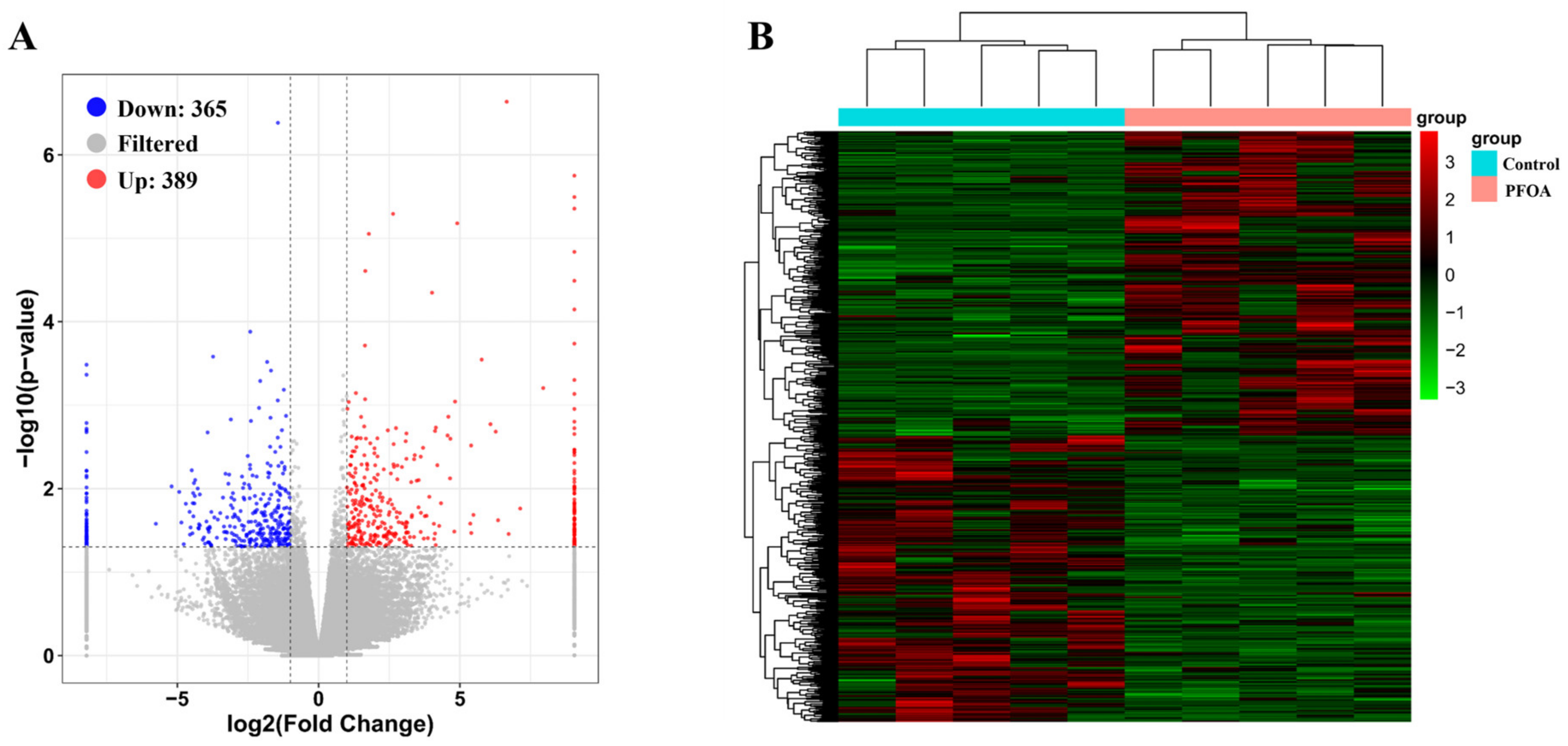
Figure 1. DEGs in the PFOA exposure group compared with the control group. (A) Volcano map. (B) Clustering thermogram. The clustering bars gather samples of similar expression patterns. The intensity of the colors shows the magnitude changes of genes.
Diversity 14 00971 g001
Figure 2. The top 20 KEGG pathways with the most significant enrichment of DEGs.
Diversity 14 00971 g002
Figure 3. The top 20 GO terms with the most significant enrichment of DEGs.
Diversity 14 00971 g003
Figure 4. Verification of transcriptome results by qPCR.
Diversity 14 00971 g004
Table 1. Liver transcriptome sequencing analysis.
CategoryC1C2C3C4C5P1P2P3P4P5
Total reads48,625,95636,803,57045,578,51448,676,03638,909,93441,953,88240,187,30238,610,86638,980,17251,300,610
Clean reads44,947,85833,650,20640,731,04844,445,61635,623,16038,395,60036,810,39835,239,34035,923,77446,437,278
Clean read proportion (%)92.4391.4389.3691.391.5591.5591.5991.2692.1590.51
Q20 (%)97.4397.1597.4397.2797.2997.497.1797.1797.1796.28
Q30 (%)93.1292.5193.292.8692.8993.0692.6192.6592.6490.76
GC (%)44.3944.3944.644.4344.4344.3744.1744.7544.3344.39
Unigene number104,143104,143104,143104,143104,143104,143104,143104,143104,143104,143
Unigene average length (bp)979.22979.22979.22979.22979.22979.22979.22979.22979.22979.22
Unigene N50 (bp)1480148014801480148014801480148014801480
Note: C represent Control; P represent PFOA.
Table 2. Gene function annotation.
DatabaseNumber of Annotated UnigenesPercentage (%)
NR28,82827.68
GO19,48018.71
KEGG15,81515.19
eggNOG18,81618.07
Swiss-Prot26,83925.77
Pfam22,20421.32
In all databases10,66110.24
Note: NR represent NCBI Non-redundant Protein Sequences; GO represent Gene Ontology KEGG represent Kyoto Encyclopedia of Genes and Genomes; EggNOG represent Evolutionary Genetics of Genes: Non-supervised Orthologous Groups; Swiss-Pro represent Swiss-Prot Protein Sequence; Pfam represent Protein Families.
Publisher’s Note: MDPI stays neutral with regard to jurisdictional claims in published maps and institutional affiliations.