1. Introduction
In recent decades, despite the large number of synthetic drugs used in modern medicine, the healthy lifestyle trend has led to the return of interest in herbal medicaments. Apart from the easier availability of these natural products in comparison to the same compounds obtained synthetically, better compliance of the patient is ensured as natural products are generally believed to be less harmful. On the other hand, natural extracts often contain large amounts of ballast compounds; therefore, the evaluation of the quality of a particular extract considering the target compound concentration is of great interest. In traditional medicine,
Rhaponticum carthamoides, also known as Maral root, has been used for centuries as a great source of toning agents that strengthen the central nervous system and increase mental and physical health [
1,
2]. The extracts were found to contain approximately 200 bioactive compounds belonging to various chemical groups such as ecdysteroids [
3,
4], flavonoids, phenolic acids, and sesquiterpene lactones [
5,
6,
7]. Among them, the greatest attention has been paid to 20-HE, tracheloside, N-feruloyl serotonin, kaempferol, hesperetin, chlorogenic acid, cynaropicrin, and quercetin due to their adaptogenic, antioxidant, anti-inflammatory, spasmolytic, immunomodulatory, anticancerogenic, antimicrobial, and free radical scavenging properties [
8,
9,
10] (
Figure 1). To check the quality of a particular extract, an evaluation of the content of all these compounds at the same time would be highly convenient; however, the issues concerning their co-elution and different ionization conditions have to be solved. The methods dealing with the separation of ecdysteroids [
11], phenolic compounds [
12,
13], flavonoids [
14], or sesquiterpene lactones [
15,
16] separately are known. More complex methods dealing with the determination of the content of species belonging to two compound classes, chlorogenic acid, and 20-HE, represent a step towards the intended evaluation of extract quality. Dealing with such a complex matrix, the stability of extracts becomes a parameter that should be considered. There have been several works studying the stability of kaempferol and quercetin in extract samples under ambient conditions, and the thermal degradation of quercetin with enhanced stability under acidic conditions [
17]. Moreover, the optimal conditions for the extraction of phenolic compounds comprise heating to 43 °C in acidic pH [
18], indicating the stability of said compounds under the given conditions.
The aim of our work was to find the optimal conditions for the simultaneous determination of eight bioactive substances from Maral root belonging to various chemical groups without time consuming sample preparation processes despite a very complex matrix. A detailed study encompassing the stability of all the main compounds of interest from Maral root under stress conditions may help to define the conditions for extract quality evaluation without distorting the results of the analysis.
2. Materials and Methods
2.1. Chemicals
The standards of 8 substances were purchased from Biopurify TCM, China (tracheloside 98%), from TRC, Canada (N-feruloyl serotonin 95%), and from Sigma-Aldrich, Czech Republic (kaempferol 97%, hesperetin 95%, chlorogenic acid 95%, cynaropicrin 98%, quercetin 98%, and 20-HE 93%). All compounds were measured as solutions in methanol (Lach-Ner, Prague, Czech Republic, HPLC gradient grade). Other solvents for HPLC were acetonitrile—ACN, isopropyl alcohol—IPA (Lach-Ner, Prague, Czech Republic, HPLC gradient grade), formic acid, ammonium formate (Lach-Ner, Prague, Czech Republic, 99%), and ultrapure water (Ultrapur Watrex system, Prague, Czech Republic). Gallic acid (Sigma-Aldrich, Prague, Czech Republic, ACS reagent), Folin–Ciocalteu reagent (Sigma-Aldrich, Prague, Czech Republic, 2M with respect to acid), sodium carbonate (Lachema, Brno, Czech Republic, 98%), aluminum chloride hexahydrate (Sigma-Aldrich, Prague, Czech Republic, 99%), and ultrapure water with conductivity and resistivity of 0.055 uS/cm and 18.18 mΩ·cm, respectively, at 25 °C were used for the determination of the total content of flavonoids and phenolic compounds in the extracts.
2.2. Preparation of Standard Solutions and Calibration Methodology
The methanolic stock solution containing 1 ± 0.1 mg/mL of each of the 8 standards of bioactive compounds was prepared. The calibration curve was verified at eight mass concentration levels within the range from 1 to 150 µg/mL for three independent peak measurements. This resulted in a linear calibration dependency for all studied compounds.
2.3. Plant Material
Dry Maral roots supplied by F-DENTAL Hodonín s.r.o. were milled (Retsch SM 200) to a particle size lower than 0.6 mm and stored in a dark and dry place. Three different extraction techniques were performed in order to obtain the highest yield of individual compounds. Maceration, one of the most common and simplest extraction methods, was performed at atmospheric pressure and room temperature for 48 h using 10 g of plant in 250 mL of ethanol. Due to possible degradation caused by light, macerated flasks were wrapped in aluminum foil and left shaken. The second method used was Soxhlet extraction (SOX), which is carried out at the boiling point of the used solvent. Dry plant material (10 g) was extracted using 250 mL of ethanol in Soxhlet apparatus for 1 h. The rotary vacuum evaporator (Rotavapor R-215, BUCHI, Switzerland) was used to remove the solvent from extracts obtained by Soxhlet extraction and maceration. In comparison to the above-mentioned methods, the pressurized liquid extraction (PLE) as a third extraction technique was performed at the highest temperatures for the shortest time period. The extraction column, filled with glass beads, cotton wool, and plant material, was placed in the oven pre-heated up to 120 °C. Ethanol, as the chosen solvent, was pumped using a pre-set flow HPLC pump into the column until the pressure reached the desired value (up to 10 MPa). After pressurization, the extraction was performed for 10 and 20 min to allow the solvent to enter the pores of the plant material and dissolve the desired substances. Each PLE experiment consisted of three cycles, resulting in three extract fractions from each experiment. After obtaining the extract, the solvent was evaporated to a constant weight using an elevated temperature (40 °C) and flowing air.
For analysis, approximately 25 mg of the obtained extracts was diluted in methanol providing the final concentration of ≈5.4 g/L. Individual samples were injected directly into HPLC, without any further treatment. All standard and real samples were stored at 4 °C in dark to avoid oxidative and light degradation.
2.4. Assessment of Total Phenolic and Flavonoid Content
In addition to determining the quantity of individual substances in the samples, the total content of phenolic and flavonoid substances was also monitored in all the samples. For the determination of the total phenolic content, an already known method using Folin–Ciocalteu reagent was applied. This spectroscopic method, also called the gallic acid equivalence (GAE) method, uses gallic acid as a standard, where the resulting concentration value is converted to an equivalent amount of gallic acid [
19].
A similar principle was used to determine the total flavonoid content in samples [
20]. The colorimetric method using quercetin as a standard to generate a standard calibration curve also required prepared samples in a 1:1 ratio with 2% aluminum chloride hexahydrate solution. The flavonoid concentrations are expressed as mg quercetin equivalent (QE)/g dried extract. Absorbance of the reaction mixtures of phenolics and flavonoids was measured against a blank at 760 nm and 420 nm, respectively, using an Evolution 220 UV-VIS spectrophotometer (Thermo Scientific, USA).
2.5. HPLC-MS Conditions
Targeted chromatographic analysis was performed using Dionex Ultimate 3000 HPLC system (Thermo Scientific, Waltham, MA, USA) with a DAD and MS detector. Separation was achieved using Synergi Polar-PR C18 column (150 mm × 4.6 mm I.D.). The column was thermostated at 25 °C and separation was performed with gradient elution. Ratio of 90:10 (v/v) of IPA:ACN was selected as component A, while the ratio of 60:40 (v/v) of ACN:water was chosen as component B with the addition of 2.5 mmol/l of ammonium formate. A gradient program was applied as follows: 0–7 min 5–50% A, 7–15 min 50% A, 15–20 min 50–95% A, 20–30 min 95% A, 30–35 min 95–5% A. The total run time with a rate flow of 1.0 mL/min was 35 min. The injection volume was set to 5 µL.
Detection and identification of individual compounds was performed by comparing the retention times along with UV and MS spectra of the real samples with standard compounds. The micrOTOF-Q III mass spectrometer (Bruker Daltonik, GmbH, Bremen, Germany) was operated using electrospray ionization (ESI) as the ionization technique in positive mode. All measurements by MS were performed with following parameters for ESI: nitrogen as nebulizer (1.6 bar) and drying gas (180 °C/8 L/min), the capillary voltage of 4200 V, the end plate offset −500 V, and the collision cell RF 350 Vpp. The mass range for the scans of MS spectra was m/z 80–1550 with low mass of 100 Da. UV detection at 254 nm was carried out using a DAD detector, as part of HPLC instrument. Collection of chromatographic and mass spectrometric data was carried out using HyStar 3.2 software (Bruker Daltonik, GmbH, Bremen, Germany) and for data processing Compass to of series 1.5 Data Analysis 4.1. (Bruker Daltonik, GmbH, Bremen, Germany) was used.
3. Results and Discussion
Based on the structure and physical and chemical properties of the plant’s secondary metabolites, several mobile phases, two stationary phases (Synergi 4µ Polar-RP 80A and Luna 3µ (C18) 100A, both 150 mm × 4.6 mm I.D.), and two ionization sources (ESI and APCI) were examined. All studied compounds showed better separation performance on the Synergi 4µ Polar-RP 80A column, providing better separation of more polar compounds than the standard C18 column. On the other hand, for ionization both sources could be used, but based on the obtained results and few published works, the ESI source was preferred due to its high ionization efficiency for phenolics, flavonoids, and other similar compounds [
21]. During the optimization process, several changes were made to evaluate the effect of the mobile phase composition and the additive concentration. After methanol was expelled as a mobile phase constituent due to non-satisfactory preliminary results, the dependence of the efficiency of the method on the ratio of acetonitrile in both components of the mobile phase was monitored. Enhancement of the MS signal was improved by the addition of an ammonium formate buffer at a concentration of 2.50 mmol/L (
Table 1). Two analytes were chosen, hesperetin and kaempferol, as they do not have baseline separation in UV spectra. The developed gradient elution profile with the required ACN ratio described in Section HPLC-MS conditions reached the highest resolution of the compounds of interest in the shortest run time.
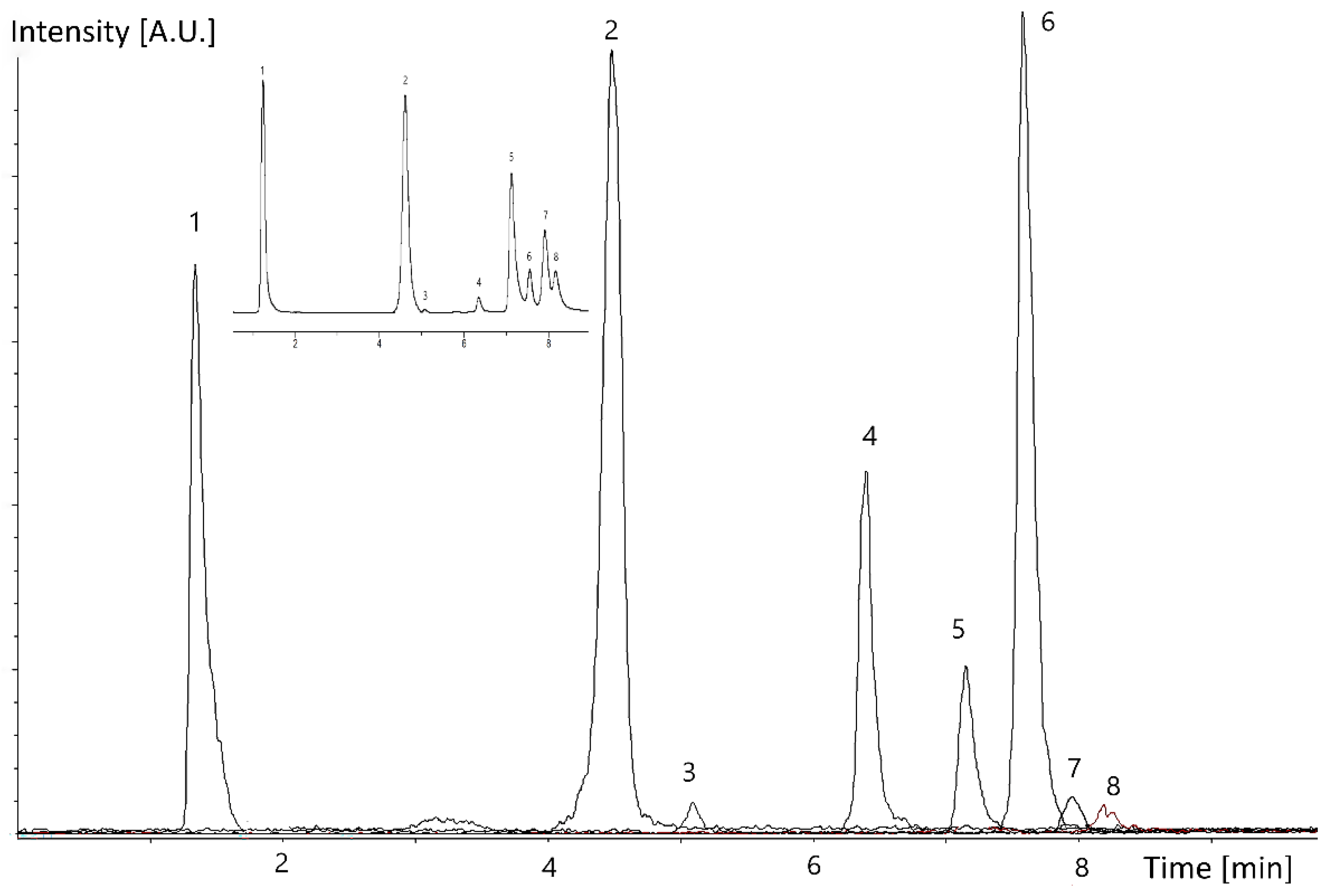
Since some compounds have a similar mass, effective and efficient separation is required (
Figure 2). Chlorogenic acid (355.3110, [M+H]
+), as the most polar compound of interest, elutes first, being well separated from the other composites. The peak of 20-HE is not perfectly separated from the peak of cyanopicrin, but the compounds may be easily distinguished based on their molar masses: cyanopicrin (347.1416, [M+H]
+) and 20-HE (481.5860, [M+H]
+). The peaks of tracheloside (551.5650, [M+H]
+) and quercetin (303.0499, [M+H]
+) are well separated provided the N-feruloyl serotonin (353.3980, [M+H]
+) content is not too high. The molecular masses are also used to identify and quantify the peaks belonging to hesperetin (303.0870, [M+H]
+), and kaempferol (287.0554, [M+H]
+). The only compounds with near values of molecular masses, quercetin and hesperetin, have retention times in the used method almost 1 min apart from each other. Although the compounds are eluted in the first 10 min, a run time of the method up to 35 min is necessary due to the elution of other components found in the extract, which by shortening the run time cause interaction and distorts the results. The key activity in chemical analysis to obtain reliable results is to check the validation parameters of the given method, such as linearity, precision, repeatability, stability, and recovery. The optimized conditions of the method along with limit of detection (LOD) and limit of quantification (LOQ), determined at signal-to-noise ratios (S/N = 3 and 10, respectively) provided the results shown in
Table 2. The molecular qualifier ions used to construct the calibration curves were selected from the mass spectrum of the target compounds using an extracted-ion chromatogram (EIC). Each point of the calibration graph corresponded to the mean value from three independent peak measurements.
The precision and accuracy of the proposed method were verified by intra-day and inter-day repeatability at a mass concentration level of 20 μg/mL for each compound. The intra-day precision was tested with six repeated injections at 100% of the test concentration, while inter-day precision was monitored every 2 weeks within half a year with a total of 12 analyses. Although some of the guidelines such as ICH, Eurachem, and IUPAC do not specify the acceptance criteria, the precision, expressed in %RSD and accuracy, determined by relative error (RE %), was within the acceptable range established by the FDA [
19] (
Table S1). According to the FDA guidance, precision should be within 15% of the nominal value (20% at LOQ level), while the accuracy criteria for an assay method is set above the range of 80 to 120% of the target concentration.
The next step, including sample analysis and recovery, required triplicate measurements of samples obtained by the three different extraction techniques described in the
Supplementary Material Table S2. Each extraction technique was performed using the same plant material. Assessment of the total phenolic and flavonoid content was performed as an assay in terms of quality control of the extraction methods, as well as control of the correct analytical measurement and analyte recovery. The content of total phenolic and total flavonoid compounds in the extracts was first tested by colorimetric methods to evaluate the efficiency of the given extraction method. In this regard, these methods give extracts of repeatable quality, whereby those obtained by the PLE and SOX methods provided material with a higher content of the compounds of interest for a shorter time. The content of each studied compound estimated as mg per g of extract by interpolation of the HPLC peaks with calibration curves constructed for the standard compounds was also in relation to the content of total phenolic and flavonoid compounds. The compounds evaluated by the present HPLC/MS method represented up to 23% of the total phenolic compounds present in the individual extracts. The validity and reliability of the method for the measuring of the bioactive compounds from the Maral root were evaluated by recovery experiment. The recovery of the method was determined using three extract samples (
Table S2). In the spiked samples, the concentration of each analyte increased by 100% and the percentage ratio between the recovered and expected concentrations was calculated. Satisfactory results expressed as a percentage suggest that the proposed HPLC-MS method is reliable for the quantification of the eight bioactive compounds found and isolated from the Maral root and provides good validation. Although analyte stability may affect the trueness and precision of the method, it is not a mandatory validation parameter except in EMA, FDA, and AOAC guidelines. On the other hand, the extracts as complex systems containing residues of the matrix may be highly influenced by stress conditions during storage. While the studied compounds proved to be stable at −30 °C for 5 days in the dark dissolved in methanol as standards and in the extracts, storage at laboratory temperature for 48 h led to extract degradation (
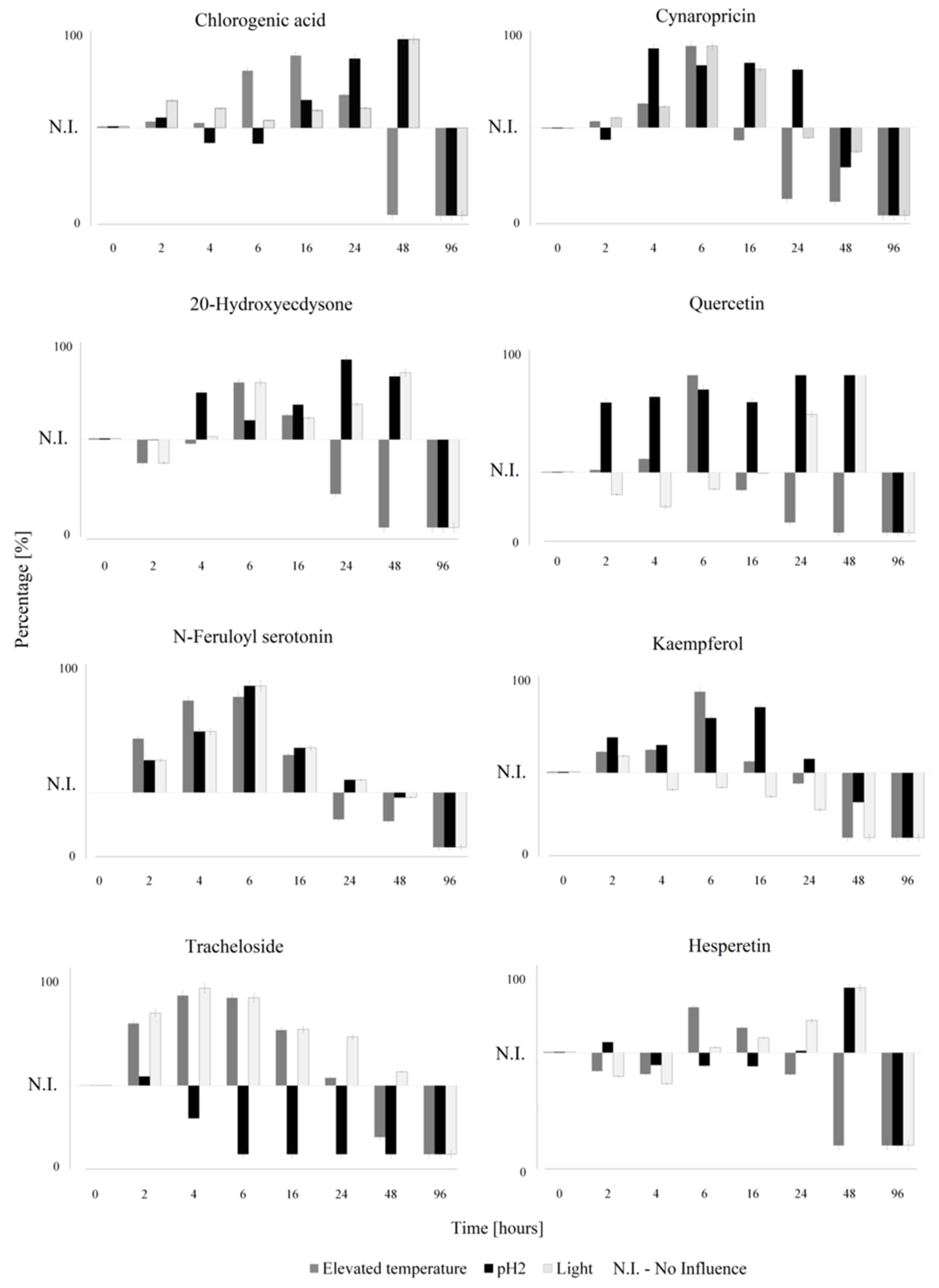
Table S3). However, the extracts showed an RSD of less than 2%, even after this time interval. Although the stability results of the individual bioactive compounds present in the extracts were satisfactory, a longer storage time under stress conditions led to degradation and increased the %RSD by over 2%. To follow the trends of the distortion of the results for the particular compounds, we applied the developed analytical method to the samples stored at elevated temperature (45 °C) and exposed to light and to the acidic environment (pH 2). The results are presented in
Figure 3, and illustrate the deviation of the compound content from the initial value over time.
The studies revealed that each group of plant secondary metabolites had individual stability and degradation kinetics, but some general comments may be deduced.
The short-time storage at elevated temperatures was not necessarily devastating for the extract quality. Even thermally less-stable compounds such as quercetin and 20-HE may be heated for a few hours without decomposing. This is important, for example, for the derivation of the time window for the use of a preheated autosampler. On the other hand, the levels of certain compounds in the samples at elevated temperatures rose due to being released from the matrix structures, positively distorting the results. Moreover, it became clear that the post-extraction composition of the material may be manipulated by heating. When the extract is intended to be rich in tracheloside and/or N-feruoyl serotonin, then short-time storage at elevated temperatures may be favorable.
On the contrary, under conditions of an acidic pH, the tracheloside was quickly lost. The remaining compounds were generally stable at an acidic pH, where the effect of stabilization was especially pronounced in the case of quercetin and cyanopicrin. The results involving higher pH values, i.e., around 12, were not processable due to complete degradation of the target compounds after one hour of exposure.
One-day exposure to daylight did not cause severe changes to the extract composition, except in the case of tracheloside. As the light exposure may lead to complete degradation, it may also cause chemical alteration which can enhance the extractability of the bound bioactive compounds [
20]. This has a great influence on obtaining extracts rich in lignans.
Summarizing the above conditions, it is possible to conclude that the right combination of critical parameters provides the highest yield of the selected bioactive compound. For example, obtaining a lignan-free extract requires adjusting the pH of the sample to a value close to pH 2, while elevated temperatures applied for 5 to 10 h along with an acidic pH provide extracts rich in phenolic and flavonoid compounds. Nevertheless, applying these conditions to the extraction method may lead to optimization from which it would be possible to obtain the selected amounts of the individual bioactive compounds.
Although the trends for the individual bioactive compounds were different and varied depending on the used conditions, degradation during a longer storage time was observable in each compound.


