Article

Identifying Early Stages of Freshwater Fish with DNA Barcodes in Several Sinkholes and Lagoons from the East of Yucatan Peninsula, Mexico

by
Adrián Emmanuel Uh-Navarrete
1,
Carmen Amelia Villegas-Sánchez
2,
José Angel Cohuo-Colli
1,
Ángel Omar Ortíz-Moreno
3 and
Martha Valdez-Moreno
1,*
1
El Colegio de la Frontera Sur, Department of Aquatic Ecology and Systematics, Av. Centenario Km. 5.5, Chetumal 77014, Quintana Roo, Mexico
2
Tecnológico Nacional de México/I. T. Chetumal, Av. Insurgentes No. 330, Chetumal 77013, Quintana Roo, Mexico
3
Comisión Nacional de Áreas Naturales Protegidas, Reserva de la Biosfera Sian Ka’an, Calle 61 # 767, Colonia Centro, Felipe Carrillo Puerto 77200, Mexico
*
Author to whom correspondence should be addressed.
Diversity 2021, 13(11), 513; https://doi.org/10.3390/d13110513
Submission received: 17 September 2021 / Revised: 18 October 2021 / Accepted: 20 October 2021 / Published: 22 October 2021
(This article belongs to the Special Issue Aquatic Organisms Research with DNA Barcodes)

Abstract

:
Our work shows the efficacy of DNA barcoding for recognizing the early stages of freshwater fish. We collected 3195 larvae and juveniles. Of them, we identified 43 different morphotypes. After DNA barcodes of 350 specimens, we ascertained 7 orders, 12 families, 19 genera, 20 species, and 20 Barcode Index Numbers, corresponding to putative species. For the first time, we reported the presence of the brackish species, Gobiosoma yucatanum in Lake Bacalar. Specimens of the genus Atherinella sp. and Anchoa sp. are possibly new species. Using both methods, morphology, and DNA barcodes, we identified 95% of the total larvae collected (2953 to species, and 78 to genus), and all of them were native. From them, the order Gobiiformes represented 87%. The most abundant species were Lophogobius cyprinoides and Dormitator maculatus, followed by Gobiosoma yucatanum and Ctenobius fasciatus. The Muyil and Chuyanché lagoons have the highest number of species. We present for the first time a short description of Cyprinodon artifrons and Floridichthys polyommus. This information conforms an indispensable baseline for ecological monitoring, to evaluate impacts, and developing management and conservation plans of biodiversity, principally in areas under human pressure such as Sian Ka’an, and Lake Bacalar, where tourism is high and growing in disorder.

1. Introduction

Quintana Roo state, in the east of the Yucatan Peninsula, Mexico, is among the places with higher levels of biodiversity. For this reason, on average this region receives 5 million visitors annually [1], and in the last decade contributed with the highest tourism income for the whole country [2]. Among the most visited natural attractions of the state are freshwater bodies such as lagoons and cenotes (sinkholes that connect to one of the most complex underground water systems in the world) [3]. However, the richness of the fishes that inhabit such ecosystems is still poorly known. Specific studies about their larvae are almost absent; the few fish larvae studies developed here have focused on marine organisms and estuarine environments [4,5,6,7,8].
The knowledge of the early stages of fish is essential as it provides information about the recruitment rates of juveniles and the size of the adult population [9,10]. Besides, these stages are helpful in the characterization of taxonomic diversity, times, and locations of spawning, and the assessment of connectivity between ecosystems [11].
The early stages of fish are considered as ichthyoplankton. They are characterized by a high rate of development and a great diversity of forms that usually differ from adults [9]. Furthermore, the morphology of the same species can change rapidly and significantly during its growth from pre-flexion to post-flexion larvae and later to the juvenile stage [12]. Hence, the identification using morphology tends to be difficult.
Given the difficulties in identifying fish larvae by traditional methods, applying multiple and complementary perspectives is advisable to identify them more precisely [13,14]. Integrative taxonomy proposes using morphological characteristics and molecular identification [15,16]. This type of work has been used successfully before, as evidenced by the study of Valdez-Moreno et al. [7], Hubert et al. [17], Baldwin, et al. [18], and Ko et al. [12], among others.
Considering the lack of information and the relevance of the studies that help understand biodiversity, this research has the goal to analyze the identities of the fish larvae that inhabit freshwater ecosystems from Quintana Roo, based on integrative taxonomy. We consider this work as a starting point towards the conservation and sustainable use of these systems.

2. Materials and Methods

2.1. Study Site and Field Sampling

We collected in 18 places; 3 of them in Sian Ka’an, a Biosphere Reserve, 9 nearby the reserve, 4 in Lake Lake Bacalar, and 2 lagoons Xul-Ha, and Huay Pix associated with Bacalar (Figure 1 and Table 1).
We collected the samples using light traps according to the methodology proposed by Elías-Gutiérrez et al. [11]. We placed only one light trap in most sites, except in Muyil, Chunyanxche, Del Padre, Siijil Noh Ha, and Chancah Veracruz, where due to their depth and area, two traps were used (one littoral and one limnetic). The sampling sites and collection dates are summarized in Table 1. Figure 1 shows the geographic locations of all sampled sites.
Once we got the samples, we immediately filtered them on a 50 μm sieve, then we fixed the material with ethanol 96% and placed it on ice. In the lab, all samples were stored for at least one week at −18 °C before processing [11,19].

2.2. Morphological Analysis

In the laboratory, the larvae were separated by morphotypes and stored in 5 mL vials with 4 mL of 96% ethanol.
For the morphological identification of the larvae, we used different identification keys as Richards [9], Fahay [20], and studies of freshwater fishes where their larval stages are described [21]. For information about what species could be found in the study area, we used Miller et al. [22], Schmitter-Soto [23], and Valdez-Moreno [24,25].
All larvae also were measured and separated according to the stage of development (preflexion, flexion, postflexion, transition, and juvenile) following the previous criteria proposed [9].

2.3. DNA Barcode Analysis

We selected 353 specimens for this study (from 1 to 5 organisms of each morphotype). All of them were photographed under a Nikon SM2 745T stereomicroscope with an Eos Rebel T7i camera.
We used a small piece of muscle (1–3 mm3) or the right eye to extract the DNA for molecular analysis. We sterilized the forceps and the material using chlorine diluted in water in a proportion 1:5 and subsequently neutralized it with 96% ethanol between each tissue or eye extraction.
For each sample’s tissue digestion, a lysis buffer was used with proteinase K, and they were allowed to digest overnight at 56 °C. The extraction was carried out through 1.0 mm PALL glass fiber plates [26]. A Cytochrome Oxidase I (COI) gene segment with an approximate length of 650 Bp [27] was amplified using the FishF1 and FishF2 primers [28,29]. Amplification was carried out with a final volume of 12.5 μL, prepared as follows: 6.5 μL of 10% trehalose, 2 μL of ultrapure water, 1.25 μL PCR buffer X10, 0.625 μL MgCl2 (50 mM), 0.125 μL of each Primer (0.01 mM), 0.06525 μL dNTP mix (10 mM), 0.625 μL Taq polymerase, and 2 μL of template of DNA. The reactions we cycled at 94 °C for a 1 min, followed by five cycles at 94 °C for 30 s, 45–50 °C for 40 s and 72 °C for 1 min, followed by 35 cycles at 94 °C for 30 s, 51–54 °C for 40 s and 72 °C for 1 min, and finally by one last cycle of 72 °C for 10 min. We visualized the PCR products in agarose gel Invitrogen TM with 4 μL of sample and 16 µL of water. We sequenced the PCR products of Cenote Cocalitos samples at the Canadian Center for DNA Barcoding (Guelph, ON, Canada), and the rest of PCR products were sent to sequence at Eurofins Scientific (Louisville, KY, USA). Finally, we edited the sequences with Codon code v.3.0.1 and uploaded them to BOLD (www.boldsystems.org) in the dataset DS-FLYP Fish larvae from Yucatan(dx.doi.org/10.5883/DS-FLYP).

2.4. Data Analysis

The sequences obtained were compared with sequences previously published using the specimen identification tool in the Barcode of Life Data System (BOLD) [30]. In addition, these sequences reached the standard to get a Barcode Index Number (BIN) [31].
We used Kimura 2-parameter model (K2P) to get the genetic divergences between the species [32] and a maximum likelihood (ML) tree, using 500 bootstrap replicates [33], provided with MEGA 7.0 software. Finally, the ML tree was simplified by using the compression feature provided by the same software [34].
The criteria to assign taxonomic level identification using BOLD was a similarity value ≥99%. The resulting BIN number allowed to assign the specimens to species level [35]. Similarities with values ≥ 94% to ≤98.4% were identified to genus, and similarities <94% were assigned to family [36].

3. Results and Discussion

3.1. Species Identification

We collected a total of 3195 larvae and juveniles. Using only morphology, we recognized 43 different morphotypes and only one species, Bathygobius soporator. We assigned the remaining 42 morphotypes to genus (Gobiosoma, Ctenogobius, and Astyanax) or family (Cyprinodontidae, Engraulidae, Gobiidae Atherinopsidae, Cichlidae, Poeciliidae, and Characidae).
These results showed how difficult the identification of the early stages of fish using only morphological characters is. There is no information available for most freshwater fish larvae [37,38]. In addition, the fragile specimens can be damaged during the manipulation and fixation process, which makes the identification more difficult [36,39]. In some cases, the larvae can lose some body parts by predation when all zooplankton are together in the light trap (obs. pers.). The result is either a high probability of erroneous taxonomic identification or simply making it impossible.
For DNA barcode analyses, we processed 350 specimens from the 43 morphotypes. We obtained 347 sequences. Their length varied between 600 and 658 base pairs (bp), after trimming the sequences to remove un-formative nucleotide sites at the 3′ and 5′ ends, except for five, which had from 171 to 585 bp. Only three specimens could not be sequenced. We did not observe insertions, deletions, or stop codons in any of the sequences.
We considered a mini-barcode sequence with 171 bp [40], belonging to Gobiosoma yucatanum with 100% similarity. This technique has been used previously, demonstrating its efficacy in identifying degraded DNA [41,42,43,44].
Our sequencing success rate was 99%. This value is similar to that reported by Frantine-Silva et al. [39], who registered a 99.81% success rate on fish eggs and larvae from the Paranapanema River in Brazil. In contrast, Almeida et al. [36] had a lower value, reporting 79.6% success in the same river with similar material. This difference could be explained because the latter authors used a low alcohol percentage to fix the samples (70%) and did not keep them in cold storage. Several authors suggest optimizing DNA fixation [11,35]. The length and quality of the sequences were similar to other studies that worked with this gene.
When comparing the 347 sequences with Bold identification system (http://www.boldsystems.org/index.php/IDS_OpenIdEngine. Accessed on 15 September 2021), 324 (93.4%) matched in BOLD reference library with >99% similarity, allowing the species-level identification [35,36] (Table 2). For the remaining 23 (6.6%), we identified them to genus level, 22 Atherinella sp., and one Anchoa sp.
All sequences represented 7 orders, 12 families, 19 genera, 20 species, and 20 BINs (Table 2 and Supplementary Materials. Gobiiformes were the order with more species (five species), followed by Cyprinodontiformes, Clupeiformes (four), and Cichliformes (three).
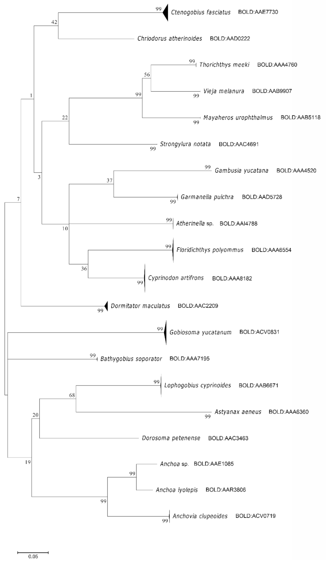
All species identified were based on similarity values that confirmed their placement under different numbers of BINs [31]. These were congruent with the K2P distance tree, and the patterns are seen in the genetic distance.
The ID tree does not show overlapping between species clusters (Figure 2). These results allowed us to make reliable species assignments [45,46].
Each BIN number was associated with one species, except Astyanax aeneus and Cyprinodon artifrons. In these two cases the BINs cannot distinguish them, due to both having congeners closely related that have recently evolved. Consequently, the genetic distance between them is small [28,35,47,48].
Most of the fish species found in this study have been reported in different aquatic systems in Yucatán and the Quintana Roo state [22,24]. Valdez Moreno et al. [25], using metabarcoding in the same places as us (except Minicenote), corroborated the presence of all the species found by us in these sites [24,25]. However, we found some interesting cases, explained in the following paragraphs.
We collected Anchoa lyolepis in the Cocalitos sinkhole in Lake Bacalar. It is considered a marine species and has been reported in the northern Gulf of Mexico and from Yucatán to Brazil [49]. In the BOLD database, there are eight specimens, one is from the same sinkhole, but the specimen is incomplete. It has no head, so morphological determination is difficult. The other samples are dried fishes collected from a Mexico City market, whose morphology appears to be this species. So, it is probable that our larvae seem to be well identified. This observation will have to be confirmed with more specimens.
The Gobiosoma yucatanum holotype was collected from the south side of the pier in Chetumal city, Quintana Roo [50]. Its distribution range includes rivers, estuaries, and inland lagoons from Mexico to Belize and Honduras [51,52]. Elías-Gutiérrez et al. [11] reported the presence of Gobiosoma sp. in the larval stage in Lake Bacalar. In the BOLD database, there are adults from Chetumal Bay, all matched with our larvae. It is the first report of the presence of G. yucatanum in Lake Bacalar. This result confirms the connectivity between the Chetumal bay and Lake Bacalar. Something similar was reported for Cyprinodon artifrons, whose adults are located in the in the reef lagoon in Xcalak and Chetumal Bay, and the larvae which were in Lake Bacalar [11].
Some specimens of the genus Atherinella matched with two species, A. alvarezi and Atherinella sp., with a 100% similarity value (Table 2). Both species have also been reported in inland waters of the Yucatán peninsula [22,23,24,53]. However, the morphological characters overlap between them. The taxonomy of freshwater atherinopsids of this region needs further studies because they possibly belong to an undescribed species [23].
One specimen collected in Huay Pix near Lake Bacalar matched with Anchoa sp. Four species of this genus were found in freshwater. However, Anchoa parva is the only one reported in the Yucatan Peninsula and Bacalar, Quintana Roo [22,23]. We do not have any barcodes for it yet. It is necessary to collect and sequence this species and compare it with our larvae to confirm its identity.
With the barcode results, we were able to recognize the different morphotypes. Then, it allowed us to identify 2681 larvae that did not enter the molecular analysis. These results are excellent because we identified 95% (3031) from all the collected larvae and juveniles. In addition, it allowed us to know the stage of development in each of them (Table 3).
Most of the larvae identified belonged to the order Gobiiformes, representing 87% of the total. The most abundant species were Lophogobius cyprinoides (992 specimens), Dormitator maculatus (655 specimens), G. yucatanum (556 specimens), and Ctenobius fasciatus (550 specimens). The rest of the species had less than 88 to 1 individual. The most widely distributed species were D. maculatus and Atherinella sp. located in four different sites (Table 3).
These results are different from those reported by other studies, based in adults. In three previous research about the fish community structure in the Sian Ka’an reserve, it was found that Cichlidae and Poeciliidae are the families with more species in this area [54,55,56]. Their presence and other families have been found in samples from these aquatic environments (obs. pers.) and using environmental DNA [24,25,57]. These differences are most likely due to different collection methods and objectives (minnow traps, dip, cast nets, hanging nets, DNA in water, and none of them collected larvae).
In our results, we reported larvae from the poeciliid Gambusia yucatana. However, their larvae were not collected with the use of the light traps. We collected some adults with a hand net in Del Padre sinkhole and put them in an aquarium. The females gave birth to some young, which were barcoded and included in this study (Table 3).
These results show that light traps have limitations due to their selectivity [58]. Some species seem to be less attracted to light, such as the poeciliids. Most likely, the parental care of cichlids such as Mayaheros uruphtalmus, Thorichthys meeki, and Vieja melanura [59] prevents their larvae from dispersing and reaching the light traps (obs. pers.). It is necessary to perform more studies about fish behavior to confirm these ideas.
The results regarding the number of species per site were variable. Cocalitos Sinkhole had nine species, Muyil lagoon had six, and Chuyanché lagoon had five. The rest of the localities presented from three to one species, while no larvae were collected in Santa Teresa, Tres Reyes 1, and Tres Reyes 2.
Although Cocalitos has the highest number of species, it is necessary to consider that this place was sampled six times compared to the others, which were visited once or twice (Table 1). Based on our results and previous studies [25], Muyil and Chuyanché lagoons have the highest number of species. The differences among the richness reported can be associated with several factors such as vegetation, shore area, and collecting method [54,56].
The analysis of the different early development stages showed that the larval stage was predominant in all species, except A. aeneus, represented by the juvenile stage. A. lyolepis and Anchoa sp. were found in the transition stage.
The most common larval stage was postflexion in 12 species, followed by the flexion stage in 6 species and preflexion in 4 species.
Atherinella sp., Anchovia clupeoides, and G. yucatanum were the species that showed four different stages during the same sampling day. Chriodorus atherinoides, A. aeneus, Dorosoma petenense, A. lyolepis, Anchoa sp., and G. yucatana were found in a single stage. The other 11 species had 2 or 3 stages (Table 3).
The high percentage of larvae in postflexion indicates that there has been a recent reproduction period. Schmitter-Soto [23] mentions that the reproduction period of C. artifrons is during spring and autumn. For T. meeki, M. urophthalmus, V. melanura and A. aeneus, it is from March to June, while D. maculatus is from September to October. In thecase of Floridichthys polyommus, its reproduction is in spring and summer [60]. Miller et al. [22] reported that the genus Atherinella and G. yucatana have long reproductive periods. Therefore, most of the species found here have reproduction periods close to or during the summer.
Some authors report that the caudal and pectoral fins are the first to develop because they are the main ones involved in the locomotion of the larvae for feeding and movement [9,61,62,63,64]. These ideas agree with the results presented here. Most larvae were found in postflexion, which developed these fins, allowing them to enter the light trap. It also explains the small number of larvae in the preflexion stage since they have more limited movement [9].
Identifying larvae of C. artifrons, F. polyommus, and Garmanella pulchra (Cyprinodontidae) is challenging because they are morphologically similar. The DNA barcodes allowed us to distinguish them.
In the following paragraphs, we present a short description of the larvae from the first two species. The minimum number of specimens reviewed for each of the stages was five.

3.2. Cyprinodon Artifrons (Hubbs, 1936)

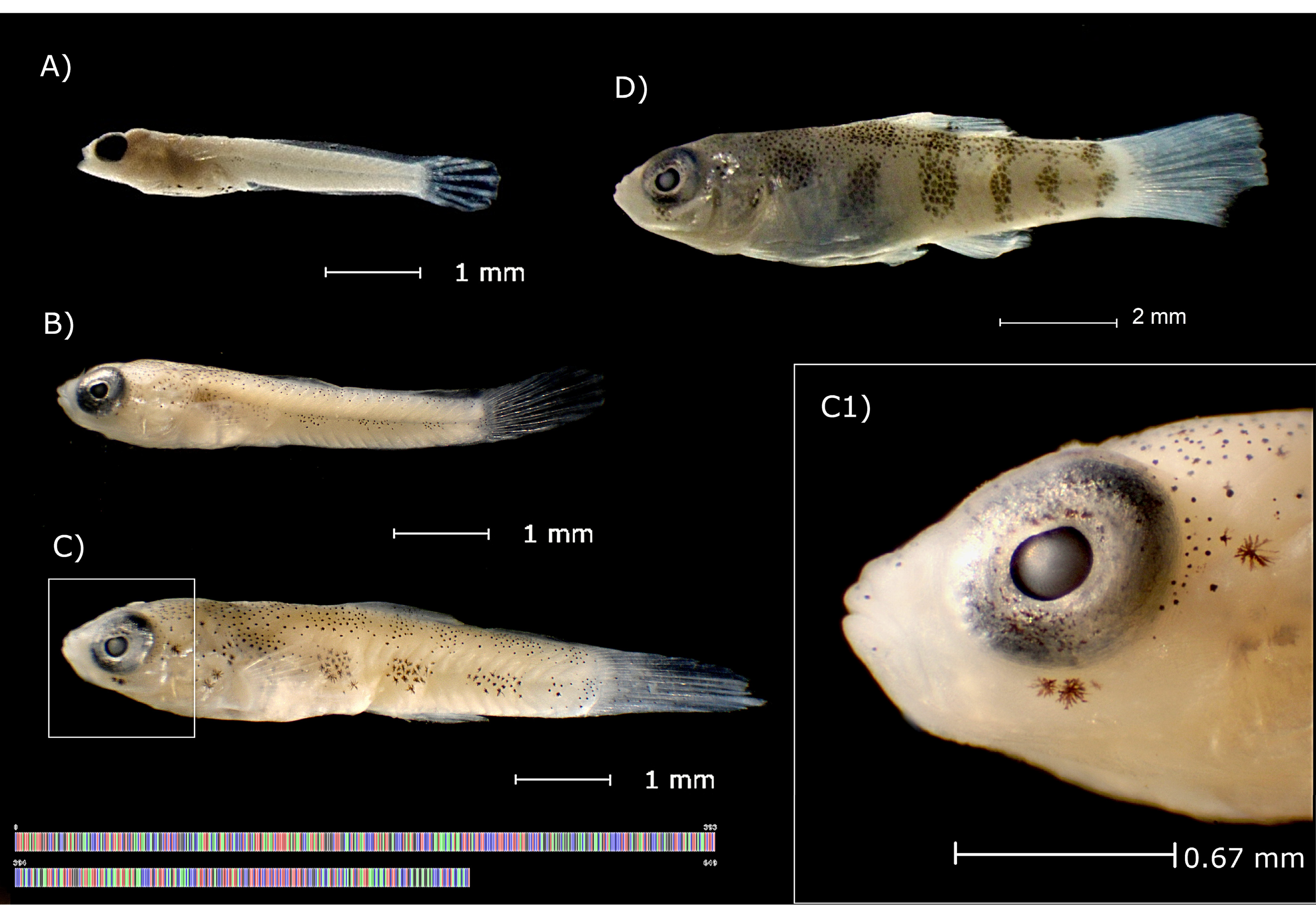
Postflexion (TL 4.2–4.6 mm): presence of finfold; up to 9 pectoral rays. Robust head and dorsally pigmented without a distinct pattern. Eyes with a round pupil. Incipient lateral pigmentation of the body with 5–7 patches of melanophores, the patches beginning above or slightly forward the anus and ending at the base of the caudal fin; some larvae with prominent abdomen have marked ventral pigmentation (linear or slightly branched), larvae without prominent abdomen show less ventral pigmentation and more isolated melanophores (Figure 3A and Figure 5A).
Postflexion (TL 5.0–5.5 mm): presence of finfold; 10–13 pectoral rays. The pupil with a small depression (barely noticeable), with 1–3 melanophores below the eye (Figure 3C1). Slight or absence of ventral pigmentation (Figure 3B and Figure 5A).
Postflexion (TL 6.0–6.9 mm): presence or absent vestigial finfold; 10–14 pectoral rays; 2–6 dorsal rays; 3–4 anal rays. Lateral pigmentation of the body with five patches of melanophores; melanophores on the dorsal part of the head and body; a patch of pigments between the fourth and fifth dorsal fin rays (Figure 3C).
Transition stage (TL 9–11 mm): no finfold; 13–15 pectoral rays; 6–10 dorsal rays; 6–8 anal rays; 4–7 pelvic rays. Head pigmented dorsally and laterally. A slightly oval pupil with further pronounced upper depression; a patch of melanophores below the eye (usually three to four). Incipient scales; pigments and melanophores on the dorsal part of the head and body; five well defined lateral bands, the second band is at the level of the anus, at this same level, is located a patch of pigments in the dorsal fin from the fourth to the seventh radius (Figure 3D).

3.3. Floridichthys Polyommus (Hubbs, 1936)

Postflexion (TL 4.5–5.5 mm): presence of finfold; up to 10 pectoral rays. Robust head and dorsally pigmented; 4.5 mm larvae without pigment below the eye, from 5.4 mm pigmentation appears below the eye. Lateral pigmentation without pattern or with four to six patches of melanophores from anus to the base of the caudal fin; prominent abdomen with large branching melanophores (Figure 4A and Figure 5B).
Postflexion (TL 6.0–6.5 mm): presence of finfold; 11–16 pectoral rays; up to 6 dorsal rays; up to 5 anal rays. A slightly oval pupil with an invagination of approximately 1/5 of the pupil diameter (Figure 4C1); pigmentation below the eye with 3–12 melanophores. Lateral pigmentation without pattern or with four to six patches of melanophores; the size of the abdomen and the ventral pigmentation begins to reduce from 6.5 mm (Figure 4B).
Postflexion (TL 7.0–8.5 mm): presence or absent vestigial finfold; 15 to 18 pectoral rays; 5–10 dorsal rays; 3–7 anal rays; up to 4 pelvic rays. The main characteristics of the eye are the oval shape of the pupil with invagination and the pigmentation below the eye. Lateral pigmentation without pattern or with four to five patches of melanophores; more dispersed pigmentation than C. artifrons in similar sizes already show pigmentation in the form of lateral bands. The abdomen with slight or totally absent pigmentation; pigment at the base of the first dorsal ray (dorsal view) (Figure 4C).
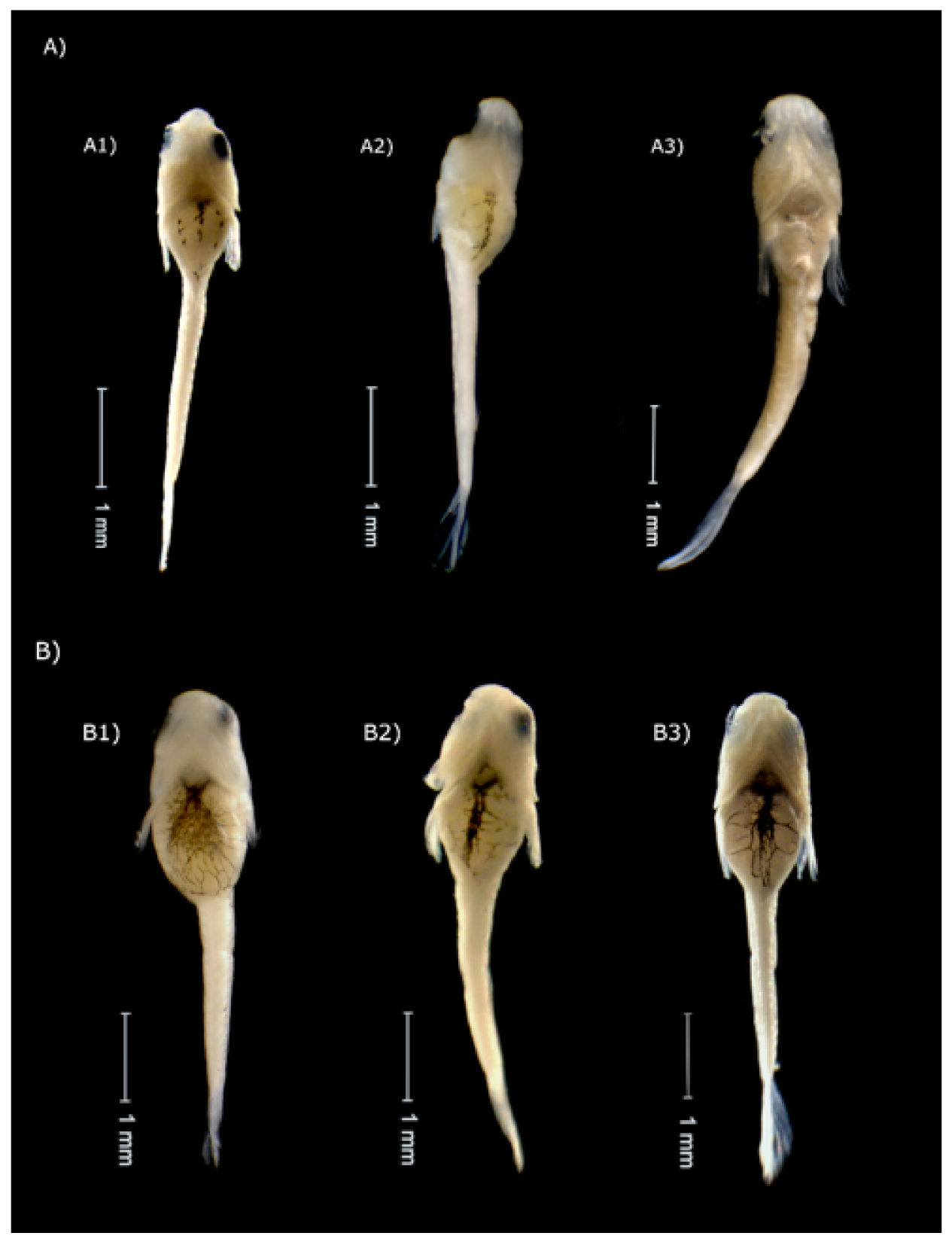
Compared to Cyprinodon artifrons, Floridichthys polyommus has a prominent abdomen and large branching melanophores (Figure 5).
The characters used to separate and describe the larvae of C. artifrons and F. polyommus principally were pigmentation below the eye, lateral pigmentation, the pupil shape and ventral pigmentation. However, we consider that the last two are the most notable and relevant.
We observed that F. polyommus presented an invagination in the pupil in sizes greater than 5.5 mm, unlike C. artifrons had minor depression and was distinguishable only up to 9.0 mm.
The pigmentation below the eyes was different for both species. In the case of C. artifrons, most of the specimens had no more than three melanophores (Figure 3C1), while in F. polyommus up to 11 melanophores were observed (Figure 4C1).
In the smallest sizes, ventral pigmentation and the belly prominent was found for both species. The pigmentation in C. artifrons showed a linear pattern (Figure 5A), while F. polyommus showing this pigmentation more intense and with a branched pattern (Figure 5B). This character has been used previously to delimit some species of the Cyprinidae family [65].
The melanophores patches in the lateral side of the body, at the size of 6.5 mm in C. artifrons are well defined; from 9.0 mm, the melanophores are grouped into bands (Figure 3). In the case of F. polyommus, the patches of lateral pigments were less distinguishable from 6.5 mm (Figure 4).
The smallest sizes were the most difficult to identify because most of the characteristics used to delimit these species appear after 5.0 mm.
It is essential to consider identifying the larvae of these species correctly. It is necessary to take all these characteristics into account as a whole and not individually.

4. Conclusions

In the present study, 95% of the specimens were identified employing DNA barcodes, providing the first report on the taxonomic composition of freshwater ichthyoplankton and their distribution in epicontinental systems in Quintana Roo state.
We report, for first time, the presence of G. yucatanum in the Lake Bacalar and the first larval description of C. artifrons (postflexion of and transition) and F. polyommus (postflexion).
Although the use of light traps has limitations due to selectivity, they have the advantage of catching species that often escape other sampling gears. However, the best approach is using various sampling gears to estimate species richness, measure biodiversity, and plan conservation strategies.
These results are the first report about some aspects of reproductive biology, and development of several species. This information is indispensable for ecological monitoring, to evaluate impacts, and developing management and conservation plans of biodiversity.
Our study confirmed the utility of the DNA barcodes for identifying larvae and juveniles by comparing their sequences with adults uploaded in BOLD.
This work confirms the importance of building DNA barcode reference libraries for all Mexican ichthyofauna.
This research is essential in areas under human pressure from different activities such as the Sian Ka’an Biosphere Reserve and the Lake Bacalar, where tourism is growing mostly in disordered ways.

Supplementary Materials

The following are available online at https://www.mdpi.com/article/10.3390/d13110513/s1, Supplementary Figure S1: ID tree of all specimens found in the dataset used for this paper. Include species name, sample ID, locality collection, BIN number.

Author Contributions

Conceptualization and methodology, M.V.-M. and A.E.U.-N.; software, M.V.-M. and A.E.U.-N.; validation, M.V.-M., A.E.U.-N. and C.A.V.-S.; formal analysis, A.E.U.-N., M.V.-M., C.A.V.-S. and J.A.C.-C.; investigation, A.E.U.-N. and M.V.-M.; resources, M.V.-M., C.A.V.-S. and Á.O.O.-M.; data curation, M.V.-M., A.E.U.-N. and J.A.C.-C.; writing—original draft preparation, M.V.-M., A.E.U.-N., C.A.V.-S. and J.A.C.-C.; writing—review and editing, M.V.-M., A.E.U.-N., C.A.V.-S. and J.A.C.-C.; visualization, A.E.U.-N., M.V.-M., C.A.V.-S. and Á.O.O.-M.; supervision, A.E.U.-N., M.V.-M. and C.A.V.-S.; project administration, M.V.-M. All authors have read and agreed to the published version of the manuscript.

Funding

This study was financed by the Global Environment Fund through the United Nations Development Programme (UNDP, Mexico), Comisión Nacional para el Conocimiento y Uso de la Biodiversidad (CONABIO) and Comisión Nacional de Áreas Naturales Protegidas (CONANP) as part of the investigation called: Programa de detección temprana piloto de especies acuáticas invasoras a través de los métodos de código de barras de la vida y análisis de ADN ambiental en la Reserva de la Biosfera Sian Ka’an within Project 00089333 “Aumentar las capacidades de México para manejar especies exóticas invasoras a través de la implementación de la Estrategia Nacional de Especies Invasoras”. Part of genetic analyzes were funded by a Tecnológico Nacional de México project (serial: 10091.21-P).

Institutional Review Board Statement

Ethical review and approval were waived for this study due to it involving material collected under the permit issued by Dirección General de Ordenamiento Pesquero y Acuícola No P2F/DGOPA-063/20.

Data Availability Statement

All data from this study are available in dataset DS-FLYP Fish larvae from Yucatan Peninsula (dx.doi.org/10.5883/DS-FLYP). In addition, all sequences are available on GenBank (www.ncbi.nlm.nih.gov).

Acknowledgments

Jorge Vidal Quijada, Heliseo Torres Otero, Gilberto Paredes Pat, Isaías Tun Santiaog, and Alejandro Tuyub Tuyub from Limones town guided us within and in the surroundings of the Sian Ka’an Reserve. Edilberto Sosa Martín supported our sampling. All ejidal commissars assisted us in their communities. Alma Estrella García Morales from the Mexican Barcode of Life (MEXBOL) node Chetumal assisted with DNA extraction, PCR reactions, and sequence edition of all material presented here. Janneth Padilla assisted us with the map.

Conflicts of Interest

The authors declare no conflict of interest. The funders had no role in the study design, analyses, interpretation of data, the writing of the manuscript, or the decision to publish the results.

References

  1. SECTUR. Results of Tourism Activity Mexico. Available online: http://www.acs-aec.org/sites/default/files/20150515_rat_a_marzo_15_en.pdf (accessed on 31 August 2021).
  2. Sánchez-Flores, E. La Contribución Del Turismo Al Crecimiento Económico: Análisis Regional En México. Transitare 2016, 2, 183–204. [Google Scholar]
  3. Martos-López, L.A. Underwater Archaeological Exploration of the Mayan Cenotes. Mus. Int. 2008, 60, 100–110. [Google Scholar] [CrossRef]
  4. Campbell, M.; Withers, K.; Tolan, J. Occurrence of Larval and Juvenile Fish in Mangrove Habitats in the Sian Ka’an Biosphere Reserve, Quintana Roo, Mexico. Gulf Caribb. Res. 2008, 20, 81–86. [Google Scholar] [CrossRef] [Green Version]
  5. Quintal-Lizama, C.; Vásquez-Yeomans, L. Asociaciones de Larvas de Peces En Una Bahía Del Caribe Mexicano. Rev. Biol. Trop. 2001, 49, 11. [Google Scholar]
  6. Sanvicente-Añorve, L.; Hernández-Gallardo, A.; Gómez-Aguirre, S.; Flores-Coto, C. Fish Larvae from a Caribbean Estuarine System in Book The Big Fish Bang, 1st ed.; Browman, H.I., Skiftesvik, A.B., Eds.; The Instituto Marine Research: Bergen, Norway, 2003; pp. 365–379. [Google Scholar]
  7. Valdez-Moreno, M.; Vásquez-Yeomans, L.; Elías-Gutiérrez, M.; Ivanova, N.; Hebert, P.D.N. Using DNA Barcodes to Connect Adults and Early Life Stages of Marine Fishes from the Yucatan. Mar. Freshw. Res. 2010, 61, 665–671. [Google Scholar] [CrossRef]
  8. Victor, B.; Vásquez-Yeomans, L.; Valdez-Moreno, M.; Wilk, L.; Jones, D.; Shivji, M.; Lara, M.; Caldow, C. The Larval, Juvenile, and Adult Stages of the Caribbean Goby, Coryphopterus Kuna (Teleostei: Gobiidae): A Reef Fish with a Pelagic Larval Duration Longer than the Post-Settlement Lifespan. Zootaxa 2010, 2346, 53–61. [Google Scholar] [CrossRef]
  9. Richards, W.J. Early Stages of Atlantic Fishes, 1st ed.; CRC Press: Boca Raton, FL, USA, 2005. [Google Scholar]
  10. Rodríguez, J.M.; Alemany, F.; García, A. A Guide to the Eggs and Larvae of 100 Common Western Mediterranean Sea Bony Fish Species; The Food and Agriculture Organization: Rome, Italy, 2017. [Google Scholar]
  11. Elías-Gutiérrez, M.; Valdez-Moreno, M.; Topan, J.; Young, M.; Cohuo-Colli, J. Improved Protocols to Accelerate the Assembly of DNA Barcode Reference Libraries for Freshwater Zooplankton. Ecol. Evol. 2018, 8, 3002–3018. [Google Scholar] [CrossRef]
  12. Ko, H.L.; Wang, Y.T.; Chiu, T.S.; Lee, M.A.; Leu, M.Y.; Chang, K.Z.; Chen, W.Y.; Shao, K.T. Evaluating the Accuracy of Morphological Identification of Larval Fishes by Applying DNA Barcoding. PLoS ONE 2013, 8, 3–9. [Google Scholar] [CrossRef]
  13. Dayrat, B. Towards Integrative Taxonomy. Biol. J. Linn. Soc. Lond. 2005, 85, 407–415. [Google Scholar] [CrossRef]
  14. Steele, P.R.; Pires, J.C. Biodiversity Assessment: State-of-the-Art Techniques in Phylogenomics and Species Identification. Am. J. Bot. 2011, 98, 415–425. [Google Scholar] [CrossRef] [Green Version]
  15. Baldwin, C.C.; Johnson, G.D. Connectivity across the Caribbean Sea: DNA Barcoding and Morphology Unite an Enigmatic Fish Larva from the Florida Straits with a New Species of Sea Bass from Deep Reefs off Curacao. PLoS ONE 2014, 9, e97661. [Google Scholar] [CrossRef] [Green Version]
  16. Krishnamurthy, K.P.; Francis, R.A. A Critical Review on the Utility of DNA Barcoding in Biodiversity Conservation. Biodivers. Conserv. 2012, 21, 1901–1919. [Google Scholar] [CrossRef]
  17. Hubert, N.; Delrieu-Trottin, E.; Irisson, J.O.; Meyer, C.; Planes, S. Identifying Coral Reef Fish Larvae through DNA Barcoding: A Test Case with the Families Acanthuridae and Holocentridae. Mol. Phylogenet. Evol. 2010, 55, 1195–1203. [Google Scholar] [CrossRef]
  18. Baldwin, C.; Brito, B.; Smith, D.; Weigt, L.; Escobar-Briones, E. Identification of Early Life-History Stages of Caribbean Apogon (Perciformes: Apogonidae) through DNA Barcoding. Zootaxa 2011, 3133, 1–36. [Google Scholar] [CrossRef]
  19. Prosser, S.; Martínez-Arce, A.; Elías-Gutiérrez, M. A New Set of Primers for COI Amplification from Freshwater Microcrustaceans. Mol. Ecol. Resour. 2013, 13, 1151–1155. [Google Scholar] [CrossRef]
  20. Fahay, M.P. Early Stages of Fishes in the Western North Atlantic Ocean: Davis Strait, Southern Greenland and Flemish Cap to Cape Hatteras; Northwest Atlantic Fisheries Organization: Dartmouth, NS, Canada, 2007. [Google Scholar]
  21. Beeching, S.C.; Pike, R.E. Ontogenetic Color Change in the Firemouth Cichlid, Thorichthys Meeki. Copeia 2010, 2010, 189–195. [Google Scholar] [CrossRef]
  22. Miller, R.R.; Minckley, W.L.; Norris, S.M. Peces Dulceacuícolas de México, 1st ed.; CONABIO: Tlalpan, DF, Mexico, 2009. [Google Scholar]
  23. Schmitter-Soto, J.J. Catalogo de Los Peces Continentales de Quintana Roo; El Colegio de la Frontera Sur: San Cistóbal de las Casas, Chiapas, Mexico, 1998. [Google Scholar]
  24. Valdez-Moreno, M.; Ivanova, N.V.; Elías-Gutiérrez, M.; Pedersen, S.L.; Bessonov, K.; Hebert, P.D.N. Using eDNA to Biomonitor the Fish Community in a Tropical Oligotrophic Lake. PLoS ONE 2019, 14, e0215505. [Google Scholar] [CrossRef] [Green Version]
  25. Valdez-Moreno, M.; Elías-Gutiérrez, M.; Mendoza-Carranza, M.; Rendón-Hernández, E.; Alarcón-Chavira, E. DNA Barcodes Applied to a Rapid Baseline Construction in Biodiversity Monitoring for the Conservation of Aquatic Ecosystems in the Sian Ka’an Reserve (Mexico) and Adjacent Areas. Diversity 2021, 13, 292. [Google Scholar] [CrossRef]
  26. Ivanova, N.V.; Dewaard, J.R.; Hebert, P.D.N. An Inexpensive, Automation-Friendly Protocol for Recovering High-Quality DNA. Mol. Ecol. Notes 2006, 6, 998–1002. [Google Scholar] [CrossRef]
  27. Hebert, P.D.N.; Cywinska, A.; Ball, S.L.; Dewaard, J.R. Biological Identifications through DNA Barcodes. Proc. R. Soc. B 2003, 270, 313–321. [Google Scholar] [CrossRef] [Green Version]
  28. Ward, R.D.; Zemlak, T.S.; Innes, B.H.; Last, P.R.; Hebert, P.D.N. DNA Barcoding Australia’s Fish Species. Philos. Trans. R. Soc. London B Biol. Sci. 2005, 360, 1847–1857. [Google Scholar] [CrossRef]
  29. Ivanova, N.V.; Zemlak, T.S.; Hanner, R.H.; Hebert, P.D.N. Universal Primer Cocktails for Fish DNA Barcoding. Mol. Ecol. Notes 2007, 7, 544–548. [Google Scholar] [CrossRef]
  30. Ratnasingham, S.; Hebert, P.D.N. Bold: The Barcode of Life Data System (Www.Barcodinglife.Org). Mol. Ecol. Notes 2007, 7, 355–364. [Google Scholar] [CrossRef] [Green Version]
  31. Ratnasingham, S.; Hebert, P.D. A DNA-Based Registry for All Animal Species: The Barcode Index Number (BIN) System. PLoS ONE 2013, 8, e66213. [Google Scholar] [CrossRef] [PubMed] [Green Version]
  32. Kimura, M. A Simple Method for Estimating Evolutionary Rates of Base Substitutions through Comparative Studies of Nucleotide Sequences. J. Mol. Evol. 1980, 16, 111–120. [Google Scholar] [CrossRef]
  33. Saitou, N.; Nei, M. The Neighbor-Joining Method: A New Method for Reconstructing Phylogenetic Trees. Mol. Biol. Evol. 1987, 4, 406–425. [Google Scholar] [CrossRef]
  34. Kumar, S.; Stecher, G.; Tamura, K. MEGA7: Molecular Evolutionary Genetics Analysis Version 7.0 for Bigger Datasets. Mol. Biol. Evol. 2016, 33, 1870–1874. [Google Scholar] [CrossRef] [Green Version]
  35. Sarmiento-Camacho, S.; Valdez-Moreno, M. DNA Barcode Identification of Commercial Fish Sold in Mexican Markets. Genome 2018, 61, 457–466. [Google Scholar] [CrossRef] [Green Version]
  36. Almeida, F.S.; Frantine-Silva, W.; Lima, S.C.; Garcia, D.A.Z.; Orsi, M.L. DNA Barcoding as a Useful Tool for Identifying Non-Native Species of Freshwater Ichthyoplankton in the Neotropics. Hydrobiologia 2018, 817, 111–119. [Google Scholar] [CrossRef]
  37. Pegg, G.; Sinclair, B.; Briskey, L.; Aspden, W. MtDNA Barcode Identification of Fish Larvae in the Southern Great Barrier Reef, Australia. Sci. Mar. 2006, 70, 7–12. [Google Scholar] [CrossRef]
  38. Loh, W.K.W.; Bond, P.; Ashton, K.J.; Roberts, D.T.; Tibbetts, I.R. DNA Barcoding of Freshwater Fishes and the Development of a Quantitative QPCR Assay for the Species-Specific Detection and Quantification of Fish Larvae from Plankton Samples. J. Fish Biol. 2014, 85, 307–328. [Google Scholar] [CrossRef] [PubMed]
  39. Frantine-Silva, W.; Sofia, S.H.; Orsi, M.L.; Almeida, F.S. DNA Barcoding of Freshwater Ichthyoplankton in the Neotropics as a Tool for Ecological Monitoring. Mol. Ecol. Resour. 2015, 15, 1226–1237. [Google Scholar] [CrossRef] [PubMed]
  40. Meusnier, I.; Singer, G.A.C.; Landry, J.-F.; Hickey, D.A.; Hebert, P.D.N.; Hajibabaei, M. A Universal DNA Mini-Barcode for Biodiversity Analysis. BMC Genom. 2008, 9, 214. [Google Scholar] [CrossRef] [PubMed] [Green Version]
  41. Bhattacharjee, M.J.; Ghosh, S.K. Design of Mini-Barcode for Catfishes for Assessment of Archival Biodiversity. Mol. Ecol. Resour. 2014, 14, 469–477. [Google Scholar] [CrossRef]
  42. Fields, A.T.; Abercrombie, D.L.; Eng, R.; Feldheim, K.; Chapman, D.D. A Novel Mini-DNA Barcoding Assay to Identify Processed Fins from Internationally Protected Shark Species. PLoS ONE 2015, 10, e0114844. [Google Scholar] [CrossRef] [Green Version]
  43. Shokralla, S.; Hellberg, R.; Handy, S.; King, I.; Hajibabaei, M. A DNA Mini-Barcoding System for Authentication of Processed Fish Products OPEN. Sci. Rep. 2015, 5, 15894. [Google Scholar] [CrossRef]
  44. Dhar, B.; Ghosh, S.K. Mini-DNA Barcode in Identification of the Ornamental Fish: A Case Study from Northeast India. Gene 2017, 627, 248–254. [Google Scholar] [CrossRef]
  45. Bénard-Capelle, J.; Guillonneau, V.; Nouvian, C.; Fournier, N.; Le Loët, K.; Dettai, A. Fish Mislabelling in France: Substitution Rates and Retail Types. PeerJ 2015, 2, e714. [Google Scholar] [CrossRef] [Green Version]
  46. Hanner, R.; Becker, S.; Ivanova, N.V.; Steinke, D. FISH-BOL and Seafood Identification: Geographically Dispersed Case Studies Reveal Systemic Market Substitution across Canada. Mitochondrial DNA 2011, 22, 106–122. [Google Scholar] [CrossRef] [Green Version]
  47. Valdez-Moreno, M.; Pool-Canul, J.; Contreras-Balderas, S. A Checklist of the Freshwater Ichthyofauna from El Petén and Alta Verapaz, Guatemala, with Notes for Its Conservation and Management. Zootaxa 2005, 1072, 43–60. [Google Scholar] [CrossRef]
  48. Anbarasi, G.; Vignesh, R.; Arulmoorthy, M.P.; Rathiesh, A.C.; Srinivasan, M. Barcoding Profiling and Intra Species Variation Within the Barcode Region of Two Estuarine Fishes Oreochromis Mossambicus and Oreochromis Niloticus. GJBB 2015, 4, 59–68. [Google Scholar]
  49. Whitehead, P.J.P.; Nelson, G.J.; Wongratana, T. FAO Species Catalogue, Clupeoid Fishes of the World (Engraulidae); FAO: Rome, Italy, 1988; pp. 305–579. [Google Scholar]
  50. Dawson, C.E. Gobiosoma (Garmannia) Yucatanum, a New Seven-Spined Atlantic Goby from Mexico. Copeia 1971, 1971, 432–439. [Google Scholar] [CrossRef]
  51. Greendfield, D.W.; Thomerson, J.E. Fishes of the Continental Waters of Belize, 1st ed.; University Press of Florida: Gainesville, FL, USA, 1997. [Google Scholar]
  52. FishBase. Available online: http://www.fishbase.org (accessed on 29 July 2021).
  53. López-Vila, J.M.; Valdez-Moreno, M.; Schmitter-Soto, J.J.; Mendoza-Carranza, M.; Herrera-Pavón, R.L. Composición y Estructura de La Ictiofauna Del Río Hondo, México-Belice, Con Base En El Uso Del Arpón. Rev. Mex. Biodivers. 2014, 85, 866–874. [Google Scholar] [CrossRef] [Green Version]
  54. Zambrano, L.; Vázquez-Domínguez, E.; Garcia-Bedoya, D.; Loftus, W.; Trexler, J. Fish Community Structure in Freshwater Karstic Water Bodies of the Sian Ka’an Reserve in the Yucatan Peninsula, Mexico. Ichthyol. Explor. Freshw. 2006, 17, 193–206. [Google Scholar]
  55. Escalera-Vázquez, L.H.; Zambrano, L. The Effect of Seasonal Variation in Abiotic Factors on Fish Community Structure in Temporary and Permanent Pools in a Tropical Wetland. Freshw. Biol. 2010, 55, 2557–2569. [Google Scholar] [CrossRef]
  56. Camargo-Guerra, T.; Escalera-Vázquez, L.H.; Zambrano, L. Fish Community Structure Dynamics in Cenotes of the Biosphere Reserve of Sian Ka’an, Yucatán Peninsula, Mexico. Rev. Mex. Biodivers. 2013, 84, 901–911. [Google Scholar] [CrossRef] [Green Version]
  57. Montes-Ortiz, L.; Elías-Gutiérrez, M. Faunistic Survey of the Zooplankton Community in an Oligotrophic Sinkhole, Cenote Azul (Quintana Roo, Mexico), Using Different Sampling Methods, and Documented with DNA Barcodes. J. Limnol. 2018, 77, 428–440. [Google Scholar] [CrossRef]
  58. Vásquez-Yeomans, L.; Vega-Cendejas, M.E.; Montero, J.L.; Sosa-Cordero, E. High Species Richness of Early Stages of Fish in a Locality of the Mesoamerican Barrier Reef System: A Small-Scale Survey Using Different Sampling Gears. Biodivers. Conserv. 2011, 20, 2379–2392. [Google Scholar] [CrossRef]
  59. Magalhães, A.L.B.; Orsi, M.L.; Pelicice, F.M.; Azevedo-Santos, V.M.; Vitule, J.R.S.; Lima-Junior, D.P.; Brito, M.F.G. Small Size Today, Aquarium Dumping Tomorrow: Sales of Juvenile Non-Native Large Fish as an Important Threat in Brazil. Neotrop. Ichthyol. 2017, 15, e170033. [Google Scholar] [CrossRef] [Green Version]
  60. Trujillo-Jiménez, P.; Sedeño-Díaz, J.E.; López-López, E. Reproductive Traits of the “Ocellated Killifish” Floridichthys Polyommus Hubbs, 1936 (Pisces: Cyprinodontidae) Inhabiting Estuary of the Champotón River (Campeche, Mexico). J. Appl. Ichthyol. 2018, 34, 806–814. [Google Scholar] [CrossRef]
  61. Koumoundouros, G.; Gagliardi, F.; Divanach, P.; Boglione, C.; Cataudella, S.; Kentouri, M. Normal and Abnormal Osteological Development of Caudal Fin in Sparus Aurata L. Fry. Aquaculture 1997, 149, 215–226. [Google Scholar] [CrossRef]
  62. Faustino, M.; Power, D.M. Development of the Pectoral, Pelvic, Dorsal and Anal Fins in Cultured Sea Bream. J. Fish Biol. 1999, 54, 1094–1110. [Google Scholar] [CrossRef]
  63. López, N.; Ruíz, C.; Landines, M. Descripcion Macroscopica Del Desarrollo Larval Del Coporo (Prochilodus Mariae). Rev. Fac. Med. Vet. Zootec. 2005, 52, 110–119. [Google Scholar] [CrossRef]
  64. Thorsen, D.H.; Hale, M.E. Development of Zebrafish (Danio Rerio) Pectoral Fin Musculature. J. Morphol. 2005, 266, 241–255. [Google Scholar] [CrossRef]
  65. Fuiman, L.A.; Conner, J.V.; Lathrop, B.F.; Buynak, G.L.; Snyder, D.E.; Loos, J.J. State of the Art of Identification for Cyprinid Fish Larvae from Eastern North America. Trans. Am. Fish. Soc. 1983, 112, 319–332. [Google Scholar] [CrossRef]
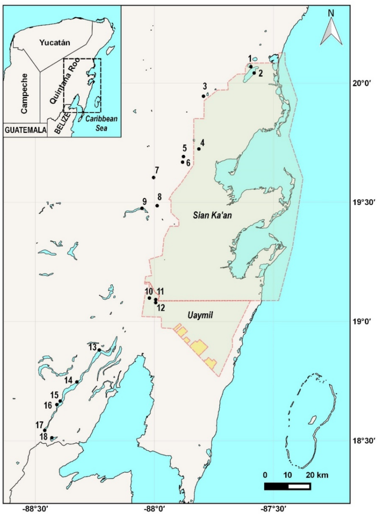
Figure 1. Study area, with the 18 sampling points: (1) Muyil, (2) Chunyaxche, (3) Km 48, (4) Santa Teresa, (5) Tres Reyes 2, (6) Tres Reyes 1, (7) Del Padre, (8) Chancah Veracruz, (9) Siijil Noh Ha, (10) El Toro, (11) Pucté 2, (12) Pucté-Cafetal, (13) Buena Vista, (14) Cayuco Maya, (15) Brujas, (16) Cocalitos, (17) Xul-ha, and (18) Huay Pix. The shaded area represents the Sian Ka’an reserve and the Uaymil protection area.
Diversity 13 00513 g001
Figure 2. ML tree showing the clustering of the 20 identified species. The numbers on the branches are the bootstrap support after 500 replicates. Numbers after the names are the BINs.
Diversity 13 00513 g002
Figure 3. Early stages of development of Cyprinodon artifrons with their DNA barcode. (A) LT = 4.6 mm; (B) LT = 5.5mm; (C) LT = 6.9 mm; (C1) Pigments below the eye and pupil shape. (D) LT = 11 mm.
Diversity 13 00513 g003
Figure 4. Early stages of development of Floridichthys polyommus with their DNA barcode. (A) LT = 5.4 mm; (B) LT = 6.4 mm; (C) LT = 7.5 mm. (C1) Pigments below the eye, invagination, and pupil shape.
Diversity 13 00513 g004
Figure 5. Ventral pigmentation patterns of (A) Cyprinodon artifrons, (A1) LT = 4.6 mm; (A2) LT = 4.5 mm; (A3) LT= 6.0 mm; and (B) Floridichthys polyommus, (B1) LT = 5.4 mm; (B2) LT = 5.5 mm; (B3) LT = 6.0 mm.
Diversity 13 00513 g005
Table 1. Collection sites * inside Sian Ka’an reserve, ** nearby of the reserve, *** inside Lake Bacalar.
NumberSiteLat NLong WNumber of Light
Traps Per Day
Date
1Muyil Lagoon *20.069−87.594224 August 2019
2Chunyaxche lagoon *20.042−87.581224 August 2019
3Km 48 sinkhole **19.943−87.794124 August 2019
4Santa Teresa sinkhole *19.723−87.813123 August 2019
5Tres Reyes 2 sinkhole **19.692−87.877123 August 2019
6Tres Reyes 1 sinkhole **19.668−87.881123 August 2019
7Del Padre sinkhole **19.604−88.003223 August 2019
8Chancah Veracruz sinkhole **19.486−87.988222 August 2019
9Siijil Noh Ha sinkhole **19.475−88.052222 August 2019
10El Toro sinkhole **19.098−88.021122 August 2019
11Pucté 2 sinkhole **19.091−87.994125 August 2019
12Pucté-Cafetal sinkhole **19.079−87.994125 August 2019
13Buena Vista ***18.88−88.231117 August 2015
14Cayuco Maya ***18.746−88.325118 August 2015
15Brujas sinkhole ***18.666−88.395128 June 2015
1 August 2019
16Cocalitos sinkhole ***18.651−88.40912 August 2015
18 April 2015
28 June 2015
19 July 2015
20 August 2015
1 December 2015
17Xul-Ha lagoon18.543−88.46115 August 2015
18Huay Pix lagoon18.512−88.43114 August 2015
Table 2. Species and genus list detected with Barcodes with their BINs numbers.
OrderFamilySpecies# Specimens% SimilarityBIN
AtheriniformesAtherinopsidaeAtherinella sp.22100BOLD:AAI4788
BeloniformesBelonidaeStrongylura notata2100BOLD:AAC4691
HemiramphidaeChriodorus atherinoides1100BOLD:AAD0222
CharaciformesCharacidaeAstyanax aeneus3100BOLD:AAA6360
CichliformesCichlidaeMayaheros urophthalmus3100BOLD:AAB5118
Thorichthys meeki8100BOLD:AAA4760
Vieja melanura3100BOLD:AAB9907
ClupeiformesClupeidaeDorosoma petenense1100BOLD:AAC3463
EngraulidaeAnchoa lyolepis199.85BOLD:AAR3806
Anchoa sp.1100BOLD:AAE1085
Anchovia clupeoides26100BOLD:ACV0719
CyprinodontiformesCyprinodontidaeCyprinodon artifrons56100BOLD:AAA8182
Floridichthys polyommus48100BOLD:AAA6554
Garmanella pulchra11100BOLD:AAD5728
PoeciliidaeGambusia yucatana5100BOLD:AAA4520
GobiiformesEleotridaeDormitator maculatus2099.36–100BOLD:AAC2209
GobiidaeBathygobius soporator7100BOLD:AAA7195
Gobiosoma yucatanum50100BOLD:ACV0831
Lophogobius cyprinoides4599.83–100BOLD:AAB6671
OxudercidaeCtenogobius fasciatus34100BOLD:AAE7730
Table 3. Species, size range, number of specimens, stage of development and collection sites: (1) Muyil, (2) Chunyaxche, (3) Km 48, (4) Santa Teresa, (5) Tres Reyes 2, (6) Tres Reyes 1, (7) Del Padre, (8) Chancah Veracruz, (9) Siijil Noh Ha, (10) El Toro, (11) Pucté 2, (12) Pucté-Cafetal, (13) Buena Vista•, (14) Cayuco Maya•, (15) Brujas•, (16) Cocalitos•, (17) Xul-ha, (18) Huay Pix. • inside Lake Bacalar. * LT = total length. ** LS = standard length.
SpecieSize Range (mm) * LT ** LSNumber of SpecimensStageCollecting Sites
123789101112131415161718
Atherinella sp.3.2–7.2 *58Preflexion larvae x xx x
11.51Postflexion larvae x x
21–22 *3Transition stage x x
27.5–42.5 *15Juvenil x
Strongylura12 **1Postflexion larvae x
notata95 **1Juvenil x
Chriodorus
atherinoides
15 **1Postflexion larvae x
Astyanax
aeneus
33.7–40.2 *3Juvenil x
Mayaheros6.5–6.9 *2Postflexion larvaex
urophthalmus14.4–17.50 *2Transition stagex
Thorichthys4.5–53Flexion larvae x
meeki6.5–7.4 *7Postflexion larvae x
Vieja melanura9.8 *1Postflexion larvae x
14.4–15 *2Transition stagex
Dorosama
petenense
4 **4Preflexion larvae x
Anchoa
lyolepis
30 **1Transition stage x
Anchoa sp.21 **1Transition stage x
Anchovia3 **1Preflexion larvae x
clupeoides7 **6Flexion larvae x
11–23 **52Postflexion larvae xxx
37–475Juvenile x
Cyprinodon4.2–6.9 *49Postflexion larvaexx x
artifrons9–11 *11Transition stagexx
Floridichthys4.5–8.5 *84Postflexion larvaexx
polyommus9–10 *3Transition stage x
24.2 *1Juvenil x
Garmanella4.2–4.5 *3Postflexion larvaexx
pulchra11–12 *2Transition stage x
14.2–20 *6Juvenilxx
Gambusia
yucatana
7.9–8.2 *6Postflexion larvae x
Dormitator5.2–13.6 *529Postflexion larvaexxx x
maculatus14.2–16.8 *26Transition stagexxx
Bathygobius2–2.2 **38Preflexion larvae x
soporator7 **2Postflexion larvae x
Gobiosoma2.2–3.5 **72Preflexion larvae x x
yucatanum3.8–4.6 **216Flexion larvae x
4.8–11 **270Posflexion larvae x x
13 **1Transition stage x
Lophogobius2.8–3.2 **157Preflexion larvae xx
cyprinoides3.4–4 **420Flexion larvae x xx
4.3–8 **415Postflexion larvae x xx
Ctenogobius3.3–3.4 **29Preflexion larvae x
fasciatus3.5–3.8 **94Flexion larvae x
4.5–11 **427Postflexion larvae xx
unidentified
organisms
164 x x
Total 3195
Publisher’s Note: MDPI stays neutral with regard to jurisdictional claims in published maps and institutional affiliations.