3. Results
A thorough scanning of the six mounted slides yielded 244 specific and infraspecific taxa, belonging to 27 orders, 45 families, and 86 genera. Only three taxa were solely identified at the genus level, and five as cf. All the diatom taxa found on
P. pulcherrimum are displayed in an iconographic catalogue (
Figure 3,
Figure 4,
Figure 5,
Figure 6,
Figure 7,
Figure 8,
Figure 9,
Figure 10,
Figure 11,
Figure 12,
Figure 13,
Figure 14,
Figure 15,
Figure 16,
Figure 17,
Figure 18,
Figure 19,
Figure 20,
Figure 21,
Figure 22,
Figure 23,
Figure 24,
Figure 25,
Figure 26,
Figure 27,
Figure 28,
Figure 29,
Figure 30,
Figure 31,
Figure 32,
Figure 33,
Figure 34,
Figure 35,
Figure 36,
Figure 37,
Figure 38,
Figure 39,
Figure 40,
Figure 41,
Figure 42,
Figure 43,
Figure 44,
Figure 45 and
Figure 46) with 615 images that show distinct focal planes, sizes and views (valve and girdle). The genera best represented were
Nitzschia (25 taxa),
Amphora (17),
Diploneis (15),
Navicula (13), and
Cocconeis (12), as they accounted for 34% of all taxa. In contrast, for 49 genera only a single taxon was recorded, representing 57% of all the records collected. Overall, 38 taxa (15.5%) are new records for Mexico’s coasts.
At the class level, 68% (25 taxa) of the new records are Bacillariophyceae, 21% (8) are Coscinodiscophyceae, and 11% (4) are Fragilariophyceae. These were included in 26 genera, of which 19 (73%) were represented by a single taxon. Nitzschia and Gyrosigma had the most taxa—five and three, respectively—while six others had two taxa each (Auricula, Diploneis, Navicula, Parlibellus, Pleurosigma, and Synedra).
Following is a systematic list of epiphytic diatoms (Bacillariophyta) of Phyllodictyon pulcherrimum from the Gulf of California. * New record for Mexican coasts. + New record for the American Continent. n = number of specimens measured. Slides containing the diatom taxa recorded are refered to the macroalgal specimen housed at the Phycological Herbarium of the Universidad Autónoma de Baja California Sur [FBCS-20172].
CLASS: COSCINODISCOPHYCEAE F.E. Round and R.M. Crawford 1990
Order: Asterolamprales F.E. Round and R.M. Crawford 1990
Family: Asterolampraceae H.L. Smith 1872
ASTEROMPHALUS C.G. Ehrenberg 1844
Asteromphalus arachne (A. Brébisson) J. Ralfs 1861
Reference illustrate (Ref. illus.): Pritchard, A. 1861, p. 837, pl. 5, Figure 66; Hasle, G.R., Syvertsen, E.E. 1996, p. 137, pl. 25.
Diameter 45 μm, areolae 7 in 10 μm (
n = 1) (
Figure 10c).
Order: Biddulphiales H. Krieger 1954
Family: Biddulphiaceae F.T. Kützing 1844
BIDDULPHIA S.F. Gray 1821
Biddulphia biddulphiana (J.E. Smith) C.S. Boyer 1900
Ref. illus.: Hustedt, F. 1930, p. 832, Figure 490; Witkowski, A.; Lange-Bertalot, H.; Metzeltin, D. 2000, p. 25, pl. 8, Figures 8 and 9 (as B. pulchella).
Size: 40–65 μm long, 33–102 μm broad, pervalvar axis 85–131 μm, areolae on the mantle 4 to 5 in 10 μm (
n = 7) (
Figure 12c–i).
Remark: Valves elliptic, swollen margin, strongly sculptured, divided into three sections by strong costae. Ends of the valve furnished with large globular process covered with fine pores, areolae arranged in longitudinal and transverse rows, girdle punctate in longitudinal lines.
Biddulphia tridentata C.G. Ehrenberg 1844 *
Ref. illus.: Schmidt, A.W.F. 1888, pl. 118, Figures 13–18.
Size: 90 μm long, 58 μm broad, areolae 5–6 in 10 μm (
n = 1) (
Figure 11d,e).
Remark: Rimoportula in B. biddulphiana are located toward the center of the valve, while in B. tridentata they are displaced to the margins. Likewise, costae in the latter show a hyaline area but no B. biddulphiana.
Biddulphia tridens (C.G. Ehrenberg) C.G. Ehrenberg 1841
Ref. illus.: Roper, F.C.S. 1859, p. 8, pl. 1, Figures 1 and 2; Foged, N. 1975, p. 15, pl. 1, Figures 6 and 7.
Size: 46–106 μm long, 31 μm broad, areolae 6 to 7 in 10 μm (
n = 3) (
Figure 11a–c,f).
Remark: Figure 11a,b show a clear difference in size and form of the pseudo-ocelii, but said variations seem insufficient to render the specimens a distinct species from
B. tridens. Although in [
29] pl. 119, the specimens from Figures 15 and 17 with short-flat pseudo-ocelii (Figure 11a,b) are considered a variety of
B. tridens, in [
30] said variation may be present individually, e.g., Figure 3, which shows classic long-rounded pseudo-ocelii, while Figure 44 (hipovalve) shows short-flat pseudo-ocelii.
LAMPRISCUS A.W.F. Schmidt 1882
Lampriscus shadboltianum (R.K. Greville) H. Peragallo and M. Peragallo 1902
Ref. illus.: Peragallo, H.; Peragallo, M. 1897–1908, p. 389, pl. 106, Figure 1; Navarro, J.N. 1981, p. 618, Figures 33–36; Vidal, L.A.; Ospino-Acosta, K.; Linares-Vargas, K.; García-Urueña, R. 2017, p. 56, Figures 5g and 6a.
Diameter 58–73 μm, pervalvar axis 100 μm, areolae 6–8 in 10 μm (
n = 6) (
Figure 14a–g).
Order: Chaetocerotales F.E. Round and R.M. Crawford 1990
Family Chaetocerotaceae J. Ralfs 1861
BACTERIASTRUM G. Shadbolt 1854
Bacteriastrum hyalinum H.S. Lauder 1864
Ref. illus.: Lauder, H.S. 1864, p. 8, pl. 3, Figure 7a,b; Bosak, S.; Šupraha, L.; Nanjappa, D.; Kooistra, W.H.C.F.; Sarno, D. 2015, p. 135, Figures 18–36.
Order: Eupodiscales V.A. Nikolaev and D.M. Harwood 2000
Family: Odontellaceae P.A. Sims, D.M. Williams and M.P. Ashworth 2018
ODONTELLA C.A. Agardh 1832
Odontella obtusa F.T. Kützing 1844
Ref. illus.: Kützing, F.T. 1844, p. 137, pl. 18/8, Figures 1–3, 6–8.
Size: 100–127 μm long, 65–92 μm broad, 5 areolae in 10 μm (
n = 4) (
Figure 13a–d).
Odontella rostrata (F. Hustedt) R. Simonsen 1987
Ref. illus.: Hustedt, F. 1939, p. 591, Figures 5–7; Simonsen, R. 1987, p. 250, pl. 373, Figures 1–11 (both as Biddulphia rostrata); Sims, P.A.; Williams, D.M.; Ashworth, M., p. 32, Figures 11–116.
Size: 19 μm long, 8–11 areolae in 10 μm (
n = 2) (
Figure 12a,b).
Remark: According to [
31],
O. aurita differs from
O. rostrata by having convex valves with elevated central areas, a valve mantle turned towards the margin of the valve, two elevations with a limited perforated plate (ocellus) on each valve, and radial rows of poroid areolae with domed cribum-like veta, with a central granule, ornamenting the valve surface. It also shows many to just a few siliceous radial ribs located between the border of the elevated central area and the mantle, twinned pores and spines relatively well developed and scattered over the valve. There are one or two labiate processes located next to the border of the central area and opposite to the elevations. These are sessile internally but have stout external tubes of varying length.
AMPHITETRAS C.G. Ehrenberg 1840
Amphitetras antediluviana C.G. Ehrenberg 1840
Ref. illus.: Jahn, R.; Kusber, W.H. 2006, p. 528, Figures 1–3.
Size: 35–45 μm long, 38 μm broad, areolae 4–6 in 10 μm (
n = 2) (
Figure 15a,b).
PSEUDICTYOTA P.A. Sims and D.M. Williams 2018
Pseudictyota dubia (T. Brightwell) P.A. Sims and D.M. Williams 2018
Ref. illus.: Hustedt, F. 1930, p. 806, Figure 46; Witkowski, A.; Lange-Bertalot, H.; Metzeltin, D. 2000, p. 42, pl. 8, Figures 4 and 5 (both as Triceratium dubium)
Size: 13–31 μm long, areolae 4–5 in 10 μm (
n = 4) (
Figure 13e–k).
Order: Coscinodiscales F.E. Round and R.M. Crawford 1990
Family Aulacodiscaceae (F. Schütt) E. Lemmermann
AULACODISCUS C.G. Ehrenberg, 1844, nom. cons.
Aulacodiscus sp.
Size: 65 μm long, pervalvar axis 31 μm (
n = 1) (
Figure 16i,j).
Remark: Because no valve view was found for this taxon, the characteristics for its specific identification could not be used. However, elements in girdle view are indicative of the genus Aulacodiscus.
Family: Coscinodiscaceae F.T. Kützing 1844
COSCINODISCUS C.G. Ehrenberg 1839
Coscinodiscus gigas C.G. Ehrenberg 1841
Ref. illus.: Peragallo, H.; Peragallo, M. 1897–1908, p. 433, pl. 118, Figure 3.
Diameter 246 μm, areolae 3 in 10 μm (
n = 2) (
Figure 5g,h).
Coscinodiscus mesoleius P.T. Cleve 1883 *,†
Ref. illus.: Cleve, P.T. 1883, p. 503, pl. 38, Figure 8; Stidolph, S.R.; Sterrenburg, F.A.S.; Smith, K.E.L.; Kraberg, A. 2012, pl. 15, Figure 32, pl. 17, Figure 98.
Diameter 63–77 μm, areolae 12 to 13 in 10 μm (
n = 2) (
Figure 6j,k).
Coscinodiscus radiatus C.G. Ehrenberg 1840
Ref. illus.: Ehrenberg, C.G. 1840, p. 68, pl. 3, Figure 1a–c; Stidolph, S.R.; Sterrenburg, F.A.S.; Smith, K.E.L.; Kraberg, A. 2012, pl. 20, Figure 22; pl. 34, Figure 2, pl. 36, Figure 36, pl. 43, Figures 110 and 111; pl. 44, Figure 2; pl. 46, Figures 1 and 4.
Family: Heliopeltaceae H.L. Smith 1872
ACTINOPTYCHUS C.G. Ehrenberg 1843
Actinoptychus minutus R.K. Greville 1866
Ref. illus.: Greville, R.K. 1866, p. 5, pl. 1, Figure 12; Siqueiros Beltrones, D.A.; Argumedo-Hernández, U. 2015, p. 114, Figure 28.
Diameter 18–25 μm (
n = 2) (
Figure 3e–j).
Actinoptychus senarius (C.G. Ehrenberg) C.G. Ehrenberg 1843
Ref. illus.: Ehrenberg, C.G. 1843, p. 400, pl. 1.1, Figure 27; pl. 1.3, Figure 21; Siqueiros Beltrones, D.A.; Argumedo-Hernández, U. 2015, p. 114, Figure 8.
Diameter 28–46 μm (
n = 4) (
Figure 4a–j).
Actinoptychus vulgaris J. Schumann 1867
Ref. illus.: Desikachary, T.V. 1988, p. 2, pl. 420, Figures 4 and 6; Moreno, J.L.; Licea, S.; Santoyo, H. 1996, p. 19, pl. 8, Figure 1; López-Fuerte, F.O.; Siqueiros Beltrones, D.A.; Navarro, J.N. 2010, p. 17, pl. 10, Figures 1 and 2.
Diameter 35–54 μm (
n = 4) (
Figure 3a–d).
ACTINOCYCLUS C.G. Ehrenberg 1837
Actinocyclus subtilis (W.W. Gregory) J. Ralfs 1861
Ref. illus.: Witkowski, A.; Lange-Bertalot, H.; Metzeltin, D. 2000, p. 22, pl. 4, Figure 1; López-Fuerte, F.O.; Siqueiros Beltrones, D.A.; Navarro, J.N. 2010, p. 14, pl. 6, Figures 1 and 2.
Diameter 31–50 μm (
n = 5) (
Figure 9a–e).
Actinocyclus tenuissimus P.T. Cleve 1878
Ref. illus.: Navarro, J.N. 1981, p. 429, Figures 28 and 29; Lobban, C.S.; Schefter, M.; Jordan, R.W.; Arai, Y.; Sasaki, A.; Theriot, E.C.; Ashworth, M.; Ruck, E.C.; Pennesi C. 2012, p. 249, pl. 5, Figures 1 and 2.
Diameter 38–58 μm, areolae 18 in 10 μm (
n = 2) (
Figure 9f–h).
Actinocyclus ochotensis A.P. Jousé 1969 *
Ref. illus.: Jousé, A.P. 1969, p. 17, pl. 2, Figures 2–5.
Diameter 34 μm, areolae 8 in 10 μm (
n = 1) (
Figure 9i–k).
AZPEITIA M. Peragallo 1912
Azpeitia nodulifera (A.W.F. Schmidt) G.A. Fryxell and P.A. Sims 1986
Ref. illus.: Fryxell, G.A.; Sims, P.A.; Watkins, T.P. 1986, p. 19, Figures 17, 18 and 30; Hasle, G.R.; Syvertsen, E.E. 1996, p. 126, pl. 21.
Diameter 20–58 μm, areolae 4–6 in 10 μm (
n = 4) (
Figure 7a–g).
Family: Hemidiscaceae N.I. Hendey, 1937 emend R. Simonsen, 1975
ROPERIA A. Grunow 1889
Roperia tesselata (F.C.S. Roper) A. Grunow 1889
Ref. illus.: Roper, F.C.S. 1858, p. 19, pl. 3, Figure 1; Hasle, G.R.; Syvertsen, E.E. 1996, p. 130, pl. 22.
Diameter 31–35 μm, areolae 7 to 8 in 10 μm (
n = 2) (
Figure 7m).
Order: Melosirales R.M. Crawford 1990
Family: Hyalodiscaceae R.M. Crawford 1990
MARGARITUM H. Moreira Filho 1968
Margaritum terebro (G. Leuduger-Fortmorel) H. Moreira Filho 1968 *
Ref. illus.: Hendey, N.I. 1957, p. 36, pl. 1, Figures 12 and 13 (as Podosira tenebro); Souza-Mosimann, R.M. de; Fernandes, L.F.; Ludwig, T.V. 1997, p. 46, Figures 1–14.
Diameter 30–32 μm (
n = 2) (
Figure 6l–o).
PODOSIRA C.G. Ehrenberg 1840
Podosira montagnei F.T. Kützing 1844
Ref. illus.: Foged, N. 1984, p. 89, pl. 18, Figure 1.
Podosira stelligera (J.W. Bailey) A.Mann 1907
Ref. illus.: Hendey, N.I. 1964, p. 90, pl. 22, Figure 6; Navarro, J.N. 1982, p. 11, pl. 2, Figures 4 and 5.
Diameter 17–42 μm (
n = 6) (
Figure 8a–k).
Podosira variegata A.W.F. Schmidt 1889 *
Ref. illus.: Schmidt, A.W.F. 1889, pl. 140, Figure 4.
Family: Melosiraceae F.T. Kützing 1844
MELOSIRA C.A. Agardh 1824
Melosira moniliformis var. octogona (A. Grunow) F. Hustedt 1927
Ref. illus.: Witkowski, A.; Lange-Bertalot, H.; Metzeltin, D. 2000, p. 35, pl. 3, Figures 1 and 2.
Order: Paraliales R.M. Crawford 1990
Family: Paraliaceae R.M. Crawford 1988
PARALIA P.A.C. Heiberg 1863
Paralia sulcata (C.G. Ehrenberg) P.T. Cleve 1873
Ref. illus.: Hendey, N.I. 1964, p. 73, pl. 23, Figure 5; Hasle, G.R.; Syvertsen, E.E. 1996, p. 91, pl. 14.
Family: Stephanodiscaceae I.V.Makarova 1986
CYCLOTELLA (F.T. Kützing) A. de Brébison 1838
Cyclotella striata (F.T. Kützing) A. Grunow 1880
Ref. illus.: Foged, N. 1984, p. 31, pl. 17 Figure 4; Hasle G.R., Syvertsen E.E. 1996, p. 34, pl. 1.
CYCLOSTEPHANOS F.E.Round 1987
Cyclostephanos sp.
Remark: According to [
27], the distinctive characteristics of this genus are—circular valves with concentric undulations and spines around the margin; valves with radiating lines of areolae, grouped into fascicles around the margin and separated by raised, rounded ridges. The central region of the valve face variously has nodules or is plain.
Cyclostephanos differs from
Cyclotella in the extension of the rows of fascicles to the center of the valve and the presence of a single ring of spines around the valve face.
Order: Thalassiosirales Z.I. Glezer and I.V. Makarova 1986
Family: Thalassiosiraceae M.V. Lebour 1930
THALASSIOSIRA P.T. Cleve 1873
Thalassiosira decipiens (A. Grunow ex H. Van Heurck) E.G. Jørgensen 1905
Ref. illus.: Navarro, J.N. 1982, p. 10, pl. 1, Figures 1 and 2; Moreno, J.L.; Licea, S.; Santoyo, H. 1996, p. 133, pl. 33, Figure 7.
Diameter 8–13 μm, areolae 9–14 in 10 μm (
n = 2) (
Figure 6f,g).
Thalassiosira eccentrica (C.G. Ehrenberg) P.T. Cleve 1904
Ref. illus.: Hendey, N.I. 1964, p. 80, pl. 24, Figure 7; Navarro, J.N. 1982, p. 10, pl. 1, Figures 3 and 4; Moreno, J.L.; Licea, S.; Santoyo, H. 1996, p. 133, pl. 33, Figures 8 and 9.
Diameter 23 μm, areolae 8 in 10 μm (
n = 1) (
Figure 6e).
Thalassiosira lineata A.P. Jousé 1968
Ref. illus.: Hasle, G.R.; Syvertsen, E.E. 1996, p. 80, pl. 10; Park, J.S.; Jung, S.W.; Lee, S.D.; Yun, S.M.; Lee, J.H. 2016, p. 410, Figure 19.
Diameter 21–22 μm, areolae 11–12 in 10 μm (
n = 2) (
Figure 6c,d).
Thalassiosira leptopus (A. Grunow) G.R. Hasle and G.A. Fryxell 1977
Ref. illus.: Hasle, G.R.; Fryxell, G.A. 1977, Figures 1–14; Hasle, G.R.; Syvertsen, E.E. 1996, pl. 10; Moreno, J.L.; Licea, S.; Santoyo, H. 1996, p. 134, pl. 33, Figure 11.
Diameter 108 μm, areolae 4 in 10 μm (
n = 1) (
Figure 6a,b).
Thalassiosira nanolineata (A. Mann) G.A. Fryxell and G.R. Hasle 1977
Ref. illus.: Mann, A. 1925, p. 68, pl. 14, Figure 4 (as Coscinodiscus nano-lineatus); Javeed, A.; Salleh, S.; Darif, A.; Mohammad, M. 2018, p. 8, Figure 2.
Diameter 19 μm, areolae 11 in 10 μm (
n = 1) (
Figure 7h–j).
Order: Plagiogrammales E.J. Cox 2015
Family: Plagiogrammaceae G. De Toni 1890
DIMEREGRAMMA J. Ralfs 1861
Dimeregramma fulvum (W. Gregory) J. Ralfs 1861 *,†
Ref. illus.: Witkowski, A.; Lange-Bertalot, H.; Metzeltin, D. 2000, p. 28, pl. 11, Figures 1 and 2.
Size: 43 μm long, 8 μm broad, 9 to 10 striae in 10 μm (
n = 1) (
Figure 17c,d).
Dimeregramma minor (W. Gregory) J. Ralfs 1861
Ref. illus.: Hendey, N.I. 1964, p. 156, pl. 27, Figure 12; Navarro, J.N.1982, p. 34, pl. 11, Figures 1–3; Witkowski, A.; Lange-Bertalot, H.; Metzeltin, D. 2000, p. 29, pl. 11, Figures 3–9.
Size: 15–32 μm long, 7 to 8 μm broad, 9 to 10 striae in 10 μm (
n = 3) (
Figure 17e–j).
TALARONEIS W.H.C.F. Kooistra and M. De Stefano 2004
PLAGIOGRAMMA R.K. Greville 1859
Talaroneis sp.
Ref. illus.: Navarro, J.N. 1982, p. 23, pl. 13, Figures 1 and 2; López-Fuerte, F.O.; Siqueiros Beltrones, D.A.; Navarro, J.N. 2010, p. 19, pl. 13, Figures 4–7 (both as Plagiogramma interruptum).
Size: 25–38 μm long, 5–6 μm broad, 20 striae in 10 μm (
n = 2) (
Figure 16a–c).
Remark: This taxon shows the combined characteristics of
Plagiogramma and
Talaroneis, but it is quite different from both of the three described species of the latter [
32,
33].
Plagiogramma interruptum in [
34] on the other hand, is very similar to our specimen, structurally and morphometrically. In [
33], the diagnostic characters of
Talaroneis are: pores in an apical pore field, round two silica flaps parallel to the valve margin proximal to the apical pore fields. While [
9] exhibits
Talaroneis sp. (Figures 9 and 10) that appears identical to our specimens, albeit thinner, but with clear subapical furrows as in ours that, in contrast with other described species of
Talaroneis, show a stauros and apparent septa in the central area. The authors of [
9] suggest that
Talaroneis sp., may be a new species; in our case given the similarity with
P. interruptum as in [
34], p. 23, pl. 13, Figures 1, 2 and [
35], p. 19, pl. 13, Figures 4–7, a review at the species and genus level may be required.
GLYPHODESMIS R.K. Greville 1862
Glyphodesmis rhombica (P.T. Cleve) R. Simonsen 1974 *
Ref. illus.: Mann, A. 1925, p. 78, pl. 16, Figures 2 and 3 (as G. acus); Simonsen, R. 1974, p. 35, pl. 23, Figure 1; Foged, N. 1978, p. 67, pl. 7, Figure 20.
Size: 26–48 μm long, 9 to 10 μm broad, 14 to 15 striae in 10 μm (
n = 3) (
Figure 25a–j).
Order: Triceratiales F.E. Round and R.M. Crawford 1990
Family: Triceratiaceae (F. Schütt) E. Lemmermann 1899
RALFSIELLA P.A. Sims, D.M. Williams and M. Ashworth 2018
Ralfsiella smithii (J. Ralfs) P.A. Sims, D.M. Williams and M. Ashworth 2018
Ref. illus.: Peragallo, H.; Peragallo, M. 1897–1908, p. 398, pl. 112, Figures 4 and 5; Hustedt, F. 1930, p. 861, Figure 513 (all as Cerataulus smithii); Sims, P.A., Williams, D.M.; Ashworth, M. 2018, p. 42, Figures 148–151.
Diameter 44 μm, 9 to 10 areolae in 10 μm (
n = 1) (
Figure 11g–j).
TRICERATIUM C.G. Ehrenberg 1839
Triceratium balearicum f. biquadrata (C. Janisch) F. Hustedt 1930
Ref. illus.: Schmidt, A.W.F. 1886, pl. 98, Figures 4–6; Hustedt, F. 1930, p. 815, Figure 477.
CLASS: FRAGILARIOPHYCEAE F.E. Round 1990
Order: Ardissoneales F.E. Round 1990
Family: Ardissoneaceae F.E. Round 1990
ARDISSONEA G. De Notaris 1870
Ardissonea formosa (C.A. Hantzsch) A. Grunow 1880
Ref. illus.: Witkowski, A.; Lange-Bertalot, H.; Metzeltin, D. 2000, p. 43, pl. 30, Figure 12.
Size: 331 μm long, 18–20 μm broad, 9 striae in 10 μm (
n = 2) (
Figure 18a).
Order: Climacospheniales F.E. Round 1990
Family: Climacospheniaceae F.E. Round 1990
CLIMACOSPHENIA C.G. Ehrenberg 1841
Climacosphenia elongata J.W. Bailey 1854
Ref. illus.: Round, F.E. 1982, Figures 4–6, 14; Stidolf, S.R.; Sterrengurg, F.A.S.; Smith K.E.L.; Kraberg, A. 2012, pl. 7, Figure 152, pl. 16, Figure 93; Al-Handal, A.Y.; Compère, P.; Riaux-Gobin, C. 2016, p. 12, pl. 6, Figures 1 and 2.
Size: 300 μm long, 18 μm broad, striae 36 in 10 μm (
n = 1) (
Figure 18b).
Order: Cyclophorales F.E. Round and R.M. Crawford 1990
Family Cyclophoraceae F.E. Round and R.M. Crawford 1990
CYCLOPHORA F. Castracane 1878
Cyclophora tenuis F. Castracane 1878
Ref. illus.: Peragallo, H.; Peragallo, M. 1897–1908, pl. 1, Figures 27–32; Navarro, J.N.1982, Figures 9–17; Navarro, J.N.; Lobban, C.S. 2009, Figures 59 and 60; Lobban, C.S.; Jordan, R.W. 2010, Figure 5b; Ashworth, M.P.; Ruck, E.C.; Lobban, C.S.; Romanovicz, D.K.; Theriot, E.C. 2012, p. 686, Figures 1, 5, 6 and 14–18.
Size: 52 μm long, 12 μm broad, striae not resolvable (
n = 1) (
Figure 37i).
Order: Fragilariales P.C. Silva 1962
Family: Fragilariaceae R.K. Greville 1833
OPEPHORA P. Pettit 1888
Opephora pacifica (A. Grunow) P. Pettit 1888
Ref. illus.: 1995, p. 244, Figures 45–53; Witkowski, A.; Lange-Bertalot, H.; Metzeltin, D. 2000 p. 72, pl. 25, Figures 18–26; Al-Handal, A.Y.; Compère, P.; Riaux-Gobin, C. 2016, p.8, pl. 4, Figures 12 and 13.
Size: 15–23 μm long, 5 to 6 μm broad, striae 9 in 10 μm (
n = 2) (
Figure 16a–c).
GEDANIELLA Chunlian Li, A.Witkowski and M.P.Ashworth 2018
Gedaniella mutabilis Chunlian Li and A.Witkowski, nom. illeg. 2018
Ref. illus.: Sabbe, K.; Vyverman, W. 1995, p. 241, Figures 13–28; Witkowski, A.; Lange-Bertalot, H.; Metzeltin, D. 2000, p. 72, pl. 25, Figures 10–17; Al-Handal, A.Y.; Compère, P.; Riaux-Gobin, C. 2016, p. 7, pl. 4, Figure 11.
Size: 15 μm long, 4 μm broad, striae 9 in 10 μm (
n = 1) (
Figure 16k).
STAUROSIRELLA D.M. Williams and F.E. Round 1987
Staurosirella guenter-grassii (A. Witkowski and H. Lange-Bertalot) E.A. Morales, C.E. Wetzel and L. Ector 2019 *
Ref. illus.: Sabbe, K.; Vyverman, W. 1995, p. 2, Figures 29–42, 66–71; Witkowski, A.; Lange-Bertalot, H.; Metzeltin, D. 2000, p. 70, pl. 24, Figures 40–44 (both as Opephora guenter-grassii).
Size: 8 μm long, 3 μm broad, striae 14 in 10 μm (
n = 1) (
Figure 16l).
SYNEDRA C.G. Ehrenberg 1830
Synedra gaillonii var. macilenta cf. (A. Grunow) H. Peragallo 1881 *
Ref. illus.: Van Heurck, H. 1881, pl. 40, Figure 1.
Size: 139–148 μm long, 8 μm broad (
n = 2) (
Figure 20a,b).
Synedra tabulata var. rostrata (H. Juhlin-Dannfelt) A. Cleve-Euler 1953 *
Ref. illus.: Juhlin-Dannfelt, H. 1882, p. 43, pl. 3, Figures 29 and 31 (as S. affinis var. rostrata); Cleve-Euler, A. 1953, p. 71, Figure 392g–h (as S. tabulata Λ hybrida).
Size: 33–38 μm long, 4 to 5 μm broad, striae 15 to 16 in 10 μm (
n = 3) (
Figure 20m–o).
HYALOSYNEDRA D.M. Williams and F.E. Round 1986
Hyalosynedra laevigata (A. Grunow) D.M. Williams and F.E. Round 1986
Ref. illus.: Foged, N. 1984, p. 97, pl. 28, Figure 13; Witkowski, A.; Lange-Bertalot, H.; Metzeltin, D. 2000, p. 62, pl. 17, Figure 22; pl. 29, Figures 6–10; pl. 30, Figures 30–23.
Size: 204 μm long, 6 μm broad, striae not resolvable (
n = 1) (
Figure 20c).
FRAGILARIA H.C. Lyngbye 1819
Fragilaria barbatula (F.T. Kützing) H. Lange-Bertalot 1993
Ref. illus.: Witkowski, A.; Lange-Bertalot, H.; Metzeltin, D. 2000, p. 79, pl. 30, Figures 13 and 14 (as Synedra barbatula).
Size: 35–73 μm long, 4–7 μm broad, striae 18–20 in 10 μm (
n = 7) (
Figure 20j,p,q).
TABULARIA (F.T. Kützing) D.M. Williams and F.E. Round 1986
Tabularia fasciculata (C.A. Agardh) D.M. Williams and F.E. Round 1986
Ref. illus.: Williams, D.M.; Round, F.E. 1986, p. 326; Witkowski, A.; Lange-Bertalot, H.; Metzeltin, D. 2000, p. 80, pl. 30, Figures 4 and 5 (as Synedra fasciculata).
Size: 30 μm long, 5 μm broad, striae 17 to 18 in 10 μm (
n = 1) (
Figure 20l).
Tabularia investiens (W. Smith) D.M. Williams and F.E. Round 1986
Ref. illus.: Cleve-Euler, A. 1953, p. 44, Figure 354a–d (as Fragilaria investiens); Williams, D.M.; Round, F.E. 1986, p. 324, Figures 39–45.
Size: 45 μm long, 3 to 4 μm broad, striae 8 to 9 in 10 μm (
n = 2) (
Figure 20g,h,k).
Tabularia parva (F.T. Kützing) D.M. Williams and F.E. Round 1986
Ref. illus.: Williams, D.M.; Round, F.E. 1986, Figures 33–38; Lobban, C.S.; Schefter, M. 2012, p. 257, pl. 13, Figures 6 and 7.
Size: 35–55 μm long, 4–7 μm broad, striae 18–20 in 10 μm (
n = 4) (
Figure 20i).
Tabularia tabulata (C.A. Agardh) P.J.M. Snoeijs 1992
Ref. illus.: Snoeijs, P.J.M. 1992, p. 343, Figures 38–48; Witkowski, A.; Lange-Bertalot, H.; Metzeltin, D. 2000, p. 81, pl. 30, Figures 1 and 2 (as Synedra tabulata var. tabulata).
Size: 113–134 μm long, 5 μm broad, striae 12 in 10 μm (
n = 3) (
Figure 20d–f).
Order: Licmophorales F.E. Round 1990
Family: Licmophoraceae F.T. Kützing 1844
LICMOPHORA C.A. Agardh 1827
Licmophora abbreviata C.A. Agardh 1831
Ref. illus.: Hustedt F. 1931–1959, p. 66, Figure 590; Witkowski, A.; Lange-Bertalot, H.; Metzeltin, D. 2000, p. 63, pl. 20, Figures 3–5.
Size: 87 μm long, 21 μm pervalvar axis, striae 16 in 10 μm (
n = 1) (
Figure 19h).
Licmophora ehrenbergii (F.T. Kützing) A. Grunow 1867
Ref. illus.: Hustedt, F. 1931–1959, p. 70, Figure 593; Witkowski, A.; Lange-Bertalot, H.; Metzeltin, D. 2000, p. 64, pls. 18, Figure 11; pl. 20, Figure 16.
Size: 72–145 μm long, 25–31 μm broad, striae 10–12 in 10 μm (
n = 2) (
Figure 19a,b).
Licmophora gracilis var. anglica (F.T. Kützing) H. Peragallo and M. Peragallo 1901
Ref. illus.: Peragallo, H.; Peragallo, M. 1901, p. 346, pl. 84. Figure 13; Witkowski, A.; Lange-Bertalot, H.; Metzeltin, D. 2000, p. 65, pl. 20, Figures 11–13.
Size: 23–28 μm long, 5 μm broad, striae 24 in 10 μm (
n = 2) (
Figure 19f,g).
Licmophora paradoxa (H.C. Lyngbye) C.A. Agardh 1828
Ref. illus.: Hustedt F. 1931–1959, p. 76, Figure 605; Witkowski, A.; Lange-Bertalot, H.; Metzeltin, D. 2000, p. 67, pl. 18, Figures 4–10.
Size: 69–75 μm long, 8 μm broad, striae 16 in 10 μm (
n = 2) (
Figure 19c,d).
Licmophora pfannkucheae M.H. Giffen 1970
Ref. illus.: Giffen, M.H. 1970, p. 278, Figures 41 and 42; Witkowski, A.; Lange-Bertalot, H.; Metzeltin, D. 2000, p. 67, pls. 18, Figure 6.
Size: 108–119 μm long, 6–8 μm broad, striae 23 in 10 μm (
n = 2) (
Figure 19e).
Order: Rhabdonematales F.E. Round and R.M. Crawford 1990
Family: Rhabdonemataceae F.E. Round and R.M. Crawford 1990
HYALOSIRA F.T. Kützing 1844
Hyalosira tropicalis J.N. Navarro 1991
Ref. illus.: Navarro, J.N.; Williams, D.M. 1991, p. 328, Figures 1–14; Witkowski, A.; Lange-Bertalot, H.; Metzeltin, D. 2000, p. 62, pl. 21, Figures 16 and 17.
Size: 11 to 12 μm long, 3–5 μm broad, striae 22–25 in 10 μm (
n = 2) (
Figure 19i–k).
RHABDONEMA F.T. Kützing 1844
Rhabdonema adriaticum F.T. Kützing 1844
Ref. illus.: Hustedt, F. 1931–1959, p. 23, Figure 552; Witkowski, A.; Lange-Bertalot, H.; Metzeltin, D. 2000, p. 76, pl. 13, Figures 10–12.
Size: 135–169 μm long, striae 10 in 10 μm (
n = 2) (
Figure 16m,n).
Order: Rhaphoneidales F.E. Round 1990
Family: Psammodiscaceae F.E. Round and D.G. Mann 1990
PSAMMODISCUS F.E. Round and D.G. Mann 1980
Psammodiscus nitidus (W. Gregory) F.E. Round and D.G. Mann 1980
Ref. illus.: Hustedt, F. 1930, p. 414, Figure 221 (as Coscinodiscus nitidus); Witkowski, A.; Lange-Bertalot, H.; Metzeltin, D. 2000, p. 75, pl. 23, Figures 12–14.
Diameter 31 μm, areolae 5 in 10 μm (
n = 1) (
Figure 10e).
Family: Rhaphoneidaceae A. Forti 1912
DELPHINEIS G.W. Andrews 1977
Delphineis minutissima (F. Hustedt) R.R. Simonsen 1987
Ref. illus.: Hustedt, F. 1939, p. 599, Figures 14 and 15; Witkowski, A.; Lange-Bertalot, H.; Metzeltin, D. 2000, p. 45, pl. 22, Figures 11–14.
Size: 17 μm long, 8 μm broad, striae 13 in 10 μm (
n = 1) (
Figure 17k).
Family: Ulnariaceae E.J. Cox 2015
FALCULA M. Voigt 1960
Falcula media M. Voigt 1960
Ref. illus.: Voigt, M. 1960, p. 87; pl. 1, Figures 6–8; pl. 2, Figures 6–10.
Size: 63–69 μm long, 7 to 8 μm broad (
n = 3) (
Figure 17a,b).
Order: Striatellales F.E. Round 1990
Family: Striatellaceae F.T. Kützing 1844
GRAMMATOPHORA C.G. Ehrenberg 1840
Grammatophora hamulifera F.T. Kützing 1844
Ref. illus.: Hustedt, F. 1931–1959, p. 40, Figure 566.
Size: 25 μm long, 6 μm broad, striae 16 in 10 μm (
n = 4) (
Figure 21l,n–r).
Grammatophora macilenta W. Smith 1856
Ref. illus.: Hustedt, F. 1931–1959, p. 47, Figure 574; Witkowski, A.; Lange-Bertalot, H.; Metzeltin, D. 2000, p. 58, pl. 15, Figures 16–18.
Size: 98–108 μm long, 8 μm broad, striae 22 in 10 μm (
n = 7) (
Figure 20r and
Figure 21a,b).
Grammatophora marina (H.C. Lyngbye) F.T. Kützing 1844
Ref. illus.: Hustedt, F. 1931–1959, p. 43, Figure 569; Witkowski, A.; Lange-Bertalot, H.; Metzeltin, D. 2000, p. 58, pl. 15, Figures 9–12.
Size: 17–77 μm long, 5 to 6 μm broad, pervalvar axis 16–42 μm, striae 15–20 in 10 μm (
n = 14) (
Figure 21k).
Grammatophora oceanica C.G. Ehrenberg 1840
Ref. illus.: Hustedt, F. 1931–1959, p. 45, Figure 573; Witkowski, A.; Lange-Bertalot, H.; Metzeltin, D. 2000, p. 59, pl. 15, Figures 13–14; pl. 16, Figure 12; pl. 17, Figures 3 and 4.
Size: 57–62 μm long, pervalvar axis 6 μm, striae 3 in 10 μm (
n = 3) (
Figure 21c–f,j).
Grammatophora undulata var. gallopagensis A. Grunow
Ref. illus.: Van Heurck, H. 1880–1881, pl. 53, Figure 20; López-Fuerte, F.O.; Siqueiros Beltrones, D.A.; Jakes-Cota U.; Tripp-Valdéz, A. 2019, p.103, Figure 2h,i.
Size: 49–116 μm long, 8–10 μm broad, striae 27 to 28 in 10 μm (
n = 5) (
Figure 21g–i,m).
Order: Toxariales F.E. Round 1990
Family: Toxariaceae F.E. Round 1990
TOXARIUM J.W. Bailey 1854
Toxarium undulatum J.W. Bailey 1854
Ref. illus.: Hustedt, F. 1931–1959, p. 224, Figure 714; Witkowski, A.; Lange-Bertalot, H.; Metzeltin, D. 2000, p. 83, pl. 31, Figures 5 and 6.
Size: 400 μm long, 3–8 μm broad, striae 10–12 in 10 μm (
n = 3) (
Figure 18c,d).
CLASS: BACILLARIOPHYCEAE E. Haeckel 1878
Order: Achnanthales P.C. Silva 1962
Family: Achnanthaceae F.T. Kützing 1844
ACHNANTHES J.B.G.M. Bory de Saint-Vincent 1822
Achnanthes cf. fimbriata (A. Grunow) R. Ross 1963
Ref. illus.: Witkowski, A.; Lange-Bertalot, H.; Metzeltin, D. 2000, p. 88, pls. 51, Figures 9 and 10; Siqueiros Beltrones, D.A.; Argumedo-Hernández, U.; López-Fuerte, F.O. 2017, p. 32, Figure 5b.
Size: 26–50 μm long, 14–16 μm broad, striae 12–16 in 10 μm (
n = 2) (
Figure 22j,l,m).
Achnanthes citronella (A. Mann) F. Hustedt 1937
Ref. illus.: Riaux-Gobin, C. 2015, p. 104, Figures 15–17, 25, 26, 33, 35–38.
Size: Long, sternum valve 35–40 μm, raphe valve 41 μm. Broad; sternum valve 18–19 μm, raphe valve 20–22 μm. Striae, sternum valve 11 in 10 μm, raphe valve 17–20 in 10 μm (
n = 7) (
Figure 22a–i).
Achnanthes groenlandica var. phinneyi C.D. McIntire and C.W. Reimer 1974
Ref. illus.: Mclntire, C.D.; Reimer, C.W. 1974, p. 172, pl. 2, Figure 3a–c; pl. 3, Figure 3a,b; Majewska, R.; De Stefano, M.; Ector, L.; Bolaños, F.; Frankovich, T.A.; Sullivan, M.J.; Ashworth, M.P.; Van de Vijver, B. 2017, p. 314, Figures 100–109.
Size: 43–54 μm long, 7 μm broad, striae 10 to 11 in 10 μm (
n = 2) (
Figure 22p,q).
Achnanthes parvula F.T. Kützing 1844
Ref. illus.: Hustedt, F. 1931–1959, p. 426, Figure 877f–i (as A. brevipes var. parvula); Witkowski, A.; Lange-Bertalot, H.; Metzeltin, D. 2000, p. 93, pl. 43, Figures 6 and 7; pl. 45, Figures 6–8, pl. 47, Figure 9.
Size: 16–21 μm long, 4–6 μm broad, striae 10 to 11 in 10 μm (
n = 2) (
Figure 22n,o,u).
Achnanthes pseudogroenlandica N.I. Hendey 1964
Ref. illus.: Witkowski, A.; Lange-Bertalot, H.; Metzeltin, D. 2000, p. 94, pl. 44, Figures 16–23; Majewska, R.; De Stefano, M.; Ector, L.; Bolaños, F.; Frankovich, T.A.; Sullivan, M.J.; Ashworth, M.P.; Van de Vijver, B. 2017, p. 314, Figures 110–136.
Size: 25 μm long, pervalvar axis 7 μm, striae 8 to 9 in 10 μm (
n = 1) (
Figure 22r,t).
Achnanthes subconstricta (M. Meister) K. Toyoda 2003 *
Ref. illus.: Toyoda, K.; Nagumo, T.; Osada, K.; Tanaka, J. 2003, p. 369; Lee, S.D.; Park, J.S.; Lee, J.H. 2011, p. 4, Figure 2G,N, Figure 5B–F.
Size: 38–75 μm long, 14 μm broad, striae 5–7 in 10 μm (
n = 2) (
Figure 22k–v).
Achnanthes yaquinensis C.D. McIntire and R.W. Reimer 1974
Ref. illus.: Mclntire, C.D.; Reimer, R.W. 1974, p. 174, pls. 2, Figure 1a,b, pl. 3, Figure 1a,b.
Size: 44 μm long, 10 μm broad, striae 9 to 10 in 10 μm (
n = 1) (
Figure 22s).
AMPHICOCCONEIS M. De Stefano and D. Marino 2002
Amphicocconeis discrepans (A.W.F. Schmidt) C. Riaux-Gobin, A. Witkowski, L. Ector and A. Igersheim 2018
Ref. illus.: Witkowski, A.; Lange-Bertalot, H.; Metzeltin, D. 2000, p. 106, pl. 41, Figures 35–40; pl. 42, Figures 26 and 27 (as Cocconeis discrepans); Riaux-Gobin, C., Ector, L., Witkowski, A. and Igersheim, A. 2018, p. 576, Figures 9–22.
Size: 19 μm long, 8 μm broad, striae 14 in 10 μm (
n = 1) (
Figure 25v).
Amphicocconeis disculoides (F. Hustedt) M. De Stefano and D. Marino 2003
Ref. illus.: Witkowski, A.; Lange-Bertalot, H.; Metzeltin, D. 2000, p. 106, pl. 42, Figures 28–33 (as Cocconeis disculoides); De Stefano, M.; Marino, D. 2003, p. 362, Figures 1–32.
Size: 15–16 μm long, 8 μm broad, striae 9–11 in 10 μm (
n = 2) (
Figure 23s,t).
ASTARTIELLA A. Witkowski, H. Lange-Bertalot and D. Metzeltin 1998
Astartiella bahusiensis (A. Grunow) A. Witkowski, H. Lange-Bertalot and D. Metzeltin 1998 *
Ref. illus.: Witkowski, A.; Lange-Bertalot, H.; Stachura, K. 1998, p. 359, Figure 80: 1–3; Witkowski, A.; Lange-Bertalot, H.; Metzeltin, D. 2000, p. 99, pl. 52, Figures 22–31.
Size: 18 μm long, 8 μm broad, striae 24 in 10 μm (
n = 1) (
Figure 17p).
KARAYEVIA F.E. Round and L. Bukhtiyarova 1998
Karayevia amoena (F. Hustedt) L. Bukhtiyarova 1999
Ref. illus.: Chang, T.P. 1992, p. 401, Figures 3a–d, 4a–h, 5a–i; Witkowski, A.; Lange-Bertalot, H.; Metzeltin, D. 2000, p. 85, pl. 51, Figures 34–36 (both as Achnanthes amoena).
Size: 12 μm long, 5 μm broad, sternum valve striae 18–19 in 10 μm, raphe valve 26 in 10 μm (
n = 5) (
Figure 17m–o).
PLANOTHIDIUM F.E. Round and L. Bukhtiyarova 1996
Planothidium campechianum (F. Hustedt) A. Witkowski, H. Lange-Bertalot and D. Metzeltin 2000
Ref. illus.: Hustedt, F. 1952, p. 389, Figures 87–90 (as Achnanthes campechianum); Witkowski, A.; Lange-Bertalot, H.; Metzeltin, D. 2000, p. 118, pl. 48, Figures 3–9.
Size: 26 μm long, 8 μm broad, striae 16 in 10 μm (
n = 1) (
Figure 25s).
Planothidium delicatulum (F.T. Kützing) F.E. Round and L. Bukhtiyarova 1996 *
Ref. illus.: Hustedt, F. 1931–1959, p. 389, Figure 836 (as Achnanthes delicatula); Witkowski, A.; Lange-Bertalot, H.; Metzeltin, D. 2000, p. 118, pl. 46, Figures 28 and 29; pl. 48, Figures 1 and 2.
Size: 17–21 μm long, 9–10 μm broad, sternum valve striae 9 in 10 μm, raphe valve 11 in 10 μm (
n = 2) (
Figure 25q,t,u).
Planothidium hauckianum (A. Grunow) F.E. Round and L. Bukhtiyarova 2008
Ref. illus.: Hustedt, F. 1931–1959, p. 388, Figure 834 (as Achnanthes hauckiana); Witkowski, A.; Lange-Bertalot, H.; Metzeltin, D. 2000, p. 120, pl. 48, Figures 39–41.
Size: 14–21 μm long, 6 to 7.5 μm broad, striae 10 to 11 in 10 μm (
n = 5) (
Figure 25w).
Planothidium lilljeborgei (A. Grunow) A. Witkowski, H. Lange-Bertalot and D. Metzeltin 2000
Ref. illus.: Hustedt, F. 1931–1959, p. 394, Figure 843 (as Achnanthes lilljeborgei); Witkowski, A.; Lange-Bertalot, H.; Metzeltin, D. 2000, p. 121, pl. 49, Figure 1; pl. 51, Figures 27–29.
Size: 20 μm long, 7 μm broad, striae 10 in 10 μm (
n = 1) (
Figure 25p).
Planothidium polare (E. Østrup) A. Witkowski, H. Lange-Bertalot and D. Metzeltin 2000
Ref. illus.: Witkowski, A.; Lange-Bertalot, H.; Metzeltin, D. 2000, p. 123, pl. 47, Figures 1–4; pl. 49, Figures 37–39 (as P. polaris).
Size: 42 μm long, 14 μm broad, striae 14 in 10 μm (
n = 1) (
Figure 25r).
Family: Cocconeidaceae F.T. Kützing 1844
COCCONEIS C.G. Ehrenberg 1838
Cocconeis californica A. Grunow 1880
Ref. illus.: Hustedt, F. 1931–1959, p. 343, Figure 796; Poulin, M.; Berard-Therriault, L.; Cardinal, A. 1984, p. 49, Figures 5–11.
Size: 10–16 μm long, 5–10 μm broad, striae 15–17 in 10 μm (
n = 5) (
Figure 23q,r).
Cocconeis contermina A.W.F. Schmidt 1894
Ref. illus.: Schmidt, A.W.F. 1894, pl. 196, Figure 21; Siqueiros Beltrones, D.A.; Argumedo Hernández, U.; Landa Cansigno, C. 2016, p. 70, Figure 59.
Size: 38 μm long, 28–30 μm broad, striae 15–17 in 10 μm (
n = 2) (
Figure 24c–f).
Cocconeis convexa M.H. Giffen 1967
Ref. illus.: Witkowski, A.; Lange-Bertalot, H.; Metzeltin, D. 2000, p. 104, pls. 37, Figures 5 and 6; pl. 41, Figures 1–4. Sar, E.A.; Romero, O.E.; Sunesen, I. 2003, p.81, Figures 2–6.
Size: 18–28 μm long, 12–22 μm broad, sternum valve striae 28–47 in 10 μm, raphe valve 22 in 10 μm (
n = 4) (
Figure 24g–j).
Cocconeis dirupta W. Gregory 1857
Ref. illus.: Kobayasi H.; Nagumo, T. 1985, p. 99, Figure 2: 16–27; Witkowski, A.; Lange-Bertalot, H.; Metzeltin, D. 2000, p. 105, pls. 39, Figures 1–5; pl. 51, Figures 5 and 8.
Size: 15–62 μm long, 8–47 μm broad, sternum valve striae 18–27 in 10 μm, raphe valve striae 18–24 in 10 μm (
n = 18) (
Figure 23a–g).
Cocconeis guttata F. Hustedt and A.A. Aleem 1951
Ref. illus.: Witkowski, A.; Lange-Bertalot, H.; Metzeltin, D. 2000, p. 108, pl. 40, Figures 13–18; Sar, E.A.; Romero, O.E.; Sunesen, I. 2003, p. 86, Figures 16–21.
Size: 23–30 μm long, 15–18 μm broad, sternum valve striae 6 to 7 in 10 μm (
n = 2) (
Figure 24o,p).
Cocconeis heteroidea C.A. Hantzsch 1863
Ref. illus.: Hustedt, F. 1931–1959, p. 356, Figure 811; Witkowski, A.; Lange-Bertalot, H.; Metzeltin, D. 2000, p. 108, pl. 35, Figures 4 and 5.
Size: 38–45 μm long, 28–31μm broad, sternum valve striae 23–26 in 10 μm, raphe valve striae 20 in 10 μm (
n = 2) (
Figure 23k–m).
Cocconeis krammeri H. Lange-Bertalot and D. Metzeltin 1996
Ref. illus.: Witkowski, A.; Lange-Bertalot, H.; Metzeltin, D. 2000, p. 109, pl. 33, Figures 1–5; pl. 34, Figures 4 and 5; pl. 42, Figure 34.
Size: 22–27 μm long, 18–14 μm broad, sternum valve striae 25–30 in 10 μm, raphe valve striae 23–28 in 10 μm (
n = 2) (
Figure 24k–n).
Cocconeis lineata C.G. Ehrenberg 1849
Ref. illus.: Ehrenberg, C.G. 1849, p. 301, pl. 5, Figure 44; p. 4, Romero, O.; Jahn, R. 2013, Figures 1–8.
Size: 55 μm long, 44 μm broad, striae 17 to 18 in 10 μm (
n = 1) (
Figure 24a,b).
Cocconeis peltoides F. Hustedt 1939
Ref. illus.: Witkowski, A.; Lange-Bertalot, H.; Metzeltin, D. 2000, p. 112, pl. 38, Figures 1–9; Sar, E.A.; Romero, O.E.; Sunesen, I. 2003, p. 91, Figures 34–41.
Size: 15–21 μm long, 9–12 μm broad, sternum valve striae 13 in 10 μm (
n = 2) (
Figure 25m,n).
Cocconeis pseudomarginata W. Gregory 1857
Ref. illus.: Hustedt, F. 1931–1959, p. 359, Figure 813; Witkowski, A.; Lange-Bertalot, H.; Metzeltin, D. 2000, p. 113, pl. 34, Figures 8 and 9; pl. 35, Figures 1–4.
Size: 42–56 μm long, 32–36 μm broad, sternum valve striae 19–22 in 10 μm, raphe valve striae 19 in 10 μm (
n = 2) (
Figure 24q–t).
Cocconeis scutellum C.G. Ehrenberg 1838
Ref. illus.: Witkowski, A.; Lange-Bertalot, H.; Metzeltin, D. 2000, p. 114, pl. 36, Figures 1–7; pl. 38, Figure 11; Sar, E.A.; Romero, O.E.; Sunesen, I. 2003, p. 95, Figures 44–50.
Size: 29 to 30 μm long, 18 μm broad, sternum valve striae 8 in 10 μm, raphe valve striae 10–12 in 10 μm (
n = 5) (
Figure 25k,l).
Cocconeis scutellum var. parva (A. Grunow) P.T. Cleve 1895
Ref. illus.: Peragallo, H.; Peragallo, M. 1897–1908, p. 20, pl. 4, Figure 3; Poulin, M.; Berard-Therriault, L.; Cardinal, A. 1984, p. 56, Figures 49–53.
Size: 12–16 μm long, 6–9 μm broad, sternum valve striae 13–16 in 10 μm, raphe valve striae 12 to 13 in 10 μm (
n = 30) (
Figure 23n–p).
Order: Bacillariales N.I. Hendey 1937
Family: Bacillariaceae C.G. Ehrenberg 1831
BACILLARIA J.F. Gmelin 1791
Bacillaria socialis (W. Gregory) J. Ralfs 1861
Ref. illus.: Poulin, M.; Berard-Therriault, L.; Cardinal, A. 1984, p. 75, Figures 2–4, 8; Witkowski, A.; Lange-Bertalot, H.; Metzeltin, D. 2000, p. 357, pl. 196, Figures 5–7; pl. 207, Figure 9.
Size: 57–156 μm long, 7 to 8 μm broad, fibulae 6 to 7 in 10 μm, striae 14 to 15 in 10 μm (
n = 9) (
Figure 44a,y,z).
CYMBELLONITZSCHIA F. Hustedt 1924
Cymbellonitzschia banzuensis J.G. Stepanek, S.E. Hamsher, S. Mayama, D.H. Jewson and J.P. Kociolek 2016
Ref. illus.: Stepanek, J.G., Hamsher, S.E., Mayama, S., Jewson, D.H. and Kociolek, J.P. 2016, p. 28, Figures 1–22.
Size: 15–38 μm long, 2 to 3 μm broad, fibulae 7–9 in 10 μm, striae 15–17 in 10 μm (
n = 5) (
Figure 44n–r).
CYLINDROTHECA L. Rabenhorst 1859
Cylindrotheca closterium (C.G. Ehrenberg) B.E.F. Reimann and J.C. Lewin 1964
Ref. illus.: Witkowski, A.; Lange-Bertalot, H.; Metzeltin, D. 2000, p. 374, pl. 212, Figures 4–6.
Size: 112–223 μm long, 7–12 μm broad, fibulae 12–37 in 10 μm (
n = 2) (
Figure 45j).
FRAGILARIOPSIS F. Hustedt 1913
Fragilariopsis doliolus (G.C. Wallich) L.K. Medlin and P.A. Sims 1993
Ref. illus.: Hasle, G.R.; Syvertsen, E.E.1996, p. 303, Figure 69; Witkowski, A.; Lange-Bertalot, H.; Metzeltin, D. 2000, p. 360, pl. 213, Figures 38 and 39.
Size: 50–65 μm long, 8 μm broad, striae 11 in 10 μm (
n = 5) (
Figure 25o).
HANTZSCHIA A. Grunow 1877
Hantzschia marina (A.S. Donkin) A. Grunow 1880
Ref. illus.: Krammer, K.; Lange-Bertalot, H. 1988, p. 132, pl. 93, Figures 1–3; Witkowski, A.; Lange-Bertalot, H.; Metzeltin, D. 2000, p. 363, pl. 178, Figures 9–11.
Size: 32–86 μm long, 5–8 μm broad, fibulae 4–6 in 10 μm, striae 7 to 8 in 10 μm (
n = 2) (
Figure 45d–g).
NAGUMOEA J.P. Kociolek and A. Witkowski 2011
Nagumoea vallus (V.A. Nikolaev) R. Majewska and B. Van de Vijver 2020 *
Ref. illus.: Nikolaev, V.A.1969, p. 30, pl. 1, Figures 3–8 (as Anaulus vallus); Sullivan, M.J. 2010, p. 175, Figures 1, 2, 4–6, 7 and 8 (as Denticula vallus).
Size: 16–20 μm long, 2 μm broad, fibulae 5 in 10 μm (
n = 2) (
Figure 17r).
NITZSCHIA A.H. Hassall 1845
Nitzschia agnita F. Hustedt 1957
Ref. illus.: Krammer, K.; Lange-Bertalot, H. 1988, p. 117, Figure 82: 1–5; Witkowski, A.; Lange-Bertalot, H.; Metzeltin, D. 2000, p. 367, pl. 210, Figures 22 and 23.
Size: 23 μm long, 5 μm broad, fibulae 17 in 10 μm (
n = 1) (
Figure 44m).
Nitzschia amabilis H. Suzuki 2010
Ref. illus.: Witkowski, A.; Lange-Bertalot, H.; Metzeltin, D. 2000, p. 387, pl. 189, Figures 13–15, pl. 190, Figures 1–6 (as Nitzschia laevis); Suzuki, H.; Nagumo, T.; Tanaka, J. 2010, p. 223, Figure 1.
Size: 8–12 μm long, 4 to 5 μm broad, fibulae 10–14 in 10 μm (
n = 2) (
Figure 44u,v).
Nitzschia angularis W. Smith 1853
Ref. illus.: Hendey, N.I. 1964, p. 281, Figure 39: 6; Witkowski, A.; Lange-Bertalot, H.; Metzeltin, D. 2000, p. 368, pl. 199, Figures 5 and 6.
Size: 142 to 143 μm long, 13–15 μm broad, fibulae 4 in 10 μm (
n = 2) (
Figure 44f,g).
Nitzschia bicapitata P.T. Cleve 1901
Ref. illus.: Fryxell, G. 2000, p. 46, Figures 1–11.
Size: 24 μm long, 5 μm broad, fibulae 12 in 10 μm, striae 25 in 10 μm (
n = 2) (
Figure 44k,l).
Nitzschia carnicobarica T.V. Desikachary and P. Prema 1987
Ref. illus.: Desikachary, T.V.; Prema, P. 1987, p. 8, Figure 304: 5; Witkowski, A.; Lange-Bertalot, H.; Metzeltin, D. 2000, p. 373, pl. 183, Figures 9 and 10.
Size: 28–38 μm long, 8–11μm broad, fibulae 11–12 in 10 μm (
n = 3) (
Figure 42m,n and
Figure 43l).
Nitzschia composita M.H. Giffen 1971 *
Ref. illus.: Giffen, M.H. 1971, p. 8, Figures 42 and 43; Witkowski, A.; Lange-Bertalot, H.; Metzeltin, D. 2000, p. 376, pl. 211, Figure 12.
Size: 42–45 μm long, 7 μm broad, fibulae 9–11 in 10 μm (
n = 2) (
Figure 45o,p).
Nitzschia costata J. Pantocsek 1892 *
Ref. illus.: Pantocsek, J. 1892, pl. 41, Figure 566.
Size: 128 μm long, 7–9 μm broad, costae 5 in 10 μm (
n = 1) (
Figure 45s).
Nitzschia distans W. Gregory 1857
Ref. illus.: Peragallo, H.; Peragallo, M. 1897–1908, p. 283, pl. 73, Figure 3; Witkowski, A.; Lange-Bertalot, H.; Metzeltin, D. 2000, p. 378, pl. 203, Figures 7–9.
Size: 69 μm long, 7 μm broad, fibulae 4 in 10 μm (
n = 1) (
Figure 44x).
Nitzschia frustulum (F.T. Kützing) A. Grunow 1880
Ref. illus.: Krammer, K.; Lange-Bertalot, H. 1988, p. 94, pl. 68, Figures 1–9; Witkowski, A.; Lange-Bertalot, H.; Metzeltin, D. 2000, p. 382, pl. 209, Figures 13–17.
Size: 7–15 μm long, 2 μm broad, fibulae 10–16 in 10 μm (
n = 2) (
Figure 44s,t).
Nitzschia fusiformis A. Grunow 1880 *
Ref. illus.: Lange-Bertalot, H.; Krammer, K. 1987, p. 20, pl. 28, Figures 4–10; Witkowski, A.; Lange-Bertalot, H.; Metzeltin, D. 2000, p. 382, pl. 197, Figures 17–20; pl. 198, Figures 1–3.
Size: 108 μm long, 5 μm broad, fibulae 10 to 11 in 10 μm, striae 24 in 10 μm (
n = 1) (
Figure 45a).
Nitzschia gracilis C.A. Hantzsch 1860
Ref. illus.: Lange-Bertalot, H. 1988, p. 93. pl. 66, Figures 1–11.
Size: 46–62 μm long, 3 to 4 μm broad, fibulae 11 to 12 in 10 μm, striae 26 in 10 μm (
n = 3) (
Figure 43p,q).
Nitzschia hybrida A. Grunow 1880
Ref. illus.: Krammer, K.; Lange-Bertalot, H. 1988, p. 61, pl. 46, Figures 3–6; Witkowski, A.; Lange-Bertalot, H.; Metzeltin, D. 2000, p. 385, pl. 191, Figures 12–14.
Size: 65–77 μm long, 5–8 μm broad, fibulae 5–8 in 10 μm (
n = 2) (
Figure 45k,m).
Nitzschia incrustans A. Grunow 1862 *
Ref. illus.: Krammer, K.; Lange-Bertalot, H. 1988, p. 26, pl. 7, Figures 9 and 10a; Witkowski, A.; Lange-Bertalot, H.; Metzeltin, D. 2000, p. 386, pls. 203, Figures 1–3.
Size: 11–15 μm long, 4 to 5 μm broad, fibulae 5 to 6 in 10 μm (
n = 3) (
Figure 43t,u).
Nitzschia incurva var. lorenziana R. Ross 1986
Ref. illus.: Krammer, K.; Lange-Bertalot, H. 1988, p. 125, pl. 86, Figures 6–10; Witkowski, A.; Lange-Bertalot, H.; Metzeltin, D. 2000, p. 392, pls. 210, Figures 24 and 25; pl. 211, Figure 3; pl. 212, Figures 1–3.
Size: 138 μm long, 7 μm broad, fibulae 7 in 10 μm, striae 18 in 10 μm (
n = 1) (
Figure 45h).
Nitzschia insignis W. Gregory 1857
Ref. illus.: Peragallo, H.; Peragallo, M. 1897–1908, p. 295, pl. 75, Figure 5; Witkowski, A.; Lange-Bertalot, H.; Metzeltin, D. 2000, p. 387, pl. 202, Figure 5, pl. 204, Figures 1–7.
Size: 119 μm long, 8 μm broad, fibulae 2–5 in 10 μm, striae 10–14 in 10 μm (
n = 4) (
Figure 42d,e and
Figure 45r).
Nitzschia lanceolata W. Smith 1853
Ref. illus.: Krammer, K.; Lange-Bertalot, H. 1988, pl. 16, Figures 1–8; Witkowski, A.; Lange-Bertalot, H.; Metzeltin, D. 2000, p. 389, pl. 194, Figures 1–5.
Size: 69 μm long, 8 μm broad, fibulae 10 in 10 μm (
n = 1) (
Figure 45n).
Nitzschia linearis W. Smith 1853
Ref. illus.: Hustedt, F. 1930, p. 409, Figure 784.
Size: 100 μm long, 5 μm broad, fibulae 8 in 10 μm (
n = 1) (
Figure 45l).
Nitzschia longa A. Grunow 1880
Ref. illus.: Peragallo, H.; Peragallo, M. 1897–1908, p. 279, pl. 72, Figure 5; Stidolph, S.R.; Sterrenburg, F.A.S.; Smith, K.E.L.; Kraberg, A. 2012, pl. 25, Figure 84.
Size: 162 μm long, 8 μm broad, fibulae 4 in 10 μm, striae 12 to 13 in 10 μm (
n = 1) (
Figure 44b,c).
Nitzschia longissima (A. Brébisson) A. Pritchard 1861
Ref. illus.: Pritchard, A. 1861, p. 783, pl. 4, Figure 23; Witkowski, A.; Lange-Bertalot, H.; Metzeltin, D. 2000, p. 391, pl. 207, Figures 6 and 7.
Size: 200–500 μm long, 5–8 μm broad, fibulae 6–10 in 10 μm (
n = 5) (
Figure 45i).
Nitzschia martiana (Agardh) H. Van Heurck 1896
Ref. illus.: Peragallo, H.; Peragallo, M. 1897–1908, p. 282, pl. 72, Figure 20; Lobban, C.S.; Mann, D.G. 1987, p. 2397, Figures 1–13.
Size: 240–262 μm long, 3 to 4 μm broad, fibulae 5 to 6 in 10 μm (
n = 2) (
Figure 45t).
Nitzschia sicula (F. Castracane) F. Hustedt 1958
Ref. illus.: Hasle, G.R.; Syvertsen, E.E.1996, p. 327, pl. 75, Figures a–d.
Size: 30 μm long, 7 μm broad, striae and fibulae 10 in 10 μm (
n = 1) (
Figure 43r,s).
Nitzschia sigma (F.T. Kützing) W. Smith 1853
Ref. illus.: Krammer, K.; Lange-Bertalot, H. 1988, pl. 23, Figures 1–9; Witkowski, A.; Lange-Bertalot, H.; Metzeltin, D. 2000, p. 404, pl. 206, Figures 1–10.
Size: 69–77 μm long, 6 to 7 μm broad, fibulae 9 in 10 μm, striae 30 in 10 μm (
n = 2) (
Figure 45b,c).
Nitzschia spathulata W. Smith 1853
Ref. illus.: Peragallo, H.; Peragallo, M. 1897–1908, p. 284, pl. 53, Figure 4.
Size: 50–135 μm long, 5–8 μm broad, fibulae 6 to 7 in 10 μm (
n = 2) (
Figure 44h,w).
Nitzschia cf. spectabilis var. americana A. Grunow 1880 *
Size: 121 μm long, 5 μm broad, fibulae 8 in 10 μm, striae 25 in 10 μm (
n = 1) (
Figure 45q).
Nitzschia valdestriata A.A. Aleem and F. Hustedt 1951
Ref. illus.: Krammer, K.; Lange-Bertalot, H. 1997, p. 121, pl. 84, Figures 9–12; Witkowski, A.; Lange-Bertalot, H.; Metzeltin, D. 2000, p. 407, pls. 203, Figures 19–21; pl. 207, Figures 14–16.
Size: 17 μm long, 3 μm broad, fibulae 8 in 10 μm, striae 14 to 15 in 10 μm (
n = 1) (
Figure 44i,j).
TRYBLIONELLA W. Smith 1853
Tryblionella bathurstensis (M.H. Giffen) D.G. Mann 1990 *
Ref. illus.: Giffen, M.H. 1970, 287 (as Nitzschia bathurstensis); Mann, D.G. 1990, p. 678.
Size: 18 μm long, 8 to 9 μm broad, fibulae 12 to 13 in 10 μm, striae 22 in 10 μm (
n = 2) (
Figure 43e,f,i).
Tryblionella coarctata (A. Grunow) D.G. Mann 1990
Ref. illus.: Peragallo, H.; Peragallo, M. 1897–1908, p. 268, pl. 69, Figures 26 and 27 (as Nitzschia puncta var. coarctata).
Size: 15–44 μm long, 7–16 μm broad, striae 12 in 10 μm (
n = 5) (
Figure 43a–d,g,h).
Tryblionella hungarica (A. Grunow) J. Frenguelli 1942
Ref. illus.: Krammer, K.; Lange-Bertalot, H. 1988, p. 42, pl. 34, Figures 1–3; Witkowski, A.; Lange-Bertalot, H.; Metzeltin, D. 2000, p. 385, pl. 188, Figures 10 and 11 (as Nitzschia hungarica).
Size: 31–58 μm long, 6 to 7 μm broad, fibulae 10 to 11 in 10 μm, striae 22 in 10 μm (
n = 3) (
Figure 43j,k).
Tryblionella lanceola A. Grunow 1878
Ref. illus.: Krammer, K.; Lange-Bertalot, H. 1988, pl. 38, Figures 11 and 12; Witkowski, A.; Lange-Bertalot, H.; Metzeltin, D. 2000, p. 388, pl. 212, Figures 13–17 (both as Nitzschia lanceola).
Size: 28 μm long, 8 μm broad, striae 9 to 10 in 10 μm (
n = 1) (
Figure 43n–o).
Order: Cymbellales D.G. Mann 1990
Family: Rhoicospheniaceae J. Chen and H. Zhu 1983
RHOICOSPHENIA A. Grunow 1860
Rhoicosphenia abbreviata (C.A. Agardh) H. Lange-Bertalot 1980
Ref. illus.: Krammer, K.; Lange-Bertalot, H. 1986, p. 381, pl. 91, Figures 20–28; Witkowski, A.; Lange-Bertalot, H.; Metzeltin, D. 2000, p. 345, pl. 212, pl. 58, Figures 4–7.
Size: 25 μm long, 4 μm broad, striae 16 to 17 in 10 μm (
n = 1) (
Figure 38p).
GOMPHOSEPTATUM L.K. Medlin 1986
Gomphoseptatum aestuarii (P.T. Cleve) L.K. Medlin 1986
Ref. illus.: Medlin, L.K.; Round, F.E. 1986, p. 212, Figures 16–18; Witkowski, A.; Lange-Bertalot, H.; Metzeltin, D. 2000, p. 222, pl. 61, Figures 17 and 18.
Size: 21 μm long, 3 μm broad, striae 14 to 15 in 10 μm (
n = 1) (
Figure 17l).
Order: Eunotiales P.C. Silva 1962
Family: Eunotiaceae F.T. Kützing 1844
COLLICULOAMPHORA D.M. Williams and G. Reid 2006
Colliculoamphora reichardtiana (A. Grunow) Williams and Reid 2006
Ref. illus.: Williams, D.M., Reid, G. 2006, p. 153, Figures 11–19; Williams, D.M. 2016, p. 81, Figures 1–14.
Size: 15 μm long, 5 μm broad, striae 15 in 10 μm (
n = 1) (
Figure 17q).
Remark: Our specimen is slightly shorter and narrower than in the references; 17–52 μm long, 7–13 μm broad, and may be mistaken for C. minima, but the number of striae in 10 μm is 18 for this taxon.
Family: Lyrellaceae D.G.Mann 1990
LYRELLA N.I. Karayeva 1978
Lyrella approximatoides (F. Hustedt) D.G. Mann 1990
Ref. illus.: Hustedt, F. 1930–1966, p. 426, Figure 1498; Foged, N. 1984, p. 60, pl. 49, Figure 1 (both as Navicula approximatoides).
Size: 58 μm long, 27 μm broad, striae 8–9 in 10 μm (
n = 1) (
Figure 28i,j).
Lyrella atlantica (A.W.F. Schmidt) D.G. Mann 1990
Ref. illus.: Hustedt, F. 1961–1966, p. 509, Figure 1555 (as Navicula lyra var. atlantica); Witkowski, A.; Lange-Bertalot, H.; Metzeltin, D. 2000, p. 231, pls. 96, Figure 6; pl. 98, Figure 5.
Size: 50 μm long, 25 μm broad, striae 11 in 10 μm (
n = 1) (
Figure 28h).
Lyrella clavata var. caribaea (P.T. Cleve) D.A. Siqueiros Beltrones 2017
Ref. illus.: Peragallo, H.; Peragallo, M. 1897–1908, p. 138, pl. 24, Figures 3 and 4 (as Navicula clavata var. caribaea); Siqueiros Beltrones, D.A.; Argumedo-Hernández, U.; López-Fuerte, F.O. 2017, Figures 2–12, 14–19, 21 and 22.
Size: 50 μm long, 24 to 25 μm broad, striae 11–12 in 10 μm (
n = 2) (
Figure 28d–g).
Lyrella hennedyi (W. Smith) A.J. Stickle and D. G. Mann 1990
Ref. illus.: Hustedt, F. 1961–1966, p. 453, Figure 1516; Witkowski, A.; Lange-Bertalot, H.; Metzeltin, D. 2000, p. 233, pl. 95, Figure 3; pl. 98, Figure 4.
Size: 77 μm long, 38 μm broad, striae 10 in 10 μm (
n = 1) (
Figure 28a–c).
Order: Mastogloiales D.G.Mann 1990
Family: Mastogloiaceae C. Mereschkowsky 1903
MASTOGLOIA G.H.K. Thwaites 1856
Mastogloia binotata (A. Grunow) P.T. Cleve 1895
Ref. illus.: Moreno, J.L.; Licea, S.; Santoyo, H. 1996, p. 89, pl. 24, Figure 3; Witkowski, A.; Lange-Bertalot, H.; Metzeltin, D. 2000, p. 240, pl. 75, Figures 15–17.
Size: 22 μm long, 14 to 15 μm broad, striae 13 in 10 μm (
n = 3) (
Figure 28k–l).
Mastogloia chersonensis A.W.F. Schmidt 1893
Ref. illus.: Schmidt, A.W.F. 1893, pl. 186, Figures 31 and 32; Hustedt, F. 1933, p. 565, Figure 999a.
Size: 28 μm long, 9 μm broad (
n = 1) (
Figure 28o).
Mastogloia ciskeiensis M.H. Giffen 1967
Ref. illus.: Giffen, M.H. 1967, p. 264, Figures 43–45; Foged, N. 1975, p. 29, pl. 16, Figures 16 and 23; Foged, N. 1978, p. 78, pl. 18, Figure 7.
Size: 32 μm long, 7 μm broad (
n = 1) (
Figure 28m,n).
TETRAMPHORA C. Mereschkowsky 1903
Tetramphora decussata (A. Grunow) J.G. Stepanek and J.P. Kociolek 2016
Ref. illus.: Peragallo, H.; Peragallo, M. 1897–1908, pl. 49, Figure 24; Lobban, C.S.; Schefter, M. 2012, p. 298, pl. 1, Figures 7–9; pl. 54, Figure 5; pl. 55, Figures 1–3 (both as Amphora decussata).
Size: 46 μm long, 8 μm broad, dorsal striae 13–15 in 10 μm, ventral striae 21–22 in 10 μm (
n = 1) (
Figure 36f–h).
Remark: Our specimen is smaller than other records, 60 μm long and 10 μm broad; the morphometrics coincide with that of Amphora acuta var. parva, but the dorsal striae are distinctively oblique relative to both raphe and fascia which differentiate our specimens from other records.
Tetramphora intermedia (P.T. Cleve) J.G. Stepanek and J.P. Kociolek 2016
Ref. illus.: Peragallo, H.; Peragallo, M. 1897–1908, p. 224, pl. 50, Figure 3, Wachnicka, A.H.; Gaiser, E.E. 2007, p. 419, Figure 118 (both as Amphora rhombica var. intermedia).
Size: 77–100 μm long, 19 μm broad, dorsal striae 13–19 in 10 μm, ventral striae 16 in 10 μm (
n = 5) (
Figure 36a–c).
Tetramphora securicula (H. Peragallo and M. Peragallo) J.G. Stepanek and J.P. Kociolek 2016 *
Ref. illus.: Peragallo, H. and Peragallo, M. 1897–1908. p. 224, pl. 50, Figure 2.
Size: 62 μm long, 15 μm broad, dorsal striae 11 to 12 in 10 μm, ventral striae 14 in 10 μm (
n = 1) (
Figure 35p).
Remark: Our specimen is broader than the one reported in [
36], who report a valve breadth of 8–10 μm. However, the authors of [
37] record it as 15 μm wide and 70 μm long, with 12 striae in 10 μm.
Order: Naviculales C.E. Bessey 1907
Family: Amphipleuraceae A. Grunow 1862
AMPHIPRORA C.G. Ehrenberg 1843
Amphiprora pseudoduplex (K. Osada and H. Kobayasi) G. Hällfors 2004
Ref. illus.: Osada, K.; Kobayasi, H. 1990, p. 165, Figures 4, 5 and 32–42 (as Entomoneis pseudoduplex).
Size: 25 μm long, 7 μm broad (
n = 1) (
Figure 37h).
HALAMPHORA (P.T. Cleve) Z. Levkov 2009
Halamphora acutiuscula (F.T. Kützing) Z. Levkov 2009
Ref. illus.: Krammer, K.; Lange-Bertalot, H. 1986, p. 348, pl. 151, Figure 6 (as Amphora coffeaeformis var. acutiuscula); Wah, T.T.; Wee, Y.C. 1988, Figures 11 and 12; Witkowski, A.; Lange-Bertalot, H.; Metzeltin, D. 2000, p. 128, pl. 161, Figures 10–13.
Size: 28 μm long, 6 μm broad, dorsal striae 14 in 10 μm (
n = 1) (
Figure 34q).
Halamphora capitata (R. Hagelstein) I. Álvarez-Blanco and S. Blanco 2014
Ref. illus.: Hagelstein, R. 1938, pl. 3, Figure 7; Wachnicka, A.H.; Gaiser, E.E. 2007, p. 415, Figures 98 and 99 (both as Amphora bigibba var. capitata).
Size: 17 μm long, 4 μm wide, dorsal striae 24 in 10 μm (
n = 1) (
Figure 34i).
Halamphora coffeiformis (C.A. Agardh) C. Mereschkowsky 1903
Ref. illus.: Witkowski, A.; Lange-Bertalot, H.; Metzeltin, D. 2000, p. 133, pl. 161, Figures 21–25 (as Amphora coffeaeformis var. coffeaeformis).
Size: 34–36 μm long, 5 to 6 μm broad, dorsal striae 13 in 10 μm, ventral striae 14 to 15 in 10 μm (
n = 2) (
Figure 34o,p).
Halamphora costata (W. Smith) Z. Levkov 2009
Ref. illus.: Witkowski, A.; Lange-Bertalot, H.; Metzeltin, D. 2000, p. 134, pl. 169, Figure 9 (as Amphora costata); Levkov, Z. 2009, p. 181, pl. 92, Figure 14.
Size: 45–89 μm long, 6–12 μm broad, dorsal striae 7 to 8 in 10 μm (
n = 6) (
Figure 33m–q).
Halamphora cuneata (P.T. Cleve) Z. Levkov 2009
Ref. illus.: Witkowski, A.; Lange-Bertalot, H.; Metzeltin, D. 2000, p. 135, pl. 167, Figures 20 and 21 (as Amphora cuneata); Levkov, Z. 2009, p. 182, pl. 105, Figures 1–6; pl. 243, Figures 5 and 6.
Size: 29–52 μm long, 5 to 6 μm broad, dorsal striae 12–14 in 10 μm (
n = 2) (
Figure 33c–f).
Remark: The morphometrics of Figure 31 c–f coincide better with Amphora maletracta var. constricta, but lack the wide hyaline area separating striae bands on the dorsal margin.
Halamphora exigua (W. Gregory) Z. Levkov 2009
Ref. illus.: Witkowski, A.; Lange-Bertalot, H.; Metzeltin, D. 2000, p. 137, pl. 161, Figures 15–17 (as Amphora exigua).
Size: 44 μm long, 6 μm broad, dorsal striae 11 in 10 μm (
n = 1) (
Figure 35g).
Halamphora wisei (M.M. Salah) I. Álvarez-Blanco and S. Blanco 2014
Ref. illus.: Simonsen, R. 1962, p. 94, pl. 3, Figure 2; Witkowski, A.; Lange-Bertalot, H.; Metzeltin, D. 2000, p. 154, pl. 162, Figures 18 and 19 (both as Amphora wisei).
Size: 16 μm long, 5 μm broad, dorsal striae 14 in 10 μm (
n = 2) (
Figure 34m,n).
Family: Berkeleyaceae D.G. Mann 1990
PARLIBELLUS E.J. Cox 1982
Parlibellus delognei (H. Van Heurck) E.J. Cox 1988
Ref. illus.: Hustedt, F. 1961–1966, p. 302, Figure 1422 (as Navicula grevillii); Witkowski, A.; Lange-Bertalot, H.; Metzeltin, D. 2000, p. 321, pl. 104, Figures 1–5.
Size: 26–34 μm long, 8–12 μm broad, striae 18–19 in 10 μm (
n = 2) (
Figure 30n–p).
Parlibellus rhombicula (F. Hustedt) A. Witkowski 2000
Ref. illus.: Hustedt, F. 1961–1966, p. 327, Figure 1422; Witkowski, A.; Lange-Bertalot, H.; Metzeltin, D. 2000, p. 325, pl. 103, Figure 3.
Size: 51–94 μm long, 11–19 μm broad, striae 16–18 in 10 μm (
n = 1) (
Figure 30g,h,k–m).
Parlibellus rhombicus (W. Gregory) E.J. Cox 1988 *
Ref. illus.: Cox, E.J. 1988, p. 25, Figures 17, 33–38.
Size: 73 μm long, 12 μm broad, striae 17–19 in 10 μm (
n = 1) (
Figure 30i,j).
Parlibellus weissflogii (A. Grunow) E.J. Cox 1988 *
Ref. illus.: Cleve, P.T. 1878, p. 7; pl. 1, Figure 9 (as Brebissonia weissflogii)
Size: 52–66 μm long, 22–31 μm broad, striae 14–18 in 10 μm (
n = 2) (
Figure 30a–f).
Family: Diadesmidaceae D.G. Mann 1990
DIPLONEIS C.G. Ehrenberg 1844
Diploneis bombus (C.G. Ehrenberg) C.G. Ehrenberg 1853
Ref. illus.: Hustedt, F. 1931–1959, p. 704, Figure 1086a–c; Witkowski, A.; Lange-Bertalot, H.; Metzeltin, D. 2000, p. 183, pl. 86, Figures 1–29; pl. 2, Figures 1–3.
Size: 102 μm long, 19 μm broad, striae 8 in 10 μm (
n = 1) (
Figure 26a).
Diploneis chersonensis (A. Grunow) P.T. Cleve 1892
Ref. illus.: Schmidt, A.W.F. 1892, pl. 174, Figure 14; Witkowski, A.; Lange-Bertalot, H.; Metzeltin, D. 2000, p. 184, pl. 86, Figure 10.
Size: 92 μm long, 31 μm broad, striae 8 in 10 μm (
n = 1) (
Figure 26b,c).
Diploneis crabro (C.G. Ehrenberg) C.G. Ehrenberg 1854
Ref. illus.: Hustedt, F. 1931–1959, p. 616, Figure 1028; Witkowski, A.; Lange-Bertalot, H.; Metzeltin, D. 2000, p. 184, pl. 93, Figures 18–21.
Size: 39–51 μm long, 15–18 μm broad, striae 7–9 in 10 μm (
n = 5) (
Figure 26d–i).
Diploneis incurvata (W. Gregory) P.T. Cleve 1894
Ref. illus.: Hustedt, F. 1931–1959, p. 593, Figure 1012b–d; Witkowski, A.; Lange-Bertalot, H.; Metzeltin, D. 2000, p. 187, pl. 86, Figures 5–6; pl. 87, Figure 4.
Size: 25–35 μm long, 8.9–12 μm broad, striae 12 to 13 in 10 μm (
n = 2) (
Figure 27o,p).
Diploneis gruendleri (A.W.F. Schmidt) P.T. Cleve 1894
Ref. illus.: Hustedt, F. 1931–1959, p. 702, Figure 1084; Navarro, J.N. 1982, p. 34, pl. 22, Figure 5; Cremer, H.; Sangiorgi, F.; Wagner-Cremer, F.; McGee, V.; Lotter, A.F.; Visscher, H. 2007, p. 35, pl. 8, Figure 76.
Size: 51 μm long, 23 μm broad, striae 9 in 10 μm (
n = 1) (
Figure 26j).
Diploneis litoralis (A.S. Donkin) P.T. Cleve 1894
Ref. illus.: Hendey, N.I. 1964, p. 226, pl. 32, Figure 9; Foged, N. 1984, p. 36, pl. 41 Figure 5.
Size: 36 μm long, 12 μm broad, striae 12 in 10 μm (
n = 2) (
Figure 27d).
Diploneis litoralis var. clathrata (E. Østrup) P.T. Cleve 1896
Ref. illus.: Hustedt, F. 1931–1959, p. 666, Figure 1062b,c; Witkowski, A.; Lange-Bertalot, H.; Metzeltin, D. 2000, p. 188, pl. 89, Figures 5 and 7–13.
Size: 22–24 μm long, 8–10 μm broad, striae 19–21 in 10 μm (
n = 2) (
Figure 27k,l).
Diploneis nitescens (W. Gregory) P.T. Cleve 1894
Ref. illus.: Hustedt, F. 1931–1959, p. 640, Figure 1047; Witkowski, A.; Lange-Bertalot, H.; Metzeltin, D. 2000, p. 189, pl. 90, Figures 1–3; pl. 94, Figure 1.
Size: 49 μm long, 18 μm broad, striae 8 in 10 μm (
n = 1) (
Figure 27q,r).
Diploneis novaeseelandiae (A.W.F.Schmidt) F. Hustedt 1937 *
Ref. illus.: Hustedt, F. 1931–1959, p. 681, Figure 1073; Witkowski, A.; Lange-Bertalot, H.; Metzeltin, D. 2000, p. 190, pl. 94, Figure 11.
Size: 31 μm long, 9 μm broad, striae 11 in 10 μm (
n = 1) (
Figure 27s).
Diploneis papula (A.W.F. Schmidt) P.T. Cleve 1894
Ref. illus.: Hustedt, F. 1931–1959, p. 680, Figure 1071a–c; Witkowski, A.; Lange-Bertalot, H.; Metzeltin, D. 2000, p. 190, pls. 86, Figures 14 and 15; pl. 89, Figures 22–25.
Size: 21 μm long, 11 μm broad, striae 14 to 15 in 10 μm (
n = 1) (
Figure 27n).
Diploneis smithii P.T. Cleve 1894
Ref. illus.: Hustedt, F. 1931–1959, p. 647, Figure 1051; Witkowski, A.; Lange-Bertalot, H.; Metzeltin, D. 2000, p. 193, pl. 88, Figures 2–5; pl. 89, Figure 1.
Size: 35–53 μm long, 19–27 μm broad, striae 8 to 9 in 10 μm (
n = 4) (
Figure 26a–c).
Diploneis suborbicularis (W. Gregory) P.T. Cleve 1894
Ref. illus.: Lobban, C.S.; Schefter, M.; Jordan, R.W.; Arai, Y.; Sasaki, A.; Theriot, E.C.; Ashworth, M.; Ruck, E.C.; Pennesi, C. 2012, p. 291, pl. 46, Figures 2–4; Pennesi, C.; Caputo, A.; Lobban, C.S.; Poulin, M.; Totti, C.2017, Figures 51–57; Park, J.; Lobban, C.; Lee, K. 2018, p. 117, Figures 74 and 75.
Size: 36 μm long, 22 μm broad, striae 10 to 11 in 10 μm (
n = 1) (
Figure 27d,e).
Diploneis suspecta (A.W.F. Schmidt) N.I. Hendey 1958 *
Ref. illus.: Schmidt, A.W.F. 1873, pl. 11, Figures 12, 13, 26, 27 (as Navicula suspecta).
Size: 30 μm long, 9 μm broad, striae 13 in 10 μm (
n = 1) (
Figure 27t).
Diploneis vacillans var. renitens (A. W. F. Schmidt) P.T. Cleve 1894
Ref. illus.: Hustedt, F. 1931–1959, p. 663, Figure 1060e–g; Witkowski, A.; Lange-Bertalot, H.; Metzeltin, D. 2000, p. 196, pl. 90, Figures 13 and 14.
Size: 30–38 μm long, 10–13 μm broad, striae 10–13 in 10 μm (
n = 6) (
Figure 27f–j).
Diploneis vacillans var. vacillans (A.W.F. Schmidt) P.T. Cleve 1894
Ref. illus.: Hustedt, F. 1931–1959, p. 662, Figure 1060a–d; Witkowski, A.; Lange-Bertalot, H.; Metzeltin, D. 2000, p. 196, pl. 89, Figure 14; pl. 90, Figures 11 and 12; pl. 91, Figures 9 and 10.
Size: 20–35 μm long, 7 to 8 μm broad, striae 18 in 10 μm (
n = 4) (
Figure 27m,u,v).
CALONEIS P.T. Cleve 1894
Caloneis elongata (A. Grunow) C.S. Boyer 1927
Ref. illus.: Navarro, J.N. 1982, p. 33, pl. 21, Figure 8; Witkowski, A.; Lange-Bertalot, H.; Metzeltin, D. 2000, p. 164, pl. 152, Figure 10.
Size: 88 μm long, 11 μm broad, striae 17 in 10 μm (
n = 1) (
Figure 17z).
Caloneis linearis (A. Grunow) C.S. Boyer 1927
Ref. illus.: Hendey, N.I. 1964, p. 230. pl. 29, Figure 3; Witkowski, A.; Lange-Bertalot, H.; Metzeltin, D. 2000, p. 166, pl. 160, Figure 12.
Size: 50–55 μm long, 6–8 μm broad, striae 20 in 10 μm (
n = 3) (
Figure 17y).
Family: Naviculaceae F.T. Kützing 1844
NAVICULA J.B.G.M. Bory de Saint-Vincent 1822
Navicula arenaria var. rostellata H. Lange-Bertalot 1985
Ref. illus.: Krammer, K.; Lange-Bertalot, H. 1985, p. 56, pl. 22, Figure 1; Witkowski, A.; Lange-Bertalot, H.; Metzeltin, D. 2000, p. 267, pl. 116, Figures 18–20; pl. 129, Figure 29.
Size: 59 μm long, 12 μm broad, striae 8 in 10 μm (
n = 1) (
Figure 31d).
Navicula cancellata A.S. Donkin 1872
Ref. illus.: Hendey, N.I. 1964, p. 203, pl. 30, Figures 18–20; Witkowski, A.; Lange-Bertalot, H.; Metzeltin, D. 2000, p. 271, pl. 138, Figures 1–3; pl. 144, Figures 1–7.
Size: 42–76 μm long, 8 μm broad, striae 6–9 in 10 μm (
n = 3) (
Figure 31e,f and
Figure 32c,g).
Navicula cluthensis W. Gregory 1854
Ref. illus.: Hustedt, F. 1961–1966, p. 651, Figure 1653a–d; Witkowski, A.; Lange-Bertalot, H.; Metzeltin, D. 2000, p. 273, pl. 100, Figure 8.
Size: 33 μm long, 22 μm broad, striae 14 in 10 μm (
n = 1) (
Figure 31j).
Navicula diversistriata F. Hustedt 1955
Ref. illus.: Hustedt, F. 1955, p. 28, pl. 9, Figures 6–9; Witkowski, A.; Lange-Bertalot, H.; Metzeltin, D. 2000, p. 275, pl. 136, Figures 1 and 2.
Size: 26–34 μm long, 11–17 μm broad, striae 10 to 11 in 10 μm (
n = 2) (
Figure 31m–p).
Navicula johanrossi M.H. Giffen 1975
Ref. illus.: Giffen, M.H. 1967, p. 268, Figures 63 and 64; Witkowski, A.; Lange-Bertalot, H.; Metzeltin, D. 2000, p. 284, pl. 129, Figure 18; pl. 137, Figures 1–10; pl. 147, Figure 7.
Size: 44 μm long, 12 μm broad, striae 12 in 10 μm (
n = 1) (
Figure 31g).
Navicula longa var. longa (W. Gregory) J. Ralfs 1861
Ref. illus.: Navarro, J.N. 1982, p. 45, pl. 28, Figure 5; Foged, N. 1984, p. 66, pl. 45, Figure 4.
Size: 105 μm long, 18 μm broad, striae 5 in 10 μm (
n = 1) (
Figure 32e,f).
Navicula longa var. irregularis F. Hustedt 1955
Ref. illus.: Hustedt, F. 1955, p. 28, pl. 9, Figure 1; Witkowski, A.; Lange-Bertalot, H.; Metzeltin, D. 2000, p. 288, pl. 135, Figures 8–12.
Size: 162 μm long, 17 to 18 μm broad, striae 5 to 6 in 10 μm (
n = 2) (
Figure 32a,b).
Navicula lusoria M.H. Giffen 1975
Ref. illus.: Giffen, M.H. 1975, p. 84, Figures 75–77; Witkowski, A.; Lange-Bertalot, H.; Metzeltin, D. 2000, p. 289, pl. 129, Figures 11–14.
Size: 21 μm long, 8 μm broad, striae 13 in 10 μm (
n = 1) (
Figure 31h,i).
Navicula palpebralis var. angulosa (W. Gregory) H. Van Heurck 1885 *,†
Ref. illus.: Gregory, W. 1856, p. 42, pl. 10, Figure 22; Witkowski, A.; Lange-Bertalot, H.; Metzeltin, D. 2000, p. 294, pl. 140, Figures 4–7.
Size: 36–39 μm long, 10 μm broad, striae 10 to 11 in 10 μm (
n = 3) (
Figure 32h,i).
Navicula pavillardi F. Hustedt 1939
Ref. illus.: Hustedt, F. 1939, p. 635, Figures 86–90; Witkowski, A.; Lange-Bertalot, H.; Metzeltin, D. 2000, p. 295, pl. 116, Figures 5 and 6; pl. 130, Figure 18; pl. 131, Figures 2–6.
Size: 31–71 μm long, 8–11 μm broad, striae 8–11 in 10 μm (
n = 2) (
Figure 31c,k,l).
Navicula pennata A.W.F. Schmidt 1876
Ref. illus.: Hendey, N.I. 1964, p. 203, pl. 30, Figure 21; Witkowski, A.; Lange-Bertalot, H.; Metzeltin, D. 2000, p. 296, pl. 141, Figures 27 and 28.
Navicula transitans P.T. Cleve 1883
Ref. illus.: Cleve, P.T. 1883, p. 467, pl. 36, Figure 31; Witkowski, A.; Lange-Bertalot, H.; Metzeltin, D. 2000, p. 309, pl. 127, Figures 6–8.
Size: 79 μm long, 13 μm broad, striae 8 in 10 μm (
n = 1) (
Figure 31a).
Navicula valida var. minuta P.T. Cleve 1883 *
Ref. illus.: Poulin, M.; Cardinal, A. 1982, p. 2840, Figure 29; Witkowski, A.; Lange-Bertalot, H.; Metzeltin, D. 2000, p. 312, pl. 128, Figures 14–16.
Size: 24 μm long, 12 μm broad, striae 9 in 10 μm (
n = 1) (
Figure 31q,r).
SEMINAVIS D.G. Mann 1990
Seminavis barbarae A. Witkowski, H. Lange-Bertalot and D. Metzeltin 2000 *,†
Ref. illus.: Witkowski, A.; Lange-Bertalot, H.; Metzeltin, D. 2000, p. 348, pl. 166, Figures 1–4 (as S. barbara).
Size: 45 μm long, 4 μm broad, dorsal striae 22 in 10 μm, ventral striae 20 in 10 μm (
n = 1) (
Figure 35m).
Seminavis basilica D.B. Danielidis 2003
Ref. illus.: Danielidis, D.B.; Mann, D.G. 2003, p. 22, Figures 1–19.
Size: 49–55 μm long, 8 μm broad, dorsal striae 26 to 27 in 10 μm, ventral striae 25 in 10 μm (
n = 1) (
Figure 35k).
Seminavis macilenta (W. Gregory) D.B. Danielidis and D.G.Mann 2002
Ref. illus.: Gregory, W. 1857, p. 510, Figure 65 (as Amphora macilenta); Danielidis, D.B.; Mann, D.G. 2002, p. 443, Figures 54–68.
Size: 68 μm long, 8 μm broad, dorsal striae 13 in 10 μm, ventral striae 13 in 10 μm (
n = 1) (
Figure 36e).
Seminavis robusta D.B. Danielidis and D.G. Mann 2002
Ref. illus.: Danielidis, D.B.; Mann, D.G. 2002, p. 440, Figures 39–53; Wachnicka, A.H.; Gaiser, E.E. 2007, p. 442, Figures 221–225.
Size: 52 μm long, 8 μm broad, dorsal striae 17 in 10 μm, ventral striae 16 in 10 μm (
Figure 35j and
Figure 36d).
TRACHYNEIS P.T. Cleve 1894
Trachyneis aspera (C.G. Ehrenberg) P.T. Cleve 1894
Ref. illus.: Hendey, N.I. 1964, p. 236, pl. 29, Figure 13; Witkowski, A.; Lange-Bertalot, H.; Metzeltin, D. 2000, p. 355, pl. 159, Figures 1–6 and 9.
Size: 62–226 μm long, 15–43 μm broad, striae 9–13 in 10 μm (
n = 10) (
Figure 29a–e).
Trachyneis velata (A.W.F. Schmidt) P.T. Cleve 1894
Ref. illus.: Hustedt, F. 1931–1959, p. 751, Figure 17; Witkowski, A.; Lange-Bertalot, H.; Metzeltin, D. 2000, p. 356, pl. 159, Figures 7 and 8.
Size: 52–75 μm long, 15–22 μm broad, striae 12–16 in 10 μm (
n = 2) (
Figure 29f,g).
Family: Plagiotropidaceae D.G.Mann 1990
PLAGIOTROPIS E. Pfitzer 1871
Plagiotropis australis (M. Peragallo) T.B.B. Paddock 1988 *,†
Ref. illus.: Peragallo, M. 1921, p. 59, pl. 3, Figure 13 (as Pseudoamphiprora australis); Paddock, T.B.B. 1988, p. 36, pl. 11, Figures 1–9.
Size: 226 μm long, 23 μm broad, striae 13 in 10 μm (
n = 1) (
Figure 34i).
Family: Pleurosigmataceae C. Mereschkowsky 1903
DONKINIA J. Ralfs 1861
Donkinia carinata (A.S. Donkin) J. Ralfs 1861
Ref. illus.: Foged, N. 1984, p. 48. pls. 40, Figures 1 and 2; pl. 41, Figure 1.
Size: 109–150 μm long, 9–22 μm broad, striae 20 in 10 μm (
n = 2) (
Figure 38j–l).
GYROSIGMA A.H. Hassall 1845
Gyrosigma balticum (C.G. Ehrenberg) L. Rabenhorst 1853
Ref. illus.: Hendey, N.I. 1964, p. 284, pl. 35, Figure 9; Foged, N. 1978, p. 73, pl. 21, Figure 1.
Size: 112 μm long, 12 μm broad, striae 14 in 10 μm (
n = 1) (
Figure 40b).
Gyrosigma parvulum F. Hustedt 1955 *
Ref. illus.: Hustedt, F. 1955, p. 34, pl. 10, Figure 10.
Size: 61–93 μm long, 5–7 μm broad, striae 20 in 10 μm (
n = 4) (
Figure 39e–g).
Remark: Our specimens are larger than those reported in [
38], 45–50 μm long, 4 μm broad, striae 40 in 10 μm.
Gyrosigma peisonis (A. Grunow) F. Hustedt 1930
Ref. illus.: Hustedt, F. 1955, p. 34, pl. 10, Figures 4 and 5; Navarro, J.N. 1982, p. 37, pl. 23, Figures 5 and 6.
Size: 104 μm long, 11 μm broad, striae 17 in 10 μm (
n = 1) (
Figure 40c).
Gyrosigma reversum (W. Gregory) N.I. Hendey 1986 *
Ref. illus.: Gregory, W. 1857, p. 530, pl. 14, Figures 105, 105b (as Pleurosigma reversum); Sterrenburg, F.A.S. 2000, p. 301, Figures 1–4.
Size: 92 μm long, 6 μm broad, striae 19 to 20 in 10 μm (
n = 1) (
Figure 40d).
Remark: Our specimen is smaller than the recorded by [
39] 160–250 μm long, 12–15 wide.
Gyrosigma tenuissimum var. hyperboreum (A. Grunow) P.T. Cleve 1894 *
Ref. illus.: Grunow, A. 1880, p. 58, pl. 4, Figure 77 (as Pleurosigma tenuissimum var. hyperborea),
Size: 127 μm long, 8 μm broad, striae 19 in 10 μm (
n = 1) (
Figure 40a).
PLEUROSIGMA W. Smith 1852
Pleurosigma formosum W. Smith 1852
Ref. illus.: Moreno, J.L.; Licea, S.; Santoyo, H. 1996, p. 113, pl. 28, Figure 18; Sterrenburg, F.A.S.; Sar, E.A.; Sunesen, I. 2014, p. 2, Figures 1a–h and 3a.
Size: 235 μm long, 29 μm broad, transapical striae parallel 16 in 10 μm, oblique striae 12 in 10 μm (
n = 1) (
Figure 41a).
Pleurosigma cf. gracile F. Hustedt 1955 *
Ref. illus.: Hustedt, F. 1955, p. 35, pl. 10, Figure 11.
Size: 120–128 μm long, 16–18 μm broad, transapical striae parallel 21 in 10 μm, oblique striae 20 in 10 μm (
n = 2) (
Figure 39d,e).
Remark: Description in [
38] gives 30 transapical parallel striae in 10 μm and 36 oblique striae 10 μm.
Pleurosigma naviculaceum A. Brébisson 1854
Ref. illus.: Shadbolt, G. 1853, p. 16, pl. 1, Figure 9; Foged, N. 1975, p. 50, pl. 17, Figure 4.
Size: 84 μm long, 15 μm broad, transapical striae parallel 19 to 20 in 10 μm, oblique striae 24 in 10 μm (
n = 2) (
Figure 40e,f).
Remark: According to [
40],
P. diverse-striatum may be differentiated from
P. inflatum because the latter shows an H-like depression on the ventral area of the valve, and by the angle of the raphe, +10–13°, whilst in
P. diverse-
striatum the angle is +17–21°. However, according to [
27],
P. naviculaceum is a synonym of
P. inflatum, whilst in [
28]
P. naviculaceum is valid.
Pleurosigma patagonicum var. paucistriatum E.A. Sar, F.A.S. Sterrenburg and I. Sunesen 2013
Ref. illus.: Sar, E.A.; Sterrenburg, F.A.S.; Lavigne, A.S.; Sunesen, I. 2013, p. 35, Figures 15a–f, 16a–f and 17a–f.
Size: 125–146 μm long; 14 to 15 broad μm, transapical striae 20 to 21 in 10 μm, oblique striae 16 to 17 in 10 μm (
n = 2) (
Figure 39a,b).
Pleurosigma rigidum W. Smith. 1853
Ref. illus.: Smith, W. 1853, p. 64, pl. 20, Figure 198; Sterrenburg, F.A.S. 2001, p. 124, Figures 7–10 and 19–22.
Size: 162 μm long; 26 broad μm, transapical striae 19 in 10 μm, oblique striae 17 in 10 μm (
n = 4) (
Figure 39c and
Figure 41b–d).
Pleurosigma subsalinum H. Peragallo 1891 *
Ref. illus.: Peragallo, H. 1891: 24; pl. 8, Figures 16 and 17.
Size: 140 μm long, 14 broad μm, transapical striae 18 in 10 μm, oblique striae 15 in 10 μm (
n = 1) (
Figure 39f).
Family: Proschkiniaceae D.G.Mann 1990
PROSCHKINIA N.I. Karayeva 1978
Proschkinia complanata (A. Grunow) D.G. Mann 1990
Ref. illus.: Hustedt, F. 1955, p. 60, pl. 9, Figure 21; Witkowski, A.; Lange-Bertalot, H.; Metzeltin, D. 2000, p. 341, pl. 60, Figures 29–32; pl. 147, Figures 8–11.
Size: 37–66 μm long, 5–8 μm broad, striae 20–40 in 10 μm (
n = 2) (
Figure 38n,o).
Family: Scoliotropidaceae C. Mereschkowsky 1903
BIREMIS D.G. Mann and E.J. Cox 1990
Biremis lucens (F. Hustedt) Sabbe, Witkowski and Vyverman 1995
Ref. illus.: Simonsen, R. 1987, p. 174, pl. 275, Figures 27–29; Witkowski, A.; Lange-Bertalot, H.; Metzeltin, D. 2000, p. 159, pl. 155, Figures 9–15.
Size: 17 μm long, 4 μm broad, striae 11 in 10 μm (
n = 1) (
Figure 17s).
Family: Sellaphoraceae C. Mereschkowsky 1902
FALLACIA A.J. Stickle and D.G. Mann 1990
Fallacia litoricola (F. Hustedt) D. G. Mann 1990
Ref. illus.: Navarro, J.N.1982, p. 45, pl. 28, Figure 6 (as Navicula litoricola); Moreno, J.L.; Licea, S.; Santoyo, H. 1996, p. 72, pl. 21, Figure 3.
Size: 23–27 μm long, 7–9 μm broad, striae 18–20 in 10 μm (
n = 3) (
Figure 17v).
Fallacia vittata (P.T. Cleve) D. G. Mann 1990
Ref. illus.: Hustedt, F. 1961–1966, p. 371, Figure 1461 (Navicula vittata).
Size: 14–26 μm long, 8–12 μm broad, striae 17 to 18 in 10 μm (
n = 2) (
Figure 17w,x).
Order: Thalassiophysales D.G. Mann 1990
Family: Catenulaceae C. Mereschkowsky 1902
AMPHORA C.G. Ehrenberg 1844
Amphora americana A.H. Wachnicka and E.E. Gaiser 2007 *
Ref. illus.: Wachnicka, A.H.; Gaiser, E.E. 2007, p. 398, Figures 29–34.
Size: 48–52 μm long, 4 μm broad, dorsal striae 27 in 10 μm (
n = 4) (
Figure 35h).
Amphora angustissima H. Heiden 1928
Ref. illus.: Simonsen, R. 1992, pl. 69, Figures 1–8; Witkowski, A.; Lange-Bertalot, H.; Metzeltin, D. 2000, p. 128, pl. 156, Figure 6.
Size: 20–40 μm long, 4–6 μm broad, striae 12–16 in 10 μm (
n = 4) (
Figure 33g–l).
Amphora beaufortiana F. Hustedt 1955
Ref. illus.: Hustedt, F. 1955, p. 38, pl. 14, Figures 1–5; Witkowski, A.; Lange-Bertalot, H.; Metzeltin, D. 2000, p. 131, pl. 168, Figures 10 and 11.
Size: 31 μm long, 5 μm broad, striae 22 to 23 in 10 μm (
n = 1) (
Figure 35l).
Amphora bigibba A. Grunow 1875
Ref. illus.: Peragallo, H.; Peragallo, M. 1897–1908, p. 227, pl. 50, Figure 36; Foged, N. 1978, p. 32, pl. 35, Figure 4; pl. 36, Figure 13.
Size: 18–22 μm long, 4 to 5 μm broad, striae 19–21 in 10 μm (
n = 4) (
Figure 34f–h,j).
Amphora bigibba var. interrupta (A. Grunow) P.T. Cleve 1895
Ref. illus.: Hustedt, F. 1955, p. 40, pl. 14, Figures 19–25; Witkowski, A.; Lange-Bertalot, H.; Metzeltin, D. 2000, p. 131, pl. 163, Figures 27–30.
Size: 50 μm long, 12 μm broad, striae 16 in 10 μm (
n = 1) (
Figure 34k,l).
Amphora cingulata P.T. Cleve 1875
Ref. illus.: Cleve, P.T. 1894–1895, p. 133, pl. 3, Figure 39; Foged, N. 1984, p. 17, pl. 52. Figure 6.
Size: 52–133 μm long, 9–22 μm broad (
n = 2) (
Figure 35a,b).
Amphora crassa W. Gregory 1857
Ref. illus.: Witkowski, A.; Lange-Bertalot, H.; Metzeltin, D. 2000, p. 134, pl. 169, Figures 5–7.
Size: 48 μm long, 8 μm broad, striae 6 in 10 μm (
n = 1) (
Figure 35o).
Amphora cf. cymbamphora B.J. Cholnoky 1960
Ref. illus.: Witkowski, A.; Lange-Bertalot, H.; Metzeltin, D. 2000, p. 136, pl. 164, Figures 26–28.
31–47 μm long, 5 to 6 μm broad, striae 17 in 10 μm (
n = 4) (
Figure 35i).
Amphora graeffeana N.I. Hendey 1973
Ref. illus.: Witkowski, A.; Lange-Bertalot, H.; Metzeltin, D. 2000, p. 138, pl. 166, Figure 24; pl. 172, Figures 6–9.
Size: 52 μm long, 11 μm broad, striae 16 in 10 μm (
n = 1) (
Figure 35e,f).
Amphora immarginata T. Nagumo 2003
Ref. illus.: Witkowski, A.; Lange-Bertalot, H.; Metzeltin, D. 2000, pl. 162, Figure 20; Nagumo 2003, pp. 22 to 23, pls. 38–42; Wachnicka, A.H.; Gaiser, E.E. 2007, p. 429, Figures 161 and 162.
Size: 37–62 μm long, 7–15 μm broad, striae 13–15 in 10 μm (
n = 4) (
Figure 34b–e).
Amphora laevissima W. Gregory 1857
Ref. illus.: Witkowski, A.; Lange-Bertalot, H.; Metzeltin, D. 2000, p. 142, pl. 168, Figures 5–7.
Size: 42–46 μm long, 6–8 μm broad (
n = 2) (
Figure 35c,d).
Amphora marina W. Smith 1857
Ref. illus.: Witkowski, A.; Lange-Bertalot, H.; Metzeltin, D. 2000, p. 144, pls. 162, Figures 8–14; pl. 166, Figures 9–11.
Size: 37–62 μm long, 7–15 μm broad, dorsal striae 13 in 10 μm, ventral striae 12 in 10 μm (
n = 1) (
Figure 34s,t).
Amphora obtusa W. Gregory 1857
Ref. illus.: Schoeman, F.R.; Archibald, R.E.M. 1987, p. 126, 127, Figures 1–12; Wachnicka, A.H.; Gaiser, E.E. 2007, p. 435, Figure 186.
Size: 42 μm long, 7 μm broad (
n = 1) (
Figure 35n).
Amphora proteus W. Gregory 1857
Ref. illus.: Witkowski, A.; Lange-Bertalot, H.; Metzeltin, D. 2000, p. 148, pls. 161, Figures 1 and 2; pl. 162, Figures 5 and 6.
Size: 62–68 μm long, 9 to 10 μm broad, dorsal striae 13 in 10 μm, ventral striae 15 in 10 μm (
n = 1) (
Figure 34u,v).
Amphora proteus var. contigua P.T. Cleve 1895
Ref. illus.: Peragallo, H.; Peragallo, M. 1897–1908, pl. 44; Wachnicka, A.H.; Gaiser, E.E. 2007, p. 429, Figures 157–160.
Size: 30 μm long, 5 μm broad, dorsal striae 17 in 10 μm, ventral striae 15 in 10 μm (
n = 1) (
Figure 34r).
Amphora studeri C. Janisch 1876
Ref. illus.: Peragallo, H.; Peragallo, M. 1897–1908, p. 214, pl. 47, Figure 18; López-Fuerte, F.O.; Siqueiros Beltrones, D.A.; Jakes-Cota U.; Tripp-Valdéz, A. 2019, p. 108, Figure 3 d, e (both as A. formosa var. studeri).
Size: 68 μm long, 9 μm broad, dorsal striae 23 to 24 in 10 μm, ventral striae 23 in 10 μm (
n = 1) (
Figure 33a,b).
Amphora spectabilis W. Gregory 1857
Ref. illus.: Witkowski, A.; Lange-Bertalot, H.; Metzeltin, D. 2000, p. 150, pl. 166, Figure 8; pl. 167, Figures 25 and 26.
Size: 88 μm long, 16 μm broad, dorsal striae 8 in 10 μm, ventral striae 15 in 10 μm (
n = 1) (
Figure 34a).
Family: Thalassiophysaceae D.G. Mann 1990
THALASSIOPHYSA P.S. Conger 1954
Thalassiophysa hyalina (R.K. Greville) T.B.B. Paddock and P.A. Sims 1981
Ref. illus.: Greville, R.K. 1865, p. 6, pl. 5, Figure 11 (as Amphiprora hyalina); Schmidt, A.W.F. 1876, pl. 40, Figures 2 and 3 (as Amphora insecta).
Size: 65 μm long, 12 μm broad (
n = 1) (
Figure 38m).
Order: Surirellales D.G. Mann 1990
Family: Auriculaceae Hendey, N.I. 1964
AURICULA F. Castracane 1873
Auricula flabelliformis M. Voigt 1960 *,†
Ref. illus.: Ruck, E.C.; Kociolek, J.P. 2004, pl. 2, Figure 5; pl. 6, Figure 3; Park, J.S.; Lee, S.D.; Kang, S.E.; Lee, J.H. 2014, p. 238, Figures 3b,c, 4i; Lobban, C.S. 2015, p.3, Figures 10 and 12.
Size: 95–108 μm long, 62–69 μm broad, 16–18 striae in 10 μm (
n = 2) (
Figure 37a).
Auricula pulchra (R.K. Greville) P.T. Cleve 1894 *,†
Ref. illus.: Cleve, P.T. 1894, p. 20, pl. 2, Figure 23.
Size: 73–83 μm long, 9–12 (45) μm broad, 22 striae in 10 μm (
n = 3) (
Figure 37b–g).
Family: Entomoneidaceae C.W. Reimer 1975
ENTOMONEIS C.G. Ehrenberg 1845
Entomoneis paludosa (W. Smith) C.W. Reimer 1975
Ref. illus.: Peragallo, H.; Peragallo, M. 1897–1908, pl. 38, Figures 12–15; Witkowski, A.; Lange-Bertalot, H.; Metzeltin, D. 2000, p. 199, pl. 109, Figures 26 and 27; pl. 173, Figure 8.
Size: 45–138 μm long, 12–25 μm broad, striae 19–23 in 10 μm (
n = 6) (
Figure 38a–i).
Family: Surirellaceae F.T. Kützing 1844
CAMPYLODISCUS C.G. Ehrenberg 1844
Campylodiscus bicostatus W. Smith 1854
Ref. illus.: Krammer, K.; Lange-Bertalot, H. 1988, p. 215, pl. 178, Figures 1–6; Al-Handal, A.Y.; Thomas, E.W.; Pennesi, C. 2018, p. 143, Figure 103.
Size: 19 μm long, 20 μm broad, striae 12 in 10 μm (
n = 1) (
Figure 46f).
Remark:
C. bicostatus is a freshwater species but is also found in marine water [
41,
42].
Campylodiscus fastuosus C.G. Ehrenberg 1845
Ref. illus.: Ruck, E.C.; Kociolek, J.P. 2004, p. 34, pls. 39, Figures 1–6; pl. 40, Figure 7; pl. 41, Figure 17; Lobban 2015, p. 3, Figures 13, 14, 16 and 18–20; Park, J.; Lobban, C.; Lee, K. 2018, p. 132, Figure 175.
Size: 38–45 μm long, 35–42 μm broad, costae 3 to 4 in 10 μm, striae 9–11 in 10 μm (
n = 2) (
Figure 46h,i).
Campylodiscus neofastuosus E.C. Ruck and T. Nakov 2016
Ref. illus.: Navarro, J.N. 1983, p. 402, Figures 69–71; Lobban, C.S.; Schefter, M.; Jordan, R.W.; Arai, Y.; Sasaki, A.; Theriot, E.C.; Ashworth, M.; Ruck, E.C.; Pennesi, C. 2012, p. 307, pl. 67, Figures 2 and 3; pl. 68, Figures 1–3 (both as Surirella fastuosa); Park, J.; Lobban, C.; Lee, K. 2018, p. 134, Figures 177–180.
Size: 35–68 μm long, 25–50 μm broad, striae around central area 24–28 in 10 μm (
n = 4) (
Figure 46a–c).
Campylodiscus ralfsii W. Smith 1853
Ref. illus.: Foged, N. 1984, p. 27, pl. 60, Figure 5; Witkowski, A.; Lange-Bertalot, H.; Metzeltin, D. 2000, p. 413, pl. 214, Figure 16; Navarro, J.N.; Lobban, C.S. 2009, p. 150, Figures 128 and 129.
Size: 38–40 μm long, 39 μm broad, costae 9 in 10 μm (
n = 2) (
Figure 46j,k).
Campylodiscus scalaris (M.H. Giffen) C.S. Lobban and J.S. Park 2018 *,†
Ref. illus.: Witkowski, A.; Lange-Bertalot, H.; Metzeltin, D. 2000, p. 416, pl. 215, Figures 4–6; Hein, M.K.; Winsborough, B.M.; Sullivan. 2008, pl. 64, Figure 4; Lobban, C.S.; Schefter, M. 2012, p. 308, pl. 68, Figures 4–6; pl. 69, Figures 1 and 2 (all as Surirella scalaris).
Size: 18 μm long, 14 μm broad (
n = 1) (
Figure 46l,m).
Campylodiscus simulans W. Gregory 1857
Ref. illus.: Schmidt, A.W.F.; Schmitz, M.; Fricke, F.; Müller, O.; Heiden, H.; Hustedt, F. 1874–1959, pl. 17, Figures 12–14.
Diameter 21 μm, 4 costae in 10 μm (margin), central striae 11 in 10 μm (
Figure 46g).
CORONIA (C.G. Ehrenberg) C.G. Ehrenberg 1912
Coronia ambigua (R.K. Greville) E.C. Ruck and M.D. Guiry 2016
Ref. illus.: Schmidt, A.W.F.; Schmitz, M.; Fricke, F.; Müller, O.; Heiden, H.; Hustedt, F. 1874–1959, pl. 18, Figures 23–26; Williams, D.M. 1988, pl. 26, Figures 1 and 2; Ruck, E.C.; Kociolek, J.P. 2004, pls. 36–38; Lobban, C.S.; Schefter, M. 2012, p. 305, pl. 63, Figures 5–7; pl. 64, Figure 1 (all as Campylodiscus ambiguus).
PSAMMODICTYON D.G. Mann 1990
Psammodictyon panduriforme (W. Gregory) D.G. Mann 1990
Ref. illus.: Witkowski, A.; Lange-Bertalot, H.; Metzeltin, D. 2000, p. 397, pl. 184, Figures 13 and 14; pl. 186, Figures 1–3 (as Nitzschia panduriformis); Lobban, C.S.; Schefter, M. 2012, p. 304, pl. 62, Figures 3 and 4.
Size: 68–136 μm long, 28–34 μm broad, striae 14 to 15 in 10 μm (
n = 7) (
Figure 42a–d,k,l,o).
Psammodictyon panduriforme var. continuum (A. Grunow) P. Snoeijis 1998
Ref. illus.: Krammer. K. Lange-Bertalot, H. 1988, pl. 38, Figures 6 and 7; Witkowski, A.; Lange-Bertalot, H.; Metzeltin, D. 2000, p. 398, pl. 183, Figure 6 (both as Nitzschia panduriformis var. continua).
Size: 12 μm long, 6 μm broad, striae 19 in 10 μm (
n = 1) (
Figure 42i,j).
Psammodictyon pustulatum (M. Voigt) C.S. Lobban 2015 *
Ref. illus.: Lobban, C.S. 2015, p. 12, Figures 118–128.
Size: 41 μm long, 19 μm broad, fibulae 9 to 10 in 10 μm, striae 16–18 in 10 μm (
n = 1) (
Figure 42e–g).
Psammodictyon roridum (M.H. Giffen) D.G. Mann 1990
Ref. illus.: Giffen, M.H. 1975, p. 90, Figures 103 and 104; Witkowski, A.; Lange-Bertalot, H.; Metzeltin, D. 2000, p. 403, pl. 184, Figures 9–12 (both as Nitzschia rorida).
Size: 49 μm long, 12 μm broad, fibulae 10 in 10 μm, striae 24 in 10 μm (
n = 1) (
Figure 42h).

 ,
, 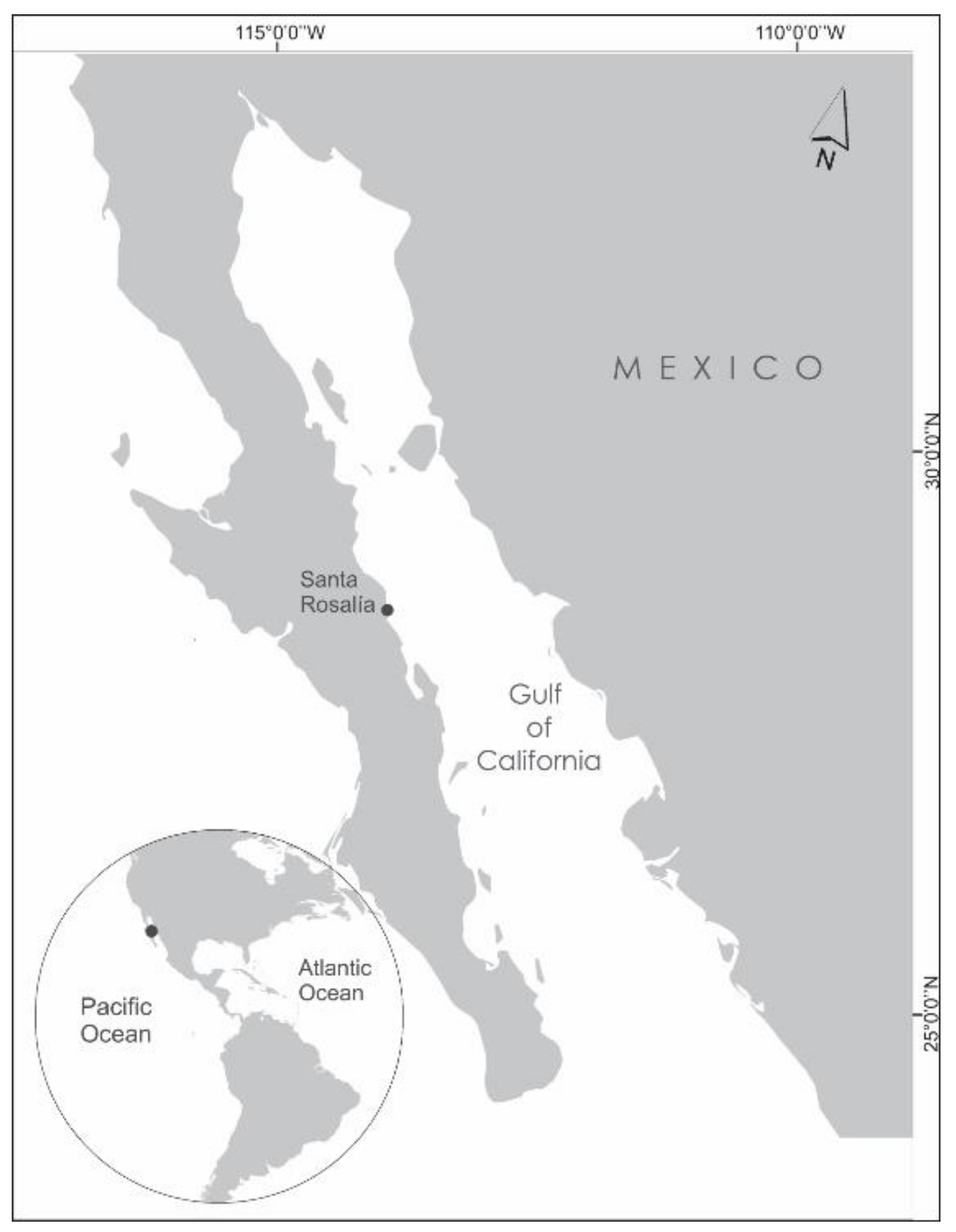
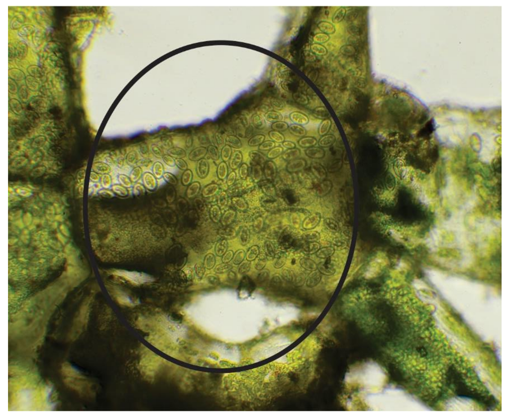
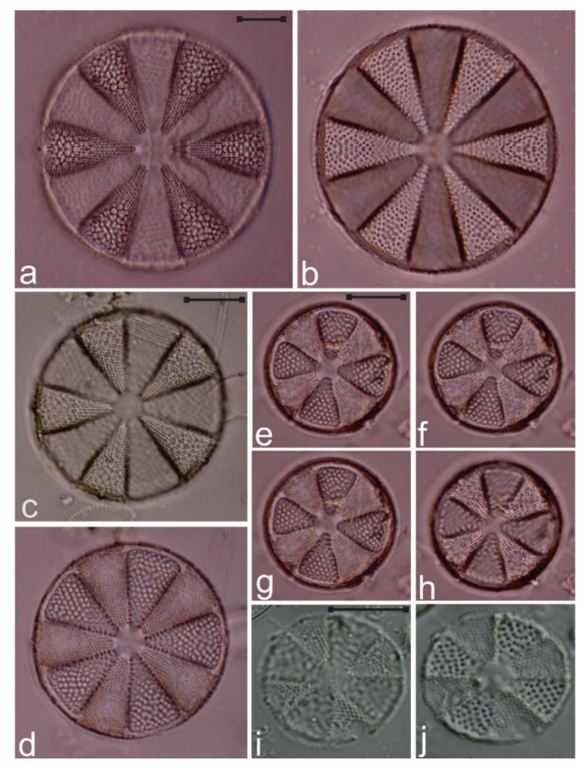
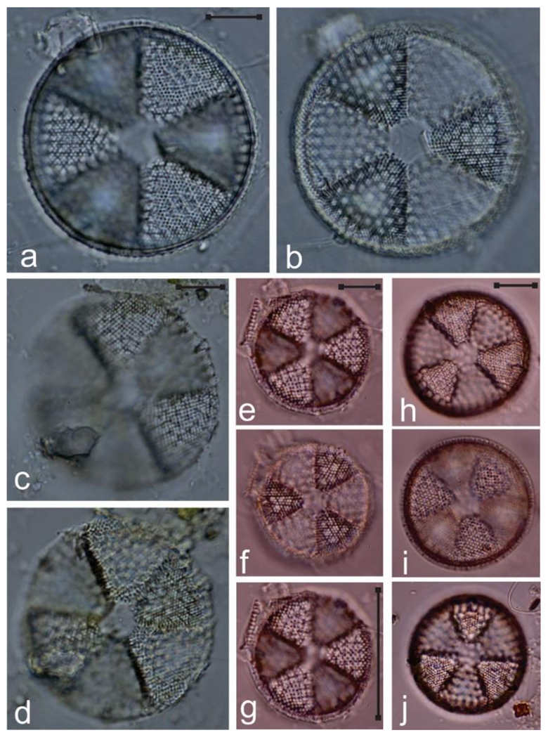
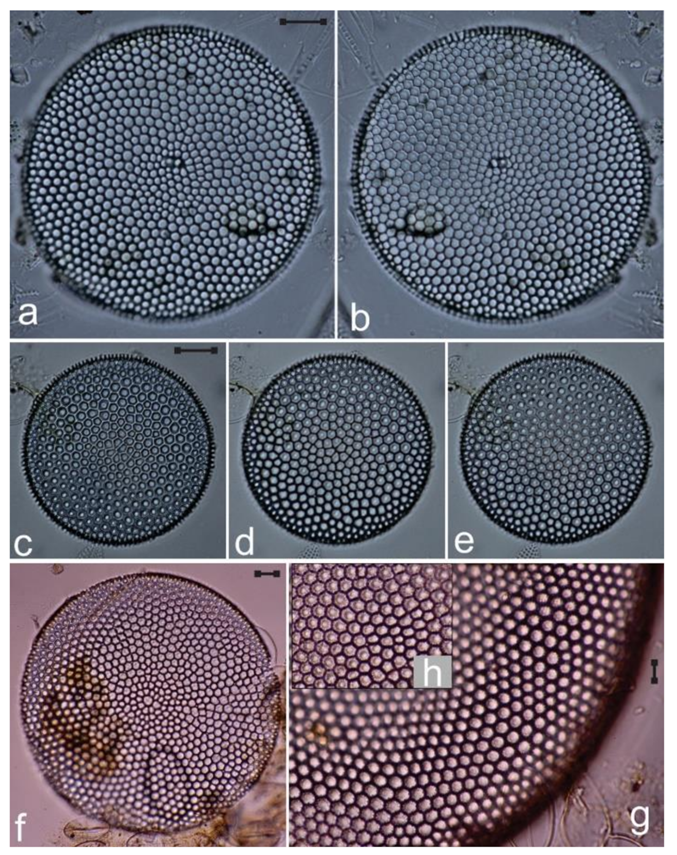
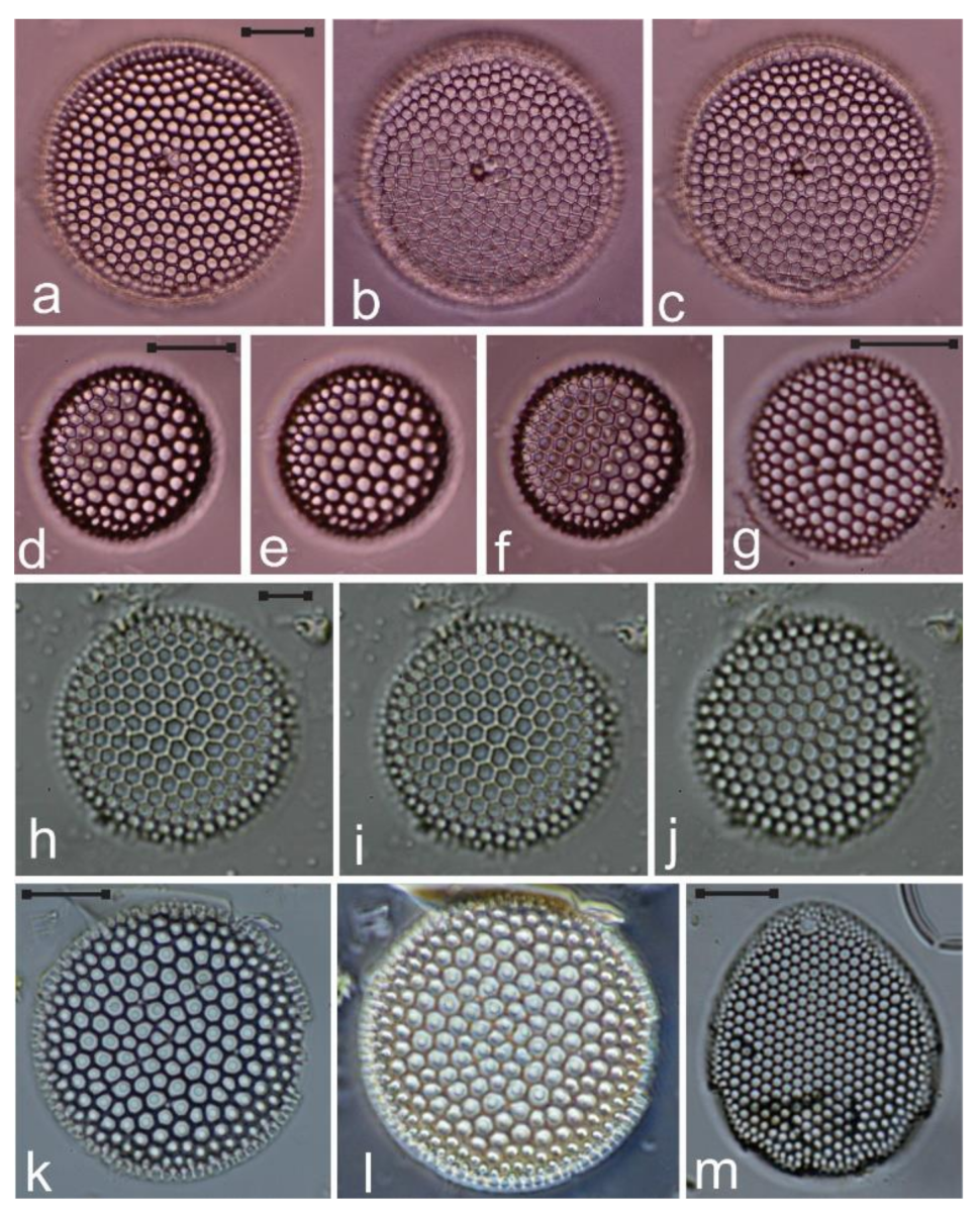
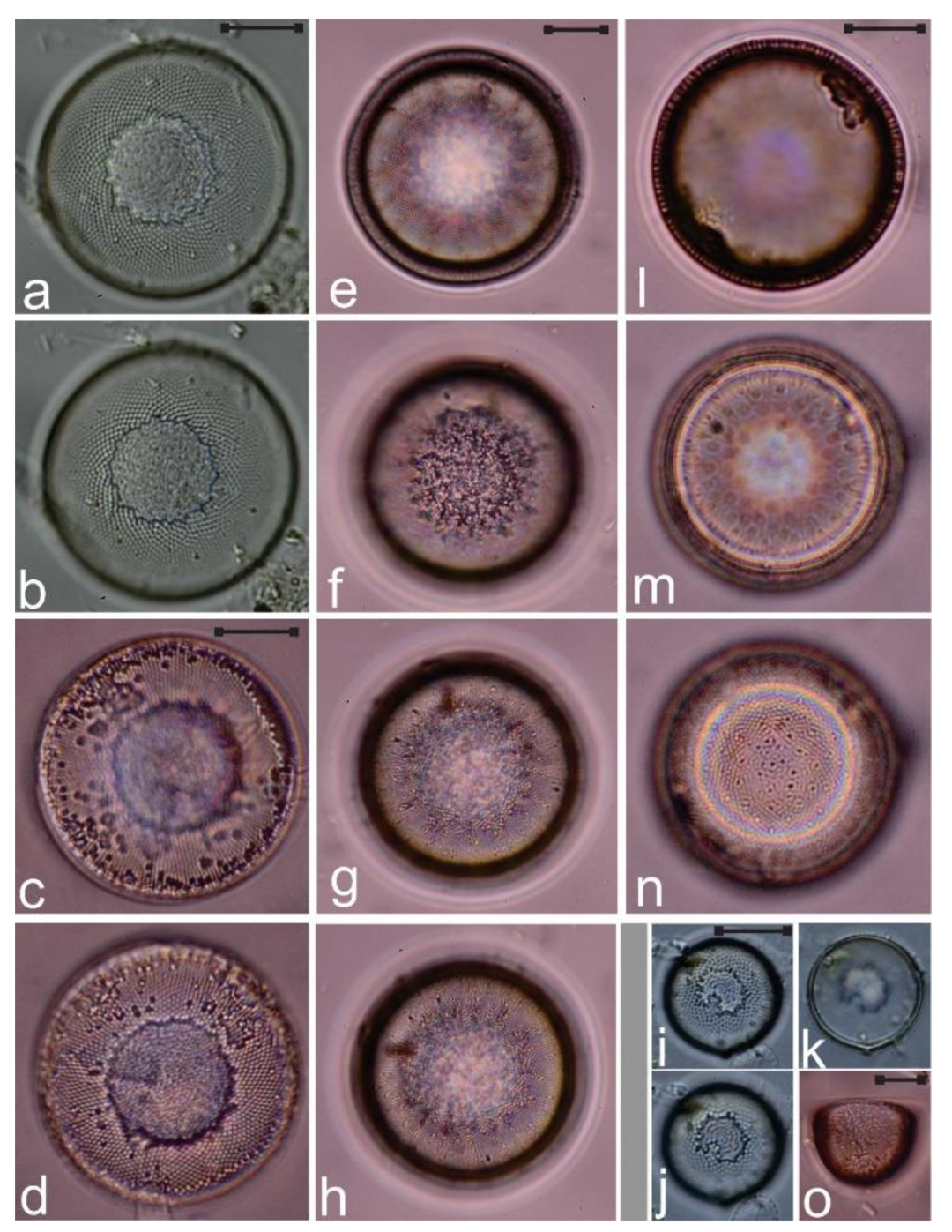
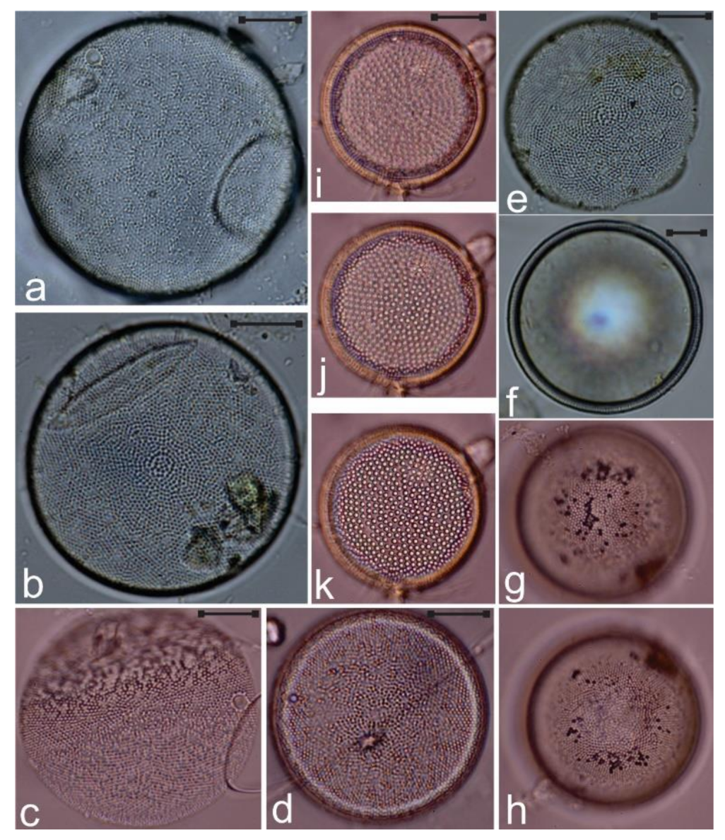
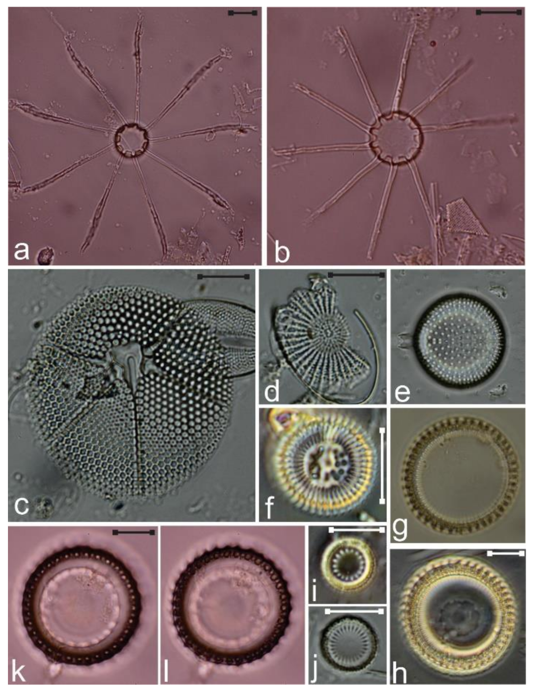
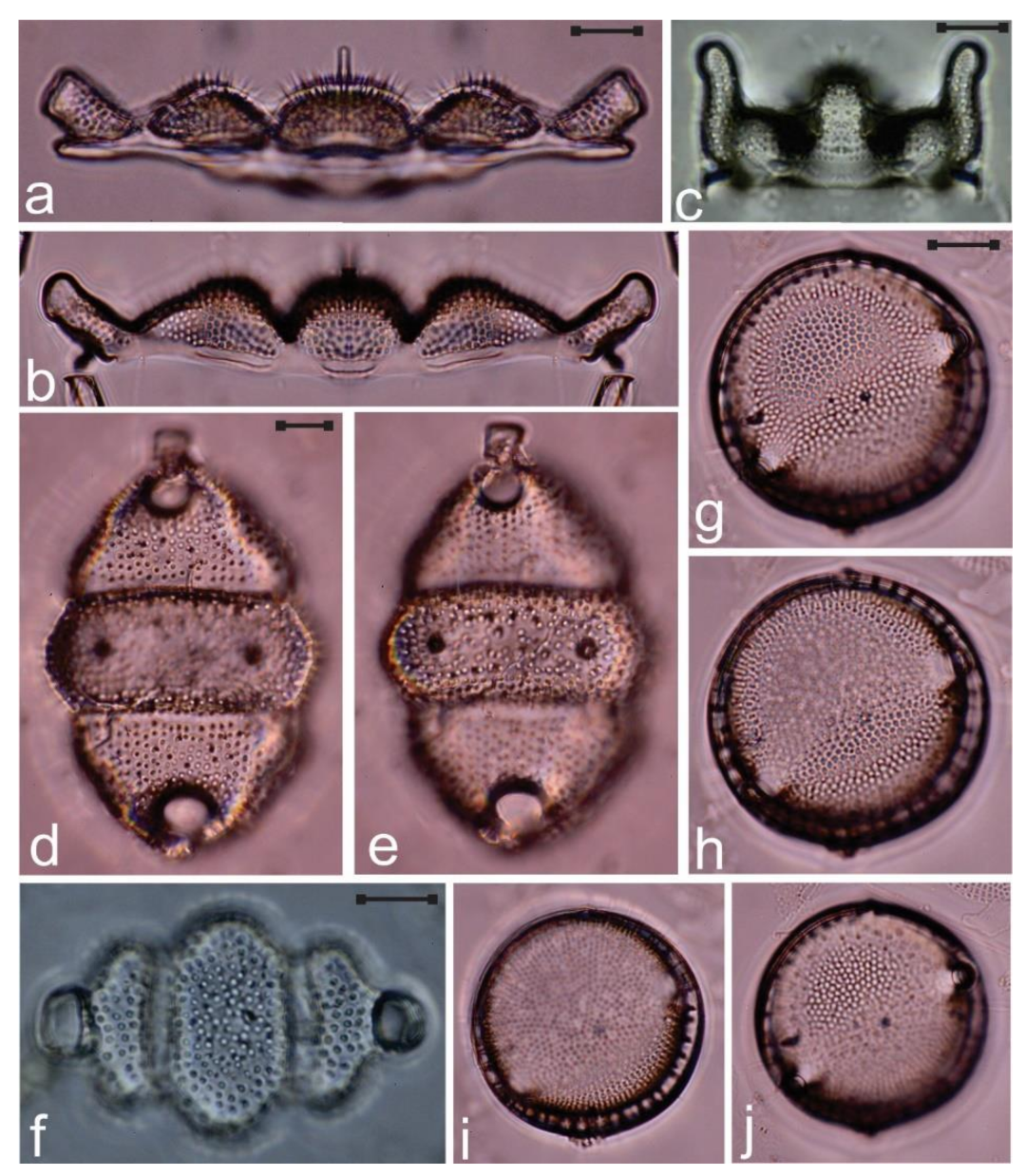
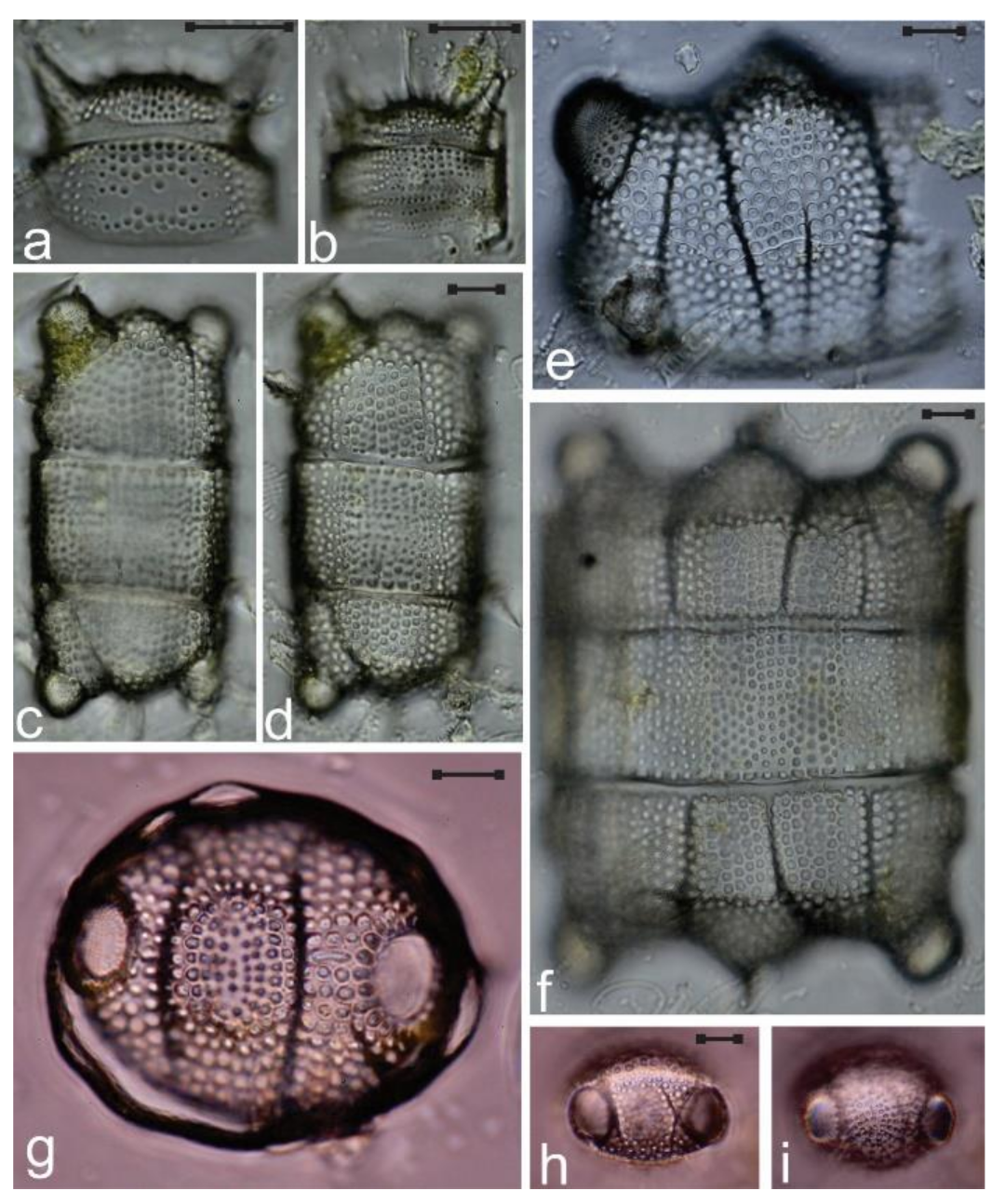
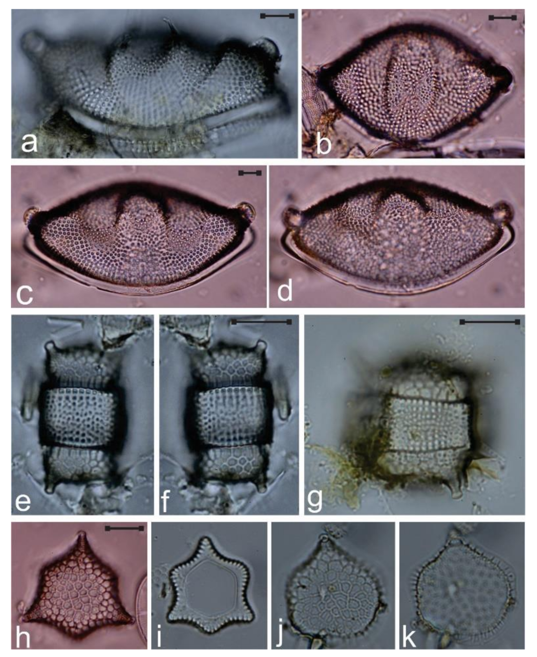
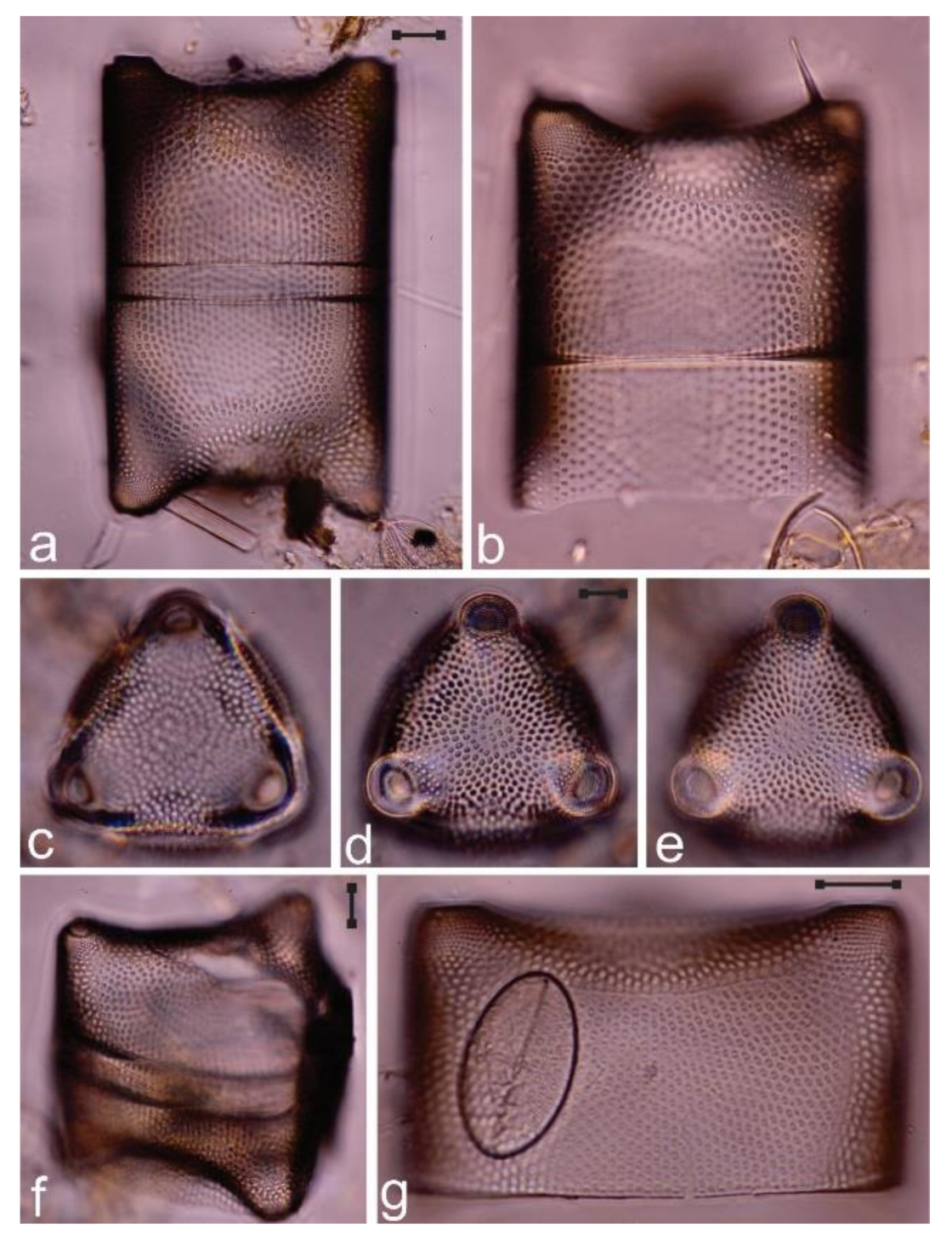
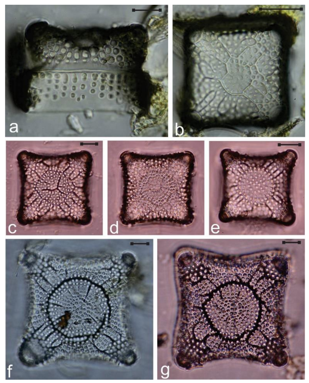
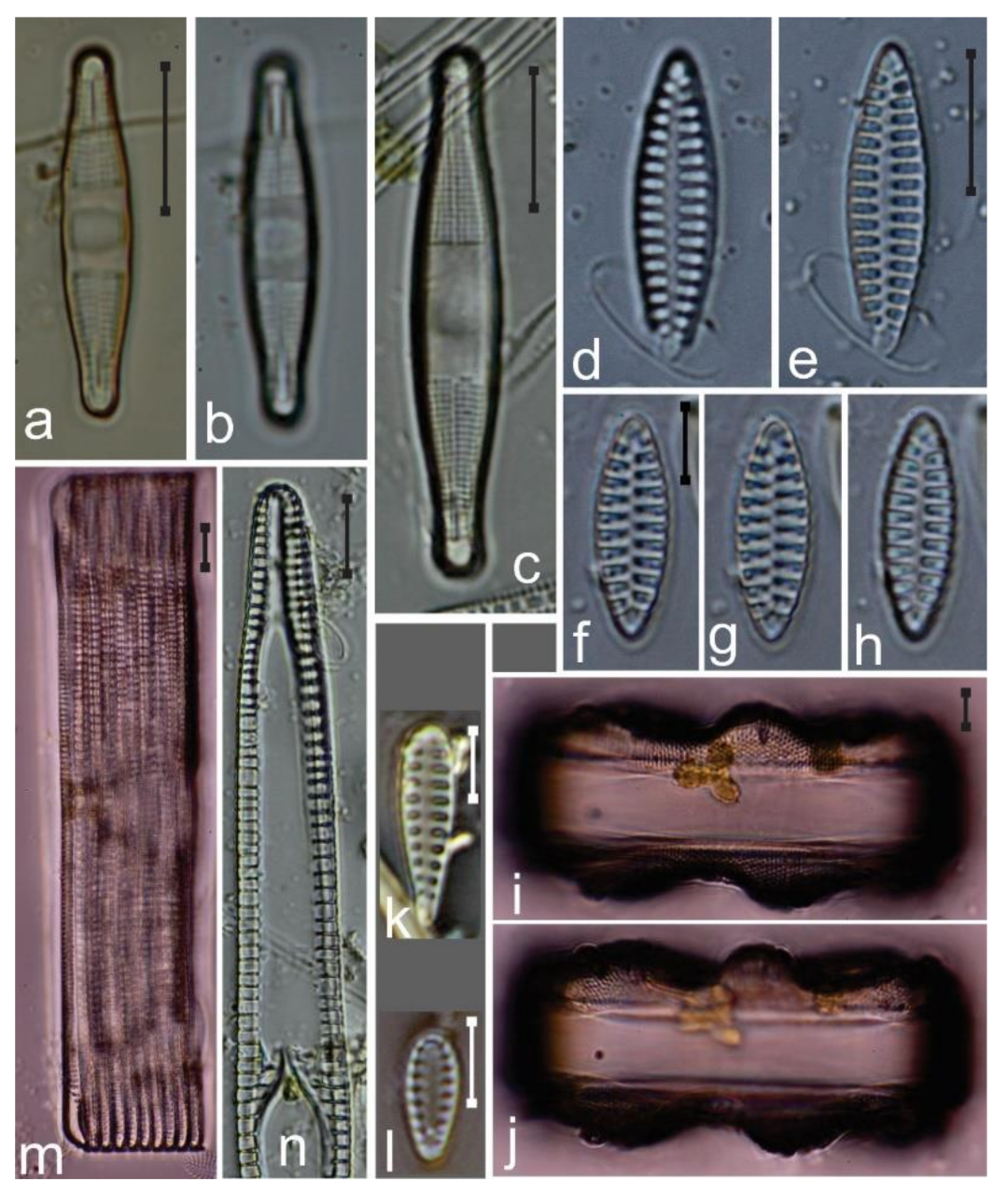
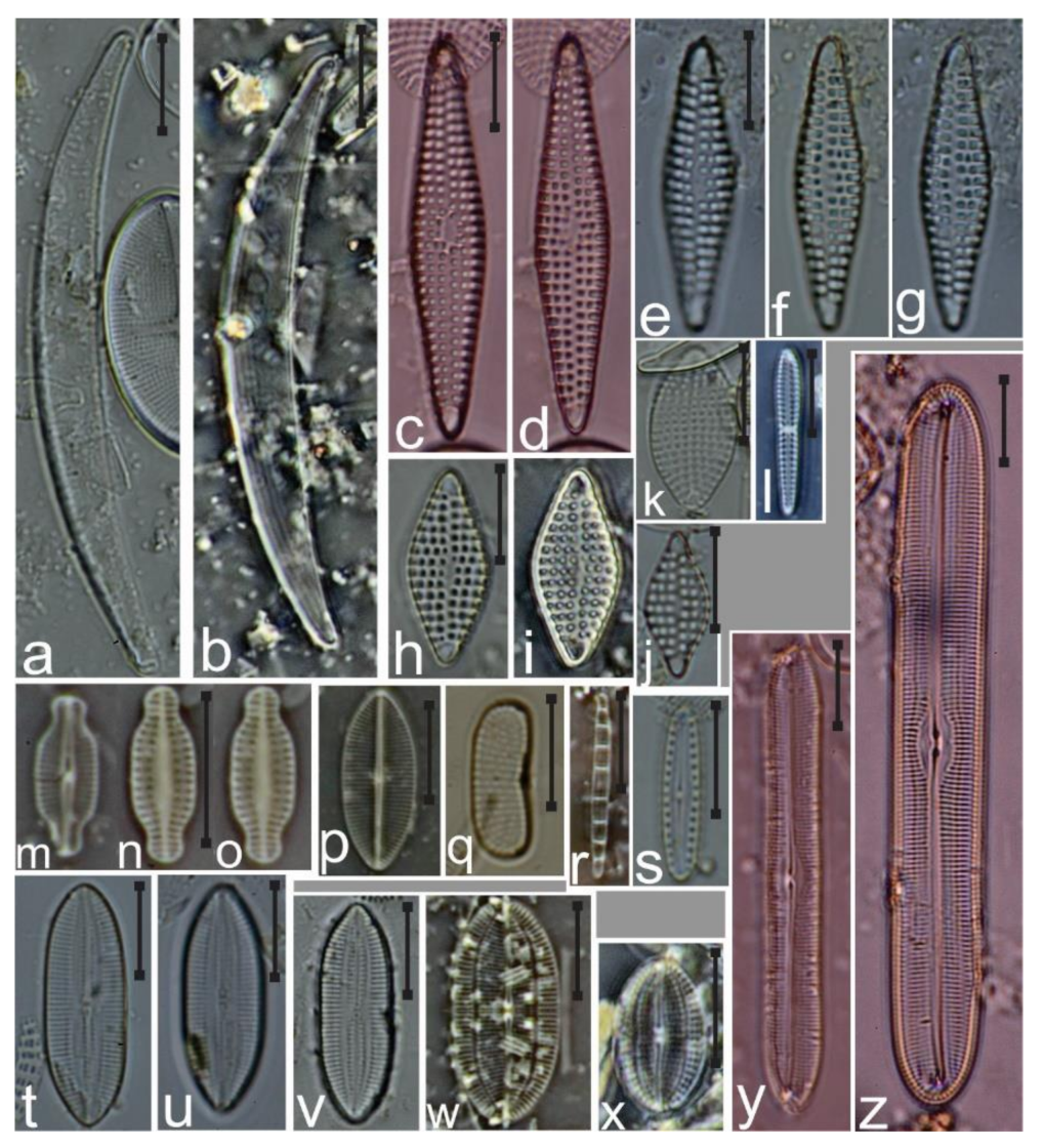
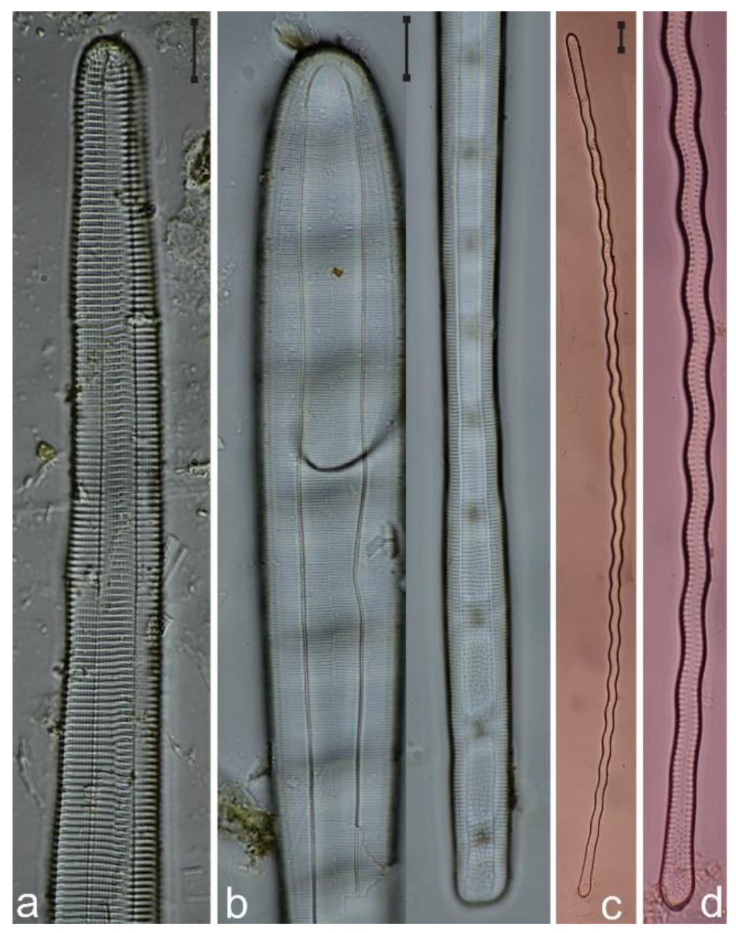
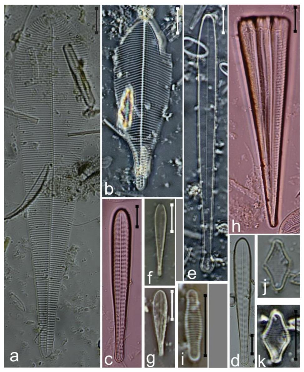
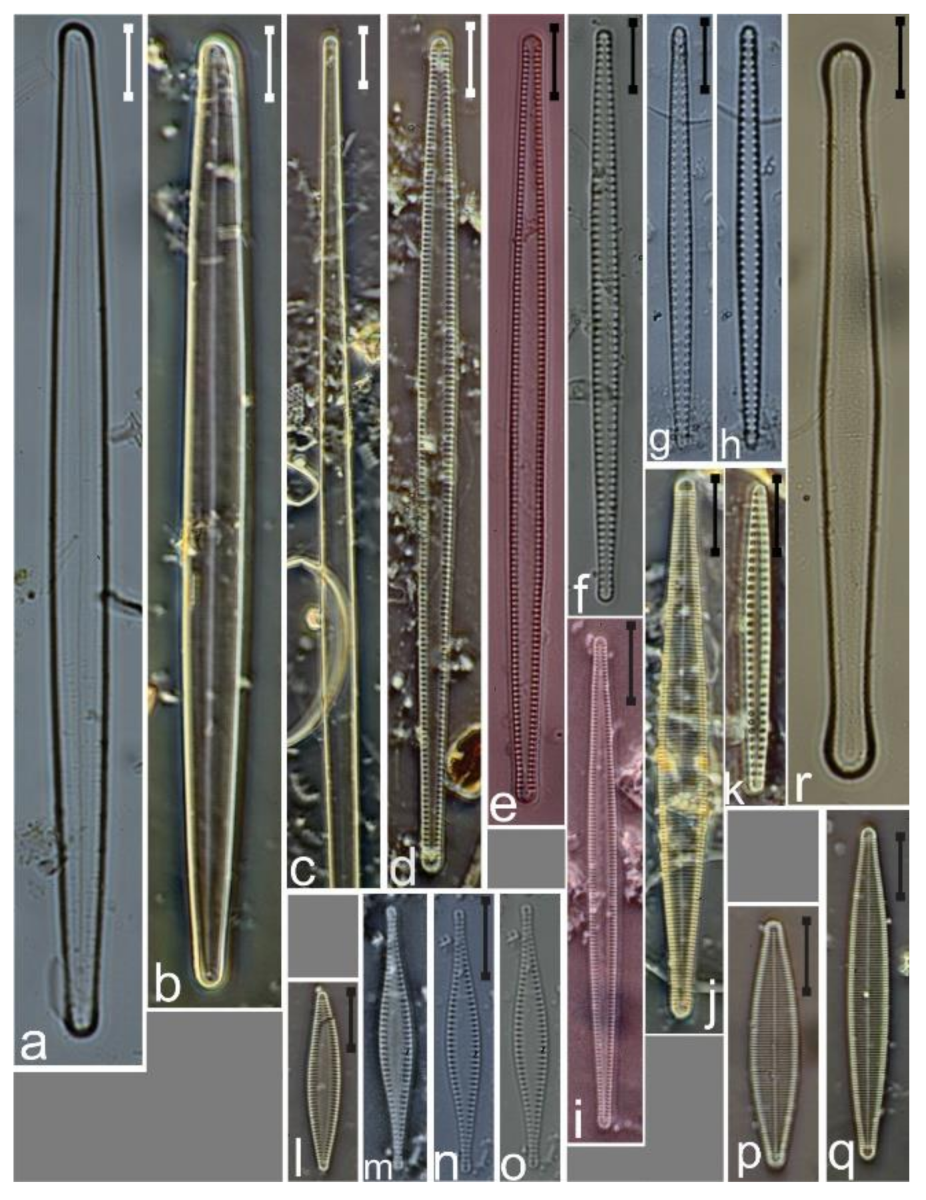
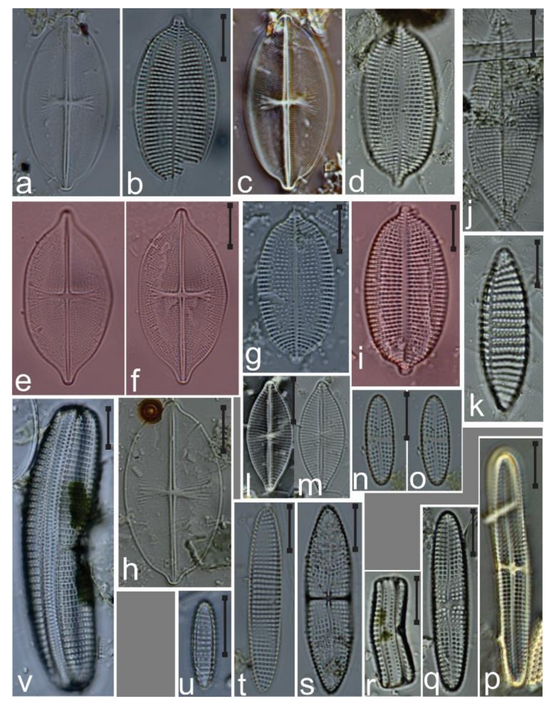
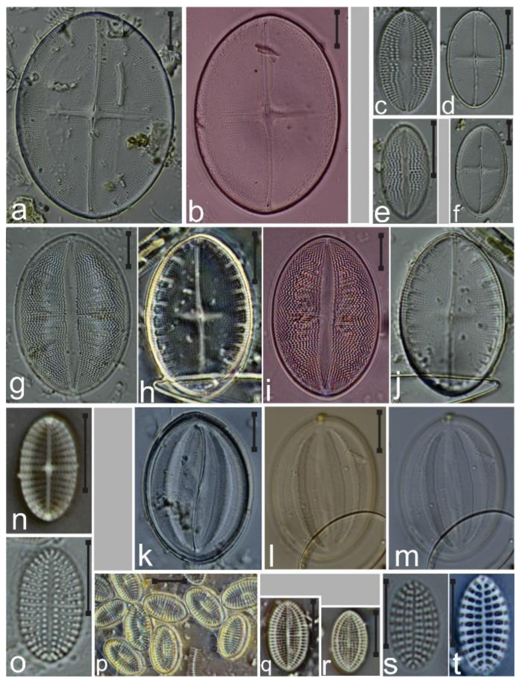
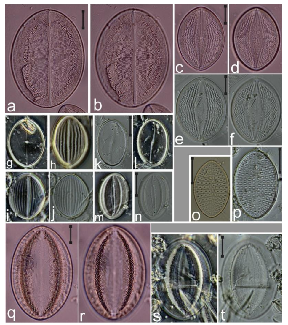
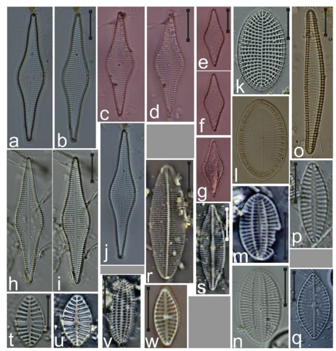
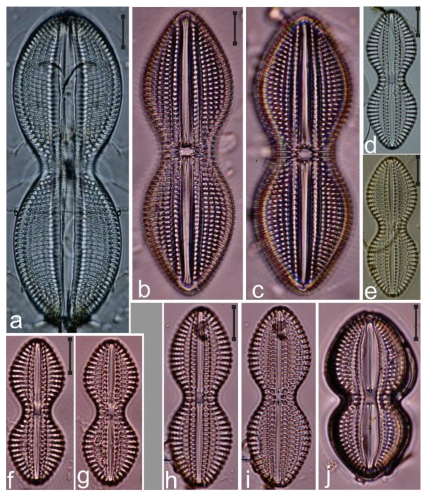
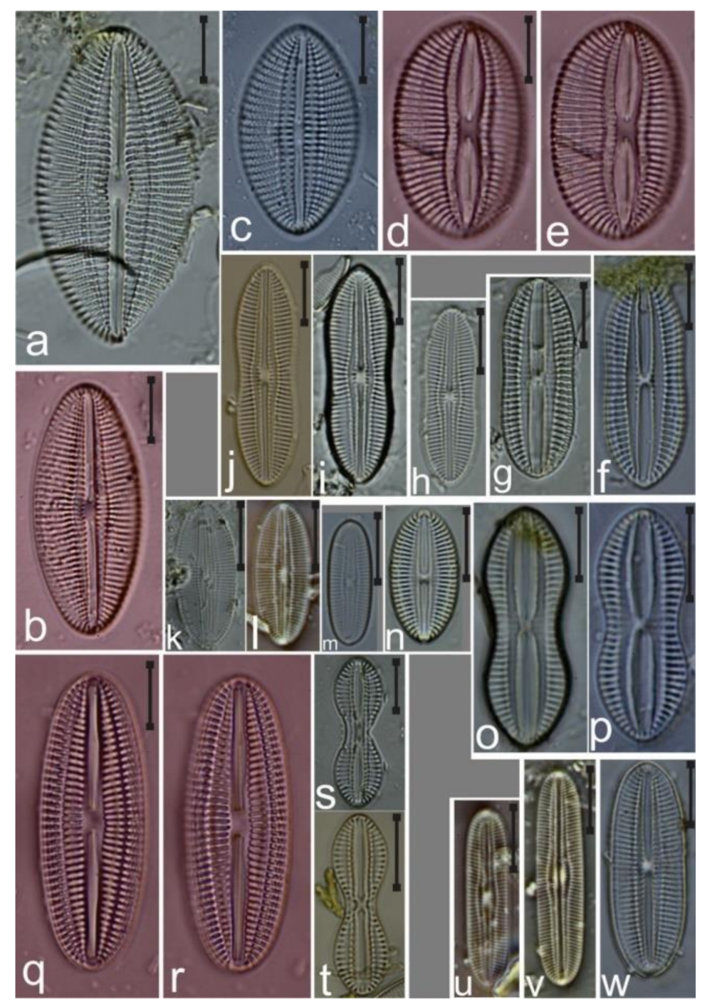
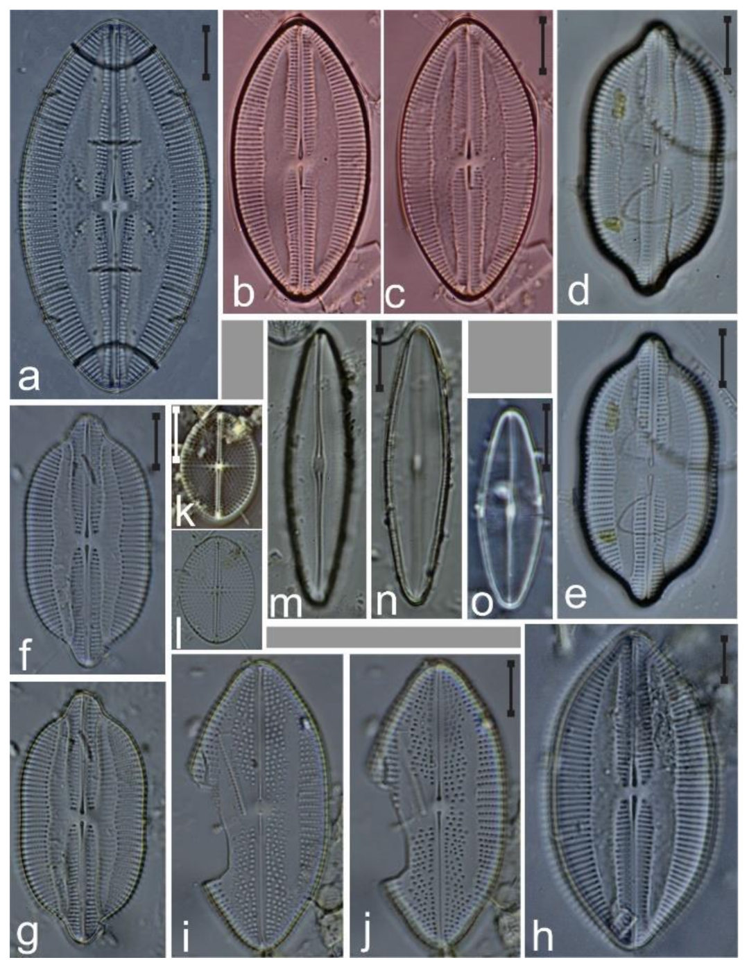
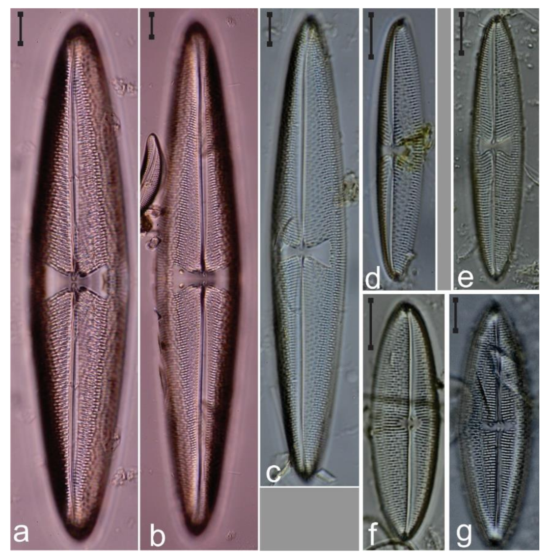
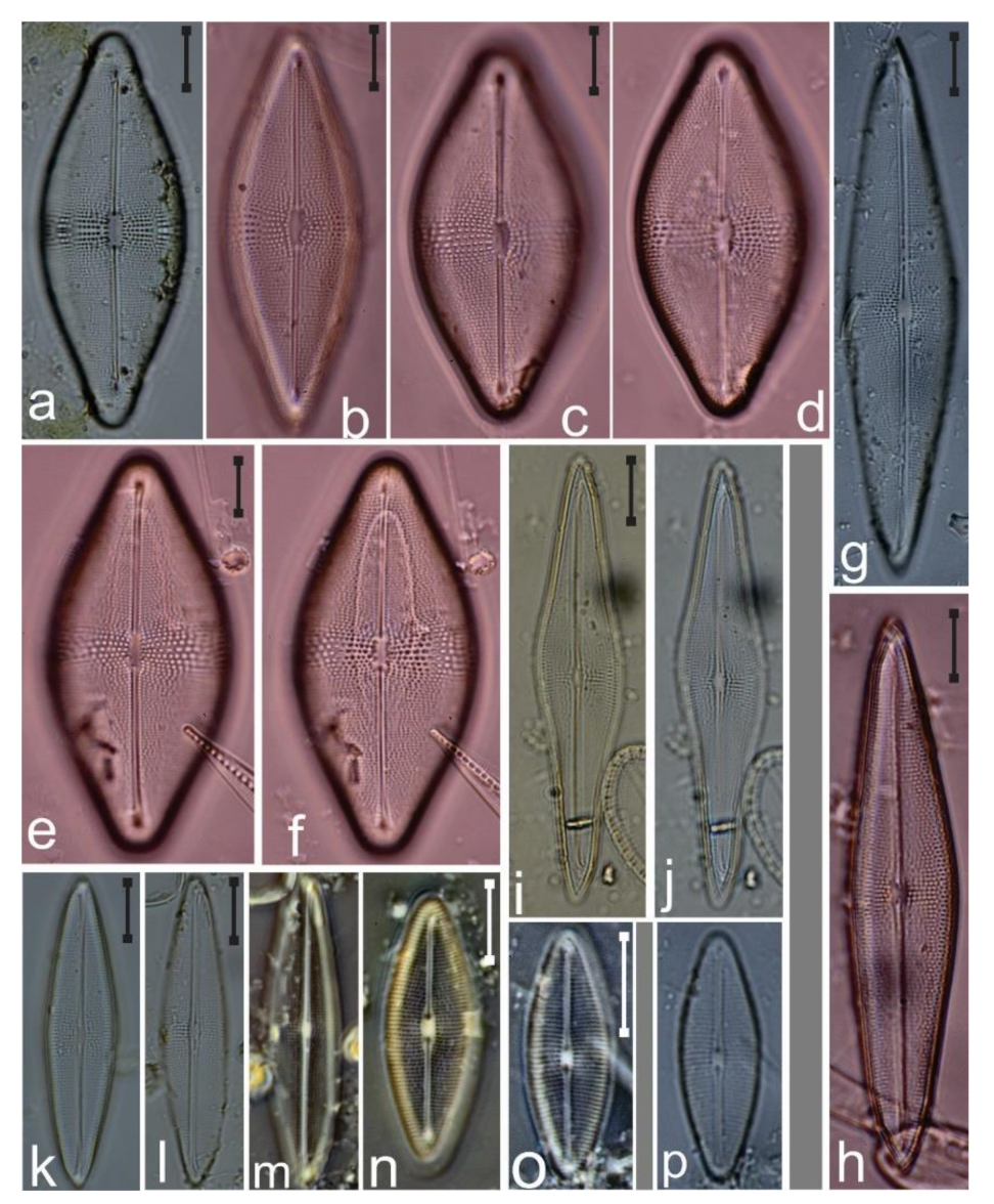
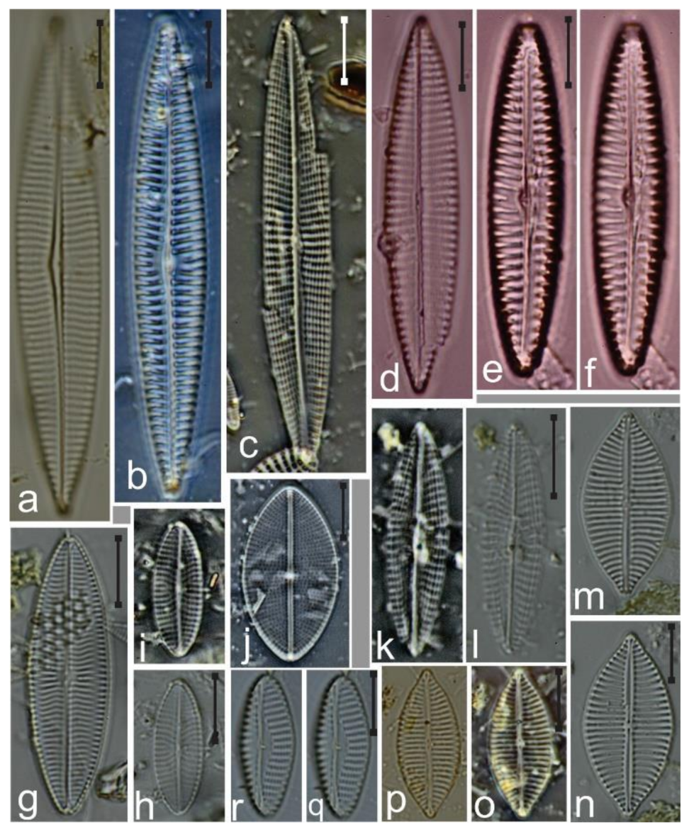
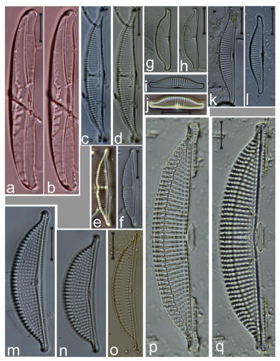
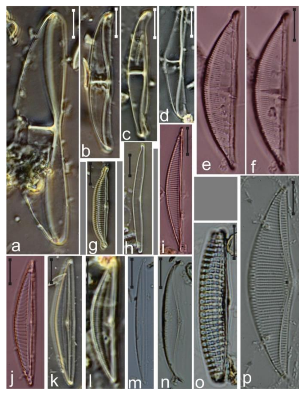
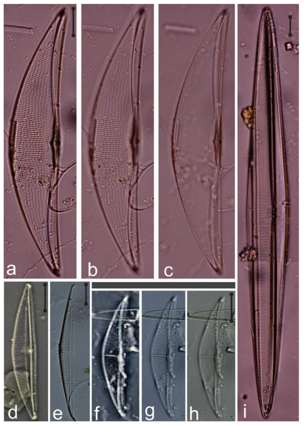
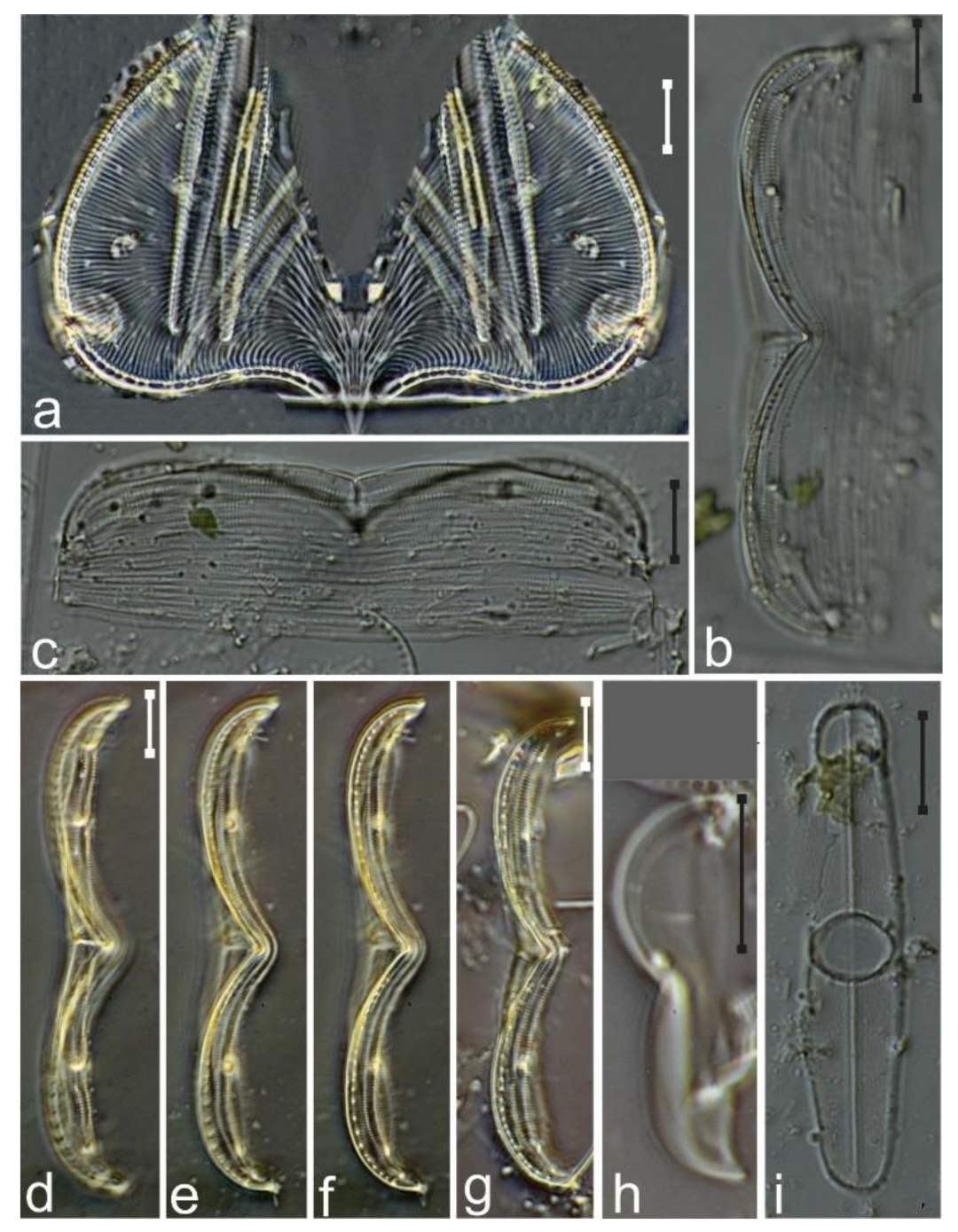
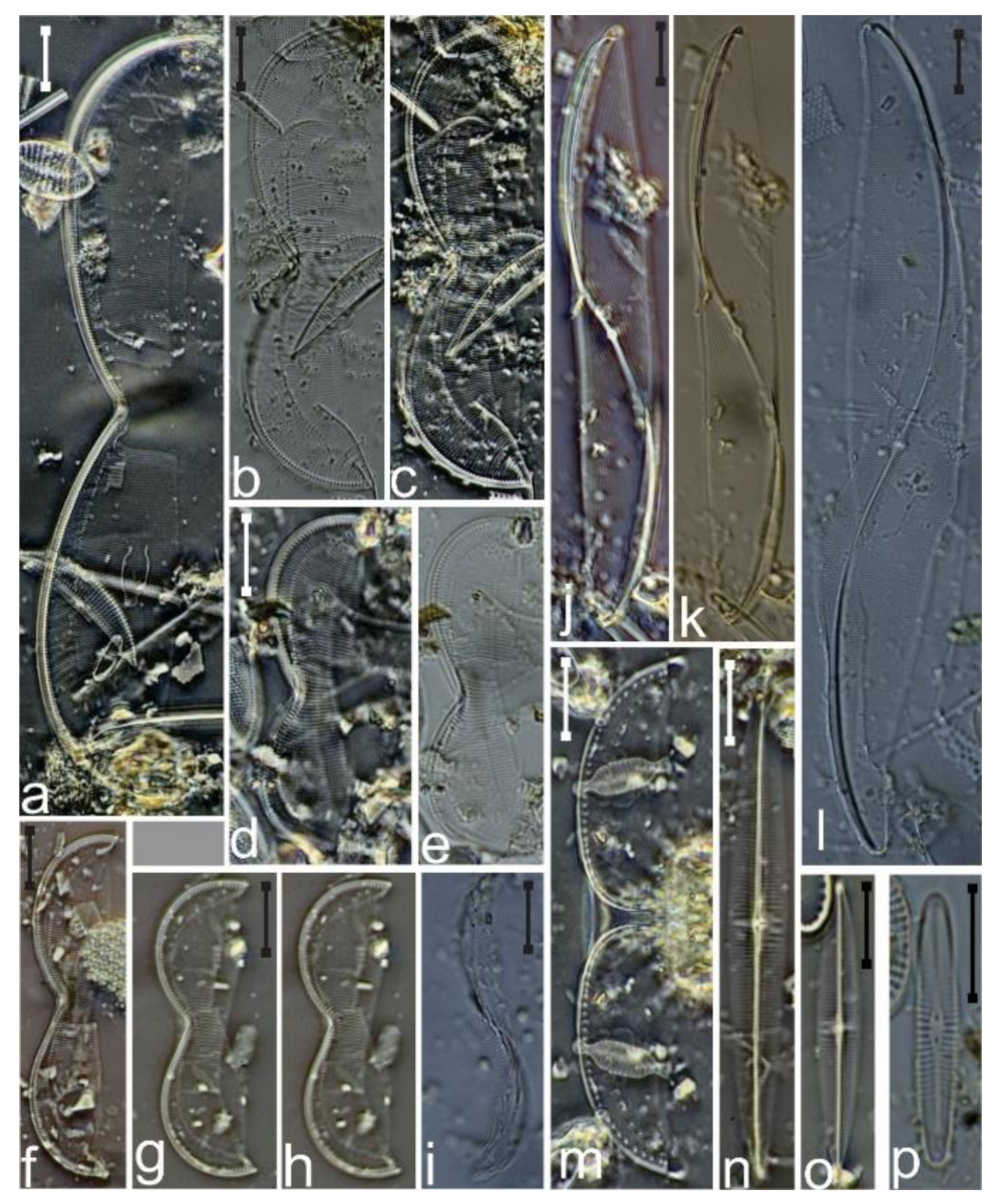
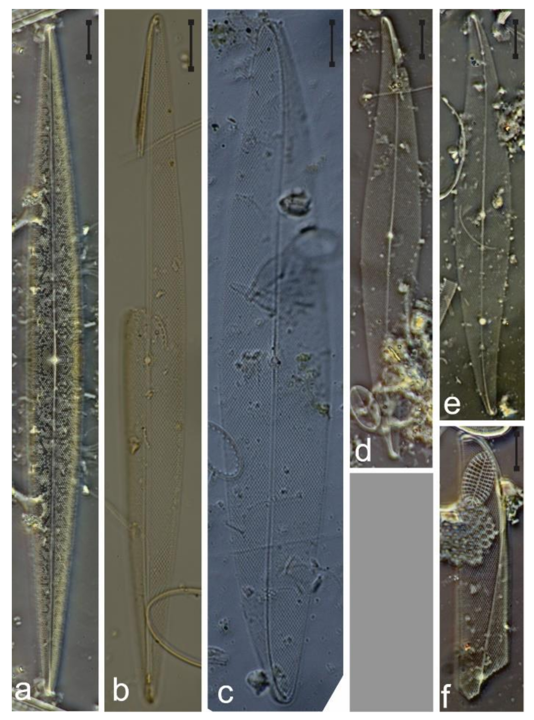
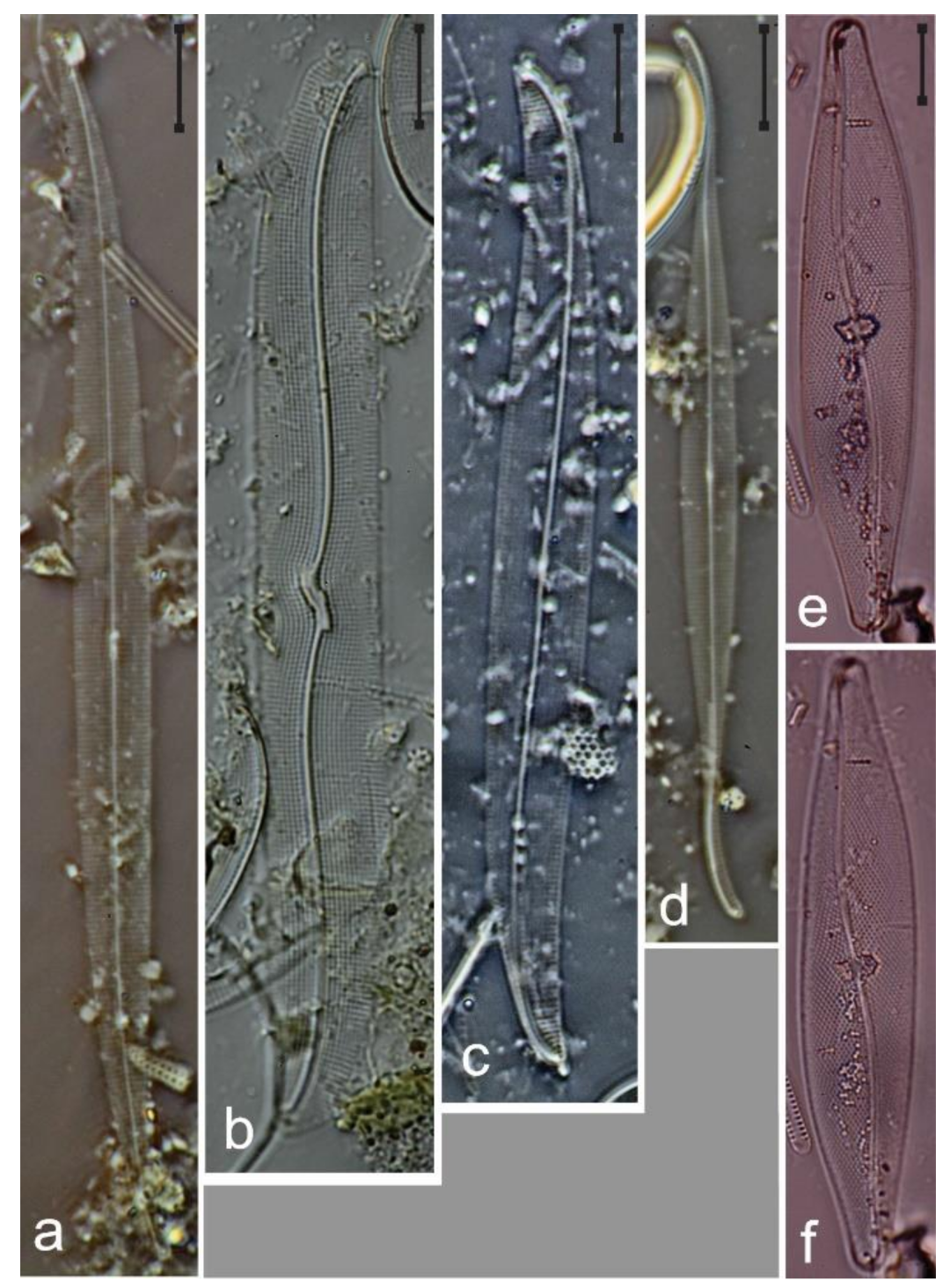
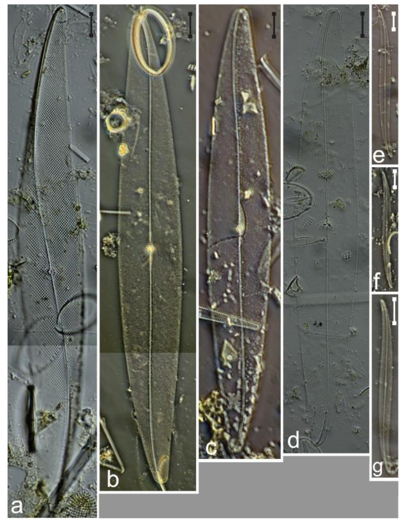
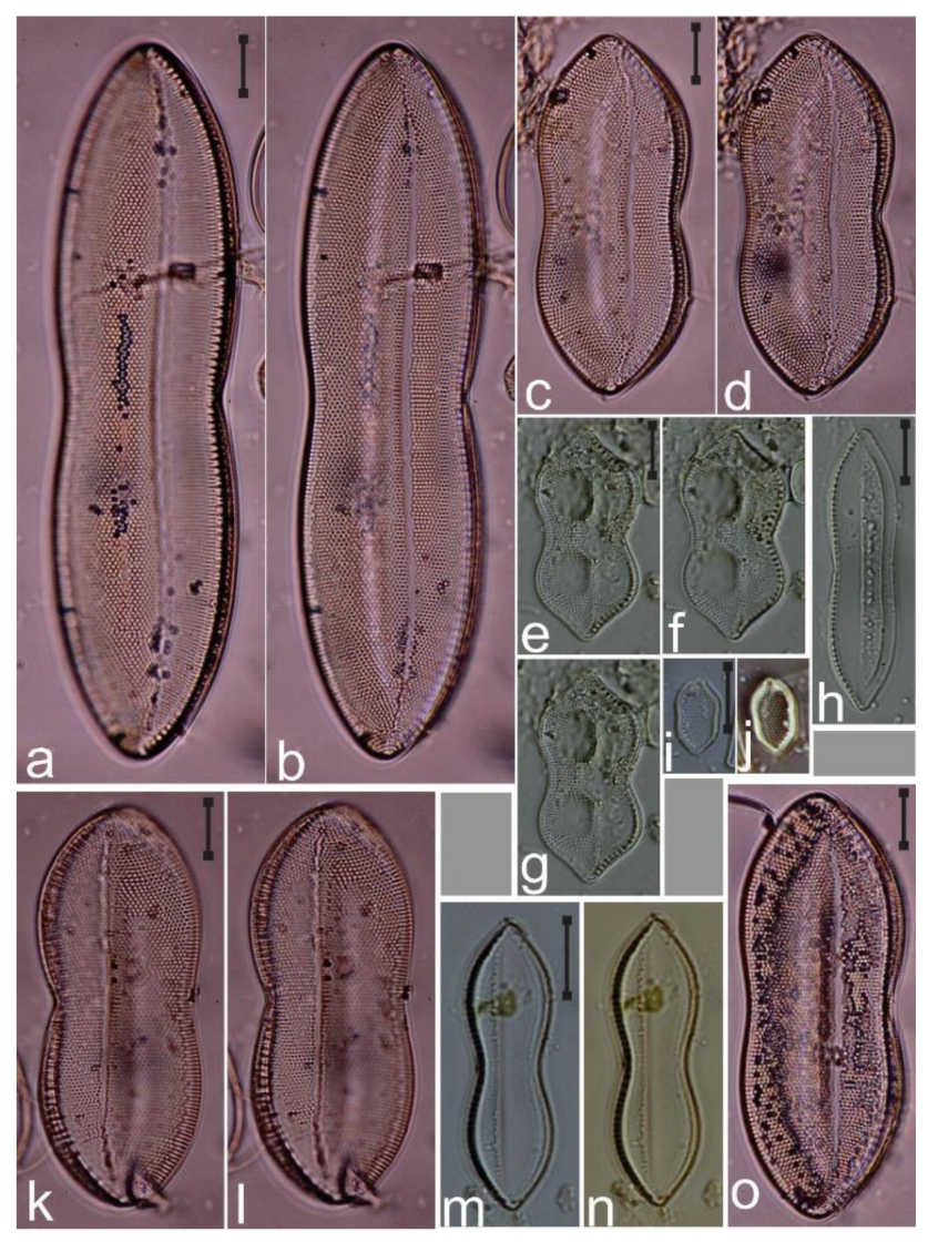
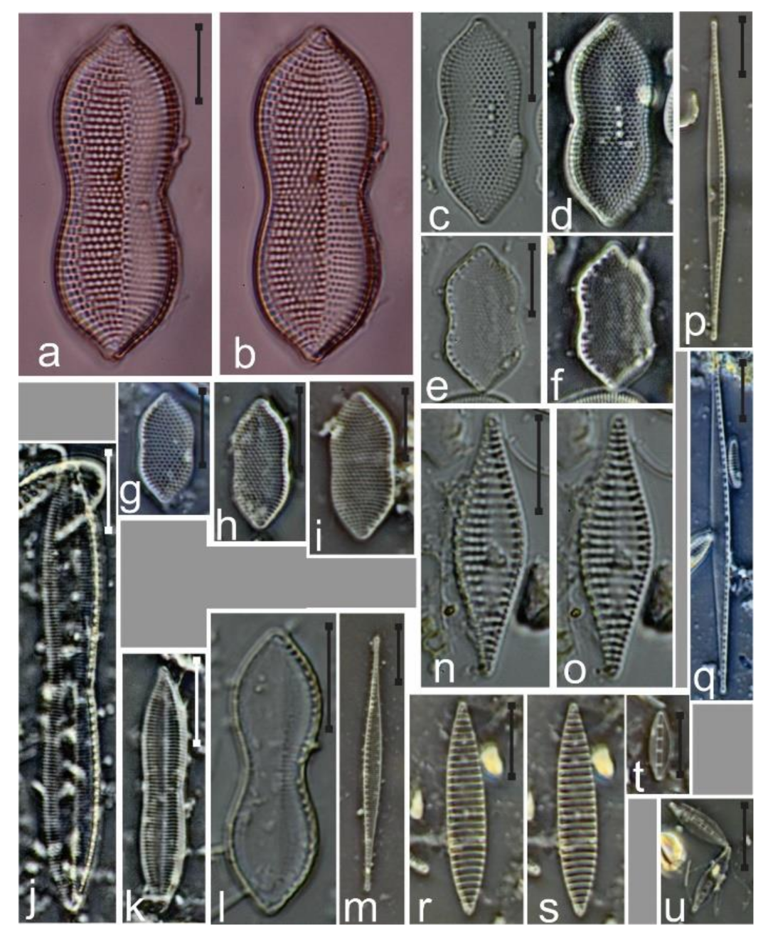
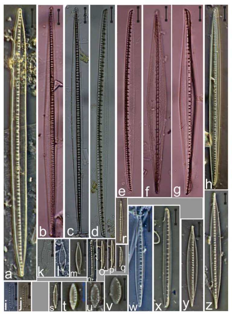
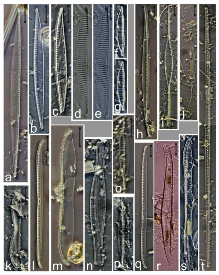
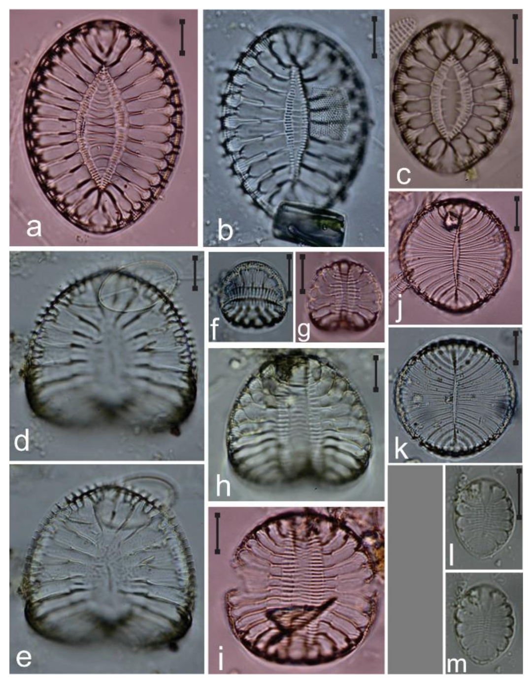


