Article

Molecular Diversity of Nematode Parasites in Afrotropical Reed Frogs (Hyperolius spp.)

1
Department of Biology, Institute of Integrated Sciences, University of Koblenz-Landau, D-56070 Koblenz, Germany
2
Department XXI (Med. Microbiology), Laboratory of Medical Parasitology, Central Military Hospital Koblenz, D-56072 Koblenz, Germany
*
Author to whom correspondence should be addressed.
Diversity 2020, 12(7), 265; https://doi.org/10.3390/d12070265
Submission received: 8 June 2020 / Revised: 29 June 2020 / Accepted: 30 June 2020 / Published: 2 July 2020
(This article belongs to the Section Animal Diversity)

Abstract

:
The diversity of nematodes infecting amphibians is understudied in tropical Africa and unknown in Rwanda. Diversity assessment is hampered by the fact that species descriptions refer mostly to morphological features that are unlinked to DNA sequences of marker genes available in public databases. In this paper, we explore the abundance and diversity of parasitic nematodes in reed frogs Hyperolius kivuensis (n = 115), H. parallelus (n = 45) and H. viridiflavus (n = 100) collected in Rwanda. Five nematode species were identified morphologically as Orneoascaris chrysanthemoides, O. schoutedeni, Gendria leberrei, Aplectana chamaeleonis and Rhabdias collaris. Corresponding DNA sequences of 18S and COI genes were determined and subsequently deposited in GenBank. Aplectana chamaeleonis showed the highest prevalence (8.7%), but O. chrysanthemoides the highest mean intensity of infection (6.0) and largest number (24) of individuals in H. kivuensis. To the best of our knowledge, all amphibian hosts are new records for these nematode species, which are known to infect a wide range of amphibian and reptile species. Our findings suggest that nematode diversity is probably lower than previously assumed due to low host specificity. As morphological species identification is often challenging, our data facilitate molecular identification of adult and specifically larval nematodes found in amphibians of Sub-Saharan Africa.

1. Introduction

Nematodes are a hyper-diverse worm taxon including free-living and parasitic species [1]. Currently, about 27,000 nematode species have been formally described based on morphological and ecological traits, but conservative estimates yield at least 100,000 species, while some authors expect more than 1 million species [2,3]. The deceptively uniform basic anatomy of nematodes complicates full appreciation of real diversity, rendering morphology-based taxonomy a time-consuming task for well-trained experts [1,4]. Recently, the use of molecular data based on COI and 18S gene sequences represents an independent approach to nematode diversity, available in public sequence databases as GenBank [1,5]. The crucial link between morphological and molecular diversity is still missing for most described nematode species, a serious drawback for the use of sequences to reliably identify voucher specimen [1,5,6].
Parasitic nematodes of vertebrates represent an excellent example for a group in which most species are exclusively identified based on their morphology [7,8]. Amphibians and reptiles have a rich diversity of nematode parasites [9,10], e.g., as many as 173 species have been recorded in the herpetofauna of Mexico [11]. They infect predominately the intestines of their hosts, but also the respiratory tract, body cavity and blood [12]. Nematode parasites may cause a range of damage or alterations, leading to modification of host behavior or metabolism, as demonstrated for lungworms Rhabdias spp. infecting toads [13,14,15]. Thus, the prevalence of nematodes may interfere with the fitness of their amphibian hosts and ultimately affect their survival [16]. A recent enigmatic amphibian decline has been attributed to the combined adverse effects of nematode parasite load and climate change [17].
In this study, we focused on the morphological and molecular diversity of the poorly studied Afrotropical nematode parasites of amphibian hosts. With very few regional exceptions such as Cameroon, Nigeria and South Africa, very little is known about species richness, geographical distribution and host specificity of Afrotropical nematodes [12,18,19,20]. Presently, identification and determination to the species level is almost exclusively based on morphological features, often relying on very old original publications and descriptions of species found only once or twice [21]. Notable exceptions are recent studies on the Afrotropical Rhabdias lungworms combining morphological and molecular evidence [22,23]. In most genera with widely distributed Afrotropical species such as Amphibiophilus, Aplectana, Falcaustra and Orneoascaris, molecular tools for species identification are still not available. Consequently, most morphological datasets are unlinked to corresponding molecular sets resulting in a very low number of reliably identified Afrotropical nematode parasites with sequence data deposited in GenBank. We present molecular and ecological data on five morphologically identified parasitic nematode species from diverse Afrotropical frog hosts. Molecular markers are used to identify them. We provide a detailed morphological diagnosis including microphotographs of the rarely found species; partial sequences of the nuclear ribosomal 18S-ITS1 locus for all species and the mitochondrial cytochrome oxidase subunit 1 (COI) for one species; and a sequences query with available GenBank entries.

2. Materials and Methods

2.1. Sampling

In total, 89 adult Kivu reed frogs (Hyperolius kivuensis, HK) and 100 common reed frogs (H. viridiflavus, HV) were sampled from a population inhabiting the cultivated wetlands (“marais”, 2.60° S 29.76° E, 1645m a.s.l.) near Huye (Butare), Rwanda [24]. Sampling dates were October 2, 2015 (n = 18 HK; n = 26 HV), October 1, 2016 (n = 4 HV), October 14, 2018 (n = 27 HV), January 10, 2017 (n = 35 HK; n = 25 HV) and March 19, 2017 (n = 26 HK; n = 18 HV), covering the complete rainy period at this locality. Complementary samples of H. kivuensis were collected in wetlands near Musanze (Ruhengeri, 1.51° S 29.65° E, 1807m a.s.l.; n = 26) on October 6, 2015 [25]. Finally, we collected two samples of Angolan reed frogs Hyperolius parallelus on October 22, 2018 (n = 21) and December 31, 2018 (n = 24) near the Cyamudongo forest (2.55° S 29.00° E, 1791 m a.s.l.). The reed frogs collected in October 2015 were transported to the laboratory in Koblenz and kept in terraria with food ad libitum until November 30, 2015. Then, they were euthanized by immersion into a 2% solution of tricaine methane-sulfonate (MS 222), except for 10 H. kivuensis individuals kept alive until May 6, 2017. All host specimens collected after 2015 were euthanized immediately after collection. The body cavity, digestive tract, lungs, kidneys and bladder were subsequently examined macroscopically and microscopically for the presence of endoparasites. Carcasses of frogs were fixed in 10% buffered formalin, transferred to 70% ethanol for long-term storage and deposited in the collection of the Zoologisches Forschungsmuseum Alexander Koenig, Bonn, Germany (ZFMK). Collection numbers are ZFMK 102849-1021962 (H. kivuensis), ZFMK 103109-103127 (H. parallelus) and ZFMK 102963-103098 (H. viridiflavus).
The detected parasites included 12 species of annelids, nematodes, monogeneans, digeneans and cestodes, but, in this study, we focused exclusively on the nematode species detected in the intestines and lungs of the frogs. We extracted six large ascaridid nematodes from the intestines of one specimen each of H. kivuensis, H. viridiflavus and H. parallelus and transferred them to Eppendorf tubes with 1.5 mL ethanol for molecular identification. One sample each of morphologically different small nematodes found in the small intestines of H. kivuensis and in the lung of H. viridiflavus were treated the same way. All remaining nematodes were fixed in 10% buffered formalin for subsequent morphological examination.
All applicable international, national and/or institutional guidelines for the care and use of animals were followed, and all procedures performed were in accordance with the ethical standards of the University of Koblenz-Landau (approval # Si 2015/01, ethic committee of FB3).

2.2. Morphological Identification of Nematode Vouchers

The macroscopically detected nematodes were first observed live using a digital Keyence microscope VHX-6000 and digitally documented using Keyence VHX-H1M1 software. Each formalin-fixed nematode was placed in a drop of lactophenol on a microscope slide, cover slipped and examined under an Olympus BX 50 microscope equipped with a high-resolution camera Olympus DP20 for documentation with the Cell Imaging software. The nematodes were identified to genus level based on the corresponding keys [7,8] and by subsequent comparison with the original descriptions. Parasite terminology is used in accordance with [26]. Vouchers specimens were deposited at the Centrum für Naturkunde (CeNak)—Center of Natural History, Universität Hamburg—Zoologisches Museum, Germany.

2.3. Nucleic Acid Extraction and Polymerase Chain Reactions

The ethanol-fixed nematodes were incubated in 1.5 mL reaction tubes, with open cap, at 40 °C in a Thermomixer (Eppendorf, Germany) until the ethanol was completely evaporated. The sample was mixed with 180 µL ATL buffer and 20 µL proteinase K, incubated at 56 °C until the specimen was completely lysed, and then further processed following the protocol for DNA purification from tissues of the QIAamp DNA mini kit (Qiagen, Germany). The extracted DNA was used to amplify parts of nuclear ribosomal locus (18S-ITS1-5.8S) and the mitochondrial cytochrome oxidase subunit 1 (COI) locus (Table 1). Polymerase chain reaction (PCR) was conducted using Taq PCR core kit as recommended by the manufacturer (Qiagen). The following programs were used: 40 cycles of 1 min at 94 °C, 1 min at 53 °C and 1 min at 72 °C for 18S-ITS1-5.8S; 40 cycles of 1 min at 94 °C, 1 min at 56 °C and 1 min at 72 °C for COI. All PCR cycles were initiated with a denaturation step for 3 min at 94 °C and terminated with an extension step of 72 °C for 7 min. Amplicons were separated on a 1% agarose gel, stained with ethidium bromide and visualized on a UV transilluminator. Prior to sequencing, PCR products were purified using QIAquick Purification Kit (Qiagen).

2.4. Sequencing and Phylogenetic Analysis

For bidirectional sequencing, the same primers as for the PCR were applied (Table 1). Assembly of DNA sequence files was conducted with DNA Baser (Heracle BioSoft, Romania) and primer sequences were clipped. Due to poor performance of reverse primer IR8 as a sequencing primer, most sequences only contain partial 18S and ITS1 regions. Sequences were deposited in GenBank (Table 2). Closest matches of sequences were identified by a BLAST search against GenBank entries [27].
Phylogenetic trees were computed from the alignments 1 using Bayesian interference in MrBayes [28]. Posterior probabilities were approximated over 3,000,000 generations. Positions containing gaps and missing data were eliminated. Trees were rooted by the sequences of outgroup species Cylicospirura petrowi (Orneoascaris tree), Ichthyobronema hamulatum (Gendria and Aplectana tree) and Strongylus vulgaris (Rhabdias tree), retrieved from GenBank. The resulting trees were manually refined using Fig Tree v1.4.2. Pairwise nucleotide comparison for COI sequences was generated using GeneDoc [29].

3. Results

The diversity of nematodes in the three representatives of the genus Hyperolius included five species with 60 specimens. Most nematodes were found within the small and large intestines (N = 59) of the frogs examined; only a single specimen was detected in the lungs. The body cavity was nematode-free in all frogs. The nematodes were morphologically identified as Orneoascaris chrysanthemoides Skrjabin, 1916 [21] (hosts: HK, HV), Orneoascaris schoutedeni (Baylis, 1940) [33] (host: HK), Gendria [Chabaudus] leberrei Bain & Philippon, 1969 [34] (host: HP), Aplectana chamaeleonis (Baylis, 1929) [35] (host: HK) and Rhabdias collaris Baker, 1987 [36] (host: HV). Infection sites, nematode number, prevalence [%], mean intensity and range of the nematodes in each of the Hyperolius species are reported in Table 3. Blast search in GenBank for corresponding gene sequences to aid molecular identification were in vain for all species.

3.1. Orneoascaris chrysanthemoides (Ascaridoidea): Morphological, Ecological and Molecular Features

The most frequent nematode species at the sampling site in Huye (Southern Province) was O. chrysanthemoides, infecting the small and large intestines of a few individuals of H. kivuensis and H. viridiflavus (Figure 1A–C). The observed features of external morphology agreed with those of the original description [37,38], specifically the presence of relatively large denticles extending over the whole border of lips and of very slender, cylindrical, long spicules in males. The only morphologically studied female O. chrysanthemoides was considerably larger than males (Body length 18.0 mm vs. 11.6–14.6 mm). The prevalence rates (2.0–3.5%) was similar to that reported for Leptopelis christyi (6% [39]), but varied during the rainy season. At the beginning of the rainy period in October, the prevalence rate was 0% in H. kivuensis and 1.1% in H. viridiflavus, increasing to 8.8% in H. kivuensis and 4.0% in H. viridiflavus in January, and decreased to zero again at the end of the rainy period. The average intensity of infection and the maximum number of nematodes per host in H. kivuensis exceeded considerably those of H. viridiflavus (Table 3). A captive-held H. viridiflavus male with two intestinal O. chrysanthemoides, but otherwise parasite-free, died 31 days after collection in the wild. Orneoascaris chrysanthemoides never co-occurred with other nematode species in any Hyperolius specimen examined, but with up to two other trematode species (metacercariae) in H. kivuensis. Voucher specimens were deposited at the Zoological Museum of the University of Hamburg (ZMH N 14025; N = 1 originating from HV, and ZMH N 14026; N = 19 from HK).
We succeeded in amplifying a partial 18S and partial ITS1 sequence of two O. chrysanthemoides nematodes collected from H. viridiflavus. These two sequences were identical, but the sequence derived from the morphologically very similar O. schoutedeni collected from H. kivuensis in Musanze (Northern Province) showed a considerable difference (see below).

3.2. Orneoascaris schoutedeni (Ascaridoidea): Morphological, Ecological and Molecular Features

At the sampling site in Musanze (Northern Province), we found three adult O. schoutedeni in the small and large intestines of a single H. kivuensis female (Figure 1D–F). The observed features of external morphology were in full agreement with those described previously [33,38], distinguishing specimens from O. chrysanthemoides by inconspicuous dentigerous ridges at the border of lips and by short spicules and prominent lips of the cloacal aperture in males. In all other morphological aspects, O. schoutedeni and O. chrysanthemoides were very similar. A female O. schoutedeni was slightly larger than two males (Body length 21.1mm vs. 11.3–19.5 mm). Prevalence (0.9%) and the maximum number of nematodes per host (n = 3) were lower than those of O. chrysanthemoides (3.5%, n =16; this study) in H. kivuensis at the Huye locality (Table 3). Orneoascaris schoutedeni did not co-occur with other nematode species, but with two trematode species (metacercariae). Voucher specimens were deposited at the Zoological Museum of the University of Hamburg (ZMH N 14024; N = 2 originating from HK).
The partial 18S and partial ITS1 sequence of one O. schoutedeni specimen showed a 3% sequence difference to those of O. chrysanthemoides. A Bayesian tree build with closest sequence matches from GenBank was computed based on a cured sequence dataset of 642 nucleotides (Figure 1G). The topology of the resulting tree is significantly supported by posterior probability values. The Orneoascaris spp. form a 100% supported sub-clade.

3.3. Gendria [Chabaudus] leberrei (Seuratoidea): Morphological, Ecological and Molecular Features

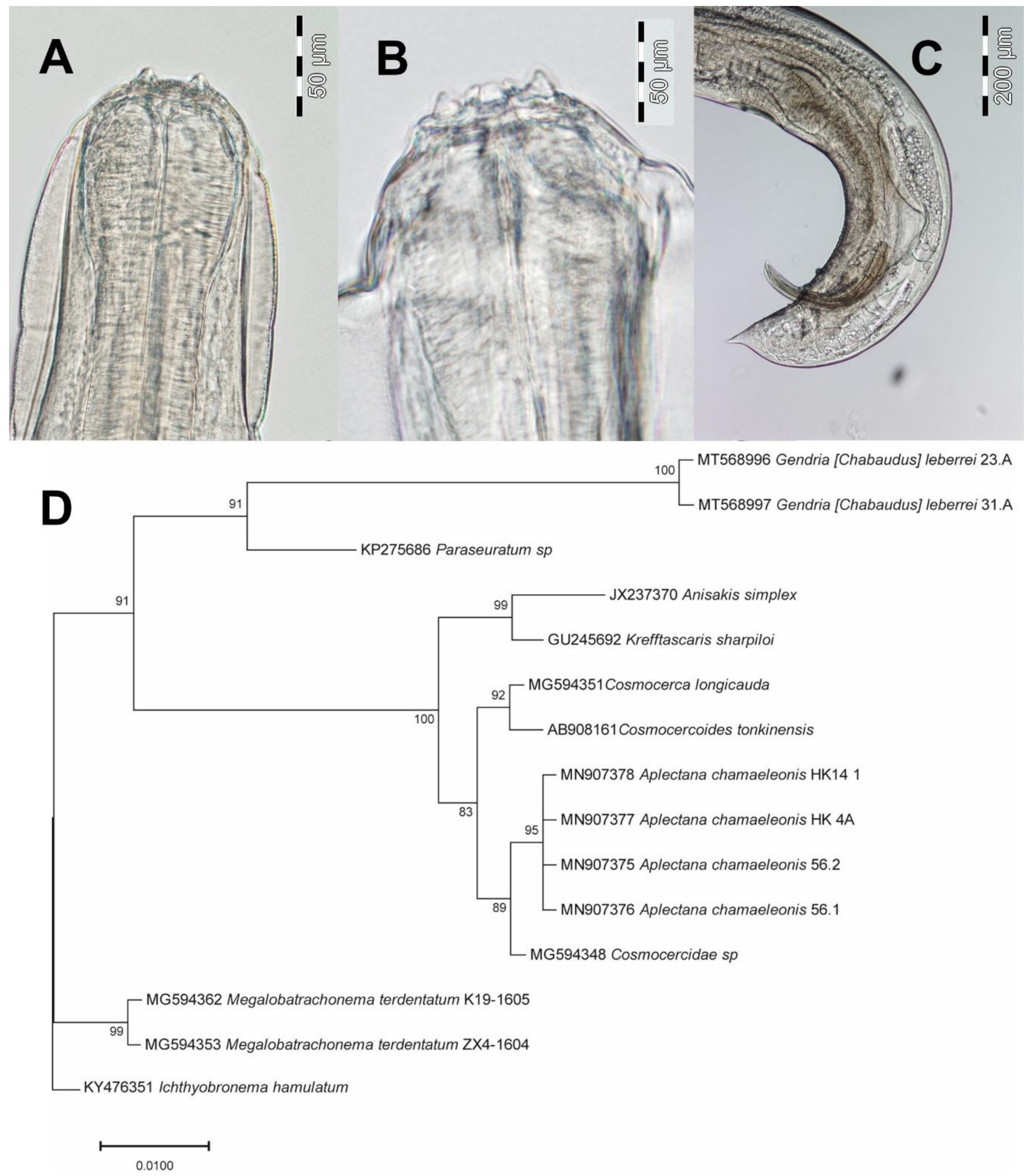
Six nematodes assignable to the genus Chabaudus [40] were collected from the small and large intestines of three H. parallelus males (Figure 2A–C). Recently, the genus Chabaudus has been placed into the synonymy of Gendria because of the absence of reliable morphological features for diagnosis [41]. As independent molecular support is not available for any view on the generic taxonomy, we refer to the nematodes identified based on their morphological features as Gendria [Chabaudus] leberrei. A female was considerably larger than two males (Body length 16.9 mm vs. 11.3 mm, 12.7 mm). The size range observed falls within the range reported for specimens collected from other anuran hosts: females 8.4–19.3 mm and males 5.6–14.6 mm [34,42,43,44]. Prevalence (6.7%) and intensity of infection (2.0; Table 3) were much lower than observed in Xenopus spp. (83.3–100% and 5.8–7.5% [42]). Gendria [Chabaudus] leberrei did not co-occur with other nematode or trematode species. Voucher specimens were deposited at the Zoological Museum of the University of Hamburg (ZMH N 14023; N = 2 originating from HP).
We succeeded in amplification of partial 18S-ITS1-5.8S sequence of two G. [Chabaudus] leberrei specimens showing 100% sequence identity. A Bayesian tree build with closest sequence matches from GenBank was computed based on a cured sequence dataset of 415 nucleotides of partial 18S sequence (Figure 2D). The topology of the resulting tree is significantly supported by posterior probability values. The Gendria specimens form a 91% supported sub-clade with the most closely related species retrieved from GenBank, the quimperiid Paraseuratum sp.

3.4. Aplectana Chamaeleonis (Cosmocercoidea): Morphological, Ecological and Molecular Features

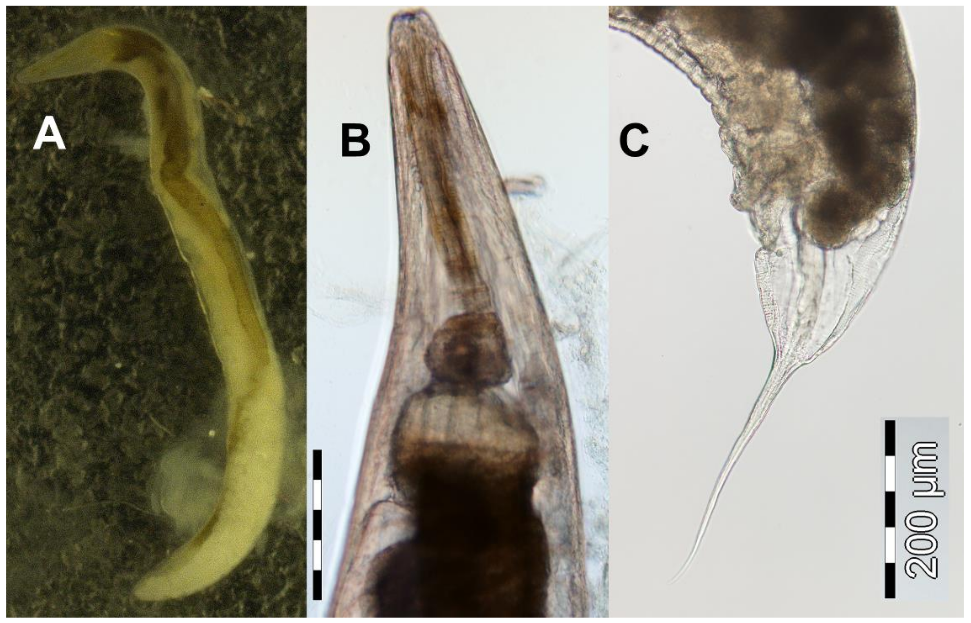
A small nematode parasite found exclusively in the large intestine of H. kivuensis was A. chamaeleonis (Figure 3A–C). The observed features of external morphology were in agreement with those described previously [45,46], specifically the position of postanal papillae in males. A female was considerably larger than five males examined (Body length 6.1 mm vs. 2.5–4.1 mm). The size range observed falls within the range reported for specimens collected from other hosts: females 3.7–6.7 mm and males 2.6–3.9 mm [35,45,46]. Prevalence (8.8%; Table 3) was much lower than reported for Leptopelis spp. (25–38% [39]), but decreased continuously from the beginning of the rainy period (11.2% in October) to the end (8.6% in January and 3.8% in March). In one host specimen each, A. chamaeleonis co-occurred either with the parasitic annelid Dero rwandae (Naididae) or with undetermined Echinostomatidae metacercariae. Long-term coexistence of nematodes and host was evident in 6 out of 10 captive-held H. kivuensis specimens in which we detected A. chamaeleonis 20 months following capture in the wild. Voucher specimens were deposited at the Zoological Museum of the University of Hamburg (ZMH N 14027; N = 19 originating from HK).
Fragments of the 18S-ITS1 sequence for four isolates of A. chamaeleonis showed 100% identical sequences. Comparison of a 475 bp-long subfragment of these sequences with the only GenBank entry of another Aplectana species (MH836325) showed 7% sequence divergence. We used a 18S rDNA fragment for computing a Bayesian tree based on closest GenBank entry matches (Figure 2). The topology of the resulting tree is supported by high probability values. The sequences of A. chamaeleonis built a subclade with a Cosmocercidae species previously collected from the smooth newt Lissotriton vulgaris [6]. Note that we failed in amplification of a fragment of the COI gene. Therefore, we cannot compare our specimens with other COI-GenBank entries of Aplectana species.

3.5. Rhabdias collaris (Rhabditoidea): Morphological, Ecological and Molecular Features

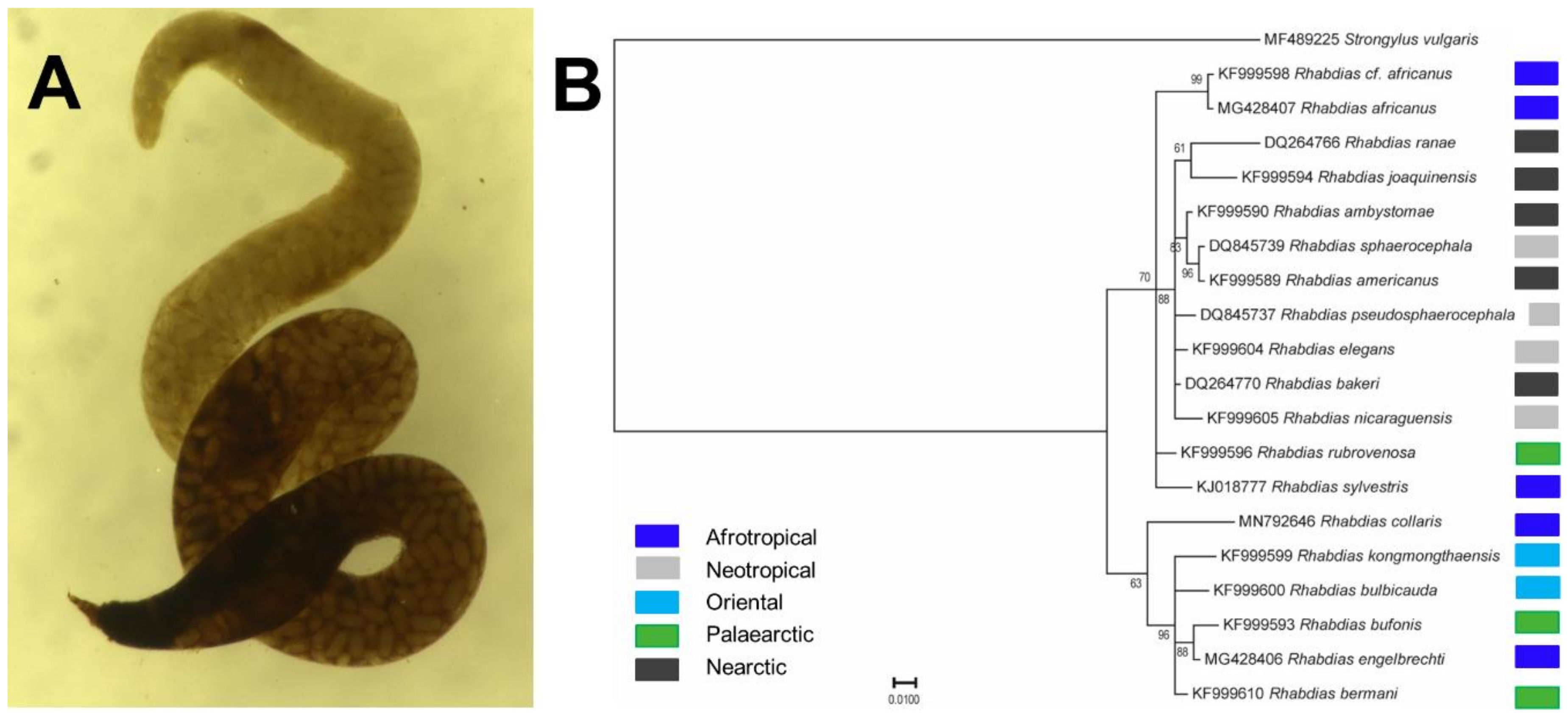
A single female R. collaris was found in the left lung of a H. viridiflavus male from the population in Huye (Table 3, Figure 4A). The lungs of all other Hyperolius specimens examined in this study were nematode-free. The observed features of external morphology were in agreement with those described previously [36], specifically the inflated and muscular cephalic end and the large uteri, filled with numerous, about 100 µm long eggs. Body length was 6.1 mm, i.e. smaller than the thus far known seven specimens (8.4–10.2 mm) collected from the lungs of Leptopelis vermiculatus and Hyperolius spec. in Amani, Tansania [36].
The topology of the Bayesian tree based on the partial ITS1 sequences (302 nucleotides) is supported by significant posterior probability values (Figure 4B). Note that Afrotropical Rhabdias spp. do not form a single clade (Figure 4B). The compared fragment of the COI gene of the six Afrotropical species of Rhabdias showed different levels of pairwise sequence variability (Table 4). Rhabdias engelbrechti and R. picardiae showed the highest level (89%) of sequence similarity to R. collaris.

4. Discussion

Our study is the first to provide molecular data on five morphologically described Afrotropical nematode species infecting reed frogs Hyperolius spp. DNA sequences facilitate identification of specimens in any developmental stage and enabling the unambiguous distinction between morphologically similar species. The reed frog species are all new host records for theses helminths. The detection of Orneoascaris schoutedeni in H. kivuensis is the first evidence for an amphibian host, while it was previously known only from reptile species [33,38]. This and previously published studies demonstrate that the nematode species reported here are capable of infecting a wide range of African amphibian and reptile host species, as summarized in Table 5. The absence of a strong host specificity facilitates the wide geographical distribution in Sub-Saharan Africa, as demonstrated by Orneoascaris chrysanthemoides currently known from Cameroon, DR Congo, Gabon, Gambia, Rwanda, Uganda and Zambia [21,37,38,39,47,48]. Considering the fragmentary nature of actual knowledge on the geographical distribution of these nematodes and the usually low prevalence in hosts, wide Sub-Saharan ranges may be characteristic for many of them.

4.1. Host–Parasite Ecology

All but one nematode species were detected in the small and large intestines of the three Hyperolius species collected in Rwanda. However, their prevalence was lower than that in other amphibian hosts [39,42]. The low prevalence and intensity of nematode infections in Hyperolius spp. may indicate either some kind of resistance against infections or alternatively short survival of infected hosts. Little is known about the impact of intestinal nematodes on the survival of anuran hosts, but the effect seems to be small [54,55]. Captive-held H. kivuensis survived infections with A. chamaeleonis more than two years, suggesting indeed a low effect on survival (this study). In contrast, a H. viridiflavus infected by two O. chrysanthemoides died a month after capture, suggesting an impact of large intestinal nematodes on survival. Experimental infections with Orneoascaris are needed to quantify the potential impact on Hyperolius survival and to access the adaption level of parasite and host.
Lung infections were limited to a single H. viridiflavus specimen by a single R. collaris, unlike the high prevalence and infection intensities by Rhabdias spp. in other African and Neotropical anuran hosts [12,15,18,19,22,56]. Lung-dwelling nematodes are known to decrease significantly the host´s fitness and cause reduced survival [13,56,57]. The proximate cause for the almost complete absence of lung infections in the Rwandan frog hosts remains enigmatic because Leptopelis and Hyperolius hosts from the neighboring Tanzania were infected by R. collaris [36].

4.2. Phylogenetic Implications

The phylogenetic relationships of Orneoascaris spp. and Gendria [Chabaudus] leberrei are currently under debate. In lip morphology, the ascaridoid genus Orneoascaris resembles Ophidascaris [38], but morphological evidence is too weak for a reliable phylogenetic hypothesis [7,8]. Our molecular data support the specific status of the morphologically very similar O. chrysanthemoides and O. schoutedeni, leaving open their relationship to O. sandoshami and an undescribed schoutedeni-like species from Bornean lizards [38]. Due to the fragmentary nature of molecular data, the enigmatic relationship of the genus Orneoascaris to other genera remains unresolved, but Contracaecum, Anisakis and Raphidascaris do not seem to be closely related (Figure 1G).
The phylogenetic relationships within the seuratioid family Quimperiidae are not well-understood [7,58,59]. In our study, Gendria [Chabaudus] leberrei and Paraseuratum sp. formed the sister group of a clade including several cosmocercoid species, whereas the quimperiid Ichthyobronema hamulatum resolved as an outgroup to both clades (Figure 2D). The association of Paraseuratum sp. with Spectatus spectatus (Cosmocercoidea: Kathlaniidae) in earlier studies [58,59], and more distantly to Megalobatrachonema terdentatum (Cosmocercoidea: Kathlaniidae [6]; this study) also indicates that the genera Gendria and Paraseuratum may be misplaced in the Quimperiidae, but actually belong to the Cosmocercidae. More molecular data on Quimperiidae genera are needed to resolve reliably taxonomic assignment of Gendria and Paraseuratum.
The cosmocercid genus Aplectana includes currently more than 50 species with increasing species descriptions, further expanding its diversity [45,60,61,62]. Unfortunately, all descriptions are based exclusively on morphological features. To the best of our knowledge, we provide the first DNA sequence corresponding to a morphologically identified Aplectana species. Phylogenetic relationships with other genus members based on molecular features remain unknown.
One of the few nematode genera with ample information on morphological and molecular features are the lung-dwelling Rhabdias parasites [23]. We contribute the first molecular data for the morphologically described R. collaris. Despite the usage of shorter sequence length, the phylogenetic tree based on 18S-ITS1 sequences obtained here for Rhabdias species is very similar to a recently published phylogenetic tree [23]. The lineage including R. collaris consists of species parasitizing different amphibian species occurring in the Afrotropical, Palearctic or Oriental regions (Figure 4B). Interestingly, our study suggests that Afrotropical Rhabdias spp. form three evolutionary lineages (R. collaris, R. africanus and R. sylvestris lineages).

5. Conclusions

The diversity of nematodes in frogs from Rwanda, a previously unstudied Afrotropical region, exclusively includes species which have already been described from other amphibian and reptile hosts. Afrotropical diversity of parasitic nematodes may be lower than previously assumed because host specificity is not strongly developed. Phylogenetic insight into the supra- and infra-generic relationships of the four Afrotropical nematode genera detected in this study requires considerably more molecular data for morphologically identified species than currently available. Here, we contribute a dataset linking morphology and molecular features of five nematode species. We recommend that experts on nematode morphology and molecular biologists should join their efforts to establish a sustainable molecular library of DNA sequence data linked to morphological identifications, which in future may serve equally for taxonomic purposes and for unravelling phylogenetic relationships.

Author Contributions

Conceptualization, U.S., J.M.D., P.S. and C.B.; methodology, U.S.; software, U.S. and C.B.; validation, U.S.; formal analysis, U.S.; investigation, U.S., J.M.D., P.S. and C.B.; resources, U.S. and C.B.; data curation, U.S. and C.B.; writing—original draft preparation, U.S., J.M.D., P.S. and C.B.; writing—review and editing, U.S., J.M.D., P.S. and C.B.; visualization, U.S. and C.B.; supervision, U.S.; and project administration, U.S. All authors have read and agreed to the published version of the manuscript.

Funding

This research received no external funding.

Acknowledgments

We are indebted to Bonny Dumbo for assistance during the collection of Hyperolius in the field. Collecting and export permits (Nos. 2/ORTPN/V.U/09, 08/RDB-T&C/V.U/12, 18/RDB-T&C/V.U/12 and 10/RDB-T&C/V.U/15) of Hyperolius for parasitological screening were issued by the Rwandan Development Board. The manuscript benefited from the comments of two anonymous reviewers.

Conflicts of Interest

The authors declare no conflict of interest.

References

  1. Martinez-Arce, A.; De Jesus-Nayarrete, A.; Leasi, F. DNA Barcoding for Delimitation of Putative Mexican Marine Nematodes Species. Diversity 2020, 12, 107. [Google Scholar] [CrossRef] [Green Version]
  2. Hugot, J.P.; Baujard, P.; Morand, S. Biodiversity in helminths and nematodes as a field of study: An overview. Nematology 2001, 3, 199–208. [Google Scholar] [CrossRef] [Green Version]
  3. Dickson, D.W.; Chen, S.; Chen, Z. Nematology: Advances and Perspectives. Nematode Morphology, Physiology and Ecology; CABI: Wallingford, UK, 2004. [Google Scholar]
  4. Dorris, M.; De Ley, P.; Blaxter, M.L. Molecular analysis of nematode diversity and the evolution of parasitism. Parasitol. Today 1999, 15, 188–193. [Google Scholar] [CrossRef]
  5. Powers, T.; Harris, T.; Higgins, R.; Mullin, P.; Sutton, L.; Powers, K. MOTUs, morphology, and biodiversity estimation: A case study using nematodes of the suborder Criconematina and a conserved 18S DNA barcode. J. Nematol. 2011, 43, 35–48. [Google Scholar]
  6. Sinsch, U.; Heneberg, P.; Těšínský, M.; Balczun, C.; Scheid, P. Helminth endoparasites of the smooth newt Lissotriton vulgaris: Linking morphological identification and molecular data. J. Helminthol. 2019, 93, 332–341. [Google Scholar] [CrossRef] [PubMed]
  7. Anderson, R.C.; Chabaud, A.G.; Willmott, S. Keys to the Nematode Parasites of Vertebrates: Archival Volume; CABI: Wallingford, UK, 2009. [Google Scholar]
  8. Gibbons, L.M. Keys to the Nematode Parasites of Vertebrates: Supplementary Volume; CABI: Wallingford, UK, 2010; Volume 10. [Google Scholar]
  9. Aho, J.M. Helminth communities of amphibians and reptiles: Comparative approaches to understanding patterns and processes. In Parasite Communities: Patterns and Processes; Esch, G.W., Bush, A.O., Aho, J.M., Eds.; Springer Netherlands: Dordrecht, The Netherlands, 1990; pp. 157–195. [Google Scholar] [CrossRef]
  10. Baker, M.R. Nematode parasitism in amphibians and reptiles. Can. J. Zool. 1984, 62, 747–757. [Google Scholar] [CrossRef]
  11. Paredes-León, R.; Garcia-Prieto, L.; Guzman-Cornejo, C.; Leon-Regagnon, V.; Perez, T.M. Metazoan parasites of Mexican amphibians and reptiles. Zootaxa 2008, 1904, 1–166. [Google Scholar] [CrossRef]
  12. Halajian, A.; Bursey, C.R.; Goldberg, S.R.; Luus-Powell, W. Helminths of Six Species of Anurans from the Republic of South Africa: Amietophrynus garmani, Amietophrynus gutturalis, Amietophrynus maculatus, Schismaderma carens (Bufonidae), Amietia angolensis, and Strongylopus grayii (Pyxicephalidae), with a Review of South African Anuran Helminths. Comp. Parasitol. 2013, 80, 80–95. [Google Scholar] [CrossRef]
  13. Finnerty, P.B.; Shine, R.; Brown, G.P. The costs of parasite infection: Effects of removing lungworms on performance, growth and survival of free-ranging cane toads. Funct. Ecol. 2018, 32, 402–415. [Google Scholar] [CrossRef] [Green Version]
  14. Goater, C.P.; Semlitsch, R.D.; Bernasconi, M.V. Effects of body size and parasite infection on the locomotory performance of juvenile toads. Bufo bufo. Oikos 1993, 66, 129–136. [Google Scholar] [CrossRef]
  15. Pizzatto, L.; Shine, R. Lungworm infection modifies cardiac response to exercise in cane toads. J. Zool. 2012, 287, 150–155. [Google Scholar] [CrossRef]
  16. Kelehear, C.; Webb, J.K.; Shine, R. Rhabdias pseudosphaerocephala infection in Bufo marinus: Lung nematodes reduce viability of metamorph cane toads. Parasitology 2009, 136, 919–927. [Google Scholar] [CrossRef] [Green Version]
  17. Leary, C.J.; Ralicki, H.F.; Laurencio, D.; Crocker-Buta, S.; Malone, J.H. Assessing the links among environmental contaminants, endocrinology, and parasites to understand amphibian declines in montane regions of Costa Rica. PLoS ONE 2018, 13, e0191183. [Google Scholar] [CrossRef] [PubMed] [Green Version]
  18. Aisien, M.; Sampson, S.A.; Amuzie, C. Anuran parasites from three biotopes in Rivers State, Nigeria. Niger. J. Parasitol. 2017, 38, 129–135. [Google Scholar] [CrossRef]
  19. Aisien, M.S.O.; Ugbomeh, A.P.; Awharitoma, A.O. Parasitic infections of anurans from a freshwater creek community in Delta State, Niger Delta of Nigeria. Helminthologia 2017, 54, 132–144. [Google Scholar] [CrossRef] [Green Version]
  20. McAllister, C.T.; Bursey, C.R.; Freed, P.S. Helminth parasites (Cestoidea, Nematoda, Pentastomida) of selected herpetofauna from Cameroon, West Africa. Acta Parasitol. 2010, 55, 90–93. [Google Scholar] [CrossRef]
  21. Skrjabin, K. Parasitic trematodes and nematodes collected by the expedition of Prof. V. Dogiel and I. Sokolov in British East Africa. In Scientific Results of the Zoological Expedition to British East Africa and Uganda Made by Prof. V. Dogiel and I. Sokolov in the Year 1914; Palala Press: Mishawaka, IN, USA, 1916; Volume 1, pp. 99–157. [Google Scholar]
  22. Junker, K.; Lhermitte-Vallarino, N.; Barbuto, M.; Ineich, I.; Wanji, S.; Bain, O. New species of Rhabdias (Nematoda: Rhabdiasidae) from afrotropical anurans, including molecular evidence and notes on biology. Folia Parasit. (Praha) 2010, 57, 47–61. [Google Scholar] [CrossRef] [PubMed]
  23. Kuzmin, Y.; Halajian, A.; Tavakol, S.; Luus-Powell, W.J.; Tkach, V.V. Description and phylogenetic position of a new species of Rhabdias Stiles et Hassall, 1905 (Nematoda: Rhabdiasidae) from the banded rubber frog, Phrynomantis bifasciatus (Smith) (Amphibia: Microhylidae), in South Africa. Folia Parasit. (Praha) 2017, 64, 35. [Google Scholar] [CrossRef] [Green Version]
  24. Sinsch, U.; Lümkemann, K.; Rosar, K.; Schwarz, C.; Dehling, J.M. Acoustic niche partitioning in an anuran community inhabiting an Afromontane wetland (Butare, Rwanda). Afr. Zool. 2012, 47, 60–73. [Google Scholar] [CrossRef]
  25. Tumushimire, L.; Mindje, M.; Sinsch, U.; Dehling, M.J. Anuran diversity of cultivated wetlands in Rwanda: Melting pot of generalists? Salamandra 2020, 56, 99–112. [Google Scholar]
  26. Bush, A.; Lafferty, K.; Lotz, J.; Shostak, A. Parasitology meets ecology on its own terms: Margolis et al. revisited. J. Parasitol. 1997, 83, 575–583. [Google Scholar] [CrossRef] [PubMed]
  27. Altschul, S.F.; Gish, W.; Miller, W.; Myers, E.W.; Lipman, D.J. Basic local alignment search tool. J. Mol. Biol. 1990, 215, 403–410. [Google Scholar] [CrossRef]
  28. Huelsenbeck, J.P.; Ronquist, F. MRBAYES: Bayesian inference of phylogenetic trees. Bioinformatics 2001, 17, 754–755. [Google Scholar] [CrossRef] [Green Version]
  29. Nicholas, K.B. GeneDoc: Analysis and visualization of genetic variation. Embnew. News 1997, 4, 14. [Google Scholar]
  30. Sinnappah, N.D.; Lim, L.-H.S.; Rohde, K.; Tinsley, R.; Combes, C.; Verneau, O. A paedomorphic parasite associated with a neotenic amphibian host: Phylogenetic evidence suggests a revised systematic position for Sphyranuridae within anuran and turtle Polystomatoineans. Mol. Phyl. Evol. 2001, 18, 189–201. [Google Scholar] [CrossRef] [PubMed]
  31. Kaci-Chaouch, T.; Verneau, O.; Desdevises, Y. Host specificity is linked to intraspecific variability in the genus Lamellodiscus (Monogenea). Parasitology 2008, 135, 607–616. [Google Scholar] [CrossRef]
  32. Folmer, O.; Black, M.; Hoeh, W.; Lutz, R.; Vrijenhoek, R. DNA primers for amplification of mitochondrial cytochrome c oxidase subunit I from diverse metazoan invertebrates. Mol. Mar. Biol. Biotechnol. 1994, 3, 294–299. [Google Scholar]
  33. Baylis, H.A. XXXVI.—On a further collection of parasitic worms from the Belgian Congo. Ann. Mag. Nat. Hist. 1940, 5, 401–417. [Google Scholar] [CrossRef]
  34. Bain, O.; Philippon, B. Recherche sur des larves de nematodes Ascaridida trouvees chez Simulium damnosum. Ann. Parasitol. Hum. Comp. 1969, 44, 147–156. [Google Scholar] [CrossRef]
  35. Baylis, H.A. XL.—Some parasitic Nematodes from the Uluguru and Usambara Mountains, Tanganyika Territory. Ann. Mag. Nat. Hist. 1929, 4, 372–381. [Google Scholar] [CrossRef]
  36. Baker, M.R. Rhabdias collaris n. sp. (Nematoda: Rhabdiasidae) from frogs of Tanzania. Syst. Parasitol. 1987, 9, 199–201. [Google Scholar] [CrossRef]
  37. Moravec, F.; Baruš, V. Some nematode parasites from amphibians and reptiles from Zambia and Uganda. Acta Soc. Zool. Bohem. 1990, 54, 177–192. [Google Scholar]
  38. Sprent, J. Ascaridoid nematodes of amphibians and reptiles: Orneoascaris. Ann. Parasitol. Hum. Comp. 1985, 60, 33–55. [Google Scholar] [CrossRef] [Green Version]
  39. Goldberg, S.R.; Bursey, C.R.; Greenbaum, E. Nematodes of Leptopelis christyi and Leptopelis karissimbensis (Anura: Arthroleptidae), from the Democratic Republic of Congo. Alytes 2016, 33, 21–24. [Google Scholar]
  40. Inglis, W.; Ogden, C. Chabaudus chabaudi gen. et sp. nov. from a fresh-water fish in Sierra Leone (Nematoda: Seuratoidea). Rev. Zool. Bot. Afr. 1965, 71, 171–176. [Google Scholar]
  41. Moravec, F.; Jirků, M. Some nematodes from freshwater fishes in central Africa. Folia Parasit. 2017, 64, 33. [Google Scholar] [CrossRef] [PubMed]
  42. Jackson, J.A.; Tinsley, R.C.; Du Preez, L.H.; Henderson, A.C. A redescription of Chabaudus leberrei (Bain & Philippon, 1969) (Nematoda: Seuratoidea) from Xenopus spp. in Swaziland. Syst. Parasitol. 2001, 50, 81–89. [Google Scholar] [CrossRef] [PubMed]
  43. Durette-Desset, M.-C.; Batcharov, G. Deux nematodes parasites d’amphibiens du Togo. Ann. Parasitol. Hum. Comp. 1974, 49, 567–576. [Google Scholar] [CrossRef] [PubMed] [Green Version]
  44. Pike, A.W. Helminth parasites of the amphibians Dicroglossus occipitalis (Günther) and Bufo regularis Reuss, in Khartoum, Republic of Sudan. J. Nat. Hist. 1979, 13, 337–376. [Google Scholar] [CrossRef]
  45. Baker, M.R. Revision of Old World species of the genus Aplectana Railliet & Henry, 1916 (Nematoda, Cosmocercidae). Bull. Mus. Natl. Hist. Nat. 1980, 2, 955–998. [Google Scholar]
  46. Chen, P. Aplectana chamaeleonis (Baylis, 1929) from a frog and a freshwater fish in Ethiopia. Ann. Parasitol. Hum. Comp. 1966, 9, 333–336. [Google Scholar] [CrossRef]
  47. Goldberg, S.R.; Bursey, C.R. Helminths from eight species of African skinks (Trachylepis: Scincidae). Com. Parasitol. 2010, 77, 236–241. [Google Scholar] [CrossRef]
  48. Rasheed, S. Some parasitic nematodes from the Cameroons (W. Africa). J. Helminthol. 1965, 39, 67–100. [Google Scholar] [CrossRef] [PubMed]
  49. Aisien, S.O.; Ugbo, A.D.; Ilavbare, A.N.; Ogunbor, O. Endoparasites of amphibians from South-Western Nigeria. Acta Parasitol. 2001, 46, 299–305. [Google Scholar]
  50. Aisien, M.S.O.; Ogoannah, S.O.; Imasuen, A.A. Helminth parasites of amphibians from a rainforest reserve in southwestern Nigeria. Afr. Zool. 2009, 44, 1–7. [Google Scholar] [CrossRef]
  51. Amuzie, C.C.; Ekerette, I.B. Variations in parasitic helminths of Amietophrynus species collected from similar habitats, Rivers State, Nigeria. Int. J. Biosci. 2017, 10, 76–79. [Google Scholar]
  52. Amuzie, C.C.; Aisien, M. Parasitic helminths of some Ptychadena species from Ogoniland, Niger Delta, Nigeria. NISEB J. 2018, 18, 68–71. [Google Scholar]
  53. Okere, S.; Joseph, F.; Amuzie, C. Endo-parasitic helminths of amphibians, Ptychadena mascareniensis and Ptychadena pumilio at Rumuji-emohua, Rivers state, Nigeria. Afr. J. Environ. Sci. 2019, 2, 71–76. [Google Scholar]
  54. Pessier, A.P.; Baitchman, E.J.; Crump, P.; Wilson, B.; Griffith, E.; Ross, H. Causes of mortality in anuran amphibians from an ex situ survival assurance colony in Panama. Zoo Biol. 2014, 33, 516–526. [Google Scholar] [CrossRef]
  55. Wiley, M.; Wu, J.; Dawson, B. A comparison of two treatments for nematode infections in the Túngara Frog, Engystomops pustulosus. J. Herpetol. Med. Surg. 2009, 19, 21–22. [Google Scholar] [CrossRef]
  56. Moretti, E.H.; Madelaire, C.B.; Silva, R.J.; Mendonça, M.T.; Gomes, F.R. The relationships between parasite intensity, locomotor performance, and body condition in adult toads (Rhinella icterica) from the wild. J. Herpetol. 2014, 48, 277–283. [Google Scholar] [CrossRef]
  57. Koprivnikar, J.; Marcogliese, D.J.; Rohr, J.R.; Orlofske, S.A.; Raffel, T.R.; Johnson, P.T.J. Macroparasite infections of amphibians: What can they tell us? Ecohealth 2012, 9, 342–360. [Google Scholar] [CrossRef] [PubMed]
  58. McElwain, A.; Warren, M.B.; Pereira, F.B.; Ksepka, S.P.; Bullard, S.A. Pathobiology and first report of larval nematodes (Ascaridomorpha sp.) infecting freshwater mussels (Villosa nebulosa, Unionidae), including an inventory of nematode infections in freshwater and marine bivalves. Int. J. Parasitol. 2019, 10, 41–58. [Google Scholar] [CrossRef] [PubMed]
  59. Sokolov, S.; Malysheva, S. Molecular characterization of Ichtyobronema hamulatum (Moulton, 1931) (Nematoda: Quimperiidae), a common parasite of burbot Lota lota (Linnaeus) (Actinopterygii: Lotidae). Helminthologia 2017, 54, 183–188. [Google Scholar] [CrossRef] [Green Version]
  60. Bursey, C.R.; Goldberg, S.R.; Kraus, A.F. New species of Aplectana (Nematoda: Cosmocercidae) in Sphenomorphus pratti from Papua New Guinea. J. Parasitol. 2011, 97, 654–660. [Google Scholar] [CrossRef]
  61. Bursey, C.R.; Goldberg, S.R.; Grismer, L.L. A new species of Aplectana (Nematoda, Cosmocercidae) in Goniurosaurus bawanglingensis (Squamata, Eublepharidae), from Hainan Province, China. Acta Parasitol. 2018, 63, 190–197. [Google Scholar] [CrossRef]
  62. Sou, S.K.; Sow, K.K.; Nandi, A.P. Aplectana hoplobatrachusia sp nov (Nematoda: Cosmocercidae) in Hoplobatrachus crassus (Jerdon, 1853) (Anura: Dicroglossidae) from Birbhum District, West Bengal, India. Zootaxa 2018, 4472, 194–200. [Google Scholar] [CrossRef]
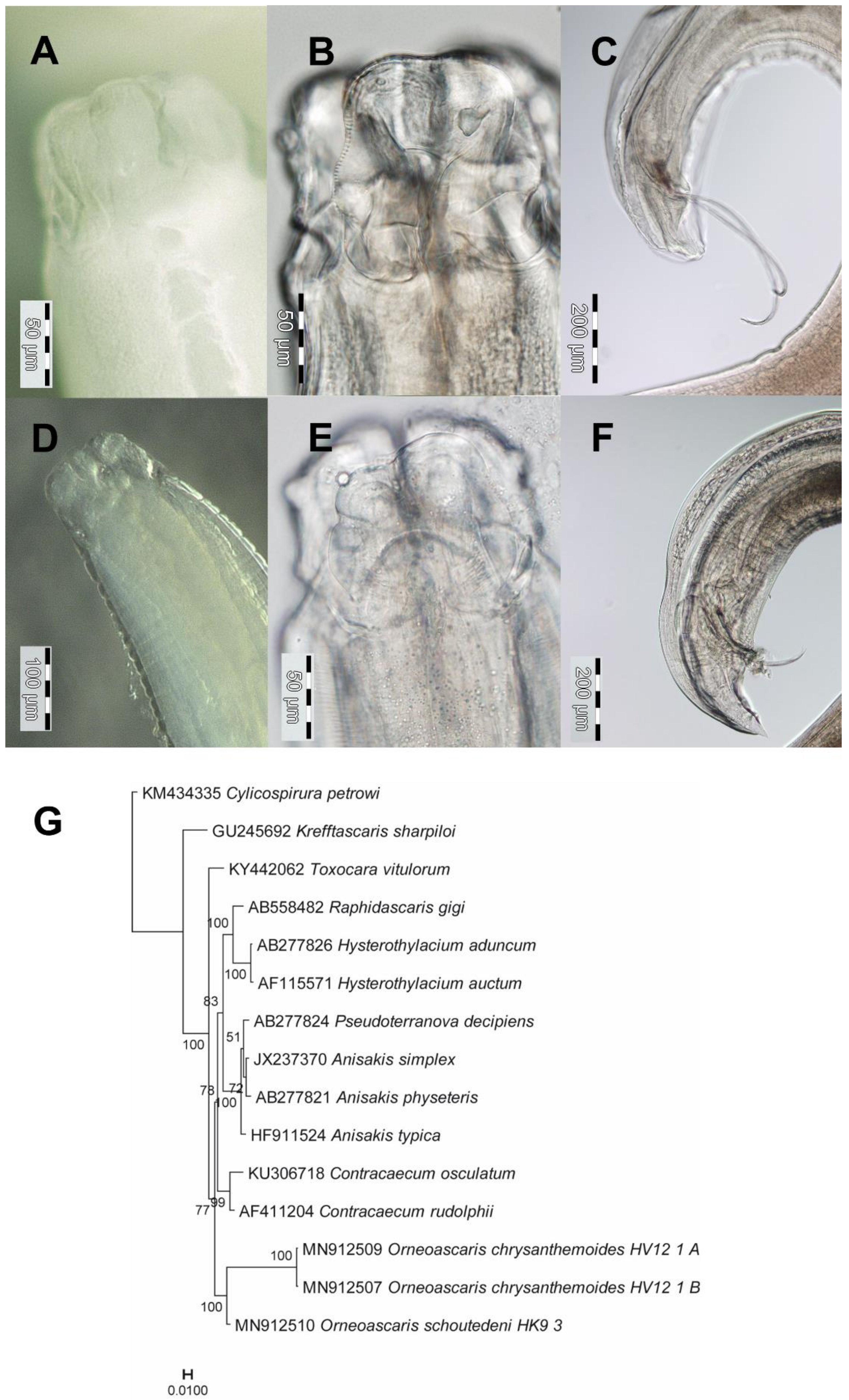
Figure 1. Orneoascaris chrysanthemoides (AC) and O. schoutedeni (DF) and their phylogenetic relationships (G). Anterior end with lateral view on the lips of live specimens (A,D). Dorsal lip with dentigerous ridges (B,E). Tail region of a male with spicules (C,F). Scales are given in each photograph. (G) Bayesian tree of partial 18S rDNA and partial ITS1 sequences of Orneoascaris spp. and other Ascaridoidea species, rooted by the spirurid Cylicospirura petrowi. Posterior probability values are indicated. Scale bar indicates substitutions per site.
Diversity 12 00265 g001
Figure 2. Gendria [Chabaudus] leberrei (AC) and phylogenetic relationships (D). Lateral view on the anterior end of a live male (A). Apical protuberances of the labia (B). Tail region of a male with spicules (C). Scales are given in each photograph. (D) Bayesian tree of 18S rRNA sequences of G. leberrei and other species with similar sequences, rooted by Ichthyobronema hamulatum. Posterior probability values are indicated. Scale bar indicates substitutions per site.
Diversity 12 00265 g002
Figure 3. (A) Live Aplectana chamaeleonis female (6.1 mm body length) from the large intestine of a H. kivuensis male. (B) Lateral view on the anterior end. (C) Lateral view on the posterior end. Scales are given in (B,C) are 200 µm. Bayesian tree of 18S-ITS1 rDNA sequences of closest matches to the sequence of Aplectana chamaeleonis is given in Figure 2.
Diversity 12 00265 g003
Figure 4. (A) Live Rhabdias collaris female (6.1 mm body length) from the lung of a H. viridiflavus male. (B) Bayesian tree of ITS1 rDNA sequences of Rhabdias nematodes deposited in GenBank including R. collaris collected from H. viridiflavus, rooted by the strongylid Strongylus vulgaris. Posterior probability values are indicated. Scale bar indicates substitutions per site. Association of parasites to biogeographic realms is color-coded.
Diversity 12 00265 g004
Table 1. Primers used for amplification and sequencing.
GenePrimerSequenceReference
18S-ITS1-5.8SS15′-ATTCCGATAACGAACGAGACT-3′[30]
IR85′-GCTAGCTGCGTTCTTCATCGA-3′[31]
COILCO14905′-GGTCAACAAATCATAAAGATATTGG-3′[32]
HCO21985′-TGATTTTTTGGTCACCCTGAAGTTTA-3′[32]
Table 2. Accession numbers of nuclear ribosomal sequences and mitochondrial COI sequences used for phylogenetic reconstruction.
SpeciesLocus
Nuclear Ribosomal SequenceCOI
Rhabdias collarisMN792646MN927222
Orneoascaris chrysanthemoides HV12_1_AMN912509-
Orneoascaris chrysanthemoides HV12_1_BMN912507-
Orneoascaris schoutedeniMN912510-
Gendria [Chabaudus] leberreiMT568996-
Gendria [Chabaudus] leberreiMT568997-
Aplectana chamaeleonis 56.2MN907375-
Aplectana chamaeleonis 56.1MN907376-
Aplectana chamaeleonis HK_4AMN907377-
Aplectana chamaeleonis HK14_1MN907378-
Table 3. Parasitism of reed frogs by nematodes in Rwanda: infection site (s, small; l, large), number of nematodes (Ntot), prevalence (presented as the number of parasitized frog hosts by the total number examined), mean intensity of infection ± SD (as mean number of parasites per individual frog) and range of nematodes recorded.
Orneoascaris chrysanthemoidesOrneoascaris schoutedeniGendria [Chabaudus] leberreiAplectana chamaeleonisRhabdias collaris
LocalityHuyeMusanzeCyamudongoHuyeHuye
Hyperolius kivuensis (n = 115)
Infection site
N specimens
Prevalence [%]
Infection intensity
Range

intestines (s, l)
Ntot = 24
3.5
6.0 ± 7.1
1–16

intestines (s, l)
Ntot = 3
0.9
3
-

-

intestines (l)
Ntot = 23
8.7
2.3 ± 2.0
1–7

-
Hyperolius parallelus (n = 45)
Infection site
N specimens
Prevalence [%]
Infection intensity
Range
--
intestines (s, l)
Ntot = 6
6.7
2.0 ± 1.7
1–4
-
-
Hyperolius viridi-flavus (n = 100)
Infection site
N specimens
Prevalence [%]
Infection intensity
Range


Intestines (s, l)
Ntot = 3
2.0
1.5 ± 0.7
1–2


-


-


-


lungs
Ntot = 1
0.7
1
1
Table 4. Percentage (upper triangle) and number (lower triangle) of identical sites based on the 596-bp-long alignment of partial sequences of COI gene of African Rhabdias species.
SpeciesR. collaris
927222 *
R. engelbrechti
MG428410 *
R. picardiae
FN434095 *
R. tanyai
FN434107 *
R. vencesi
FN434104 *
R. africanus
MG428411 *
R. collaris08989888786
R. engelbrechti532091889089
R. picardiae5365460879087
R. tanyai52653052308587
R. vencesi523537540509087
R. africanus5155335245225240
* GenBank accession numbers.
Table 5. Currently known amphibian and reptile host species of the parasitic nematodes detected in Hyperolius frogs. Hosts identified in this study are shown in bold letters.
Orneoascaris chrysanthemoidesOrneoascaris schoutedeniGendria [Chabaudus] leberreiAplectana
chamaeleonis
Rhabdias
collaris
Type hostSclerophrys spec.
(Toad)
Varanusniloticus
(Nile monitor)
Sclerophrysregularis
(Toad)
Kinyongia matschiei
(Chamaeleon)
Leptopelisvermiculatus
(Frog)
Amphibian hostsHyperolius
kivuensis
H. viridiflavus
Amietia nutti
Arthroleptis
stenodactylus
Breviceps
macrodactylus
Leptopelis christyi
L. karissimbensis
Sclerophrys
regularis
S. maculatus
S. superciliaris
Trichobatrachus
robustus
Xenopus laevis
Hyperolius
kivuensis
Hyperolius
parallelus
Aubria subsigillata
Hoplobatrachus
occipitalis
Ptychadena
mascareniensis
P. pumilio
Sclerophrys
camerunensis
S. maculatus
Xenopus laevis
X. muelleri
Hyperolius
kivuensis
Amietia angolensis
Bufotes viridis
Cacosternum capense
Epidalea calamita
Hylarana spec.
Leptopelis christyi,
L. karissimbensis
Ptychadena
oxyrhynchus
Scotobleps gabonicus
Sclerophrys rangeri
Sclerophrys spp.
Schismadera carens
Trichobatrachus robustus
Vandijkophrynus
angusticeps
Hyperolius
viridiflavus
Hyperolius spec.
Reptile hostsVaranus niloticus
Chameleo dilepis
Trachylepis affinis
Kinixys erosa
Natrix olivaceus
Bitis cornuta
Dispholidus typus
Causus
rhombeatus
Crocodylus
Niloticus
Varanus
exanthematicus
---
References[21,37,38,39,47,48][33,38][18,34,42,49,50,51,52,53][12,35,39,45,46][36]