Short Note

3-(4-Hydroxynaphthalen-1-yl)-3-(1-hydroxynaphthalen-2-yl)isobenzofuran-1(3H)-one

EaStCHEM School of Chemistry, University of St Andrews, North Haugh, St Andrews, Fife KY16 9ST, UK
*
Author to whom correspondence should be addressed.
Molbank 2026, 2026(4), M2195; https://doi.org/10.3390/M2195
Submission received: 20 May 2026 / Revised: 24 June 2026 / Accepted: 29 June 2026 / Published: 1 July 2026
(This article belongs to the Section Structure Determination)

Abstract

3-(4-hydroxynaphthalen-1-yl)-3-(1-hydroxynaphthalen-2-yl)isobenzofuran-1(3H)-one is a previously unknown derivative of the well-known acid/base indicator naphtholphthalein. We report the synthesis and the molecular structure of the title compound, as determined by single-crystal X-ray diffraction. 1H and 13C NMR spectroscopy data, IR spectroscopy data, and mass spectrometry data are provided.

1. Introduction

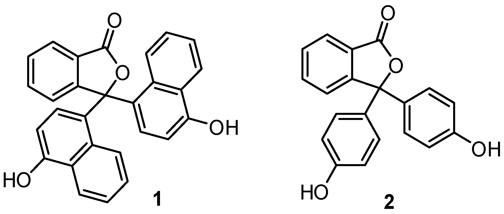
Naphtholphthalein (Figure 1, 1) is a pH indicator that is used in titrimetric analysis, it is particularly useful for titrations in aqueous ethanol [1,2,3]. naphtholphthalein differs from phenolphthalein (Figure 1, 2) by showing a colourless to teal colour change, rather than colourless to pink, in dilute solutions of sodium hydroxide [1].
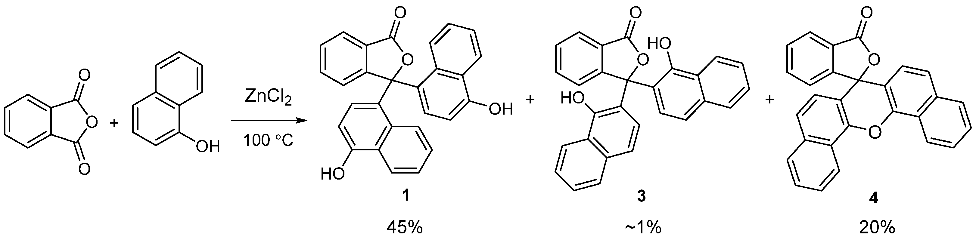
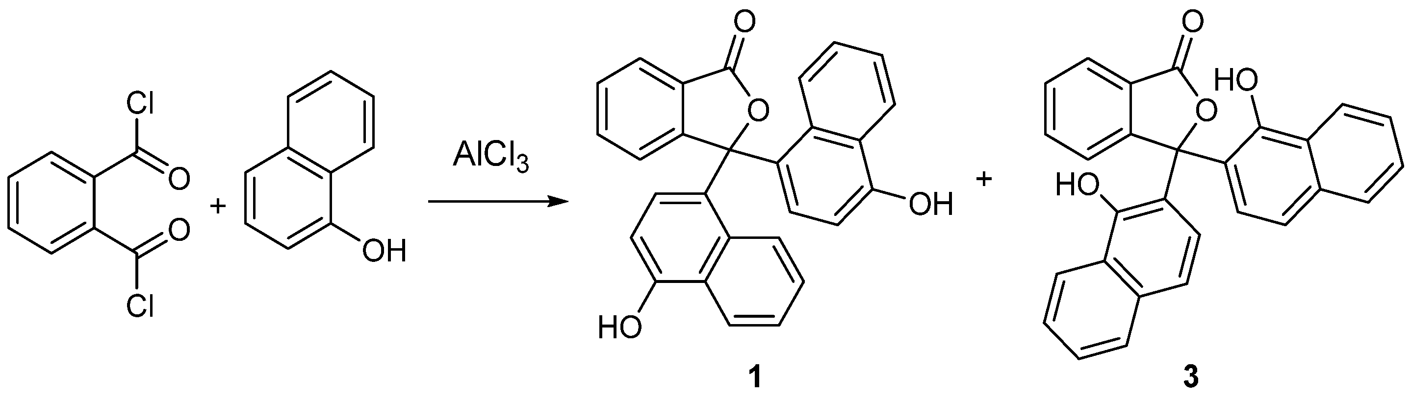
Naphtholphthalein is usually prepared by heating 1-naphthol with phthalic anhydride under acidic conditions; however, this compound is known to be harder to prepare under these conditions than other phenolphthalein derivatives [4,5,6,7]. Several studies on the synthesis of naphtholphthalein have been conducted, and it has been found that, depending on the conditions used, as many as three significant products can be obtained. For example, Copisarow reported in 1920 that the reaction of phthalic anhydride with 1-naphthol in hot zinc chloride afforded naphtholphthalein (1), the naphtholphthalein isomer 3 and the fluoran by-product 4 (Scheme 1) [5]. The latter compound is often referred to as “Grabowski’s anhydride”; this compound is likely formed by heating isomer 3 under strongly acidic conditions. Around the same time, Csányi conducted a detailed study on the reactions of 1-naphthol with phthalic anhydride and phthaloyl chloride (Scheme 2) [6]. A key finding from this work was that the reaction of phthaloyl chloride with 1-napthol in the presence of aluminium chloride can also form naphtholphthaleins 1 and 3; it was also found that compound 3 was converted to fluoran 4 at elevated temperature. Both Csányi and Copisarow reported that the naphtholphthalein isomer 3 formed a green solution when dissolved in alkaline solutions, this is in contrast to the teal colour observed when naphtholphthalein is treated under the same conditions [5,6].
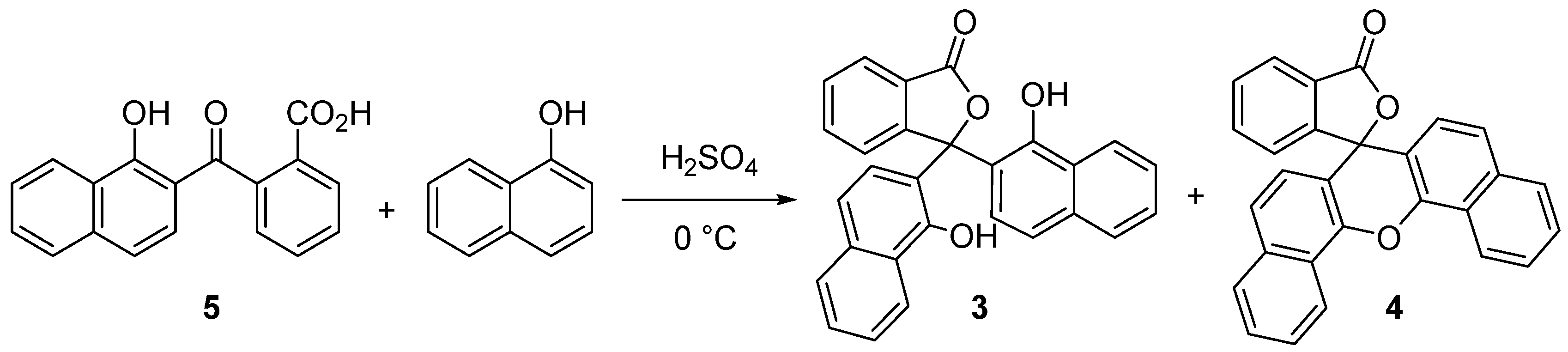
Later work by Schulenburg sought to make naphtholphthalein isomer 3 on a larger scale and with improved purity; however, a different approach was adopted [7]. Schulenburg reacted naphtholphthalic acid 5 with 1-naphthol in concentrated sulfuric acid (Scheme 3). Small quantities of fluoran 4 were obtained from the reaction, although it was also reported that heating the product (3) to its melting point resulted in the formation of fluoran 4 as the major product [7].
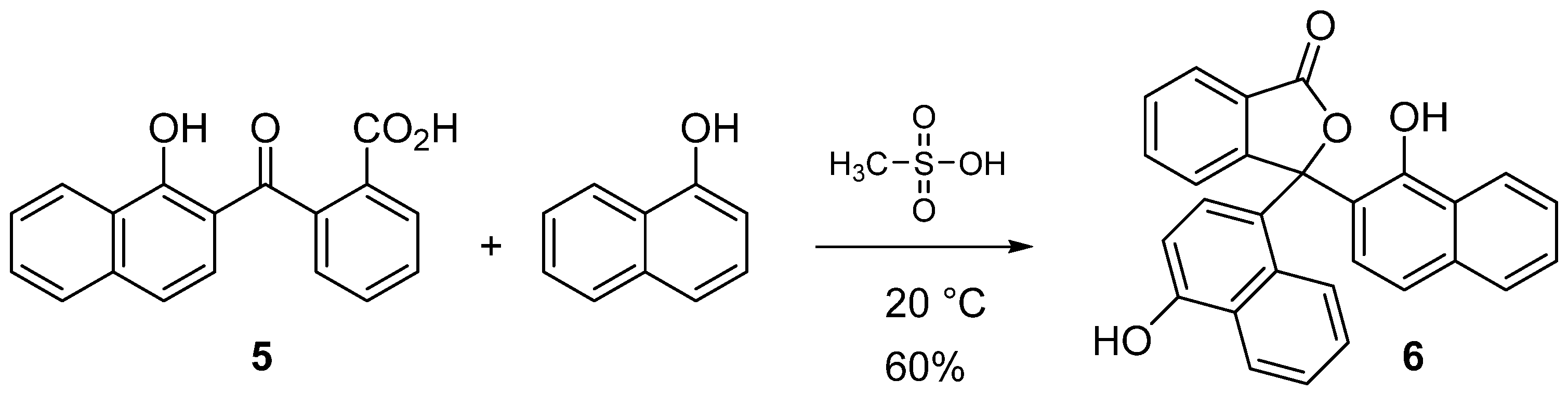
In this paper, we describe results from our recent studies on the reaction of naphtholphthalic acid 5 with 1-naphthol in methanesulfonic acid. The conditions of these reactions are based on recent work by Sabnis, who originally reported that methanesulfonic acid could serve as a convenient alternative to concentrated sulfuric acid for the synthesis of phenolphthalein derivatives [8]. We wanted to determine whether naphtholphthalein isomer 3 formed, as was seen in Schulenburg’s work [7], or if a hitherto unknown isomer was the major product from this reaction. Our findings are that naphtholphthalein isomer 6 is the main product, although traces of fluoran 4 were also detected (Scheme 4). This outcome does not fully align with Schulenburg’s previous observations when the reaction was conducted in concentrated sulfuric acid [7]. To our knowledge, the work herein is the first report of the isolation of naphtholphthalein isomer 6. The result of our study is perhaps not unexpected because Schulenburg acknowledged that isomer 6 could form in theory; however, this compound was not reported as a product in his paper from 1920 [7]. Studies from the early 20th century precede the introduction of spectroscopic methods and modern X-ray crystallography, so it was more difficult at that time to determine the composition of complex mixtures and to differentiate isomers. Our experiments have allowed us to acquire NMR spectra and IR spectra for naphtholphthalein isomer 6; furthermore, the molecular structure of compound 6 was determined by single-crystal X-ray diffraction. It is possible that Csányi, Copisarow and Schulenburg prepared compound 6 in their experiments, and work is currently underway in our laboratory to repeat the reactions described these earlier studies [5,6,7]. The data acquired from the products obtained from these reactions will be compared with the results of the study reported herein.

2. Results and Discussion

2.1. Synthesis and Spectroscopy

Reaction of 1-naphthol with naphtholphthalic acid 5 in methanesulfonic acid (20 °C) was allowed to proceed for 1 h. The crude product was then easily isolated by filtration after the reaction was quenched with water. The crude material was obtained as a brown-coloured solid; however, this contained unreacted 1-naphthol and traces of fluoran 4. Removal of the impurities was achieved by mixing the crude product in hot dichloromethane and filtering off the remaining solid (the impurities, such as 1-naphthol, are significantly more soluble than the product). Samples of the product could be crystallised from ethanol. The effect of increasing the reaction time was investigated, it was found that extending the reaction time beyond 1 h led to an increase in the amount of fluoran 4 formed.
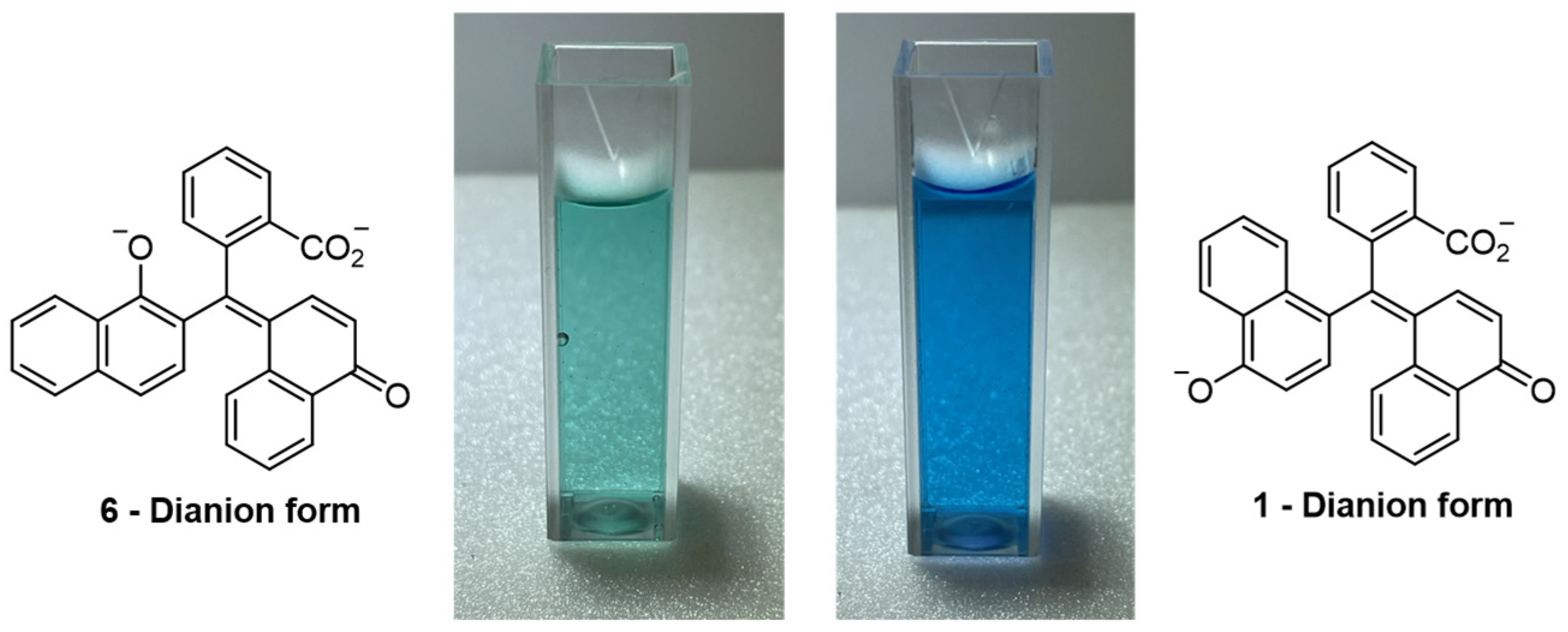
The reactions of compounds 1 and 6 in basic solution were compared. Compound 1 forms a teal-coloured solution in dilute sodium hydroxide. It has been found that compound 6 results in a green-coloured solution under the same conditions [1]. Figure 2 shows the results of adding drops of 0.5% ethanolic solutions of compounds 1 or 6 to a 3 mL aliquot of 0.1 M sodium hydroxide. Previous work suggests that the naphtholphthalein isomer 3 forms a green solution when added to aqueous solutions of sodium hydroxide; our results show a similar outcome with basic solutions of compound 6 [5,6]. A set of UV-visible absorption spectra of compounds 1 and 6 dissolved in dilute sodium hydroxide has been provided in the Supporting Information document (Supporting Information Figure S1).
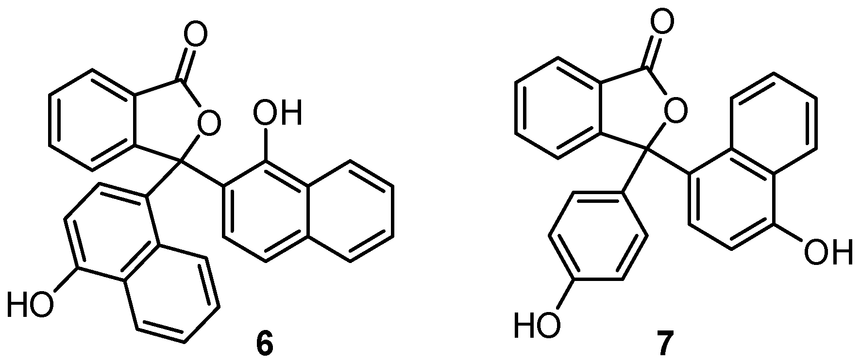
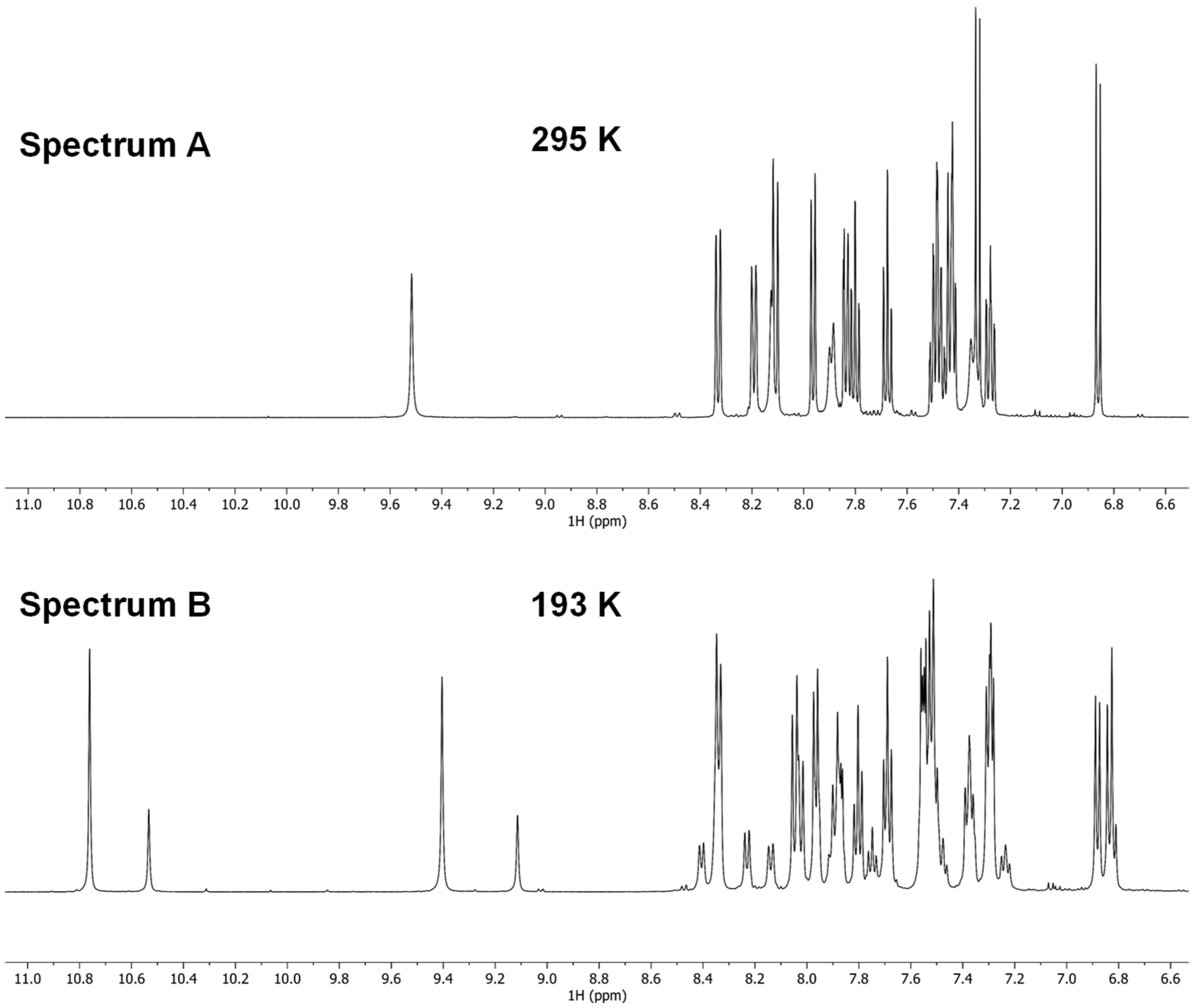
1H and 13C NMR NMR spectra were acquired in deuterated acetone and deuterated dimethyl sulfoxide. The former gave the best signal dispersion of the 1H NMR signals; therefore, deuterated acetone was selected for NMR spectroscopy experiments (Supporting Information Figures S2–S8). The 1H NMR spectrum of 6 (Supporting Information Figures S2 and S3) is consistent with the structure. There were two signals attributed to the hydroxyl groups (9.52 and 8.13 ppm), and 2D NMR techniques allowed the isobenzofuranone protons and the naphthol substituent proton signals to be identified (Supporting Information Figure S3). A notable feature in the 1H NMR spectrum acquired at room temperature was that the H26, H27 and H5 signals were broad. This observation is consistent with restricted rotational freedom of the substituents connected to the spiro-carbon (C-3). These results also align with previous NMR spectroscopy studies of compound 7 (Figure 3), a key finding from those experiments being that a preferred conformation could be identified when the sample was cooled to 179 K [9]. In contrast, under the same conditions, phenolphthalein (1) showed no conformational preference.
As part of our study, a sample of compound 6 was cooled to 193 K. The resulting 1H NMR spectrum suggested that two conformers were present in a roughly 2:1 ratio (a comparison of 1H NMR spectra acquired at 295 K and 193 K is presented in Figure 4). This observation is consistent with earlier work focused on compound 7, which has a similar structure [9]. In that study it was reported that the presence of a α-naphthol substituent led to restricted rotation around the bonds that connected the substituents to the phthalide ring. At present, the identities of the conformers at lower temperature have not been confirmed; these are currently under further investigation.
The ATR-IR spectrum of compound 6 was also obtained (Supporting Information Figure S9); this shows a distinctive signal at 1732 cm−1 that is consistent with the carbonyl group within the isobenzofuranone moiety. In addition, there are two signals corresponding to the two different hydroxyl groups from each phenol substituent, these are observed at 3467 cm−1 and 3237 cm−1.

2.2. The Crystal Structure of 6

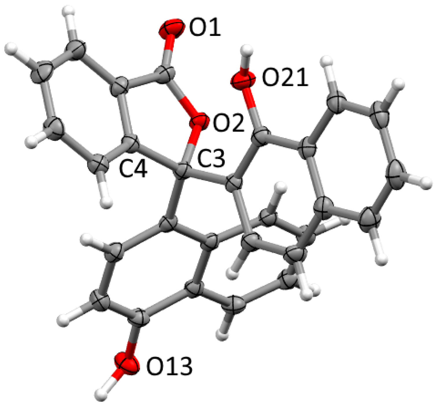
Crystals of 6 suitable for X-ray diffraction were obtained by slow evaporation from a mixture of ethanol and water. The compound crystallises in the monoclinic space group P21/n with one molecule of 6 and a disordered ethanol solvate in the asymmetric unit. The observed connectivity is consistent with the spectroscopic data (Figure 5). The spiro carbon (C3) of the phthalide ring shows distortion from ideal tetrahedral geometry, with the angle constrained by the ring (O2-C3-C4) at 101.78(11)°, and other angles similarly larger. The adjacent O2-C3 bond is longer than typical for γ-lactone structures (c.a. 1.46 Å) [10,11] at 1.4908(17) Å, similarly to related phthalein structures [12,13].
The packing of 6 in the crystal structure is dominated by hydrogen bonding interactions. Repeated hydrogen bonds from the 4-hydroxylnahthalen-1-yl hydroxyl (O13) to the carbonyl (O1) of adjacent molecules (H···O 1.682(17) Å, O···O 2.6517(16) Å) results in helical C 10 chains running along [0 1 0] (Supporting Information Figure S11). The other hydroxylnahthaleneyl hydroxyl (O21) forms C 2 2 ( 12 ) hydrogen-bonded chains to the other hydroxyl group (O13) via the hydroxyl of the major disordered component of the ethanol solvate (H···O 1.71(2) & 1.858(19) Å, O···O 2.6238(18) & 2.7810(19) Å). These hydrogen bonds link the [0 1 0] chains into sheets in the (1 0 1) plane. The minor component of the solvate forms a discrete hydrogen bond to the phthalein carbonyl. The hydrogen-bonded sheets further pack into the overall 3D structure supported by a weaker CH···π interaction from C8 to the C14-19 ring (H···centroid 2.7626(6) Å, C-H···centroid 162.26(11)°).
The known structure most similar to 6 is that of 3-(3,8-dimethoxynaphthalen-1-yl)-3-(3,8-dimethoxynaphthalen-2-yl)-2-benzofuran-1(3H)-one [14]. It shows a near-identical arrangement of the phthalide ring and one naphthyl group, analogous to C10-C18 in the structure of 6. The second naphthyl ring in 3-(3,8-dimethoxynaphthalen-1-yl)-3-(3,8-dimethoxynaphthalen-2-yl)-2-benzofuran-1(3H)-one is arranged in nearly the same plane as that of C20-C27 but rotated by approximately 180°. In the absence of hydroxy groups to form hydrogen bonds, the packing is dominated by CH···π interactions and non-conventional CH···O hydrogen bonds.
In summary, this study presents a simple synthetic route to a previously unknown acid/base indicator (compound 6) which is an isomer of naphtholphthalein. The title compound was prepared by mixing naphtholphthalic acid 5 and 1-naphthol for 1 h in methanesulfonic acid. 1H and 13C NMR datasets for compound 6 were acquired, IR and mass spectra were also recorded. As part of this work, the molecular structure of compound 6 was determined by single-crystal X-ray diffraction.

3. Experimental Section

Naphtholphthalic acid 5 was prepared and purified by a previously reported procedure [15]. Melting points were recorded on a Stuart SMP3 melting point apparatus (tolerance ±1.5 °C at 300 °C) (Mettler Toledo Ltd., Leicester, UK). The melting point apparatus was calibrated using samples of phenolphthalein (m.p. 261–263 °C). IR spectra were recorded on a Nicolet Summit FTIR instrument with Everest diamond ATR accessory (Fisher Scientific, Loughborough, UK). NMR spectra were obtained for 1H at 500.13 MHz and for 13C at 125.77 MHz using a Bruker AVIII_HD 500 instrument (Bruker UK Ltd., Coventry, UK). Spectra were run at 25 °C in CD3COCD3. Chemical shifts are reported in ppm to a high frequency of the reference, and coupling constants J are reported in Hz. The HRMS data were acquired from the University of St Andrews Mass Spectrometry Service.

3.1. 3-(4-Hydroxynaphthalen-1-yl)-3-(1-hydroxynaphthalen-2-yl)isobenzofuran-1(3H)-one (6)

A mixture of 2-((1-hydroxy-2-naphthyl)carbonyl)benzoic acid (naphtholphthalic acid 5, 0.30 g, 1.0 mmol) and 1-naphthol (0.14 g, 1.0 mmol) in methanesulfonic acid (2.00 g) was stirred for 1 h at room temperature (21–22 °C). The reaction was quenched with water (10 mL), and the resulting crude product was precipitated as a brown-coloured solid. The solid was filtered off under suction and washed with water (4 × 5 mL). After drying, DCM (3 mL) was added to the crude product, the mixture was boiled for 5 min and the remaining solid was filtered off under suction to afford product 6 (0.25 g, 0.6 mmol, 60%). Colourless X-ray quality crystals of 6 were grown by slow evaporation from ethanol. m.p. 239–241 °C. IR (ATR) 3467, 3237, 1732, 1585, 1352, 1253, 1118 cm−1; 1H NMR (500 MHz, CD3COCD3); 9.52 (1H, bs, OH), 8.33 (1H, d, J 14-15 = 8.3 Hz, 14-H), 8.19 (1H, d, J 22-23 = 8.0 Hz, 22-H), 8.13 (1H, bs, OH), 8.11 (1H, d, J 16-17= 8.8 Hz, 17-H), 7.96 (1H, d, J 7-8 = 7.6 Hz, 8-H), 7.89 (1H, d, J 5-6 = 7.6 Hz, 5-H), 7.83–7.85 (1H, m, 25-H), 7.80 (1H, td, J 5-6 = 7.6 Hz, J 6-7 = 7.6 Hz, J 6-8 = 1.1 Hz, 6-H), 7.68 (1H, td, J 6-7 = 7.6 Hz, J 7-8 = 7.6 Hz, J 5-7 = 1.1 Hz, 7-H), 7.41–7.51 (4H, m, 15-H, 23-H, 24-H, 26-H), 7.35 (1H, bs, 27-H), 7.33 (1H, d, J 11-12 = 8.0 Hz, 11-H), 7.26–7.29, m, 16-H), 6.86 (1H, d, J 11-12 = 8.0 Hz, 12-H); 13C NMR (126 MHz, CD3COCD3); 169.4 (C=O), 154.5 (ArCq), 152.5 (ArCq), 149.6 (ArCq), 134.6 (ArCq), 133.9 (ArCq), 132.8 (ArCq), 129.4 (ArCH), 127.8 (ArCH), 127.3 (ArCH), 127.2 (ArCq), 127.1 (ArCH), 126.8 (ArCH), 126.6 (ArCH), 126.2 (ArCq), 126.1 (ArCq), 125.9 (ArCH), 125.6 (ArCH), 125.4 (ArCH), 125.3 (ArCH), 124.6 (ArCH), 123.3 (ArCq), 122.6 (ArCH), 121.3 (ArCH), 120.3 (ArCH), 106.2 (ArCH), 91.9 (Cq). HRMS (ESI+) m/z (%) Calcd. for C28H18O4Na 441.1094, found 441.1097 [M + Na] (100).

3.2. X-Ray Structure Determination of 6

X-ray diffraction data were collected using a Rigaku FR-X High Brilliance Microfocus RA generator/confocal optics [Mo Kα radiation (λ = 0.71073 Å)] with XtaLAB P200 diffractometer (Rigaku Corporation, Tokyo, Japan). Data were collected (using a calculated strategy) and processed (including correction for Lorentz, polarization, and absorption) using CrysAlisPro [16]. The structure was solved by dual-space methods (SHELXT) [17] and refined by full-matrix least-squares against F2 (SHELXL-2019/3) [18]. Non-hydrogen atoms were refined anisotropically, and hydrogen atoms were refined using a riding model except for the hydrogen atoms on the hydroxyl oxygens O13, O21, and O31A; these were located from the difference Fourier map and refined isotropically, subject to distance restraints. The hydrogen atom on the minor component of the disorder (O31B) was placed in a calculated position and refined using a riding model. The solvate showed positional disorder, the non-hydrogen atoms of the minor part were placed using FragmentDB [19,20] and their positions and anisotropic displacement parameters allowed to refine with geometric (DFIX, DANG) and displacement parameter (ISOR, RIGU) restraints. A strong (non-default) thermal similarity restraint (SIMU) between the non-hydrogen atoms in the major and minor components of the disorder was required to achieve similarity of displacement parameters. Refinement of the minor component with isotropic displacement parameters resulted in less similarity of displacement parameters between major and minor components with the same SIMU restraint, so the anisotropic model was retained. All calculations were performed using the Olex2 interface [21].
Crystal data for C30H24O5, M = 464.49 g mol−1, monoclinic, a = 9.1996(2), b = 11.9748(3), c = 21.2399(5) Å, β = 100.735(2), U = 2298.91(9) Å3, T = 100 K, space group P21/n (no. 14), Z = 4, 48,885 reflections measured, 5563 unique (Rint = 0.1262), which were used in all calculations. The final R1 [I > 2σ(I)] was 0.0509 and wR2 (all data) was 0.1407. Data have been deposited at the Cambridge Crystallographic Data Centre as CCDC 2554594. These data can be obtained free of charge via http://www.ccdc.cam.ac.uk/getstructures.

Supplementary Materials

The following supporting information can be downloaded online: Figure S1: UV-vis spectra of the dianion forms of compounds 1 and 6; Figure S2: 500 MHz (CD3COCD3) 1H NMR spectrum of compound 6; Figure S3: 500 MHz (CD3COCD3) 1H NMR spectrum of compound 6 (expansion); Figure S4: 1H-1H COSY NMR spectrum of 6; Figure S5: 1H-1H TOCSY NMR spectrum of 6; Figure S6: 126 MHz (CD3COCD3) 13C DEPTQ NMR spectrum of 6; Figure S7: 1H-13C HSQC NMR spectrum of 6; Figure S8: 1H-13C HMBC NMR spectrum of 6; Figure S9: IR spectrum of 6; Figure S10: ESI-mass spectrum of 6. Figure S11; Helical C(10) chains running along [0 1 0].

Author Contributions

Synthetic steps, crystallisation trials, and preliminary analysis were conducted by N.V., I.A.S., B.A.C. and I.L.J.P.; N.V., T.L. and I.A.S. collected the NMR spectroscopy data; A.P.M. and D.B.C. collected the X-ray data and solved the structure; D.B.C., A.P.M. and I.A.S. wrote the paper. All authors have read and agreed to the published version of the manuscript.

Funding

This research received no external funding.

Data Availability Statement

CCDC 2554594 contains the supplementary crystallographic data for this paper. These data can be obtained free of charge from The Cambridge Crystallographic Data Centre via www.ccdc.cam.ac.uk/structures. Additional data supporting the findings of this study are available from the corresponding author upon reasonable request.

Acknowledgments

The authors are very grateful to Georg Hähner from the University of St Andrews for his help in checking the translation of some of the documents for this study. The authors would also like to express gratitude to the University of St Andrews School of Chemistry for use of laboratory facilities and provision of materials. We acknowledge support for the St Andrews Single-Crystal X-Ray Diffraction Service from the University of St Andrews Strategic Equipment Fund. We acknowledge support by the St Andrews NMR facility, and the Mass Spectrometry facility, funded by EPSRC ([EP/X034747/1).

Conflicts of Interest

The authors declare no conflicts of interest.

References

  1. Sabnis, R.W. Developments in the chemistry and applications of phthalein dyes. Part 1: Industrial applications. Color. Technol. 2018, 134, 187–205. [Google Scholar] [CrossRef]
  2. Poethke, W. Konduktometrisehe Studien. Fresenius Z. Für Anal. Chem. 1931, 86, 399–422. [Google Scholar] [CrossRef]
  3. Fox, J.J.; Ellis, B.A. Analytical Chemistry. Annu. Rep. Prog. Chem. 1932, 29, 220–238. [Google Scholar] [CrossRef]
  4. Werner, E.A. III—The Preparation of α-Naphtholphthalein. J. Chem. Soc. Trans. 1918, 113, 20–21. [Google Scholar] [CrossRef]
  5. Copisarow, M. XXVI—Phthaleins and Fluorans. J. Chem. Soc. Trans. 1920, 117, 209–218. [Google Scholar] [CrossRef]
  6. Csányi, W. Über die Kondensation von Phthalylchlorid mit α-Naphthol. Berichte Dtsch. Chem. Ges. A/B 1919, 52, 1788–1793. [Google Scholar] [CrossRef][Green Version]
  7. Schulenburg, W. Beiträge zur Kenntnis des α-Naphthol-phthaleins und seiner Äther. Berichte Dtsch. Chem. Ges. A/B 1920, 53, 1445–1457. [Google Scholar] [CrossRef][Green Version]
  8. Sabnis, R.W. A facile synthesis of phthalein dyes. Tetrahedron Lett. 2009, 50, 6261–6263. [Google Scholar] [CrossRef]
  9. Ghelli, S.; Rastelli, G.; Barlocco, D.; Rinaldi, M.; Tondi, D.; Pecorari, P.; Costi, M.P. Conformational Analysis of Phthalein Derivatives Acting as Thymidylate Synthase Inhibitors by Means of 1H NMR and Quantum Chemical Calculations. Bioorg. Med. Chem. 1996, 4, 1783–1794. [Google Scholar] [CrossRef] [PubMed]
  10. Allen, F.H.; Watson, D.G.; Brammer, L.; Orpen, A.G.; Taylor, R. International Tables for Crystallography Vol. C; Fuess, H., Hahn, T., Wondratschek, H., Müller, U., Shmueli, U., Prince, E., Authier, A., Kopský, V., Litvin, D.B., Rossmann, M.G., et al., Eds.; John Wiley & Sons: Hoboken, NJ, USA, 2006; pp. 790–811. [Google Scholar] [CrossRef]
  11. Sugira, H.; Kato, T.; Senda, H.; Kunimoto, K.K.; Kuwae, A.; Hanai, K. Crystal Structure of Phenolphthalein. Anal. Sci. 1999, 15, 611–612. [Google Scholar] [CrossRef]
  12. Fitzgerald, L.J.; Gerkin, R.E. Phenolphthalein and 3’,3″-Dinitrophenolphthalein. Acta Crystallogr. Sect. C Cryst. Struct. Commun. 1998, 54, 535. [Google Scholar] [CrossRef] [PubMed]
  13. Chalmers, B.A.; Cordes, D.B.; McKay, A.P.; Patterson, I.L.J.; Pearson, R.J.; Sequeira-Shuker, J.H.; Smellie, I.A.; Vladymyrova, N. Single-Crystal X-Ray Diffraction Studies of Derivatives of Phenolphthalein (3,3-Bis(4-hydroxyphenyl)isobenzofuran-1(3H)-one). Crystals 2025, 15, 901. [Google Scholar] [CrossRef]
  14. Yang, Y.; Strongin, R.M.; Fronczek, F.R. CCDC 1404537; CSD Communication: Omagh, UK, 2015. [Google Scholar] [CrossRef]
  15. Park, I.S.; Heo, E.-J.; Kim, J.-M. A photochromic phenoxyquinone based cyanide ion sensor. Tetrahedron Lett. 2011, 53, 2454–2457. [Google Scholar] [CrossRef]
  16. CrysAlisPro v1.171.43.144a Rigaku Oxford Diffraction; Rigaku Corporation: Tokyo, Japan, 2023.
  17. Sheldrick, G.M. SHELXT—Integrated space-group and crystal structure determination. Acta Crystallogr. Sect. A Found. Adv. 2015, 71, 3–8. [Google Scholar] [CrossRef] [PubMed]
  18. Sheldrick, G.M. Crystal structure refinement with SHELXL. Acta Crystallogr. Sect. C Struct. Chem. 2015, 71, 3–8. [Google Scholar] [CrossRef] [PubMed]
  19. Kratzert, D.; Holstein, J.J.; Krossing, I. DSR: Enhanced modelling and refinement of disordered structures with SHELXL. J. Appl. Crystallogr. 2015, 48, 933–938. [Google Scholar] [CrossRef] [PubMed]
  20. Kratzert, D.; Krossing, I. Recent improvements in DSR. J. Appl. Crystallogr. 2018, 51, 928–934. [Google Scholar] [CrossRef]
  21. Dolomanov, O.V.; Bourhis, L.J.; Gildea, R.J.; Howard, J.A.K.; Puschmann, H. OLEX2: A complete structure solution, refinement and analysis program. J. Appl. Crystallogr. 2009, 42, 339–341. [Google Scholar] [CrossRef]
Figure 1. Structures of naphtholphthalein (1), phenolphthalein (2).
Molbank 2026 m2195 g001
Scheme 1. Reaction of phthalic anhydride with 1-naphthol mixed with ZnCl2.
Molbank 2026 m2195 sch001
Scheme 2. Csányi’s reaction of phthaloyl chloride with 1-naphthol in the presence of AlCl3.
Molbank 2026 m2195 sch002
Scheme 3. Schulenberg’s reaction of naphtholphthalic acid 5 with 1-naphthol in concentrated sulfuric acid.
Molbank 2026 m2195 sch003
Scheme 4. Reaction of naphtholphthalic acid 5 with 1-naphthol in methanesulfonic acid.
Molbank 2026 m2195 sch004
Figure 2. Solutions of compounds 1 and 6 in 0.1 M sodium hydroxide.
Molbank 2026 m2195 g002
Figure 3. Structures of naphtholphthalein (6) and phenolphthalein derivative (7).
Molbank 2026 m2195 g003
Figure 4. 1H NMR spectra of compound 6 in CD3COCD3. Spectrum A acquired at 295 K. Spectrum B acquired at 193 K.
Molbank 2026 m2195 g004
Figure 5. X-ray structure of compound 6. Thermal ellipsoids drawn at 50% probability, and solvate omitted for clarity.
Molbank 2026 m2195 g005
Disclaimer/Publisher’s Note: The statements, opinions and data contained in all publications are solely those of the individual author(s) and contributor(s) and not of MDPI and/or the editor(s). MDPI and/or the editor(s) disclaim responsibility for any injury to people or property resulting from any ideas, methods, instructions or products referred to in the content.