Short Note

Methyl α-d-Tagatopyranoside

Graduate School of Biomedical Sciences, Nagasaki University, 1-14 Bunkyo-machi, Nagasaki 852-8521, Japan
*
Author to whom correspondence should be addressed.
Molbank 2025, 2025(3), M2046; https://doi.org/10.3390/M2046
Submission received: 28 July 2025 / Revised: 9 August 2025 / Accepted: 12 August 2025 / Published: 14 August 2025

Abstract

d-Tagatose, classified as a rare sugar, exhibits notable biological activities, including its function as a low-calorie sweetener. The three-dimensional configuration of carbohydrates is crucial for elucidating their functional properties. Numerous studies have reported the X-ray crystallographic structures of d-tagatose and its derivatives bearing a free anomeric hydroxy group. However, there are no reports on the X-ray crystallographic structure of d-tagatosides featuring a glycosidic linkage at the anomeric position. In this study, we synthesized methyl α-d-tagatopyranoside from d-tagatose and successfully determined its X-ray crystallographic structure, revealing its 5C2 conformation.

1. Introduction

Alternative sugars are important from the perspective of low-calorie sweeteners and their functional properties. Among these, rare sugars such as d-allulose (d-psicose) and d-tagatose have attracted considerable attention because of their notable biological activities, including their role as low-calorie sweeteners and ability to suppress blood glucose levels [1,2,3,4,5,6,7]. d-Tagatose, a C4-epimer of d-fructose, possesses 92% of the sweetness of sucrose but only 38% of its calories [8]. Owing to its mutarotation, d-tagatose exists in five different structures, namely, α- and β-d-tagatopyranoses, α- and β-d-tagatofuranoses, and an open-chain form (Figure 1). Therefore, there have been several attempts at derivatization of d-tagatose by controlling these structures, including synthesis of 1,2:3,4-di-O-isopropyidene-α-d-tagatofuranose [9,10,11], α-selective glycosidation of 3,4-O-isopropyidene-d-tagatofuranosyl donor [12], and synthesis of penta- and tetra-O-benzoyl-α-d-tagatopyranoses [13].
Acquiring comprehensive structural information is essential to understand the biological effects of d-tagatose. Nevertheless, owing to the mutarotation of d-tagatose, investigating its conformation in solution poses a considerable challenge [14]. Consequently, X-ray crystallographic analysis offers a valuable method for obtaining conformational data on d-tagatose. For example, X-ray crystallographic structures of α-d-tagatopyranose [15,16], 1,2:3,4-di-O-isopropyidene-α-d-tagatofuranose [9], and 1,3,4,5-tetra-O-acetyl-α-d-tagatopyranose [17] have been reported. These examples are all pyranose or furanose forms, featuring a free anomeric hydroxy group. However, to the best of our knowledge, there are no reports on the X-ray crystallographic structures of its glycosides. The simplest d-tagatopyranoside is methyl d-tagatopyranoside, which was first documented by Khouvine et al. [18]. Subsequently, Angyal et al. conducted NMR analyses of both methyl α-d-tagatopyranoside and β-d-tagatopyranoside [14,19]. Thus, the available structural information on methyl α-d-tagatopyranoside is limited. Herein, we report the synthesis of methyl α-d-tagatopyranoside (1) in a single step from d-tagatose, along with its X-ray crystallographic structure, marking the first successful example of d-tagatopyranosides.

2. Results and Discussion

Methyl α-d-tagatopyranoside (1) was synthesized using commercially available d-tagatose, as depicted in Scheme 1. Treatment of d-tagatose in MeOH with Amberlyst 15dry, a polymeric acid catalyst, resulted in the formation of methyl α-d-tagatopyranoside (1) in 90% yield, along with other isomers of methyl glycosides, including methyl β-d-tagatopyranoside (5%). Amberlyst 15dry offers a safer and more manageable alternative for the removal of acid catalysts than the previously employed methanolic hydrogen chloride in the synthesis of glycoside 1 [18,19]. The 13C NMR spectrum of product 1 in D2O solution was consistent with the literature (Figure S2b) [14]. Further spectroscopic analyses, including 1H NMR, 1H-1H COSY, HSQC, and HMBC spectra, as well as IR and MS spectra, confirmed the structure of compound 1. However, the NOESY spectrum of compound 1 did not provide three-dimensional information because of the excessively close or overlapping correlation peaks (Figure S6).
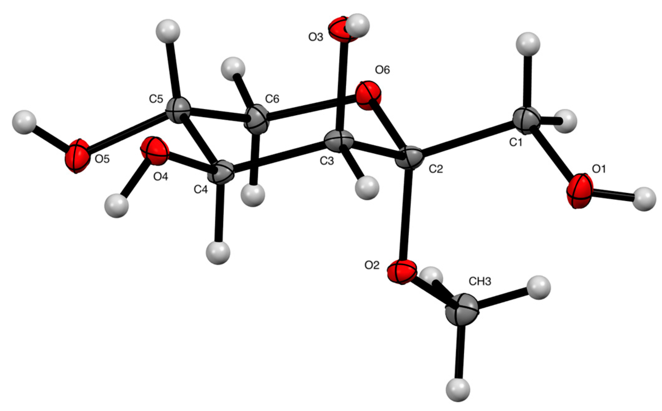
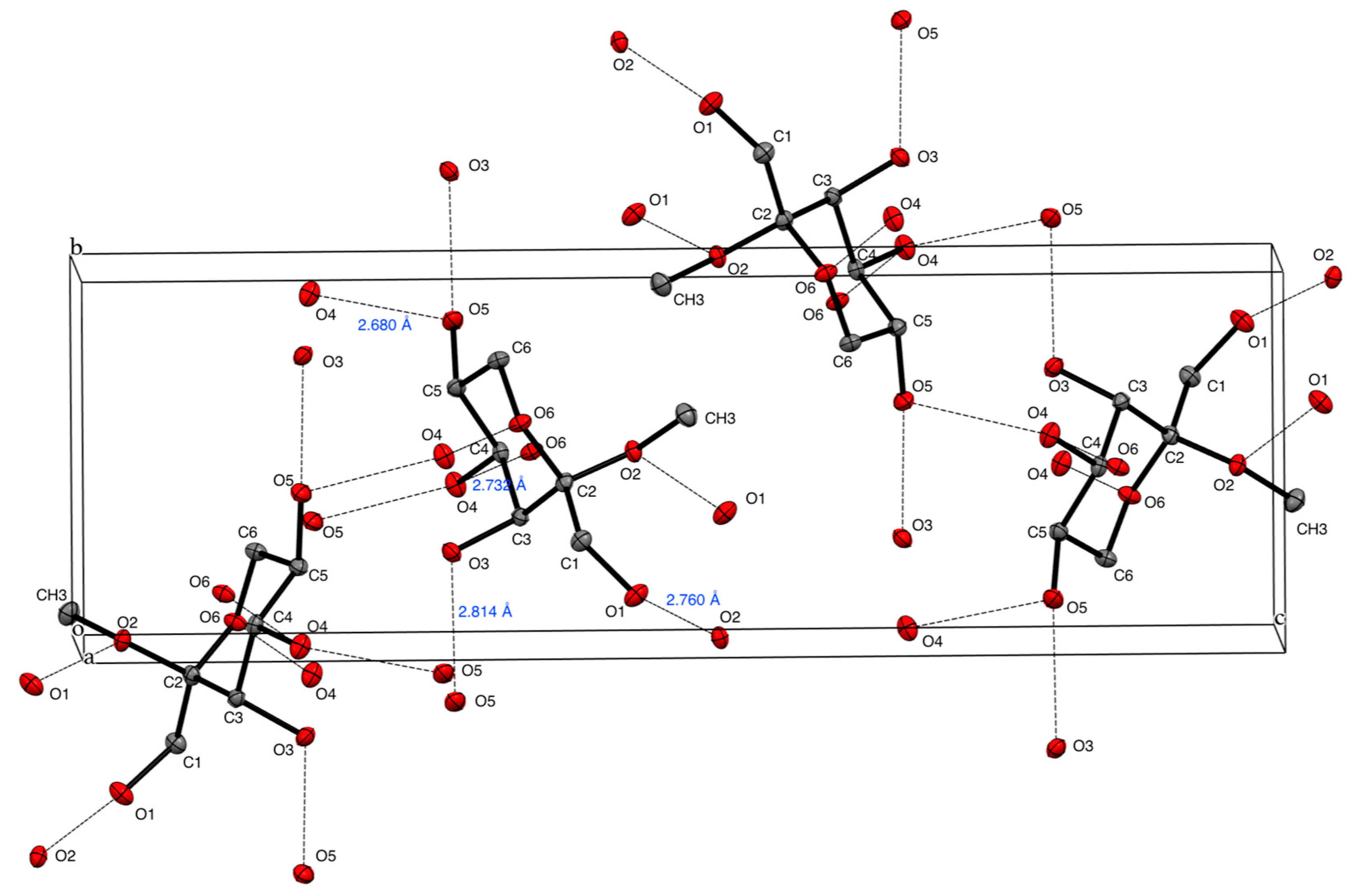
Slow evaporation of the solvent from a solution of compound 1 in MeOH resulted in the formation of colorless crystals that were suitable for X-ray diffraction analysis. The structure of compound 1 was solved and refined using OLEX2 [20] in conjunction with SHELXT [21] and SHELXL [22]. Figure 2 presents an ORTEP drawing of the X-ray crystallographic structure of compound 1, indicating a 5C2 conformation. Figure 3 and Table 1 detail the intermolecular hydrogen bond parameters of compound 1, which include four intermolecular hydrogen bonds in compound 1: O1-H···O2′, O3-H···O5′, O4-H···O6′, and O5-H···O4′.
The torsion angles of compound 1 are summarized in Table 2, along with those of α-d-tagatopyranose (2) [16]. Owing to the presence of an exo-anomeric effect in compound 1 [23], the methyl glycoside adopts a gauche conformation, characterized by a torsion angle ϕ (O6-C2-O2-CH3) of +61.1°. The methyl glycoside in compound 1 significantly influenced the orientation of the C1 hydroxy group, resulting in a torsion angle of O1-C1-C2-O2 of –49.5°, compared to +50.8° in compound 2. Consequently, the C1 hydroxy group of compound 1 was positioned antiperiplanar to the O6 atom, whereas in compound 2, it was positioned gauche to the O6 atom. Additionally, a difference of approximately 10° was observed in the torsion angle of O4-C4-C5-O5 between compounds 1 and 2. These findings provide valuable insights into the biological effects of d-tagatose and its related compounds, as the orientation of the hydroxy groups is crucial for interactions with receptors or other molecules.

3. Materials and Methods

3.1. General Procedure and Method

Optical rotations were measured on a JASCO DIP-370 polarimeter (JASCO Corporation, Tokyo, Japan) using MeOH as a solvent. 1H NMR and 13C NMR spectra were recorded on a JEOL JNM-ECZ400R (400 MHz and 100 MHz) spectrometers (JEOL Ltd., Tokyo, Japan) or a Varian NMR System 500PS SN (500 MHz and 125 MHz) spectrometers (Agilent Inc., Santa Clara, CA, USA). Chemical shifts (δ) are reported in parts per million (ppm). Tetramethylsilane was used as the internal reference (0.00 ppm in CD3OD) for 1H NMR spectra, while the central solvent peak was used as the reference (49.0 ppm in CD3OD) for 13C NMR spectra. The IR spectra were recorded on a Shimadzu IRAffinity-1 FT-IR spectrophotometer (Shimadzu Corporation, Kyoto, Japan). High-resolution mass spectra (HRMS) were obtained on a JEOL JMS-T100TD using electrospray ionization (ESI) (JEOL Ltd., Tokyo, Japan) in time-of-flight (TOF) mode. Analytical thin-layer chromatography (TLC) was performed with Merck Millipore precoated TLC plates (MilliporeSigma, Burlington, VT, USA), silica gel 60 F254, and layer thicknesses of 0.25 mm. Compounds were observed in UV light at 254 nm and then visualized by staining with p-anisaldehyde stain. Flash column chromatography separations were performed on Kanto Chemical silica gel 60N, spherical neutral, with particle sizes of 40–50 μm. All moisture-sensitive reactions were conducted under an inert atmosphere. Reagents and solvents were of commercial grade and were used as supplied, unless otherwise noted.

3.2. Methyl α-d-Tagatopyranoside (1)

Amberlyst 15dry (200 mg) was introduced to a suspension of d-tagatose (1.80 g, 10.0 mmol) in MeOH (10 mL) at room temperature, and the reaction mixture was stirred at room temperature for four days. After filtration through filter paper and removal of the solvent, the residue was purified by flash column chromatography on silica gel (15% MeOH in CHCl3) to yield 1 (1.74 g, 90%) as a colorless crystalline solid along with a mixture of the other isomers (100 mg, 5%), such as methyl β-d-tagatopyranoside, in the form of a colorless syrup. An aliquot of 1 was recrystallized from MeOH by slow evaporation of the solvent. White crystalline. m.p. 129–131 °C. Rf = 0.26 (20% MeOH in CHCl3). [α]29D +50.1 (c 1.00, MeOH) {lit. [19] [α]22.5D +41 (c 0.16, MeOH)}. 1H NMR (400 MHz, CD3OD) δ: 3.86 (1H, d, J3,4 = 3.3 Hz, H-3), 3.80 (1H, ddd, J5,6b = 10.5, J4,5 = 9.5 Hz, J5,6a = 5.7 Hz, H-5), 3.71 (1H, dd, J4,5 = 9.5, J3,4 = 3.3 Hz, H-4), 3.69 (1H, d, J1a,1b = 12.0 Hz, H-1a), 3.64 (1H, dd, J6a,6b = 10.7 Hz, J5,6a = 5.7 Hz, H-6a), 3.58 (1H, d, J1a,1b = 12.0 Hz, H-1b), 3.27 (1H, dd, J6a,6b = 10.7 Hz, J5,6b = 10.5 Hz, H-6b), 3.25 (3H, s, OCH3). 1H NMR (500 MHz, D2O) δ: 3.90 (1H, d, J3,4 = 3.1 Hz, H-3), 3.84 (1H, ddd, J5,6b = 10.3, J4,5 = 9.6 Hz, J5,6a = 5.4 Hz, H-5), 3.80 (1H, dd, J4,5 = 9.6, J3,4 = 3.1 Hz, H-4), 3.76 (1H, dd, J6a,6b = 10.7 Hz, J5,6a = 5.4 Hz, H-6a), 3.67 (2H, s, H-1), 3.35 (1H, dd, J6a,6b = 10.7 Hz, J5,6b = 10.3 Hz, H-6b), 3.27 (3H, s, OCH3). 13C{1H} NMR (100 MHz, CD3OD) δ: 102.8 (C-2), 72.9 (C-4), 70.8 (C-3), 67.8 (C-5), 64.5 (C-6), 58.8 (C-1), 48.0 (CH3). 13C{1H} NMR (125 MHz, D2O) δ: 102.4 (C-2), 71.5 (C-4), 69.5 (C-3), 66.7 (C-5), 63.4 (C-6), 57.4 (C-1), 48.4 (CH3). IR (film): 3400 (br), 2953 cm–1. HRMS (ESI) m/z: [M + Na]+ calcd for C7H14O6Na, 217.0688; found, 217.0697. CCDC 2472885 contains the supplementary crystallographic data for this paper. These data can be obtained free of charge via http://www.ccdc.cam.ac.uk/conts/retrieving.html (or from the CCDC, 12 Union Road, Cambridge CB2 1EZ, UK.; Fax: +44-1223-336033; E-mail: deposit@ccdc.cam.ac.uk).

4. Conclusions

The synthesis and structural characterization of methyl α-d-tagatopyranoside (1) have been documented. X-ray crystallographic analysis of compound 1 unambiguously determined its three-dimensional structure, facilitating structural comparison between glycoside 1 and α-d-tagatopyranose (2) in the solid state.

Supplementary Materials

The following supporting information can be downloaded: Figure S1. 1H NMR spectra of tagatopyranoside 1; Figure S2: 13C NMR spectra of tagatopyranoside 1; Figure S3: 1H-1H COSY spectrum of tagatopyranoside 1; Figure S4: HSQC spectrum of tagatopyranoside 1; Figure S5: HMBC spectrum of tagatopyranoside 1; Figure S6: NOESY spectrum of tagatopyranoside 1; Table S1. Crystal and diffraction parameters of tagatopyranoside 1; Figure S7. Packing diagram of tagatopyranoside 1.

Author Contributions

Conceptualization, A.U.; methodology, A.U.; validation, Y.H., A.I., and A.U.; formal analysis, Y.H., A.I., M.T., and A.U.; investigation, Y.H., A.I., and A.U.; writing—original draft preparation, A.U.; writing—review and editing, Y.H., A.I., M.T., and A.U.; visualization, Y.H. and A.U.; supervision, A.U.; project administration, A.U.; funding acquisition, A.U. All authors have read and agreed to the published version of the manuscript.

Funding

This research received no external funding.

Data Availability Statement

The original contributions presented in this study are included in the article/Supplementary Material. Further inquiries can be directed to the corresponding author.

Acknowledgments

This work was the result of using research equipment shared in the MEXT Project for promoting the public utilization of advanced research infrastructure (program for supporting the introduction of the new sharing system), Grant Number JPMXS0422500320.

Conflicts of Interest

The authors declare no conflicts of interest.

Abbreviations

The following abbreviations are used in this manuscript:
COSYHomonuclear Correlation Spectroscopy
HSQCHeteronuclear Single Quantum Coherence
HMBCHeteronuclear Multiple Bond Coherence
NOESYNuclear Overhauser Effect Spectroscopy

References

  1. Granstrom, T.B.; Takata, G.; Tokuda, M.; Izumori, K. Izumoring: A novel and complete strategy for bioproduction of rare sugars. J. Biosci. Bioeng. 2004, 97, 89–94. [Google Scholar] [CrossRef] [PubMed]
  2. Matsuo, T.; Suzuki, H.; Hashiguchi, M.; Izumori, K. d-Psicose is a rare sugar that provides no energy to growing rats. J. Nutr. Sci. Vitaminol. 2002, 48, 77–80. [Google Scholar] [CrossRef]
  3. Hayashi, N.; Yamada, T.; Takamine, S.; Iida, T.; Okuma, K.; Tokuda, M. Weight reducing effect and safety evaluation of rare sugar syrup by a randomized double-blind, parallel-group study in human. J. Funct. Foods 2014, 11, 152–159. [Google Scholar] [CrossRef]
  4. Izumori, K. Izumoring: A strategy for bioproduction of all hexoses. J. Biotechnol. 2006, 124, 717–722. [Google Scholar] [CrossRef] [PubMed]
  5. Lu, Y.; Levin, G.V.; Donner, T.W. Tagatose, a new antidiabetic and obesity control drug. Diabetes Obes. Metab. 2008, 10, 109–134. [Google Scholar] [CrossRef]
  6. Livesey, G.; Brown, J.C. d-Tagatose Is a Bulk Sweetener with Zero Energy Determined in Rats. J. Nutr. 1996, 126, 1601–1609. [Google Scholar] [CrossRef] [PubMed]
  7. Levin, G.V. Tagatose, the New GRAS Sweetener and Health Product. J. Med. Food 2002, 5, 23–36. [Google Scholar] [CrossRef] [PubMed]
  8. Levin, G.V.; Zehner, L.R.; Saunders, J.P.; Beadle, J.R. Sugar substitutes: Their energy values, bulk characteristics, and potential health benefits. Am. J. Clin. Nutr. 1995, 62, 1161S–1168S. [Google Scholar] [CrossRef]
  9. Watkin, D.J.; Glawar, A.F.G.; Soengas, R.; Skytte, U.P.; Wormald, M.R.; Dwek, R.A.; Fleet, G.W.J. 1,2:3,4-Di-O-isopropylidene-α-d-tagatofuranose. Acta Crystallogr. Sect. E 2005, 61, o2891–o2893. [Google Scholar] [CrossRef]
  10. Yoshihara, A.; Haraguchi, S.; Gullapalli, P.; Rao, D.; Morimoto, K.; Takata, G.; Jones, N.; Jenkinson, S.F.; Wormald, M.R.; Dwek, R.A.; et al. Isomerization of deoxyhexoses: Green bioproduction of 1-deoxy-d-tagatose from l-fucose and of 6-deoxy-d-tagatose from d-fucose using Enterobacter agglomerans strain 221e. Tetrahedron: Asymmetry 2008, 19, 739–745. [Google Scholar] [CrossRef]
  11. Baráth, M.; Lin, C.-H.; Tvaroška, I.; Hirsch, J. Development of transition state analogue inhibitors for N-acetylglycosyltransferases bearing d-psico- or d-tagatofuranose scaffolds. Chem. Pap. 2015, 69, 348–357. [Google Scholar] [CrossRef]
  12. Makura, Y.; Ueda, A.; Matsuzaki, T.; Minamino, T.; Tanaka, M. α-Selective glycosidation of d-tagatofuranose with a 3,4-O-isopropylidene protection. Tetrahedron 2019, 75, 3758–3766. [Google Scholar] [CrossRef]
  13. Hu, Y.; Iyoshi, A.; Makura, Y.; Tanaka, M.; Ueda, A. 1,3,4,5-Tetra-O-benzoyl-α-d-tagatopyranose. Molbank 2025, 2025, M2041. [Google Scholar] [CrossRef]
  14. Angyal, S.J.; Bethell, G.S. Conformational analysis in carbohydrate chemistry. III* The 13C N.M.R. spectra of the hexuloses. Aust. J. Chem. 1976, 29, 1249–1265. [Google Scholar] [CrossRef]
  15. Takagi, S.; Rosenstein, R.D. The structure of α-d-tagatose and comparison with crystal structures of other ketohexoses. Carbohydr. Res. 1969, 11, 156–158. [Google Scholar] [CrossRef]
  16. Punzo, F.; Watkin, D.J.; Fleet, G.W.J. α-d-Tagatopyranose. Acta Crystallogr. Sect. E 2009, 65, o1393–o1394. [Google Scholar] [CrossRef] [PubMed]
  17. Miura, D.; Fujimoto, T.; Tashiro, M.; Machinami, T. Crystal Structure of 1,3,4,5-Tetra-O-acetyl-α-d-tagatopyranose. X-Ray Struct. Anal. Online 2014, 30, 3–4. [Google Scholar] [CrossRef]
  18. Khouvine, Y.; Arragon, G.; Tomoda, Y. Ketoses. II. The structure of α-d-tagatose. Bull. Soc. Chim. Fr. Mem. 1939, 6, 354–359. [Google Scholar]
  19. Angyal, S.; Bodkin, C.; Mills, J.; Pojer, P. Complexes of carbohydrates with metal cations. IX. Synthesis of the methyl d-tagatosides, d-psicosides, d-apiosides and d-erythrosides. Aust. J. Chem. 1977, 30, 1259–1268. [Google Scholar] [CrossRef]
  20. Dolomanov, O.V.; Bourhis, L.J.; Gildea, R.J.; Howard, J.A.K.; Puschmann, H. OLEX2: A complete structure solution, refinement and analysis program. J. Appl. Crystallogr. 2009, 42, 339–341. [Google Scholar] [CrossRef]
  21. Sheldrick, G. SHELXT–Integrated space-group and crystal-structure determination. Acta Crystallogr. Sect. A 2015, 71, 3–8. [Google Scholar] [CrossRef] [PubMed]
  22. Sheldrick, G. Crystal structure refinement with SHELXL. Acta Crystallogr. Sect. C 2015, 71, 3–8. [Google Scholar] [CrossRef] [PubMed]
  23. Lemieux, R.U.; Koto, S.; Voisin, D. The Exo-Anomeric Effect. ACS Symp. Ser. 1979, 87, 17–29. [Google Scholar] [CrossRef]
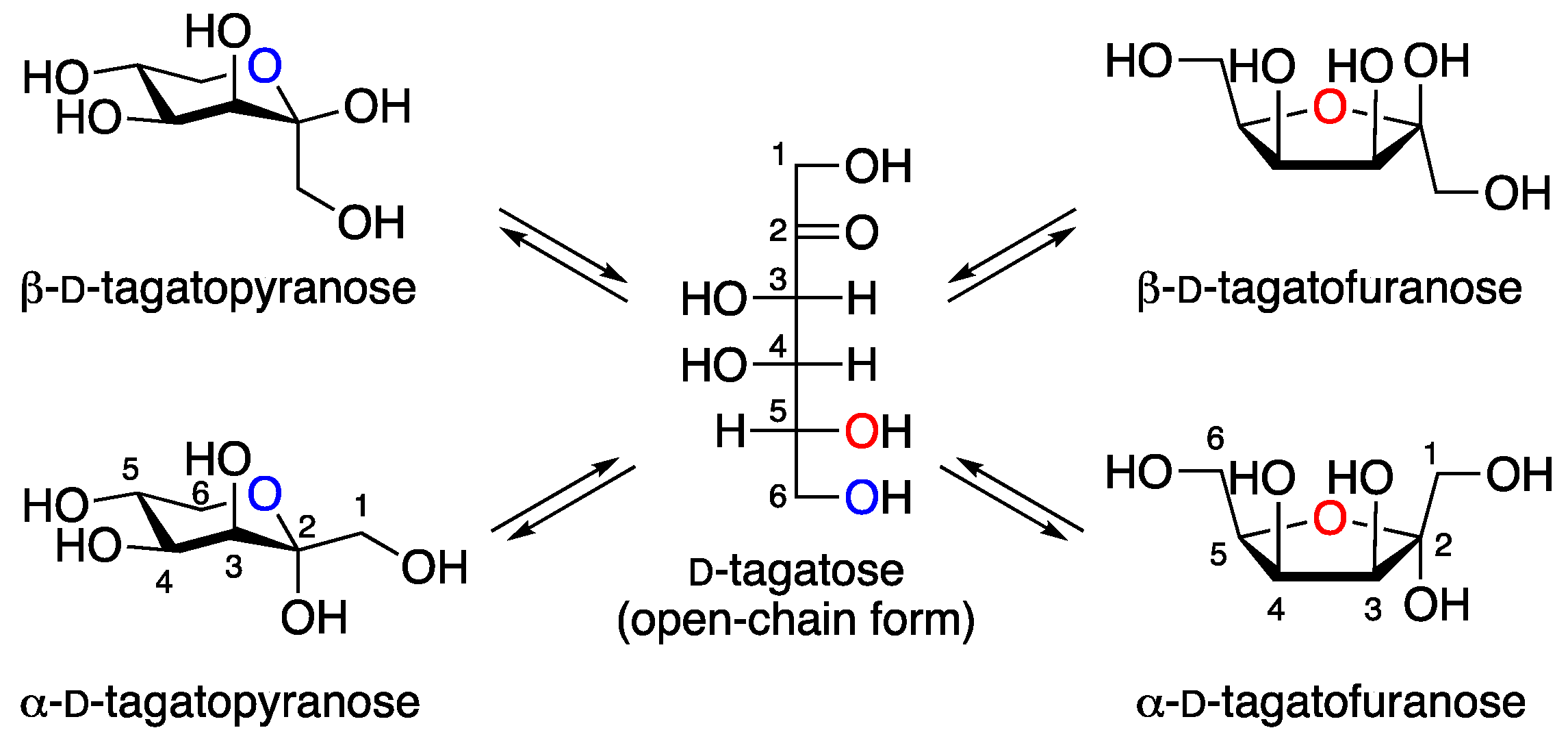
Figure 1. Structures and mutarotation of D-tagatose.
Molbank 2025 m2046 g001
Scheme 1. Synthesis of methyl α-d-tagatopyranoside (1) from d-tagatose.
Molbank 2025 m2046 sch001
Figure 2. An ORTEP drawing of compound 1 (ellipsoids at 50% probability).
Molbank 2025 m2046 g002
Figure 3. Packing diagram of compound 1. Hydrogen bonds are indicated by dashed lines, and hydrogen atoms are omitted for clarity.
Molbank 2025 m2046 g003
Table 1. Hydrogen bond parameters of compound 1.
DonorAcceptorDistance (Å)Angle (°)Symmetry
D–HAD–HH···AD···AD–H···Aoperations
O1-HO2′0.8401.9272.760170.9–1/2+x, 1/2−y, 1−z
O3-HO5′0.8401.9822.814171.0x, −1+y, z
O4-HO6′0.8401.9022.732169.51+x, y, z
O5-HO4′0.8521.8712.680157.82−x, 1/2+y, 1/2−z
Table 2. Comparative analysis of torsion angles between glycoside 1 and α-d-tagatopyranose (2).
DescriptionTorsion Angles (°)
12 1
O1-C1-C2-O2–49.550.8
O2-C2-C3-O3177.6174.6
O3-C3-C4-O4–57.2–57.9
O4-C4-C5-O5–71.6–62.1
O5-C5-C6-O6–169.3–175.7
ϕ (O6-C2-O2-CH3)61.1N/A 2
1 The torsion angles of 2 were obtained from the literature [16]. 2 N/A = not applicable.
Disclaimer/Publisher’s Note: The statements, opinions and data contained in all publications are solely those of the individual author(s) and contributor(s) and not of MDPI and/or the editor(s). MDPI and/or the editor(s) disclaim responsibility for any injury to people or property resulting from any ideas, methods, instructions or products referred to in the content.