Short Note

Cocrystal of Codeine and Cyclopentobarbital

Institute of Pharmacy, University of Innsbruck, Innrain 52c, A-6020 Innsbruck, Austria
*
Author to whom correspondence should be addressed.
Molbank 2023, 2023(3), M1722; https://doi.org/10.3390/M1722
Submission received: 30 July 2023 / Revised: 5 September 2023 / Accepted: 6 September 2023 / Published: 11 September 2023
(This article belongs to the Section Structure Determination)

Abstract

:
The two-component compound formed by codeine and cyclopentobarbital was produced using grinding techniques and through evaporation from alcoholic solutions. The cocrystal nature of this phase was established unequivocally through single crystal X-ray structure determination. The asymmetric unit contains one formula unit. In the cyclopentobarbital molecule, the cyclopentenyl ring is disordered over two positions related by a rotation of approximately 180° about its C—C bond to the pyrimidine ring. The two NH groups of the cyclopentobarbital molecule form N—H⋯N and N—H⋯O bonds to piperidine and hydroxyl groups, respectively, belonging to different codeine molecules. In addition, the hydroxyl and methoxy groups of neighboring codeine molecules are linked by O—H⋯O interactions, resulting in a H-bonded framework structure of codeine and cyclopentobarbital molecules. The cocrystal was also characterized using thermal analysis, X-ray powder diffraction and IR spectroscopy.

Graphical Abstract

1. Introduction

Codeine (I), (5a,6a)-7,8-didehydro-4,5-epoxy-3-methoxy-17-methylmorphinan-6-ol (Scheme 1), is a natural alkaloid of the opium poppy plant Papaver somniferum, first discovered in 1832 by Pierre Jean Robiquet [1]. Codeine is contained in the WHO model list of essential drugs [2]. With an annual production (2013) of about 361,000 kg, it is the most widely used narcotic drug in medical practice. Codeine is applied as an analgesic for the treatment of mild to moderate pain, as an antitussive (cough depressant) and as an antidiarrheal agent [3,4,5].
The List of Narcotic Drugs under International Control (Yellow List) not only contains the free base of codeine, but also 20 codeine multicomponent systems [6]. These are primarily salts with mineral acids (e.g., HCl, phosphate, sulphate) and common organic acids (glucuronic, salicylic, acetic and barbituric acid) where the pKa difference between codeine (pKa = 8.2) and the respective acid is higher than 3.5. Another five entries in the Yellow List concern combinations of codeine with 5,5-disubstituted derivatives of barbituric acid, all associated with a small pKa difference (between 0.2 and 1.1). This suggests that the corresponding two-component systems of codeine and the respective barbiturate exist as cocrystals [7]. Cocrystals are ‘solids that are crystalline materials composed of two or more different molecular and/or ionic compounds generally in a stoichiometric ratio which are neither solvates nor simple salts’ [8,9].
One of the barbiturates concerned is cyclopentobarbital (II), 5-allyl-5-(cyclopent-2-en-1-yl)pyrimidine-2,4,6(1H,3H,5H)-trione (Scheme 1; alternative names: cyclopal, dormisan). Cyclopentobarbital was invented in the 1940s [10,11], and possesses sedative and anticonvulsant properties with a slow onset of action. Its primary application was as an anesthetic in veterinary medicine.
The two-component system of codeine/cyclopentobarbital is associated with a pKa difference of just 0.7. It is therefore expected to exist as a cocrystal form, which will be denoted henceforth as (I)·(II). The current study was carried out to unequivocally establish the chemical nature of this phase by means of single-crystal X-ray structure determination, which was accompanied by a characterization with differential scanning calorimetry, hot-stage microscopy and FT-IR spectroscopy.

2. Results

2.1. Crystal Structure

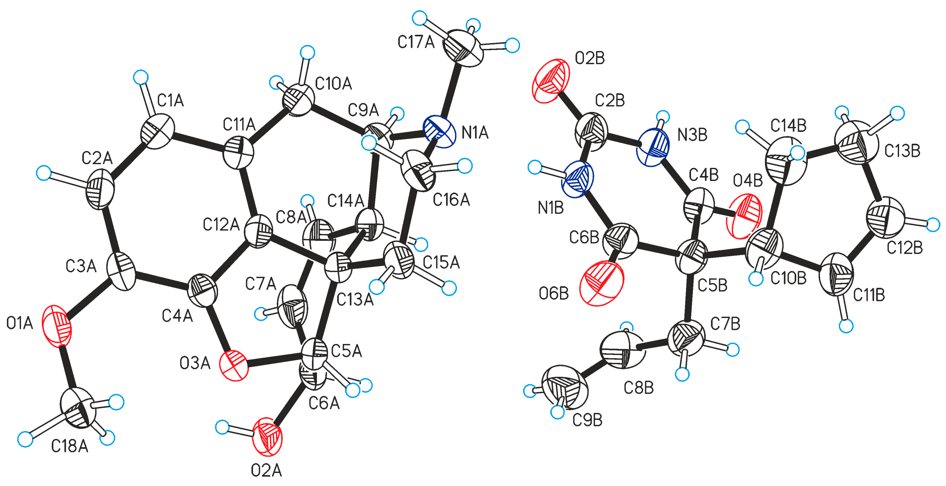
Crystal data and refinement details are collected in Table 1. The asymmetric unit of the orthorhombic crystal structure contains one formula unit (Figure 1), i.e., one codeine and one cyclopentobarbital molecule. The hydrogen positions of both NH groups in the molecule of (II), and thus the cocrystal nature of (I)·(II), were confirmed unequivocally.
The molecular structure of (I) is largely inflexible. The two mean planes defined by the rings A/B/C and D/E (Scheme 1) are nearly orthogonal, forming an angle of 88.20(6)°, which illustrates the presence of the characteristic T conformation of the opiate family [12]. The Cambridge Structural Database (CSD; version 5.44, June 2023) [13] contains nine unique examples of crystal structures containing the neutral codeine molecule or its cation form, i.e., CSD codes CDNECR [14], CODHBH [15], EZEWAK [16], OPOQAN], ZZZRFQ01, ZZZTZQ02 [17], QITZOK [18], QUBSEM [12] and ZZZTSE03 [19]. In this group, the angle characterizing the T geometry of the codeine skeleton varies in a narrow range between 86.0° and 90.0° (Table S1 of the Supporting Information). The orientation of the methoxy group can be described in terms of the torsion angle C4A—C3A—O1A—C18A, denoted as τ in Scheme 1. Its value of 25.2(6)° indicates that the methoxy group is twisted out of the plane of the phenyl ring (A) and oriented towards ring B (see Scheme 1). Similar conformations (τ = 25.4° and τ = 36.2°) have also been reported for both independent codeine molecules in the monophosphate hemihydrate [12]. By contrast, all other codeine structures from the CSD listed above exhibit an orientation of the methoxy group towards the phenyl ring, indicated by absolute τ values between 128.3° and 178.8° (Table S1 of the Supporting Information). This fundamental difference to (I)·(II) may be interpreted as a trade-off effect between an optimal molecular geometry on the one hand and an optimal intermolecular H-bond geometry on the other hand.
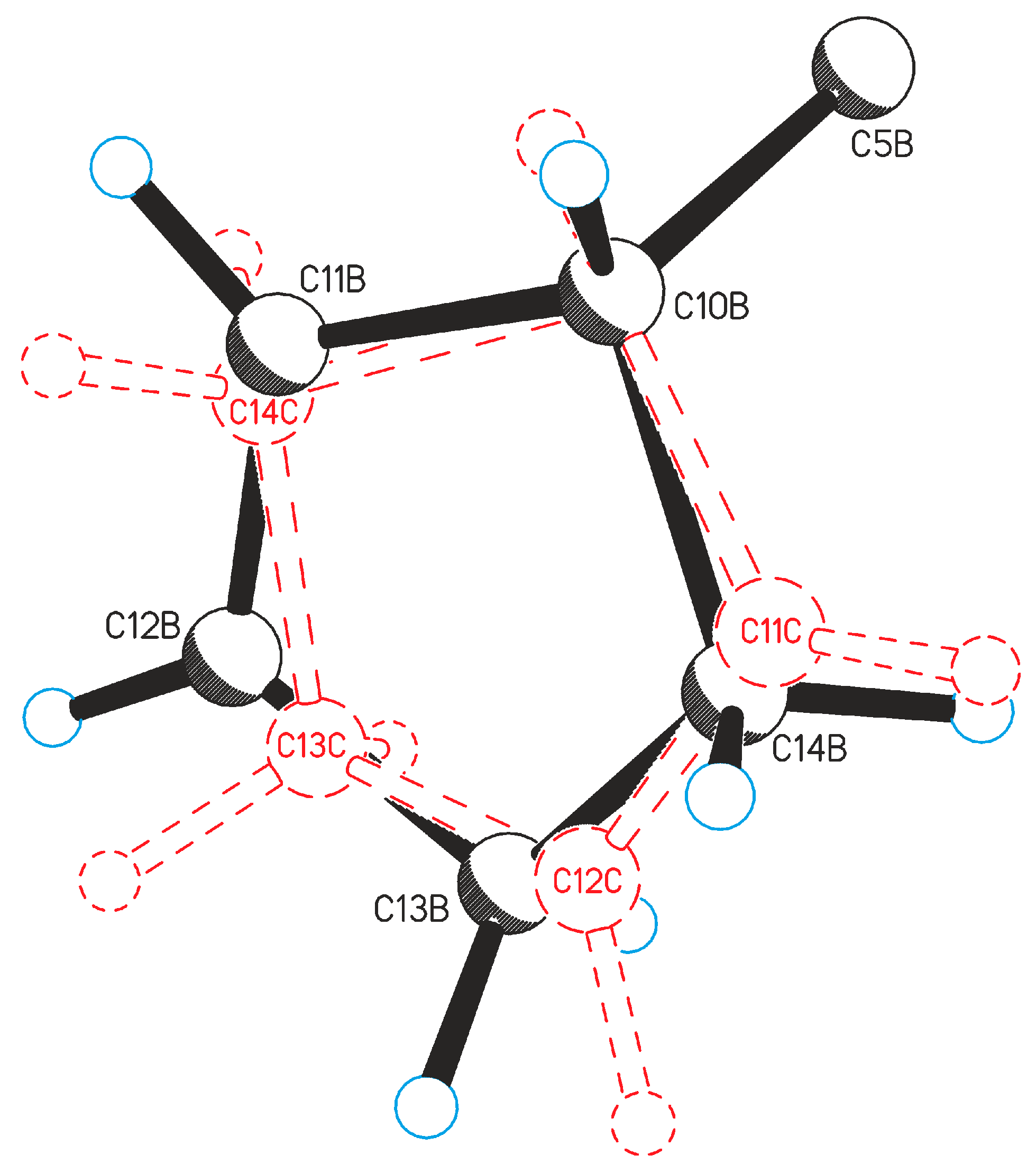
In the molecule of (II), the central six-membered ring is nearly planar (rmsd 0.013 Å). The value of the torsion angle C8B—C7B—C5B—C10B (defined as ψ in Scheme 1) is 179.9(4)°, indicating that the allyl and cyclopentenyl substituents bonded to C5B adopt a trans conformation along the C7B—C5B bond. The cyclopentenyl was found to be disordered over two orientations, with the two disorder components (occupancy ratio 0.65:0.35) being related by a rotation of approximately 180° about the C5B—C10B bond to the pyrimidine ring (Figure 2). Although the existence of four polymorphic forms of cyclopentobarbital (II) has been reported [20], the crystal structure of only one of these has been determined so far [21]. Its two independent molecules differ fundamentally in terms of the torsion angle ψ (Scheme 1). One molecule displays the same trans geometry of the ring substituents (ψ = 169.1°) as the cocrystal (I)·(II), whilst the corresponding arrangement in the second molecule is gauche (ψ = −44.0°). In addition, the cyclopentenyl group of the first molecule shows a disorder similar to that found in (I)·(II).
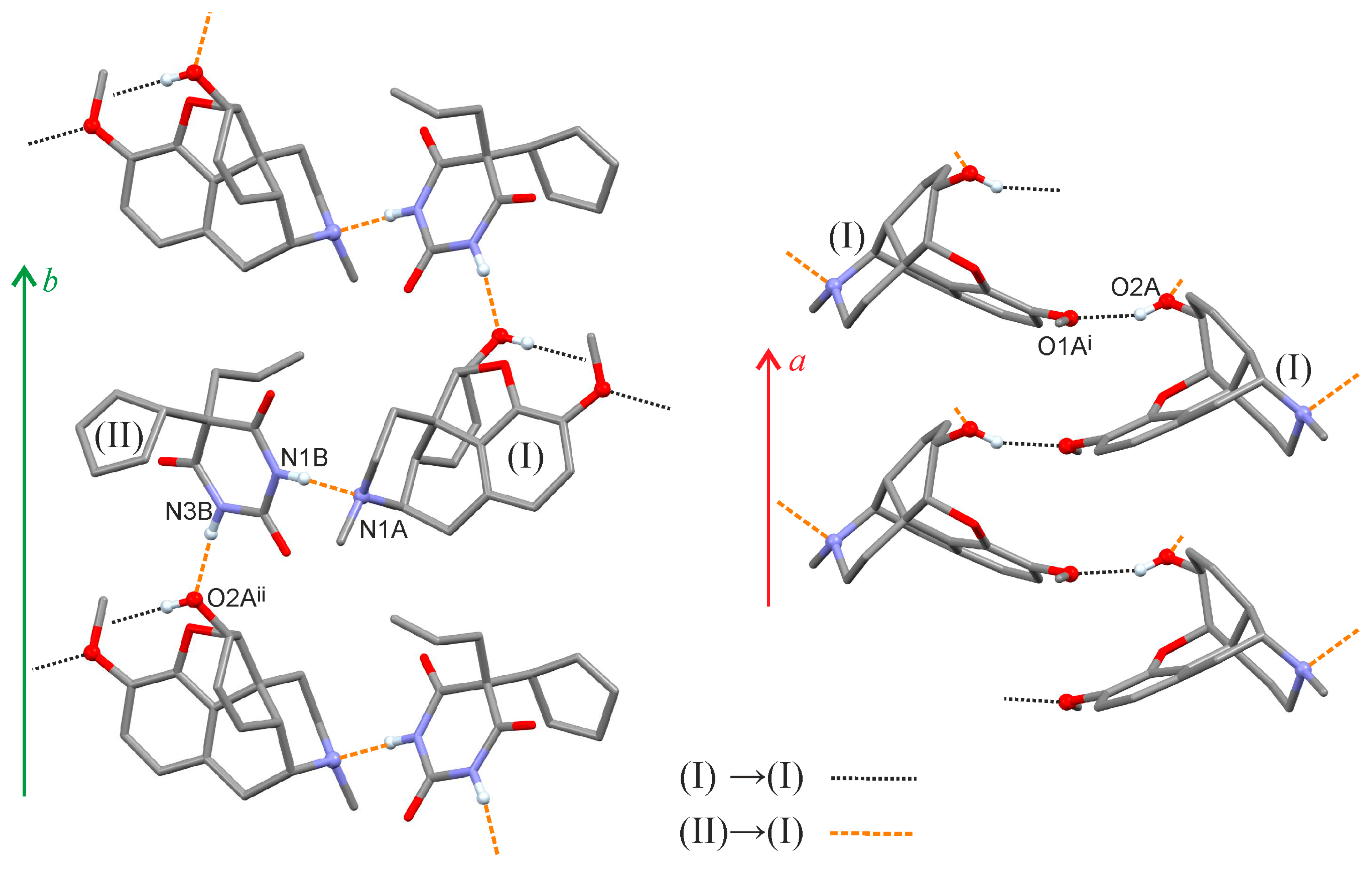
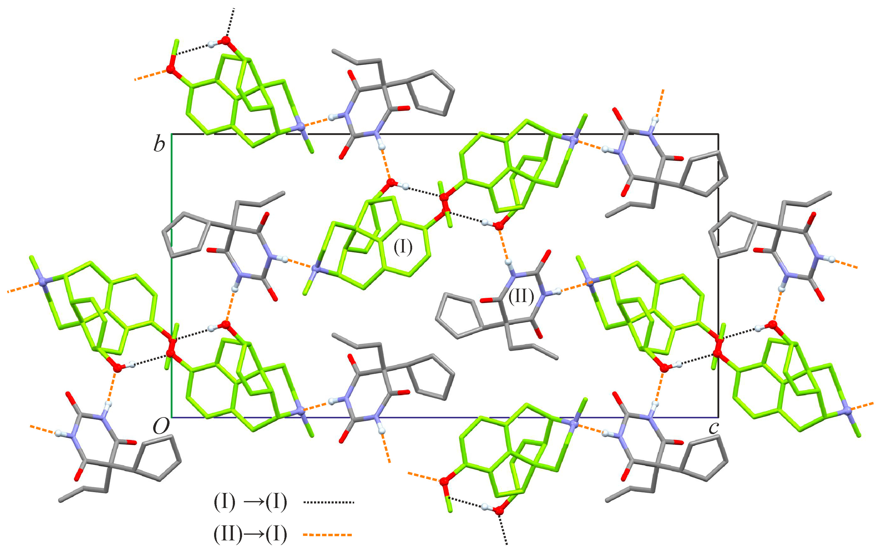
The central six-membered ring of (II) bears two NH groups, which form N1B—H1B⋯N1A and N3B—H3B⋯O2A(−x, y − 1/2, −z + 1/2) bonds to the piperidine and hydroxyl groups belonging to two different codeine molecules (Table 2). These two interactions result in a H-bonded chain of alternating cyclopentobarbital and codeine molecules along the crystallographic b axis (Figure 3, left). Additionally, the OH group of each codeine molecule forms an O2A—H2A⋯O1A(x − 1/2, −y + 1/2, −z) interaction with the methoxy-O atom of a second codeine molecule related to the former by a two-fold screw operation along the a axis (Figure 3, right). Altogether, a H-bonded framework structure is formed (Figure 4). This framework can also be considered a topological net with H-bonds as the vertices and the molecules of (I) and (II) serving as four- and two-connected nodes, respectively. In this context, single-point connections link each node of (I) to two neighboring nodes of (I), as well as two neighboring nodes of (II), whereas no connections exist between any two nodes of (II).

2.2. Hot-Stage Microscopy

On heating prismatic single crystals of (I)·(II), sublimation occurs at approximately 120 °C. Condensation droplets form at 128 °C, and the melting of the cocrystal ensues at 139 °C (Figure S2b of the Supporting Information). The melting equilibrium can be adjusted at 142 °C. Seeds of the cocrystal in the melt were appear between 100 and 110 °C, with a maximum growth rate of roughly 140 µm min−1 (Figure S3 of the Supporting Information).
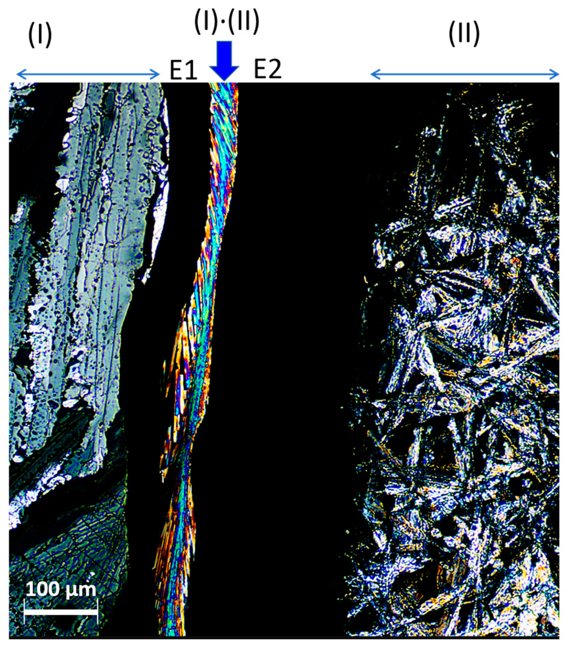
The investigation of contact preparations (see Section 3.3) of the two components (I) and (II) confirmed the formation of the cocrystal from the melt. The eutectic temperatures of the cocrystal (I)·(II) with its two mother compounds were also determined. On heating, the eutectic E2 between II (polymorph I [20]) and the cocrystal occurs as a black strip under polarized light, at 115 °C. The eutectic E1 between (I) and the cocrystal occurs at 128 °C. Cyclopentobarbital (II) crystallizes as form I from the melt and this new phase melts completely at 133 °C. The cocrystal (I)·(II), appearing as a birefringent ribbon between the two (eutectic) liquids (Figure 5), melts between 135 and 139 °C, leaving codeine as the only crystalline phase (Figure S4 of the Supporting Information).

2.3. Differential Scanning Calorimetry (DSC)

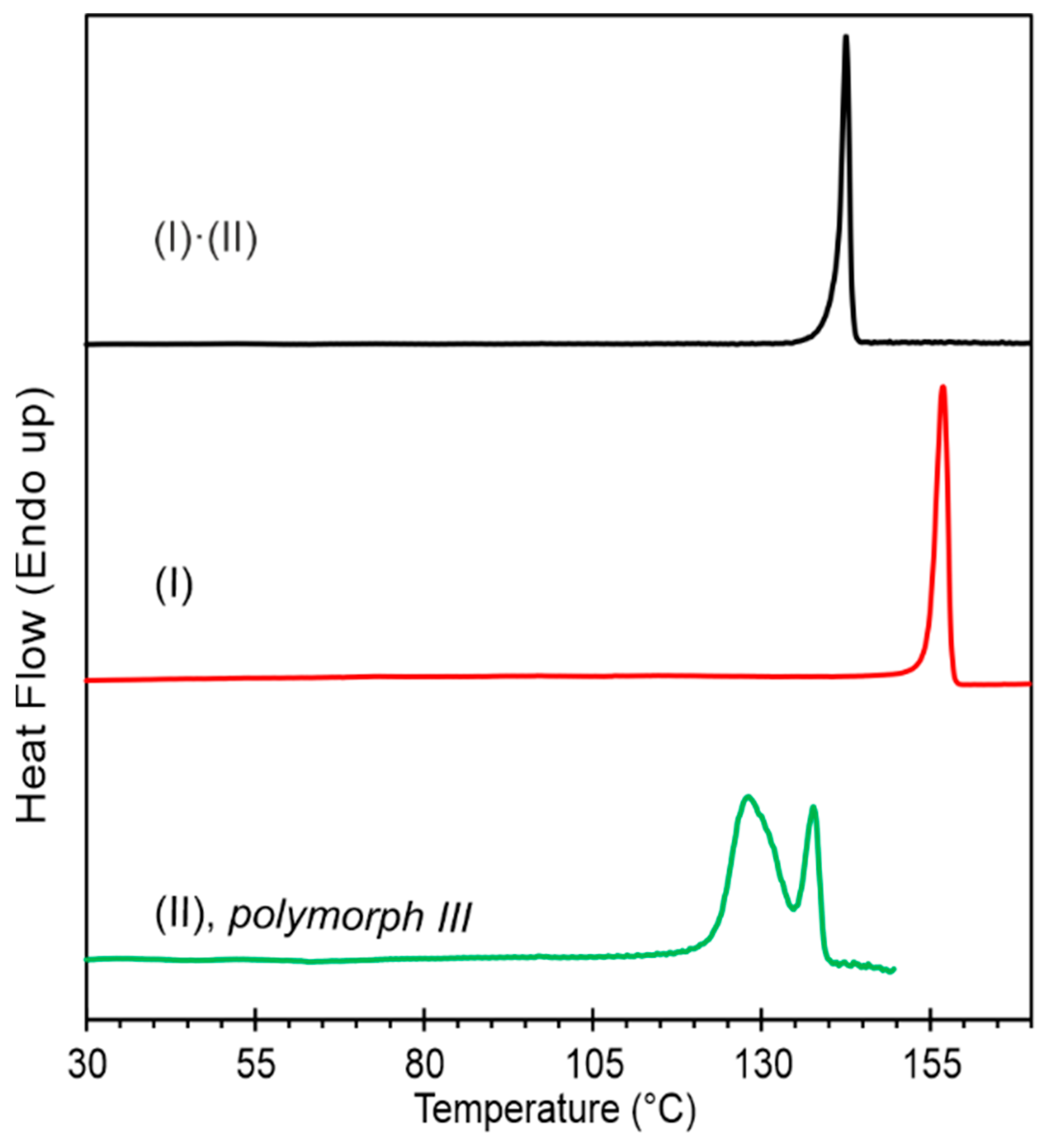
The cocrystal showed one sharp melting endotherm at 141.2 °C, with a fusion enthalpy of 47.6 kJ mol−1 (Figure 6). No recrystallization of the cocrystal occurred on the cooling of the melt. Thermoanalytical data of the cocrystal (I)·(II) are collected in Table 3.
The DSC trace for codeine anhydrate (I) showed melting at 156.6 °C (Tonset) [17]. Polymorph III of cyclopentobarbital (II) displayed incongruent melting behavior. The melting endotherm of polymorph III at 123.4 °C was followed by the crystallization and subsequent melting of polymorph I. Polymorph I of (II) melts at 135.2 °C (Tonset) [20].

3. Materials and Methods

3.1. Preparation of the Cocrystal (I)·(II)

Codeine (I). First, 20 g of codeine phosphate hemihydrate (Siegfried, Zofingen, Switzerland) was dissolved in 21.5 g of water and 100 ml of NaOH was added to the resulting solution. Drying of the precipitation product for 30 min at 130 °C yielded the codeine-free base (I) (yield 92.3%).
Cyclopentobarbital (II) was obtained from Alltech, State College, PA, USA.
Cocrystal (I)·(II). The two components were mixed in a 1:1 stoichiometric ratio and dissolved in either EtOH or 2-PrOH. A beige powder or a yellow sticky mass was obtained after the evaporation of the solvent at ambient conditions. The treatment of the sticky mass, with a few added drops of an antisolvent (n-heptane or n-hexane), in a mortar with a pestle yielded the cocrystal (I)·(II). Colourless prismatic crystals of (I)·(II) suitable for a single crystal structure determination were obtained via slow evaporation from a 2-PrOH solution containing equimolar amounts of (I) and (II). The cocrystal product was characterized by powder X-ray diffraction, FTIR spectroscopy, hot-stage microscopy (see Figures S1–S5 of the Supporting Information) and differential scanning calorimetry.

3.2. Single-Crystal Structure Determination

Intensity data were collected at 173 K, using Cu radiation (λ = 1.54184 Å), on an Oxford Diffraction Gemini-R Ultra diffractometer. The data were corrected for absorption effects by means of a comparison of equivalent reflections. The crystal structure was solved using Direct Methods with SHELXT [22] and refined with least-squares techniques using SHELXL [23]. H atoms were identified in difference-Fourier maps and those bonded to carbon atoms were refined using a riding model with Uiso parameters set to 1.2Ueq of the parent C atom (1.5Ueq for the methyl groups C17 and C18). The H positions in NH and OH groups were refined with distance restraints, N—H = 0.88(1) Å and N—O = 0.84(1) Å, and their Uiso(H) parameters were refined freely. The two disorder components of the cyclopentenyl ring (i.e., C11B > C14B/C11C > C14C) were refined using restraints on chemically equivalent 1,2- and 1,3-distances. The bond distances of the pair C14B—C15B and C14C—C15C were restrained to 1.55(1) Å, whilst those of the pair C11B—C12B and C11B—C12B were restrained to 1.32(1) Å. Soft restraints (SIMU) were applied on the anisotropic displacement parameters of the atoms in the two disorder components. The absolute structure was assigned with reference to the known absolute structure of the codeine molecule [absolute structure (Flack) parameter −0.3(3)]. CCDC 2278182 contains the supplementary crystallographic data for this paper. These data can be obtained free of charge from the Cambridge Crystallographic Data Centre via www.ccdc.cam.ac.uk/structures.

3.3. Hot Stage Microscopy and Contact Preparation Method

Hot stage microscopy investigations were performed with a Reichert Thermovar® polarization microscope equipped with a Kofler hot stage (both Reichert, Vienna, Austria). The temperature was measured with a digital thermometer (Fluke 51 II) and the temperature calibration was performed with WHO melting point standards.
The contact preparation allows the quick identification of whether two meltable compounds form either a eutectic, a cocrystal (molecular compound) or a solid solution. The preparation was performed according to A. Kofler and L. Kofler [24,25], starting by melting a small amount of the higher melting component between a glass slide and a cover slip using a Kofler hot-bench (Reichert, Vienna, Austria). The sample should be allowed to crystallize on cooling down and should fill the gap between the slide and cover slip only partially. The lower-melting component was then placed on the opposite side of the preparation (empty space between glass slides) and was also melted on a hot bench until its melt came into contact with the higher melting component, resulting in a mixture of the two components within the contact zone. The second component should crystallize on cooling together with the cocrystal in the contact zone. To induce the crystallization of the cocrystal (I)·(II), the contact zone was seeded with seeds of (I)·(II) harvested from the edges of the cover slip.

Supplementary Materials

Figure S1: Powder X-ray diffractograms; Figure S2: Photomicrographs of the cocrystal (I)·(II); Figure S3: Microscopic film preparation showing the growth of the cocrystal (I)·(II) in the melt; Figure S4: Polarized-light photomicrographs of a contact preparation; Figure S5: FTIR spectra; Table S1: Torsion angle τ (°) and the angle formed by the mean planes defined by the rings A/B/C and D/E (°) in solid forms of codeine.

Author Contributions

Conceptualization, U.J.G.; formal analysis, J.S., T.G. and U.J.G.; investigation, J.S. and T.G.; writing—original draft preparation, T.G.; writing—review and editing, T.G., J.S. and U.J.G. All authors have read and agreed to the published version of the manuscript.

Funding

This research received no external funding.

Data Availability Statement

CCDC 2278182 contains the supplementary crystallographic data for this paper. These data can be obtained free of charge from the Cambridge Crystallographic Data Centre via www.ccdc.cam.ac.uk/structures.

Acknowledgments

We thank Judith Ulmer for assistance with crystallization experiments.

Conflicts of Interest

The authors declare no conflict of interest.

References

  1. Wisniak, J. Pierre-Jean Robiquet. Educ. Quim. 2013, 24, 139–149. [Google Scholar] [CrossRef]
  2. World Health Organization. World Health Organization Model List of Essential Medicines: 22nd List (2021); World Health Organization: Geneva, Switzerland, 2021. [Google Scholar]
  3. Eddy, N.B.; Friebel, H.; Hahn, K.-J.; Halbach, H. Codeine and its alternates for pain and cough relief: 1. Codeine, exclusive of its antitussive action. Bull. World Health Organ. 1968, 38, 673–741. [Google Scholar] [CrossRef] [PubMed]
  4. Eddy, N.B.; Friebel, H.; Hahn, K.-J.; Halbach, H. Codeine and its alternates for pain and cough relief: 3. The antitussive action of codeine—Mechanism, methodology and evaluation. Bull. World Health Organ. 1969, 40, 425–454. [Google Scholar] [PubMed]
  5. Atkinson, A.J.; Adler, H.F.; Ivy, A.C. Motility of the human colon: The normal pattern, dyskinesia and the effekt of drugs. J. Am. Med. Soc. 1943, 121, 646–652. [Google Scholar] [CrossRef]
  6. International Narcotics Control Board. List of Narcotic Drugs under International Control: Yellow List; International Narcotics Control Board: Vienna, Austria, 2020. [Google Scholar]
  7. United States Food and Drug Administration; Center for Drug Evaluation and Research. Guidance for Industry: Regulatory Classification of Pharmaceutical Co-Crystals. 2018 Revision 1. Available online: https://www.fda.gov/files/drugs/published/Regulatory-Classification-of-Pharmaceutical-Co-Crystals.pdf (accessed on 20 July 2023).
  8. Aitipamula, S.; Banerjee, R.; Bansal, A.K.; Biradha, K.; Cheney, M.L.; Choudhury, A.R.; Choudhury, A.R.; Desiraju, G.R.; Dikundwar, A.G.; Dubey, R.; et al. Polymorphs, Salts, and Cocrystals: What’s in a Name? Cryst. Growth Des. 2012, 12, 2147–2152. [Google Scholar] [CrossRef]
  9. Zhang, C.; Xiong, Y.; Jiao, F.; Wang, M.; Li, H. Redefining the Term of “Cocrystal” and Broadening Its Intention. Cryst. Growth Des. 2019, 19, 1471–1478. [Google Scholar] [CrossRef]
  10. Centolella, A.P.; Nelson, J.W.; Kolloff, H.G. Barbiturates Containing the Δ2-Cyclopentenyl Group. J. Am. Chem. Soc. 1943, 65, 2091–2092. [Google Scholar] [CrossRef]
  11. Vander Brook, M.J.; Cartland, G.F. A Pharmacologic Study of 5-Allyl-5-Δ2-cyclopentenyl Barbituric Acid (cyclopal). J. Pharmacol. Exp. Ther. 1944, 80, 119–125. [Google Scholar]
  12. Langes, C.; Gelbrich, T.; Griesser, U.J.; Kahlenberg, V. Codeine dihydrogen phosphate hemihydrate. Acta Crystallogr. C 2009, 65, o419–o422. [Google Scholar] [CrossRef] [PubMed]
  13. Groom, C.R.; Bruno, I.J.; Lightfoot, M.P.; Ward, S.C. The Cambridge Structural Database. Acta Crystallogr. B 2016, 72, 171–179. [Google Scholar] [CrossRef]
  14. Arzeno, H.B.; Barton, D.H.R.; Davies, S.G.; Lusinchi, X.; Meunier, B.; Pascard, C. Synthesis of 10(S)-methylcodeine and 10(S)-methylmorphine. Nouv. J. Chim. 1980, 4, 369–375. [Google Scholar]
  15. Kartha, G.; Ahmed, F.R.; Barnes, W.H. Refinement of the crystal structure of codeine hydrobromide dihydrate, and establishment of the absolute configuration of the codeine molecule. Acta Crystallogr. 1962, 15, 326–333. [Google Scholar] [CrossRef]
  16. Rosenberger, L.; von Essen, C.; Khutia, A.; Kühn, C.; Georgi, K.; Hirsch, A.K.H.; Hartmann, R.W.; Badolo, L. Crystalline sponge affinity screening: A fast tool for soaking condition optimization without the need of X-ray diffraction analysis. Eur. J. Pharm. Sci. 2021, 164, 105884. [Google Scholar] [CrossRef] [PubMed]
  17. Braun, D.E.; Gelbrich, T.; Kahlenberg, V.; Griesser, U.J. Insights into Hydrate Formation and Stability of Morphinanes from a Combination of Experimental and Computational Approaches. Mol. Pharm. 2014, 11, 3145–3163. [Google Scholar] [CrossRef] [PubMed]
  18. Runčevski, T.; Petruševski, G.; Makreski, P.; Ugarkovic, S.; Dinnebier, R.E. On the hydrates of codeine phosphate: The remarkable influence of hydrogen bonding on the crystal size. Chem. Commun. 2014, 50, 6970–6972. [Google Scholar] [CrossRef]
  19. Scheins, S.; Messerschmidt, M.; Morgenroth, W.; Paulmann, C.; Luger, P. Electron Density Analyses of Opioids:  A Comparative Study. J. Phys. Chem. A 2007, 111, 5499–5508. [Google Scholar] [CrossRef] [PubMed]
  20. Brandstätter-Kuhnert, M.; Aepkers, M. Molekülverbindungen, Mischkristallbildung und neue Polymorphiefälle bei Barbituraten. Microchim. Acta 1962, 50, 1041–1054. [Google Scholar] [CrossRef]
  21. Zencirci, N.; Gelbrich, T.; Kahlenberg, V.; Griesser, U.J. Crystallization of metastable polymorphs of phenobarbital by isomorphic seeding. Cryst. Growth Des. 2009, 9, 3444–3456. [Google Scholar] [CrossRef]
  22. Sheldrick, G.M. SHELXT—Integrated space-group and crystal-structure determination. Acta Crystallogr. A 2015, 71, 3–8. [Google Scholar] [CrossRef]
  23. Sheldrick, G.M. Crystal structure refinement with SHELXL. Acta Crystallogr. C 2015, 71, 3–8. [Google Scholar] [CrossRef]
  24. Kofler, A. Mikro-Thermoanalyse organischer Zweistoffsysteme. Naturwissenschaften 1943, 31, 553–557. [Google Scholar] [CrossRef]
  25. Kofler, L.; Kofler, A. Thermo-Mikro-Methoden zur Kennzeichnung organischer Stoffe und Stoffgemische; Wagner: Innsbruck, Austria, 1954. [Google Scholar]
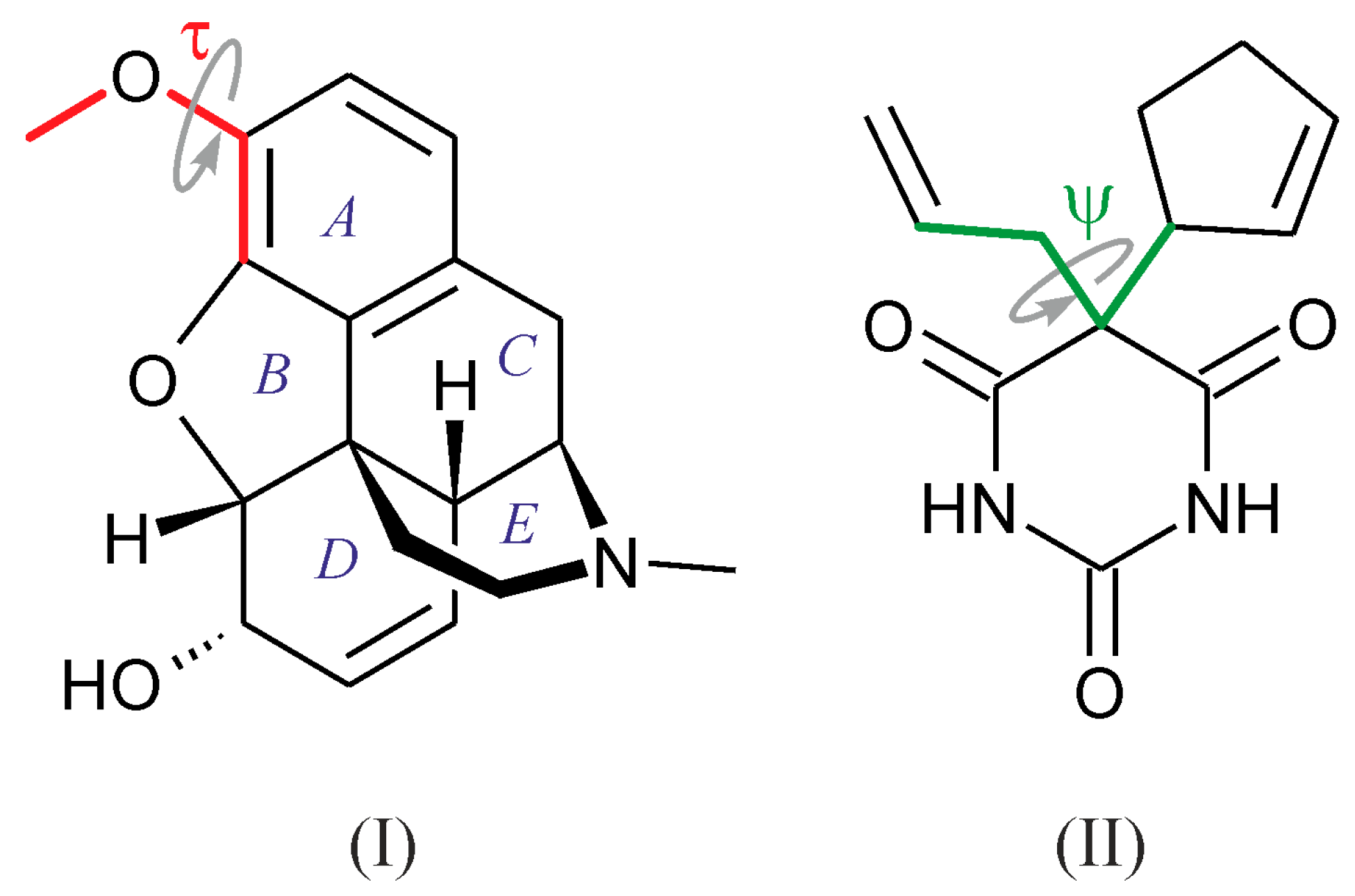
Scheme 1. Structural formulas of codeine (I) and cyclopentobarbital (II); definition of the torsion angle τ and rings AE for (I) and the torsion angle ψ for (II).
Molbank 2023 m1722 sch001
Figure 1. Asymmetric unit of (I)·(II) with non-H atoms depicted as ellipsoids at the 50% probability level and H atoms drawn as spheres of random size (minor disorder omitted for clarity).
Molbank 2023 m1722 g001
Figure 2. Disorder of the cyclopentenyl ring involving two components, (C10B > C14B; solid lines) and (C10B, C11C > C14C; dashed lines), related by a rotation about the bond C5B—C10B to the pyrimidine ring.
Molbank 2023 m1722 g002
Figure 3. Two fragments of the H-bonded framework of (I)·(II). (Left): N1B—H1B⋯N1A and N3B—H3B⋯O2Aii bonds resulting in a chain of alternating cyclopentobarbital and codeine molecules which propagates parallel to the b axis. (Right): O2A—H2A⋯O1Ai bonded chain formed exclusively via codeine molecules and extending along the a axis (H atoms not engaged in H-bond interactions omitted for clarity). Symmetry codes: (i) x − 1/2, −y + 1/2, −z; (ii) −x, y − 1/2, −z + 1/2.
Molbank 2023 m1722 g003
Figure 4. Framework of H-bonded codeine (I) and cyclopentobarbital (II) molecules, viewed along the crystallographic a axis [molecules of (I) highlighted green; H atoms not engaged in H-bond interactions omitted for clarity].
Molbank 2023 m1722 g004
Figure 5. Polarized-light photomicrograph of a contact preparation of codeine (I) with cyclopentobarbital (II; polymorph I). At the center, the contact zone consisted of the central cocrystal phase (I)·(II) (arrow) and two surrounding eutectics (E1, E2).
Molbank 2023 m1722 g005
Figure 6. DSC traces of the cocrystal (I)·(II), codeine anhydrate (I) and cyclopentobarbital (II) (polymorph III); heating rate: 10 K min−1.
Molbank 2023 m1722 g006
Table 1. Crystal data and structure refinement.
Compound(I)·(II)
Moiety formulaC18H21NO3 · C12H14N2O3
Empirical formulaC30H35N3O6
Formula weight533.61
Temperature (K)173(2)
Wavelength (Å)1.5418
Crystal systemOrthorhombic
Space groupP212121
a (Å)6.9914(3)
b (Å)14.1455(7)
c (Å)27.1767(11)
Unit cell volume (Å3)2687.7(2)
Z/Z’4/1
Reflections collected/Rint9191/0.0447
Data/restraints/parameters4466/133/404
Goodness-of-fit on F21.027
R1 [I > 2 σ(I)]0.0500
wR2 (all data)0.1289
Largest diff. peak and hole (e · Å−3)0.197 and −0.189
CCDC no.2278182
Table 2. Hydrogen bonds (Å and °).
D—H⋯AdD—HdH⋯AdD⋯A<(DHA)
O2A—H2A⋯O1Ai0.838(14)1.99(3)2.784(4)157(6)
N1B—H1B⋯N1A0.886(13)1.974(15)2.859(5)176(4)
N3B—H3B⋯O2Aii0.881(13)1.945(18)2.811(5)167(5)
Symmetry codes: (i) x − 1/2, −y + 1/2, −z; (ii) −x, y − 1/2, −z + 1/2.
Table 3. Thermoanalytical data of the cocrystal (I)·(II).
ParameterValue a
Tfus (°C) b
   DSC (onset)141.1 ± 0.2
   HSM (melting equilibrium)142
ΔfusH (kJ mol−1) c47.6 ± 0.1
ΔfusS (J mol−1 K−1) d115.0 ± 0.3
a Errors calculated as 95% confidence interval. b Melting point. c Enthalpy of fusion. d Entropy of fusion.
Disclaimer/Publisher’s Note: The statements, opinions and data contained in all publications are solely those of the individual author(s) and contributor(s) and not of MDPI and/or the editor(s). MDPI and/or the editor(s) disclaim responsibility for any injury to people or property resulting from any ideas, methods, instructions or products referred to in the content.