Short Note

3-Morpholino-7-[N-methyl-N-(4′-carboxyphenyl)amino]phenothiazinium Chloride

Department of Chemical Science and Technologies, University of Rome Tor Vergata, Via della Ricerca, Scientifica snc, 00133 Rome, Italy
*
Author to whom correspondence should be addressed.
Molbank 2022, 2022(4), M1493; https://doi.org/10.3390/M1493
Submission received: 13 September 2022 / Revised: 7 November 2022 / Accepted: 8 November 2022 / Published: 14 November 2022

Abstract

:
The synthesis of 3-morpholino-7-[N-methyl-N-(4′-carboxyphenyl)amino]phenothiazinium chloride is reported here. Interestingly, non-symmetric phenothiazinium salt is functionalized with a carboxylic acid group that allows the easy and stable anchoring on metal oxides. In addition, the morpholine unit reduces the dye aggregation tendency; thus, improving its potential applications in the biomedical and photo-electrocatalytic field.

1. Introduction

Since their introduction into medicine, phenothiazines have been widely used as antibacterial, antifungal and insecticidal agents [1], as well as active compounds in different optoelectronic applications [2]. Phenothiazinium salts are heteroaromatic compounds coming from the oxidation of phenothiazine, that are commonly used as sensitizers in photovoltaic devices [3,4], redox mediators in catalytic reactions [5], intercalating agents [6] and photoactive species in photodynamic therapy [7].
As for other polycyclic-conjugated dyes [8,9,10,11], the fascinating properties of phenothiazinium salts are strictly related to the fully delocalized system, that confers distinctive electronic and electrochemical properties.
Recently, several phenothiazinium salts, having different substituents in 3- and 7- positions, have been synthetized to modulate their electronic properties and to reduce their aggregation tendency [12]. In fact, dye aggregation is known to limit phenothiazinium application in the energy and biomedical field. Accordingly, the synthesis of novel phenothiazinium salts functionalized with auxochrome units with hydrophilic groups led to appropriate molecules for internalization and staining human ovarian cancer cells [13]; similarly, phenothiazinium salts functionalization with (trimethoxysilyl)alkyl amino group(s) in position 3- and 7- resulted in being strategic to anchor dyes onto surfaces or nanoparticles, thus improving their efficacy for biomedical and antibacterial applications [14]. Additionally, complexes formed by phenothiazinium Schiff base ligands and Ag nanoparticles showed excellent antibacterial activity against E. coli and S. aureus strains [15].
In this short note, the synthesis of a new non-symmetric phenothiazinium salt (i.e., 3-morpholino-7-[N-methyl-N-(4′-carboxyphenyl)amino]phenothiazinium chloride, 3) is presented. The introduction of a carboxylic acid anchoring group allows for the easy and stable dye anchoring on several metal oxides (as TiO2 [16], SnO2 [17], NiO [18]), thus improving their application in photoelectrochemical devices, used in the biomedical field. In addition, the morpholine unit reduces the aggregation tendency of the dye, by limiting π–π stacking interactions.

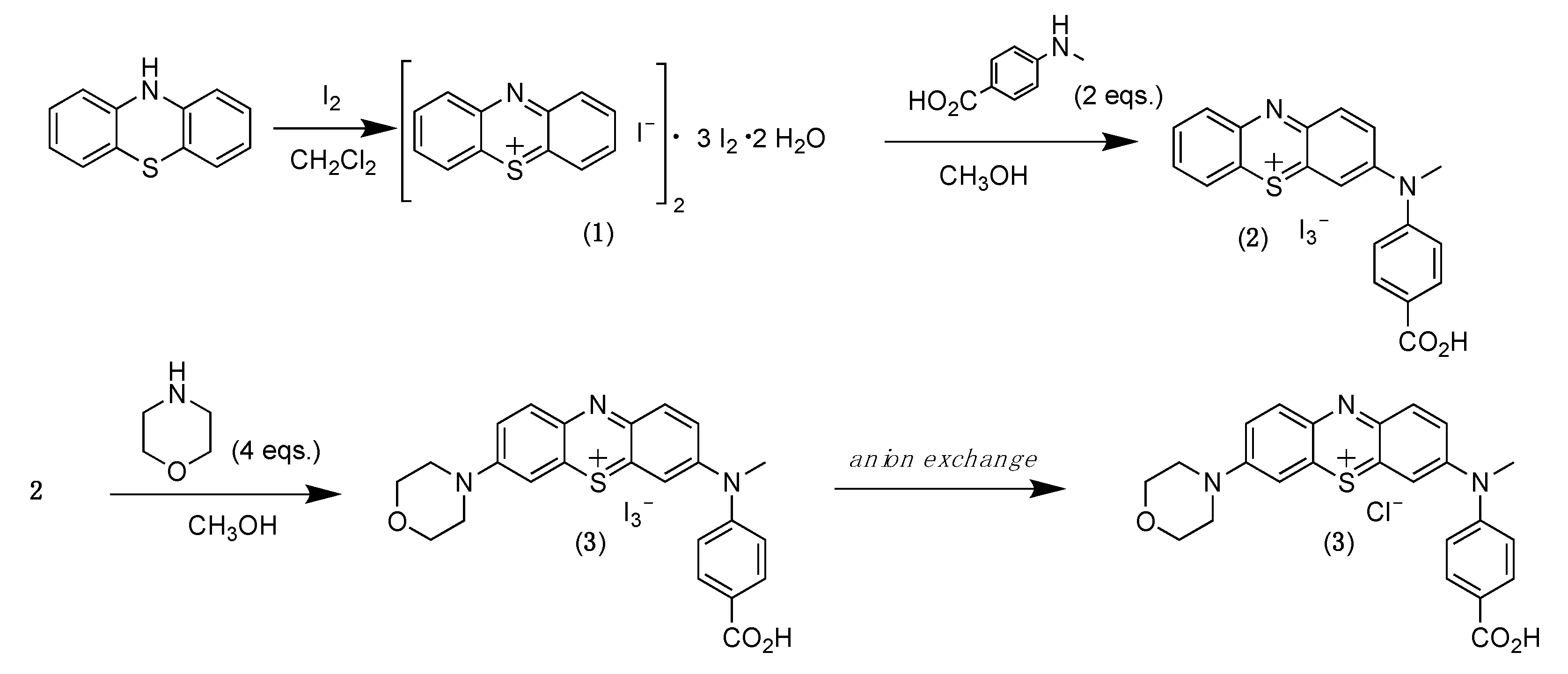
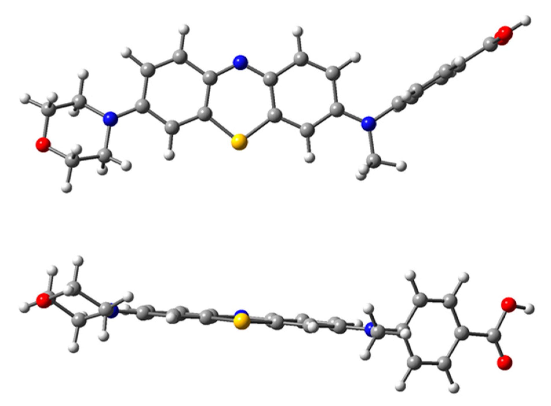
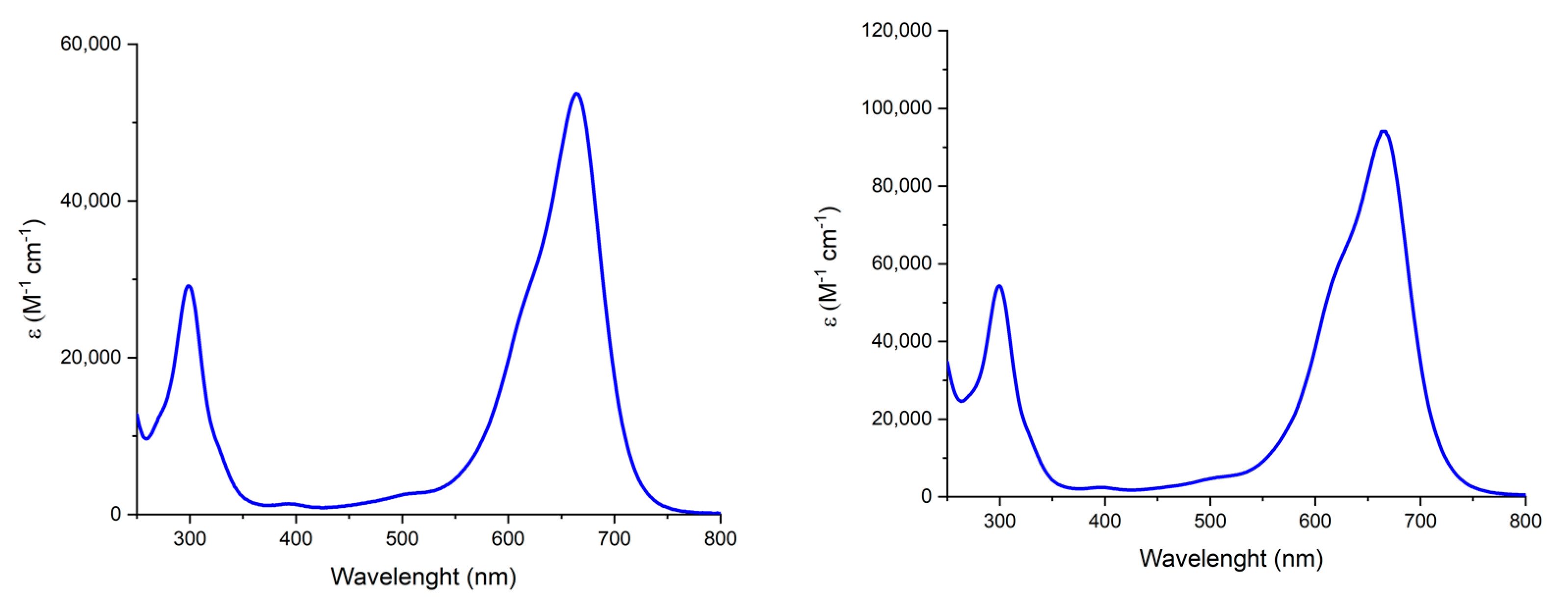
2. Results and Discussion

The Strekowski’s procedure for synthesis of new phenothiazinium salts is a versatile approach that can provide a wide series of phenothiazinium derivatives with different functionalities [19].
The synthetic strategy (Scheme 1) involves the oxidation of phenothiazine by molecular iodine, to obtain a product which structure consists of two phenothiazinium cations, two iodide counter ions, three molecules of iodine and two molecules of water. Such compound is—for simplicity—called phenothiazinium tetraiodide hydrate (1). The compound 1 can undergo nucleophilic addition by amines in position 3- and 7-. Interestingly, 3-monosubstituted phenothiazinium salts can be functionalized in position 7- with a different amine, to afford non-symmetrically substituted phenothiazinium derivatives.
The synthesis of the title compound (3) was performed reacting 1 with 2 equivalents of 4-(methylamino)benzoic acid [19], to obtain the mono-substituted phenothiazinium salt 2. To note, to afford the 3,7-disubstituted derivative (i.e., 3,7-bis(methyl-(4-carboxy)phenylamino)phenothiazinium iodide), four equivalents of the proper amine (4-(methylamino)benzoic acid) would be needed.
Compound 2 was fully characterized by 1H NMR spectroscopy in DMSO-d6; the singlet at 3.91 ppm and the signals in the aromatic region clearly confirm the mono-functionalization of the reagent.
Compound 3 was then obtained after reaction of 2 with 4 equivalents of morpholine as secondary amines [19]. The structure was confirmed by spectroscopic analyses. 1H NMR spectrum in DMSO-d6 shows morpholine signals in the region between 4 and 3.5 ppm.
DFT calculations performed with WB97XD functional and 6-31G+(d,p) basis set [12] indicated that the most stable conformation of 3 shows the phenyl ring arranged in equatorial-like conformation, while the methyl group is perpendicularly located with respect to the phenothiazinium core (Figure 1). The conformation with the phenyl ring perpendicularly accommodated to the polycyclic ring (Figure S4, Supplementary Materials) resulted 0.3 kcal⋅mol−1 higher in energy. The morpholine unit, in chair conformation, contributes to limit the aggregation tendency of the dye, by limiting π–π stacking interactions [12]. As a matter of fact, the UV-vis absorption spectra of a 2 × 10⁻⁵ M solution of 3 in H2O and in 1M NaCl solution (Figure 2) are characterized by a broad and intense band between 500 and 800 nm, which is typical of the monomeric form of phenothiazinium salts [12]. On the contrary, aggregation was previously observed for other phenothiazinium salts, as methylene blue, in the same experimental conditions [12].
In conclusion, the synthesis of a new non-symmetric phenothiazinium salt having a carboxylic acid anchoring group and a morpholine unit has been performed. Thanks to the low aggregation propensity of the dye, it may exhibit improved biomedical and photoelectrochemical properties.

3. Materials and Methods

All commercial reagents and solvents were purchased from Sigma Aldrich/Merck Life Science (KGaA, Darmstadt, Germany), with the highest degree of purity. Phenothiazine was crystallized from toluene prior to use. The absorption spectra were recorded with a UV/Vis 2450 Shimadzu spectrophotometer (Kyoto, Japan). 1H NMR experiments were carried out using a Bruker Avance (600.13 MHz, Bruker, Billerica, MA, USA) and all data were processed with TopSpin software (Bruker, Billerica, MA, USA). MS-ESI analyses have been performed with a LC-MSD-trap-SL ESI + FI. DFT calculations have been performed with Gaussian 16 rev. A03 [20].
Synthesis of phenothiazinium triiodide hydrate (1) [19]. A solution of iodine (3.82 g, 15.06 mmol) in dichloromethane (75 mL) was added dropwise, within 1 h, to a 25 mL solution of phenothiazine (1 g, 5.02 mmol) dissolved in dichloromethane. The reaction mixture was stirred at room temperature, for 3 h, and the resulting precipitate was collected by filtration and washed with 200 mL of dichloromethane. The black-blue powder was dried under vacuum for 3 h, to give 1 in quantitative yield (3.58 g, 4.95 mmol). Each structural unit consists of two phenothiazinium cations, two iodide counter ions, three molecules of iodine and two molecules of water.
Synthesis of 3-[N-methyl-N-(4-carboxyphenyl)amino]phenothiazinium triiodide (2) [19]. Phenothiazinium tetraiodide hydrate (0.5 g, 0.69 mmol) was dissolved in 10 mL of methanol. A solution of 4-(methylamino)benzoic acid (210 mg, 1.39 mmol) in methanol (2 mL) was added dropwise and the reaction mixture was stirred at room temperature until PTZ+I4 was consumed. The reaction mixture was monitored by TLC on silica gel (eluent: 3% aqueous NH4OAc/CH3OH 1:17 v/v). After 17 h, the resulting product was collected by filtration and washed with diethyl ether. A total of 202 mg of the product were obtained (0.28 mmol, 40% yield).
1H NMR in DMSO-d6: δ 8.39–8.33 (dd, J1 = 7.80 Hz, J2 = 1.66 Hz, 1H), δ 8.25–8.18 (d, J = 8.59 Hz, 2H), δ 8.03–7.91 (m, 2H), δ 7.77–7.70 (d, J = 8.56 Hz, 2H), δ 7.70–7.64 (d, J = 8.82 Hz, 2H), δ 6.57–6.49 (d, J = 8.85 Hz, 2H), δ 3.91 (s, 3H). Mass spectrum (ESI+) m/z calcd. 347.08; found 347.20. Anal. Calcd. for C20H15I3N2O2S: C, 32.99; H, 2.08; N, 3.85; Found: C, 32.84; H, 2.13; N, 4.13. UV-vis in CH3OH [λmax, nm (ε, M−1 cm−1)]: 584 (16,600); 437 (13,900); 303 (50,300).
Synthesis of 3-morpholino-7-[N-methyl-N-(4′-carboxyphenyl)amino]phenothiazinium chloride (3) [19]. Morpholine was dried over anhydrous Na2CO3 prior to use. A 0.28 M solution of morpholine in methanol, prepared by dissolving 240 mg of morpholine (2.79 mmol) in 10 mL of MeOH, was diluted with 25 mL of methanol containing 0.5 g (0.69 mmol) of 2. The reaction mixture was kept for 30 min under stirring, in the dark, at room temperature. The product was collected by filtration and washed with diethyl ether. The resulting blue powder (303 mg) was passed through a strongly basic anion exchange resin in order to substitute iodide with chloride (eluent CH3OH/H2O 1:1 v/v). The obtained product (140 mg, 0.27 mmol, 39% yield) was fully characterized.
1H NMR (700 MHz, DMSO-d6): δ 8.12 (d, J = 8.44 Hz, 2H), δ 8.03 (d, J = 9.40 Hz, 1H), δ 7.96 (d, J = 9.38 Hz, 1H), δ 7.86 (d, broad, J = 2.48 Hz, 1H), δ 7.84-7.80 (dd, J1 = 9.90 Hz, J2 = 2.48 Hz, 1H), δ 7.66 (d, J = 2.59 Hz, 1H), δ 7.58 (d, J = 8.45 Hz, 2H), δ 7.24-7.20 (dd, J1 = 9.46 Hz, J2 = 2.59, 1H), δ 3.97 (t, broad, 4H), δ 3.82 (t, broad, 4H), δ 3.65 (s, 3H). 13C NMR (700 MHz, DMSO-d6): δ 166.92, δ 154.02, δ 153,08, δ 148.47, δ 148.45, δ 139.13, δ 137.74, δ 137.60, δ 136.00, δ 135.94, δ 133.77, δ 131.85, δ 126.77, δ 121.10, δ 120.55, δ 108.75, δ 107.86, δ 66.43, δ 48.70, δ 41.99. Mass spectrum (ESI+) m/z calcd. 432.14; found 432. Anal. Calcd. for C24H22ClN3O3S: C, 55.22; H, 5.41; N, 8.05; S, 6.14; Found: C, 55.55; H, 5.46; N, 7.82; S, 5.92. UV-vis in H2O [λmax, nm (ε, M−1 cm−1)]: 663 (53,800); 298 (29,000).

Supplementary Materials

The following are available online, Figure S1: 1H NMR of 3 in DMSO-d6, Figure S2: 13C NMR of 3 in DMSO-d6, Figure S3: Geometry optimization of conformer α of compound 3 in the vacuum, Figure S4: Geometry optimization of conformer β of compound 3 in the vacuum.

Author Contributions

Conceptualization, P.G.; methodology, P.G., M.T. and V.C.; validation, P.G., F.S. and M.T.; investigation, M.T., P.G. and F.S.; resources, P.G.; data curation, M.T., F.S., F.V. and P.G.; writing—original draft preparation, M.T. and F.S.; writing—review and editing, F.S., F.V., P.G. and V.C.; supervision, P.G. and V.C.; project administration, P.G.; funding acquisition, P.G. All authors have read and agreed to the published version of the manuscript.

Funding

This research was funded by University of Rome Tor Vergata, grant HYPHOTOCAT project, “Beyond borders”.

Data Availability Statement

The data presented in this study are available on request from the corresponding author.

Conflicts of Interest

The authors declare no conflict of interest.

References

  1. Mitchell, S.C. Phenothiazine: The Parent Molecule. Curr. Drug Targets 2006, 61, 1181–1189. [Google Scholar] [CrossRef] [PubMed]
  2. Al-Busaidi, I.J.; Haque, A.; Al Rasbi, N.K.; Khan, M.S. Phenothiazine-based derivatives for optoelectronic applications: A review. Synth. Met. 2019, 257, 116189. [Google Scholar] [CrossRef]
  3. Kamat, P.V.; Lichtln, N.N. Electron Transfer in the Quenching of Protonated Triplet Methylene Blue by Ground-State Molecules of the Dye. J. Phys. Chem. 1981, 85, 814–818. [Google Scholar] [CrossRef]
  4. Lal, C. Use of mixed dyes in a photogalvanic cell for solar energy conversion and storage: EDTA–thionine–azur-B system. J. Power Sources 2007, 164, 926–930. [Google Scholar] [CrossRef]
  5. Ye, J.; Baldwin, R.P. Catalytic reduction of myoglobin and hemoglobin at chemically modified electrodes containing methylene blue. Anal. Chem. 1988, 60, 2263–2268. [Google Scholar] [CrossRef] [PubMed]
  6. Tuite, E.; Norden, B. Sequence-Specific Interactions of Methylene Blue with Polynucleotides and DNA: A Spectroscopic Study. J. Am. Chem. Soc. 1994, 116, 7548–7556. [Google Scholar] [CrossRef]
  7. Gorman, S.A.; Bell, A.L.; Griffiths, J.; Roberts, D.; Brown, S.B. The synthesis and properties of unsymmetrical 3,7-diaminophenothiazin-5-ium iodide salts: Potential photosensitisers for photodynamic therapy. Dyes Pigments 2006, 71, 153–160. [Google Scholar] [CrossRef]
  8. Shen, L.; Zhang, S.; Ding, H.; Niu, F.; Chu, Y.; Wu, W.; Hu, Y.; Hu, K.; Hua, J. Pure organic quinacridone dyes as dual sensitizers in tandem photoelectrochemical cells for unassisted total water splitting. Chem. Commun. 2021, 57, 5634–5637. [Google Scholar] [CrossRef] [PubMed]
  9. Sabuzi, F.; Lentini, S.; Sforza, F.; Pezzola, S.; Fratelli, S.; Bortolini, O.; Floris, B.; Conte, V.; Galloni, P. KuQuinones Equilibria Assessment for Biomedical Applications. J. Org. Chem. 2017, 82, 10129–10138. [Google Scholar] [CrossRef] [PubMed]
  10. Huang, R.; Phan, H.; Herng, T.S.; Hu, P.; Zeng, W.; Dong, S.-Q.; Das, S.; Shen, Y.; Ding, J.; Casanova, D.; et al. Higher Order π-Conjugated Polycyclic Hydrocarbons with Open-Shell Singlet Ground State: Nonazethrene versus Nonacene. J. Am. Chem. Soc. 2016, 138, 10323–10330. [Google Scholar] [CrossRef] [PubMed]
  11. Wu, H.; Wang, S.; Ding, J.; Wang, R.; Zhang, Y. Effect of π-conjugation on solid-state fluorescence in highly planar dyes bearing an intramolecular H-bond. Dye. Pigment. 2020, 182, 108665. [Google Scholar] [CrossRef]
  12. Tiravia, M.; Sabuzi, F.; Cirulli, M.; Pezzola, S.; Di Carmine, G.; Cicero, D.O.; Floris, B.; Conte, V.; Galloni, P. 3,7-Bis(N-methyl-N-phenylamino)phenothiazinium Salt: Improved Synthesis and Aggregation Behavior in Solution. Eur. J. Org. Chem. 2019, 2019, 3208–3216. [Google Scholar] [CrossRef]
  13. Stoean, B.; Gaina, L.; Cristea, C.; Silaghi-Dumitrescu, R.; Branzanic, A.M.V.; Focsan, M.; Fischer-Fodor, E.; Tigu, B.; Moldovan, C.; Cecan, A.D.; et al. New methylene blue analogues with N-piperidinyl-carbinol units: Synthesis, optical properties and in vitro internalization in human ovarian cancer cells. Dyes Pigments 2022, 205, 110460. [Google Scholar] [CrossRef]
  14. Kirla, H.; Henry, D.J. Synthesis and characterization of novel silane derivatives of phenothiazinium photosensitisers. Dye. Pigment. 2022, 199, 110087. [Google Scholar] [CrossRef]
  15. Kannaiyan, S.; Easwaramoorthy; Kannan, K.; Andal, V. Green synthesis of Phenothiazinium Schiff base and its nano silver complex using egg white as a catalyst under solvent free condition. Mater. Today-Proc. 2022, 55, 267–273. [Google Scholar] [CrossRef]
  16. Zhang, L.; Cole, J.M. Anchoring Groups for Dye-Sensitized Solar Cells. ACS Appl. Mater. Interfaces 2015, 7, 3427–3455. [Google Scholar] [CrossRef] [PubMed]
  17. Volpato, G.A.; Marasi, M.; Gobbato, T.; Valentini, F.; Sabuzi, F.; Gagliardi, V.; Bonetto, A.; Marcomini, A.; Berardi, S.; Conte, V.; et al. Photoanodes for water oxidation with visible light based on a pentacyclic quinoid organic dye enabling proton-coupled electron transfer. Chem. Commun. 2020, 56, 2248–2251. [Google Scholar] [CrossRef] [PubMed]
  18. Bonomo, M.; Sabuzi, F.; Di Carlo, A.; Conte, V.; Dini, D.; Galloni, P. KuQuinones as sensitizers of NiO based p-type dye sensitized solar cells. New J. Chem. 2017, 41, 2769–2779. [Google Scholar] [CrossRef]
  19. Strekowski, L.; Hou, D.-F.; Wydra, R.L.; Schinazi, R.F. A synthetic route to 3-(dialkylamino)phenothiazin-5-ium salts and 3,7-disubstituted derivatives containing two different amino groups. J. Heterocycl. Chem. 1993, 30, 1693–1695. [Google Scholar] [CrossRef]
  20. Frisch, M.J.; Trucks, G.W.; Schlegel, H.B.; Scuseria, G.E.; Robb, M.A.; Cheeseman, J.R.; Scalmani, G.; Barone, V.; Petersson, G.A.; Nakatsuji, H.; et al. Gaussian 16, Revision A.03; Gaussian, Inc.: Wallingford, CT, USA, 2016. [Google Scholar]
Scheme 1. Synthesis of 3.
Molbank 2022 m1493 sch001
Figure 1. Optimized geometry of the most stable conformer of 3 in the vacuum: front (top) and side (bottom) view; geometry optimization performed with WB97XD functional, 6-31G+(d,p) basis set.
Molbank 2022 m1493 g001
Figure 2. UV-vis absorption spectra of 2 × 10⁻⁵ M solution of 3 in H2O (left) and in 1M NaCl solution (right).
Molbank 2022 m1493 g002
Publisher’s Note: MDPI stays neutral with regard to jurisdictional claims in published maps and institutional affiliations.

