Short Note

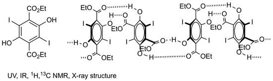
Diethyl 2,5-Dihydroxy-3,6-diiodoterephthalate

EaStCHEM School of Chemistry, University of St Andrews, North Haugh, St Andrews, Fife KY16 9ST, UK
*
Author to whom correspondence should be addressed.
Molbank 2022, 2022(2), M1369; https://doi.org/10.3390/M1369
Submission received: 5 May 2022 / Revised: 17 May 2022 / Accepted: 20 May 2022 / Published: 23 May 2022
(This article belongs to the Section Structure Determination)

Abstract

:
The title compound has been characterised for the first time by the full range of spectroscopic methods, and its X-ray structure shows hydrogen bonded stacks with iodine atoms aligned.

Graphical Abstract

1. Introduction

The simple aromatic compound diethyl 2,5-dihydroxy-3,6-diiodoterephthalate 1 (Scheme 1) was first reported in the literature in 1899 [1]. Rather remarkably, there have been no further references to it since then, and the only experimental data for it is a melting point. In the course of studies on new linkers for metal organic framework (MOF) materials, we prepared the compound, intending to hydrolyse it to give the so-far unknown dihydroxydiiodoterephthalic acid. In fact, we were unable to hydrolyse 1 to the corresponding diacid, and this prompted further investigation of its structure including analysis by NMR and X-ray diffraction. In this paper, we present full characterisation of 1 for the first time, including UV, IR 1H and 13C NMR spectra and its X-ray structure (see Supplementary Materials).

2. Results

Compound 1 was readily prepared by reaction of the dibromo analogue 2 [2] with an excess of potassium iodide in boiling ethanol for 6 h. Its UV spectrum (see Supporting Material) showed four strong absorptions in the range 200–354 nm, while its IR spectrum showed a strong C=O absorption at 1686 cm−1. The NMR spectra were in agreement with expectation and featured a 1H signal at 9.55 ppm for OH as well as a remarkably shielded 13C signal for C–I at 88.6 ppm. This can be compared with a value of 110 ppm for C–Br in compound 2 and results from the well-known heavy atom shielding effect of iodine.
Crystals of compound 1 as prepared were directly suitable for X-ray diffraction, and the resulting molecular structure is shown in Figure 1. This showed the expected high degree of steric congestion around the central six-membered ring, resulting in the ester groups being almost orthogonal to the plane of the benzene ring.
In the crystal, adjacent molecules had iodine atoms in a similar orientation but with alternating positions of the hydroxyl and ester groups, allowing efficient hydrogen bonding (Figure 2, Table 1).
Overall, the crystal structure consisted of stacks of molecules with the iodine atoms aligned and joined by hydrogen bonds alternately at the top and bottom (Figure 3). This is a type of structure that has been described before for halogenated dihydroxyterephthalate esters. In fact, the solid-state structure of such compounds has been of considerable interest ever since the early observation by Hantzsch of different coloured forms of dimethyl dichlorodihydroxyterephthalate [3]. A summary of the different structures determined for such compounds together with the CSD reference codes and literature references is shown in Figure 4.
In diethyl dihydroxyterephthalate 3, the molecules are planar and form chains featuring both intra- and intermolecular hydrogen bonding [4]. This is also the case for the dihydro analogue 4, for which three separate, but essentially identical, structures have been reported [5,6,7]. Introduction of a bulky substituent such as aryloxy at the remaining ring positions removes the intermolecular hydrogen bonding, and compounds 58 retain only the two intramolecular hydrogen bonds [8].
When we come to ring halogenated derivatives, the situation is considerably more interesting and provides an explanation on a molecular level for Hantzsch’s early observation. Thus, the yellow form of dimethyl dichlorodihydroxyterephthalate 9 has the molecules approximately planar and forming chains featuring both intra- and intermolecular hydrogen bonding, as for 3 and 4 [9]. There is also a further “pale yellow” form that has ester groups twisted out of plane by around 40°, but still retains the same chain structure. However, Hatzsch’s white form of 9 has the ester groups orthogonal to the ring with no intramolecular hydrogen bonding, and actually has the same stack structure as we have found for 1 [9]. A similar structure has also been reported for dimethyl dibromodihydroxyterephthalate 10 [11]. When we come to diethyl dibromodihydroterephthalate 2, the situation is slightly different again with two reports of the stack structure lacking intramolecular hydrogen bonding [8,12], but also a different structure that has a chain of molecules in which one ester group is in plane and involved in both intra- and intermolecular hydrogen bonding, but the second ester group is orthogonal and not involved in hydrogen bonding [12].
In summary, the X-ray crystal structure of 1, the first for a dihydroxydiiodoterephthalate, has the ester groups orthogonal to the benzene ring and adopts the hydrogen bonded stack structure also previously described for the related dichloro and dibromo esters 2, 9 and 10.

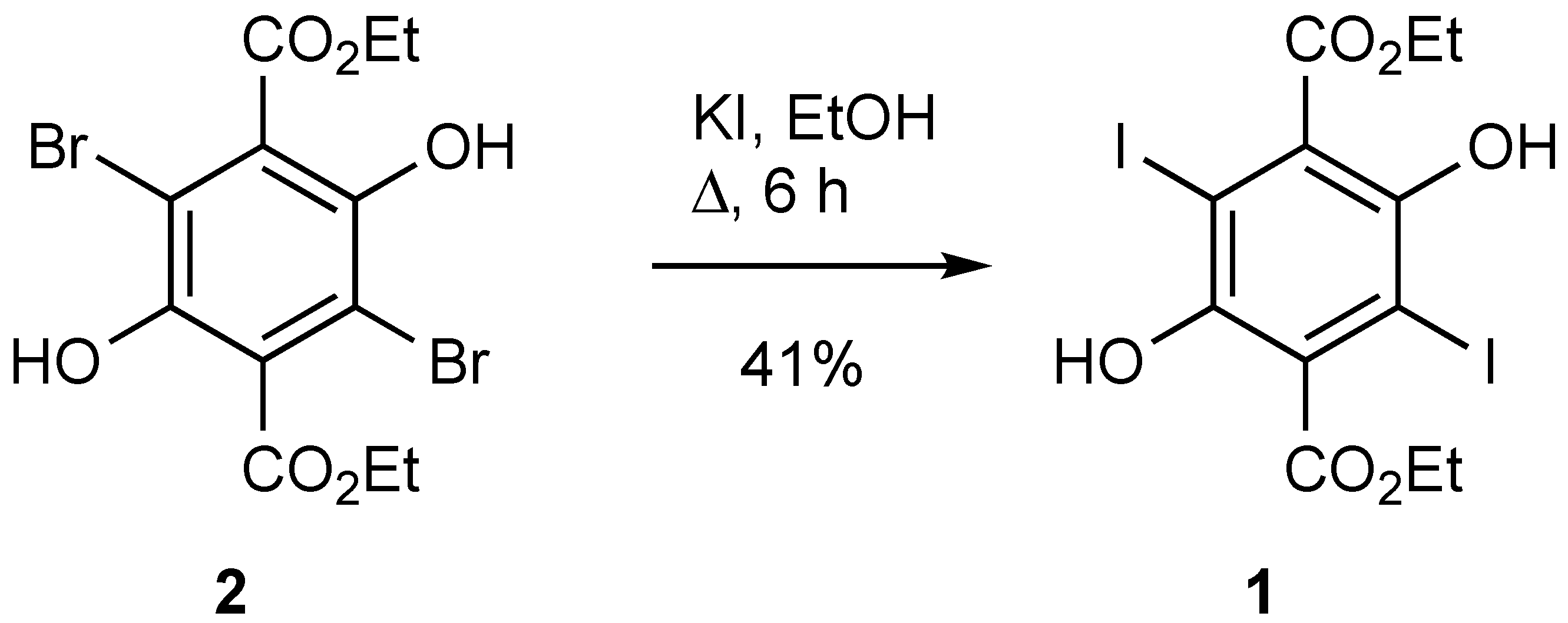
3. Experimental Section

Melting points were recorded on a Reichert hot-stage microscope (Reichert, Vienna, Austria) and are uncorrected. IR spectra were recorded on a Perkin-Elmer 1420 instrument (Perkin-Elmer, Waltham, MA, USA). NMR spectra were obtained for 1H at 300 MHz and for 13C at 75 MHz using a Bruker AV300 instrument (Bruker, Billerica, MA, USA). Spectra were run at 25 °C on solutions in CD3SOCD3 with internal Me4Si as the reference. Chemical shifts are reported in ppm to high frequency of the reference, and coupling constants J are in Hz.

Diethyl 2,5-Dihydroxy-3,6-Diiodoterephthalate (1)

A solution of diethyl 3,6-dibromo-2,5-dihydroxyterephthalate 2 [2] (4.0 g, 9.7 mmol) and potassium iodide (4.83 g, 29.1 mmol) in ethanol (40 mL) was heated under reflux for 6 h. The mixture was hot-filtered, and upon cooling to room temperature, a solid was formed which was filtered off and washed with ethanol (3 × 15 mL) to give product 1 (2.0 g, 41%) as faintly pink needles, mp 168–170 °C (lit. [1] 167 °C). UV–Vis (MeCN): λmax (log e) 354 (3.85), 290 (4.04), 230 (3.97), 200 (4.04); IR (ATR) 1686 (C=O), 1412, 1306, 1273, 1188, 1142, 1009, 860, 829, 737, 681 cm−1; 1H NMR (300 MHz, CD3SOCD3) 9.55 (2H, br s, OH), 4.30 (4H, q, J 7.1), 1.30 (6H, t, J 7.1); 13C NMR (75 MHz, CD3SOCD3) 165.9 (C=O), 146.9 (C–OH), 131.5 (C–CO2Et), 88.6 (C–I), 61.5 (CH2), 13.9 (CH3).
Crystal data for C12H12I2O6, M = 506.03 g mol−1, colourless plate, crystal dimensions 0.278 × 0.085 × 0.032 mm, orthorhombic, space group Pbca (No. 61), a = 8.2343(2), b = 18.9496(6), c = 19.3066(6) Å, α = β = γ = 90°, V = 3012.54(15) Å3, Z = 8, Dcalc = 2.231 g cm−3, T = 173 K, R1 = 0.0261, Rw2 = 0.0537 for 2594 reflections with I > 2σ(I), and 191 variables. Data were collected using graphite monochromated Mo Kα radiation λ = 0.71073 Å and have been deposited at the Cambridge Crystallographic Data Centre as CCDC 2,169,373. The data can be obtained free of charge from the Cambridge Crystallographic Data Centre via http://www.ccdc.cam.ac.uk/getstructures. The structure was solved by direct methods and refined by full-matrix least-squares against F2 (SHELXL, Version 2018/3 [13]).

Supplementary Materials

The following are available online. Figure S1: UV–Vis spectrum of 1; Figure S2: IR spectrum of 1; Figure S3: 1H NMR spectrum of 1; Figure S4: 13C NMR spectrum of 1. Cif and check-cif files for 1.

Author Contributions

N.S. prepared the compound and ran the spectra; A.M.Z.S. collected the x-ray data and solved the structure; R.A.A. designed the study, analysed the data, and wrote the paper. All authors have read and agreed to the published version of the manuscript.

Funding

This research received no external funding.

Conflicts of Interest

The authors declare no conflict of interest.

References

  1. Guinchard, J. Ueber einige Derivate des Succinylobernsteinsäreesters. Ber. Dtsch. Chem. Ges. 1899, 32, 1742–1744. [Google Scholar] [CrossRef] [Green Version]
  2. Böniger, M. Ueber desmotrope Derivate des Succinylobernsteinsäureäthers. Ber. Dtsch. Chem. Ges. 1888, 21, 1758–1765. [Google Scholar] [CrossRef] [Green Version]
  3. Hantzsch, A. Die Chromoisomerie der p-Dioxy-terephthalsäure-Derivate als Phenol-Enol-Isomerie. Ber. Dtsch. Chem. Ges. 1915, 48, 797–816. [Google Scholar] [CrossRef] [Green Version]
  4. Näther, C.; Bock, H.; Seitz, W.; Nagel, N. Diethyl 2,5-dihydroxyterephthalate at 200K. Acta Crystallogr. Sect. C 1996, 52, 2631–2633. [Google Scholar] [CrossRef]
  5. Mez, H.-C.; Rihs, G. Chemistry of succinylsuccinic acid derivatives. Part II The crystal and molecular structure of diethyl succinylsuccinate. Helv. Chim. Acta 1973, 56, 2766–2772. [Google Scholar] [CrossRef]
  6. Frischmann, L.; Langhals, H.; Polborn, K. CCDC 263834: Experimental Crystal Structure Determination; The Cambridge Crystallographic Data Centre (CCDC): Cambridge, UK, 2005. [Google Scholar] [CrossRef]
  7. Chandrashaker, V.; Ptaszek, M.; Taniguchi, M.; Lindsey, J.S. Synthesis of diverse acyclic precursors to pyrroles for studies of prebiotic routes to tetrapyrrole macrocycles. New J. Chem. 2016, 40, 8786–8808. [Google Scholar] [CrossRef]
  8. Benniston, A.C.; Winstanley, T.P.L.; Lemmetyinen, H.; Tkachenko, N.V.; Harrington, R.W.; Wills, C. Large Stokes shift fluorescent dyes based on a highly substituted terephthalic acid core. Org. Lett. 2012, 14, 1374–1377. [Google Scholar] [CrossRef] [PubMed]
  9. Byrn, S.R.; Curtin, D.Y.; Paul, I.C. The X-ray crystal structures of the yellow and white forms of dimethyl 3,6-dichloro-2,5-dihydroxyterephthalate and a study of the conversion of the yellow form to the white form in the solid state. J. Am. Chem. Soc. 1972, 94, 890–898. [Google Scholar] [CrossRef]
  10. Yang, Q.C.; Richardson, M.F.; Dunitz, J.D. Internal molecular motion as forerunner of a phase change involving conformational isomerization. J. Am. Chem. Soc. 1985, 107, 5535–5537. [Google Scholar] [CrossRef]
  11. Kingsbury, C.J.; Abrahams, B.F.; Robson, R. CCDC 1567624: Experimental Crystal Structure Determination; The Cambridge Crystallographic Data Centre (CCDC): Cambridge, UK, 2017. [Google Scholar] [CrossRef]
  12. Näther, C.; Nagel, N.; Bock, H.; Seitz, W.; Havlas, Z. Structural, kinetic and thermodynamic aspects of the conformational dimorphism of diethyl 3,6-dibromo-2,5-dihydroxyterephthalate. Acta Crystallogr. Sect. B 1996, 52, 697–706. [Google Scholar] [CrossRef]
  13. Sheldrick, G.M. A short history of SHELXL. Acta Crystallogr. Sect. A 2008, 64, 112–122. [Google Scholar] [CrossRef] [PubMed] [Green Version]
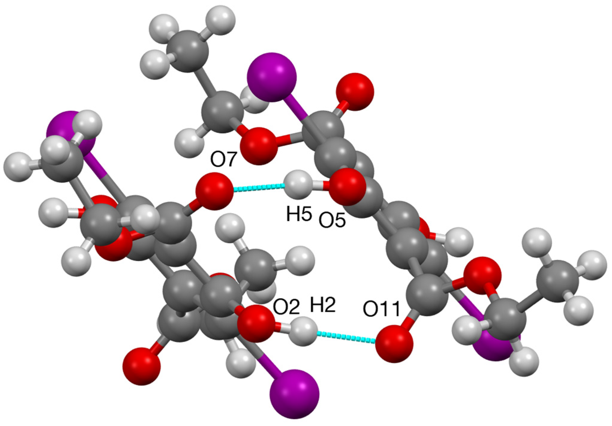
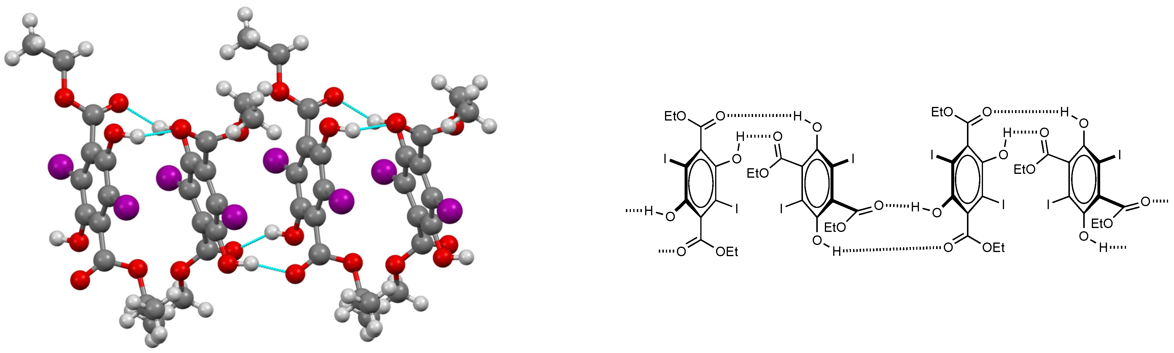
Scheme 1. Synthesis and structure of 1.
Molbank 2022 m1369 sch001
Figure 1. Two views of the molecular structure of 1 showing esters orthogonal to ring (thermal ellipsoids at 50% level). Bond lengths C(1)–I(1) 2.096(3), C(4)–I(2) 2.094(3) Å; torsion angles C(5)–C(6)–C(11)–O(12) −92.6(4), C(2)–C(3)–C(7)–O(8) 109.9(3).
Molbank 2022 m1369 g001
Figure 2. The basic hydrogen bonding pattern.
Molbank 2022 m1369 g002
Figure 3. Structure of hydrogen-bonded stacks and schematic representation.
Molbank 2022 m1369 g003
Figure 4. Reported X-ray structures of dihydroxyterephthalates and substituted derivatives [4,5,6,7,8,9,10,11,12].
Molbank 2022 m1369 g004
Table 1. Hydrogen bonding parameters for 1 (Å, °).
D—H…AD—HH…AD…AD—H…A
O(2)–H(2)…O(11)0.98(3)1.90(3)2.778(4)149(4)
O(5)–H(5)…O(7)0.98(2)1.79(2)2.746(3)163(4)
Publisher’s Note: MDPI stays neutral with regard to jurisdictional claims in published maps and institutional affiliations.

